
Diverse applications of DNA origami as a cross-disciplinary tool
Lingyun
Xiao
,
Xiaoxue
Hu
,
Zhaoyu
Zhou
,
Xiaolin
Xie
,
Shujing
Huang
,
Min
Ji
,
Aobo
Xu
* and
Ye
Tian
 *
*
College of Engineering and Applied Sciences, State Key Laboratory of Analytical Chemistry for Life Science, Nanjing Jinling Hospital, Affiliated Hospital of Medical School, Nanjing University, Nanjing 210023, China. E-mail: ytian@nju.edu.cn; 495896458@qq.com
First published on 17th March 2025
Abstract
As knowledge from a single discipline is no longer sufficient to keep pace with the growing complexity of technological advancements, interdisciplinary collaboration has become a crucial driver of innovation. DNA nanotechnology exemplifies this integration, serving as a field where cross-disciplinary communication is particularly prominent. Since its introduction by Rothemund in 2006, DNA origami has proved to be a powerful tool for interdisciplinary research, offering exceptional structural stability, programmability, and addressability. This review provides an overview of the development of DNA origami technology, highlights its major advances, and explores its innovative applications across various disciplines in recent years, showcasing its vast potential and future prospects. We believe DNA origami is poised for even broader applications, driving progress across multiple fields.
 Ye Tian | Ye Tian holds a B.S. degree from Nanjing University, a M.S. degree from Institute of Chemistry, Chinese Academy of Sciences, and a PhD. degree from Stony Brook University. He was a postdoc at Columbia University and Brookhaven National Lab, before becoming a Professor at Nanjing University in 2017. His research interests include DNA origami technology, self-assembly of nanoparticles, and applying DNA assemblies into biology. |
1 Introduction
Since the advent of the 21st century, relying solely on single-discipline knowledge has become increasingly inadequate for addressing the complex demands of scientific and technological progress. The integration of diverse disciplines has gradually provided greater inspiration for cutting-edge research and has become a significant driving force behind innovation in science and technology. Interdisciplinary research not only fosters the cross-fusion of ideas, methods, and technologies across different fields but also gives rise to numerous emerging disciplines. DNA nanotechnology, introduced by Seeman in 1982,1 has since evolved into a dynamic arena for interdisciplinary integration, encompassing biology, chemistry, materials science, and other fields.2–7 DNA molecules serve as an ideal template for interdisciplinary research due to their stable structure, predictable interactions, ease of modification with various inorganic and organic materials, and straightforward synthesis. Consequently, multiple disciplines have converged within this field. After more than two decades of progress, DNA origami technology, proposed by Rothemund in 2006, injected new vitality into this domain.8 This innovative approach utilized the single-stranded DNA (ssDNA) of the M13mp18 phage, composed of more than 7 kilobases, as a scaffold, which was precisely folded by hundreds of staple strands. Through this technique, DNA undergoes a transformation from a self-assembled one-dimensional (1D) double helix to multidimensional structures, unlocking potential functional applications. The coding nature of DNA is founded on the Watson–Crick base-pairing principle of complementarity, along with its characteristic double-helix structure featuring a hydrophobic interior and negatively charged exterior. Additionally, DNA's dynamic hybridization ability, virtually unrestricted oligonucleotide sequence synthesis, and chemical stability have established it as an exceptionally successful template for programmed molecular self-assembly.9 Over the last two decades, the mechanisms underlying DNA origami assembly have been thoroughly investigated and clarified. Today, various complex DNA origami-based nanomaterials can be synthesized with high precision.10–12 As a nanostructure based on a bottom-up self-assembly strategy, DNA exhibits stable properties, excellent addressability, and remarkable programmability, making it an increasingly valuable tool for interdisciplinary research.Here, we highlight recent advancements in DNA origami within the interdisciplinary realm of DNA nanotechnology. We begin by outlining the development of DNA origami techniques and subsequently provide a comprehensive summary of the latest breakthroughs in their applications across various fields, showcasing their versatility and transformative potential. In materials science, DNA origami has enabled the fabrication of functionalized nanomaterials, such as DNA–silica composites, metal nanowires, and nanostructure substrates, by integrating precise self-assembly with chemical and physical methods. These advances open new possibilities for developing multifunctional nanoscale devices. In biology, DNA origami has significantly enhanced the sensitivity of detecting a wide range of substances, including short oligonucleotides, microRNAs, lipids, enzymes, and ions. In drug delivery systems, these improvements facilitate targeted therapies and enable the creation of nanoreactors for studying biological processes. In optics, DNA origami supports the development of nanophotonic systems and enhances techniques such as surface-enhanced Raman scattering (SERS). In robotics, DNA origami has enabled the construction of nanoscale robots capable of performing precise tasks, including medical interventions. Additionally, DNA origami demonstrates potential in computing and information science, contributing to innovations such as DNA-integrated circuits and biocomputing. Finally, we address both the challenges and future prospects of interdisciplinary applications in this domain.
2 DNA origami
First proposed by Rothemund in 2006, the DNA origami technique is a bottom-up strategy for the precise construction of nanoscale structures.8 The resulting nanostructures consist of hundreds of different staples and a long scaffold, offering excellent addressability and programmability. Over the past 20 years, scientists have continuously explored and innovated in various directions, including design principles, design tools, assembly mechanisms, and application areas, thereby driving the continuous advancement of DNA origami technology. The most direct outcome of this progress is the increasing complexity and refinement of DNA origami patterns, evolving from the initial symmetrical two-dimensional (2D) shapes like squares and smiling faces8 to asymmetrical 2D patterns such as Chinese maps13 and dolphins,14 multilayer three-dimensional (3D) structures,15 hollow 3D structures,16 and wireframe structures incorporating multi-arm junctions.17 Additionally, dynamic structures capable of undergoing configuration transformations in response to external stimuli have been developed.18 The design and exploration of these origami structures have fostered the integration of DNA nanotechnology with other fields, expanding the applicability of DNA nanostructures across interdisciplinary domains. In this section, we outline the evolution of DNA origami structures and highlight new structures or systems that have recently been applied.2.1 DNA origami structures
Although atomic force microscopy (AFM) and scanning tunneling microscopy have enabled the manipulation of individual atoms, their stringent temperature and vacuum conditions limit their practicality. To control the growth of small molecules at the nanoscale and use them as fundamental units for multilevel structures, DNA, which self-assembles under relatively mild conditions, emerged as an ideal raw material for the bottom-up synthesis of nanomaterials with ordered molecular arrangements. The original DNA origami technology was designed to create highly complex nanostructures. Rothemund utilized parallel double helices to fill predetermined patterns based on the precise specificity of base pairing, producing a series of 2D basic patterns (Fig. 1A).8 Additionally, variations in DNA origami staples allow for precise addressing and fluorescent labeling of specific sites, enabling the display of various images or text at a given resolution on the rectangular 2D origami template (Fig. 1B).8 Building on this strategy, Qian et al. introduced a more intricate geographic pattern by designing block structures to fill asymmetrical areas, exemplified by the detailed borders of the Chinese map and its islands (Fig. 1C).13 Han and collaborators expanded DNA origami strategies using partially complementary double-stranded DNA (dsDNA) and cohesive interactions of parallel crossovers. Unlike the traditional “staple” model, this novel construction method eliminates the need for auxiliary strands, allowing compact shapes, such as hearts and diamonds, to be formed by simply folding ssDNA (Fig. 1D).19 | ||
| Fig. 1 DNA origami graphics obtained by different strategies. [A] Folding paths and AFM images of symmetric star and smiley face patterns. Scale bars: 100 nm. Reproduced from ref. 8 with permission from Springer Nature, copyright 2006. [B] Scaffold design and AFM image of the highlighted patterns on rectangular DNA origami structure. Reproduced from ref. 8 with permission from Springer Nature, copyright 2006. [C] Design of Chinese map origami graphic and AFM image. Shape is of ∼150 nm length, ∼120 nm width, and 2 nm height. Reproduced from ref. 13 with permission from Springer Nature, copyright 2006. [D] Various origami self-folding models with corresponding AFM images. Scale bars: 50 nm. Reproduced from ref. 19 with permission from AAAS, copyright 2017. [E] Scaffold folding path (upper right) and AFM images for flower-and-bird pattern. Scale bar: 100 nm. Reproduced from ref. 20 with permission from AAAS, copyright 2019. [F] AFM images of mesh patterns consisting of multi-arm junctions. Scale bars: 20 nm (model) and 50 (zoom-in) and 150 nm (zoom-out). Reproduced from ref. 21 with permission from AAAS, copyright 2022. | ||
Beyond parallel raster stacking, a novel origami strategy directly constructs polygons using helix bundles composed of multiple dsDNA. Zhang and co-workers first created a variety of complex 2D wireframe origami structures using double crosses (DX) and double helix bundles (2HB) (Fig. 1E).17 To assemble 2D geometries with more vertices and arbitrary edge lengths and curvatures, Jun et al. introduced a wireframe origami program based on automatic PERDIX generation (Fig. 1F).20 By simply drawing the edges of a desired structure, the program generates optimized graph-theoretic scaffold routing, enabling the rapid and robust creation of nanoscale templates for drug delivery, nanophotonics, and biomolecular sensing. In contrast to the initial single-chain folding approach, Wang et al. employed a 6HB based on a multilayer honeycomb design to enhance the stiffness and planarity of 2D origami structures.21 By maximizing the number of crossovers at each neighboring edge, they effectively counteracted the unintended curvature typically observed in a liquid-phase environment.
The dense DNA helix design facilitates the construction of higher-dimensional structures, extending beyond conventional staple-based connections between adjacent parallel scaffolds in the plane (Fig. 2A). DNA helix bundles are arranged in specific stacking patterns to form various hollow structures with storage space.22 These bundles are predominantly stacked in honeycomb and square configurations. Douglas et al. leveraged both of these as template interfaces to develop the open-source caDNAno software, which automates staple design and sequence filling within a framework of preset geometrical parameters, including double-helix diameter (2.0 nm), base pair length (0.34 nm), and helix period (10.5 base pairs).23 Using caDNAno, researchers designed monoliths, square nuts (Fig. 2B), railed bridges, slotted crosses, stacked crosses, and numerous other structures. Dietz et al. extended the scope beyond highly symmetrical structures made from straight DNA helix bundles by introducing controlled bending and twisting through the strategic addition or removal of base pairs.24 A 3 × 6 DNA helix bundle was designed to achieve a quasi-continuous bend ranging from 0° to 180°, progressing through seven incremental stages at 30° intervals (Fig. 2C). In contrast, Han and his collaborators pursued a different approach, producing in-plane curvature by constructing concentric circles of DNA while adjusting crossover positions to control out-of-plane curvature, ultimately achieving high-curvature nanostructures with low symmetry.25 Using this strategy, representative nanospheres, ellipsoids, and nanoflasks were designed (Fig. 2D). Similar to wireframe structures, DNA origami has inspired new approaches for 3D construction. Benson et al. applied graph theory-based wiring algorithms and relaxation simulations for automated wiring, enabling the creation of complex structures that would have been challenging to achieve through traditional parallel alignment of DNA bundles.16 Non-closed structures such as notched rings, spirals, and asymmetrical complex geometries—including rabbits—were designed in Autodesk Maya, and these hollow structures were visualized using cryo-electron microscopy (Fig. 2E). The development of new design software and construction strategies has injected fresh momentum into DNA origami, facilitating the creation of a diverse array of highly intricate multilayered structures while providing a solid theoretical foundation and expanded design space for future applications.
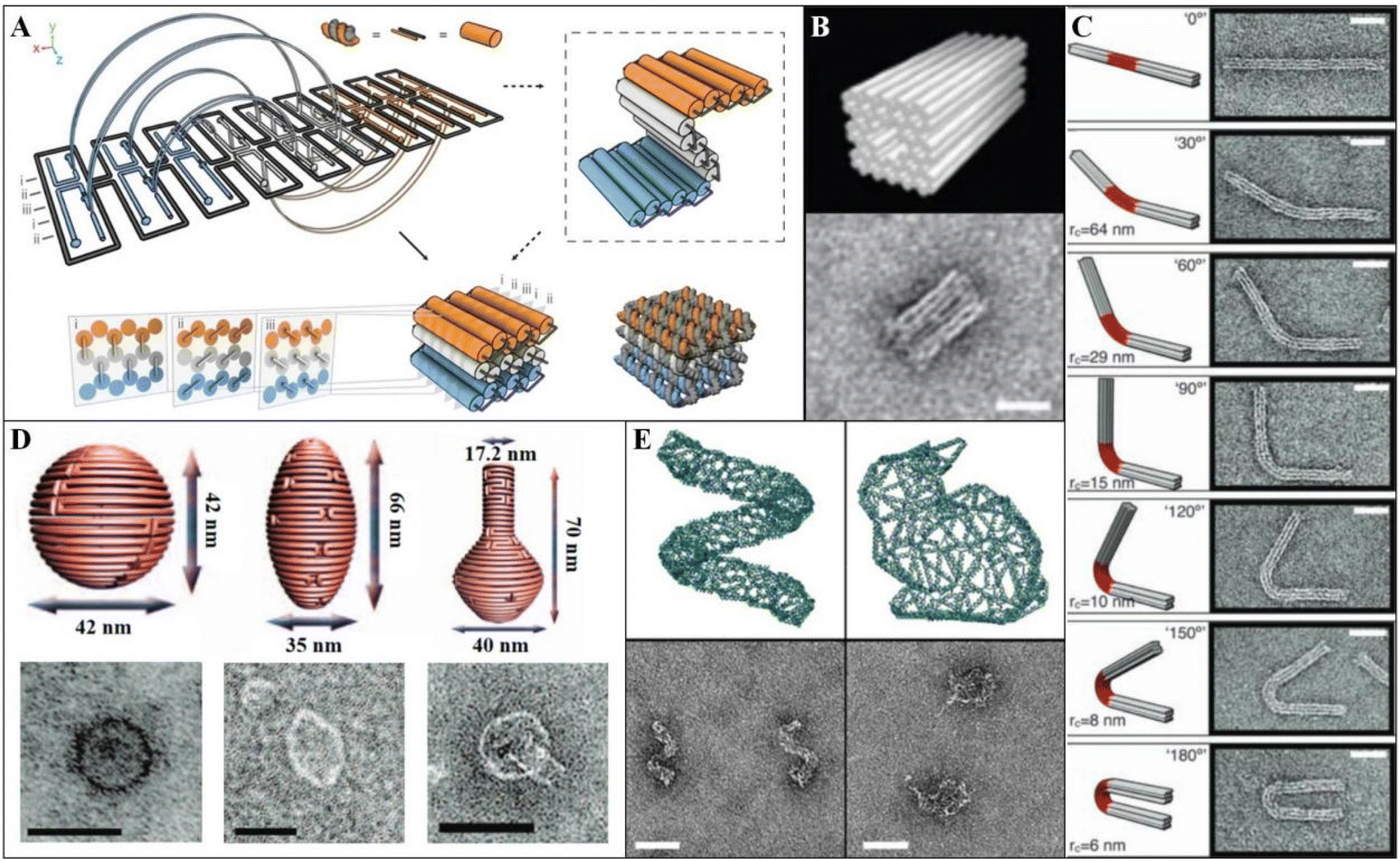 | ||
| Fig. 2 DNA origami nanogeometry and 3D structures. [A] Schematic diagram of the construction strategy of 3D DNA origami. Reproduced from ref. 22 with permission from Springer Nature, copyright 2009. [B] Diagram and transmission electron microscope (TEM) image of square nut origami model. Scale bar: 20 nm. Reproduced from ref. 23 with permission from Oxford University Press, copyright 2009. [C] Schematic diagram and TEM image of the 3 × 6 helix bundle with different bending angles. Scale bars: 20 nm. Reproduced from ref. 24 with permission from AAAS, copyright 2009. [D] Model diagrams and TEM images of the DNA origami structures with 3D curvature. Scale bars: 50 nm. Reproduced from ref. 25 with permission from AAAS, copyright 2011. [E] 3D hollow origami wiring schematic and TEM image. Scale bars: 50 nm. Reproduced from ref. 16 with permission from Springer Nature, copyright 2015. | ||
2.2 DNA origami assemblies
As the diversity of DNA origami structures has expanded significantly, researchers have set their sights on the next challenge in DNA nanotechnology: fabricating larger DNA structures. The maximum size of DNA origami is constrained by the 7249-nucleotide length of the M13mp18 phage, limiting 2D structures to 78 × 78 nm2 and 3D structures to 24.7 × 24.7 × 24.7 nm3.26 To further increase the size of DNA origami structures, new scaffolds and techniques have been introduced, including phiX174,27 λ-phage,28 PCR amplification scaffolds,29 and the conversion of dsDNA to ssDNA by pyromorphism.26 These advancements have expanded the size of 2D origami monoliths to an area of 25![[thin space (1/6-em)]](https://www.rsc.org/images/entities/char_2009.gif) 704 nm2, with a side length of up to 300 nm.30,31
704 nm2, with a side length of up to 300 nm.30,31
Adaptations based on DNA scaffolds have led to a broader design space but also increased costs and by-products. Nonetheless, synthesizing large structures with nanometer-scale precision remains a challenge. The assembly of origami monomers with attached connectors into layered, advanced structures represents an innovative strategy for constructing micrometer-scale lattices. This approach leverages the combinatorial arrangement of monomers in one-, two-, or multiple-unit combinations to achieve complex configurations. The construction principles of assembly connectors fall primarily into two categories: DNA hybridization8,32 and blunt-end stacking.33 Wang et al. designed their assembly based on the sides of 6HB wireframe origami, creating bundle vacancies and extended sticky ends to form a superstructure using triangular wireframe origami as the core element (Fig. 3A).34 This parallel-crossing assembly enables the construction of a compact, rigid 2D planar superstructure with potential applications in nanoscale lithography. Another assembly strategy, DNA reverse parallel ligation, is commonly used to hybridize the sticky ends of protruding single strands. Wang and colleagues developed a hexagonal origami tile that can be assembled into large 2D planes or honeycomb tubes of varying diameters by adjusting the number of complementary and gap bases in the sticky-end regions (Fig. 3B).35 Incorporating Au nanoparticles (AuNPs) into the honeycomb lattice enabled the ordered arrangement of Au superlattices, exhibiting optical metamaterial properties and plasmonic resonances. Similarly, Liu et al. applied longer sticky ends to the vertices of square DNA origami structures, indirectly controlling monomer loop formation and planar quality by modulating the number of T bases in the spacer region of the sticky ends (Fig. 3C).36
 | ||
| Fig. 3 Examples of DNA origami assemblies. [A] Schematic and TEM image of triangular array assembly. Scale bar: 100 nm. Reproduced from ref. 34 with permission from ACS, copyright 2022. [B] Sticky ends guide the assembly of DNA origami tiles into tubular or planar structures. Reproduced from ref. 35 with permission from ACS, copyright 2016. [C] T sequence regulates SDO ring formation and planar assembly. Scale bars: 500 nm. Reproduced from ref. 36 with permission from ACS, copyright 2024. [D] Schematic and TEM images of the 3D assembly of DNA origami octahedra. Reproduced from ref. 37 with permission from Springer Nature, copyright 2020. [E] Schematic and TEM images of DNC 2D assembly. Scale bar: 50 nm. Reproduced from ref. 39 with permission from ACS, copyright 2020. [F] Schematic and TEM images of 3D meta-DNA assemblies Scale bar: 100 nm. Reproduced from ref. 44 with permission from Springer Nature, copyright 2020. | ||
Designs incorporating long T sequences between dsDNA bundles and sticky ends provide a flexible approach for attaching 3D hollow frameworks, enabling versatile structural configurations. Tian et al. reported a series of frame structures composed of 6HB or 10HB, co-assembled with various nanoparticles (NPs) such as AuNPs, quantum dots (QDs), and proteins (Fig. 3D).37 This approach offers a general strategy for creating arrays of nano-objects using a consistent assembly process. Building on this foundation, a total of 85 DNA origami frame/AuNP superlattices were synthesized through the co-crystallization of multiple octahedral frameworks.38 The DNA origami nano-chamber (DNC), designed by Lin et al., encoded specific connections along three orthogonal axes while preserving cavities for molecular associations (Fig. 3E).39 Bayrak et al. employed a similar strategy to produce highly homogeneous gold nanowires, with diameters of 20–30 nm, through ionic growth by tightly connecting chambers loaded with 5 nm AuNPs in a 1D arrangement.40 Berengut et al. designed chiral nanotubes with varying degrees of twist and diameters by modifying alternating dihedral angles to control intrinsic sheet curvature.41 Wickham et al. coaxially assembled DNA origami barrels to create precisely addressable decamers, functioning as 3D pegboards that isolate reaction environments inside and outside the barrels.42
To construct micron-sized DNA structures, researchers have explored various methods to amplify scaffolds30 and optimize annealing conditions.43 Yao et al. pioneered the use of submicron-sized six-helix bundle DNA origami nanostructures as building blocks. By mimicking DNA strand assembly behavior, they created micron-scale complex origami structures. Three self-assembly strategies—single-stranded tile assembly, single-stranded origami, and multi-arm junction tiles—were validated with meta-DNA, leading to the formation of larger 3D polyhedra and ribbons exceeding 5 μm in length (Fig. 3F).44
In addition to these methods, the degrees of freedom of adsorbed DNA origami monomers can be constrained to a 2D plane through interfaces such as solid–liquid,45,46 liquid–air,47 and lipid–liquid.48 This surface-assisted strategy on mica sheets facilitates hierarchical DNA origami assembly via electrostatic interactions. By introducing an abundance of monovalent sodium ions, divalent magnesium ions at the mica–DNA interface are replaced, weakening the binding effect on DNA origami nanostructures. This reduction in binding enhances DNA origami mobility, allowing for more effective diffusion and alignment along the 2D surface.45,46 Xin et al. utilized surface-assisted assembly on mica sheets to fabricate 2D lattices featuring densely packed rows with macroscopic-scale control. The ordered DNA origami lattice, extending up to 18.75 cm2, was validated using fluorescence microscopy. Each DNA origami triangle was conjugated to a QD via streptavidin, and the uniformity of QD fluorescence served as an indicator of the lattice's structural order.49 As a mask for molecular lithography, ordered monolayers of DNA origami structures can guide the adsorption of various proteins, including single-stranded annealing proteins like Redβ and Sak, the iron storage protein ferritin, and the blood protein bovine serum albumin (BSA).50
2.3 DNA origami purification
Purification is a critical step in obtaining high-quality DNA origami structures while preventing undesired aggregate formation. Agarose gel electrophoresis (AGE) is a widely used, high-resolution purification method that separates by-products based on their varying migration rates, allowing the recovery and enrichment of desired products by disrupting the gel.51 However, AGE purification can result in significant sample loss. As an alternative, the polyethylene glycol (PEG) purification method has been proposed, offering a straightforward approach capable of achieving a purification level of over 99%.52 Furthermore, to remove functional or fluorescent groups without introducing additional impurities, ultrafiltration filters provide adequate recovery and are well suited for purifying small quantities effectively.53 For DNA–peptide polymers, purification can be performed using reversed-phase high-performance liquid chromatography, where buffer exchanges yield pure samples; however, this method is cumbersome.54 Ye et al. introduced a purification technique based on magnetic bead pull-down reactions to remove incompletely assembled structures, effectively separating fully assembled superstructures from excessive substructures.55 By executing a multi-step pull-down reaction, desired superstructures can be meticulously isolated from assemblies or other inorganic NPs. W/W-droplet fractionation purification methods enable the rapid separation of macroscopic phases and enclosed molecules within seconds under centrifugation. This versatile technique can be applied to DNA origami structures of various sizes.56 Additionally, other purification methods include size exclusion chromatography (SEC), gel filtration using rotary columns, and gradient ultracentrifugation. When selecting the appropriate purification method for a specific application, factors such as yield, purity, concentration, volume, residue, efficiency, and the method's impact on the sample should be carefully evaluated and compared.3 Interdisciplinary applications
DNA origami provides an extraordinary ability to build micron-scale materials with sub-nanometer addressability. As a highly versatile platform characterized by significant programmability, it offers numerous chemically modified sites through pre-designed sequences, allowing precise control of nanoscale entities such as metal NPs, QDs, fluorescent dyes, and proteins. As a bridge, nanostructures fabricated using this bottom-up approach have the potential to facilitate advancements across various disciplines, leading to new discoveries and novel technological approaches in fields like materials science, biology, optics, robotics, and information science. This section discusses the diverse applications of DNA origami in these fields in recent years.3.1 Interdisciplinary with material science
DNA is a synthetic biomaterial with self-assembling capabilities, and the presence of a phosphate backbone in the molecule imparts negative electrical properties. The elaborate high-dimensional structures created through DNA origami provide an excellent platform for fabricating advanced nanomaterials, exemplifying a quintessential case of interdisciplinary innovation. Silicon dioxide, metal nanoparticles, and metal oxide nanoparticles are integrated into complex structural or superlattice templates with the aid of DNA structures through various chemical methods, including chemical growth, atomic layer deposition (ALD), etching, and physical techniques like metal deposition and photolithography. These approaches collectively open up new possibilities for fabricating multifunctional devices at the nanoscale.In 2009, Jin et al. introduced a novel approach for structural analysis under an electron microscope by constructing a DNA–silica complex (DSC).57 By anchoring the trimethylene group of N-trimethoxysilylpropyl-N,N,N-trimethylammonium chloride (TMAPS) to the silicon atoms and the cationic ammonium group in the DSC, in the presence of the condenser quaternary ammonium group, the silane sites interacted with tetraethoxysilane (TEOS) to form a hard silica shell.58 Auyeung et al. decoupled crystal parameters, such as symmetry and lattice spacing, of DNA-assembled superlattice crystals from the liquid-phase environment by silica encapsulation and obtained stable 10 nm gold nanoparticle lattices that did not deform, collapse, or dissociate under normal conditions.59 The use of TMAPS and TEOS in DNA-encapsulated silica technology has been extensively studied, offering both theoretical insights and practical foundations for nanofabrication based on customized DNA origami templates.60–62
DNA origami, with its unique addressability as a structural material, has undergone extensive research and has proved to be an excellent customized template for fabricating DNA–silica composites. A surface-assisted two-step method was employed to silicify DNA origami by forming a silica shell of approximately 3 nm thickness on both small DNA origami structures and 2D lattices. The silica shell serves to protect the encapsulated DNA origami, ensuring that it maintains structural integrity even after being removed from a liquid-phase environment (Fig. 4A).63 Nguyen et al. proposed an analogous sol–gel reaction to silicify larger 3D DNA origami crystals, resulting in silica shells with enhanced structural rigidity (Fig. 4B).64 Jing et al. presented a DNA origami silicification (DOS) method for depositing silica onto the surface of DNA frameworks, allowing the formation of DNA–silica composites with more complex, programmable morphologies not limited to monolayers or multilayers of DNA origami with honeycomb lattice arrangements.65 Shang et al. prepared silica patterns specifically on DNA origami templates designed with protruding strands at fixed positions. Differences in binding energies between prehydrolyzed products (PP)-dsDNA and PP-DNA origami were exploited, leading to preferential adsorption of PP onto the protruding dsDNA regions rather than the DNA origami surface.66 Wassermann et al. reported a method that enables the specific addressing of silica cladding, allowing for the creation of dynamic silica nanostructures with precisely controllable shapes and sizes (Fig. 4C).67 Wang et al. used a six-helix bundle DNA origami to explore how the presence of structural surface ssDNA and dsDNA affects the DNA origami during the silicification process. Crosslinking and aggregation of the origami structures were effectively prevented by ssDNA, allowing a thicker silica shell to form over a longer reaction time. Meanwhile, dsDNA facilitated the formation of silica on the surface of the DNA origami.68 Additionally, Wang et al. achieved rapid silicification of DNA origami at a graphene interface, yielding a distortion-free 2D DNA origami–silica composite with twice the fidelity of bare DNA origami.69
 | ||
| Fig. 4 Preparation of DNA origami silica composites. [A] DNA origami template-based guided growth of silica composites. Reproduced from ref. 63 with permission from Springer Nature, copyright 2018. [B] Schematic of DNA origami helix bundle package silicon. Reproduced from ref. 64 with permission from Wiley, copyright 2019. [C] Dynamic structure of 18HB with variable morphology before and after silicification. Reproduced from ref. 67 with permission from Wiley, copyright 2023. | ||
Similarly, DNA origami acts as a structural template for the bottom-up synthesis of conducting metal nanowires. Continuous, complex metallic nanostructures are achieved through chemical growth between small metallic nanoparticles facilitated by reducing agents.70 Common structures include metal nanowires used in electrical applications, such as gold,71 silver,72 palladium,73 platinum,74 copper,75 nickel,76 tellurium,76 and cobalt nanowires,77 which are typically formed through wet chemistry mediated by DNA templates. However, this approach has the limitation of providing only a restricted degree of control over the nanowires’ shape. Yin et al. encapsulated gold nanoparticles in DNA origami molds containing cavities and introduced silver nitrate and ascorbic acid to limit the growth of silver monomers on the surface of the gold seeds, obtaining a series of gold-core–silver-shell structures with relatively controllable shapes (Fig. 5A).78 This fabrication concept was later extended to casting triangular and disc-shaped structures and has also been applied to the synthesis of large-scale gold nanostructures.78–80
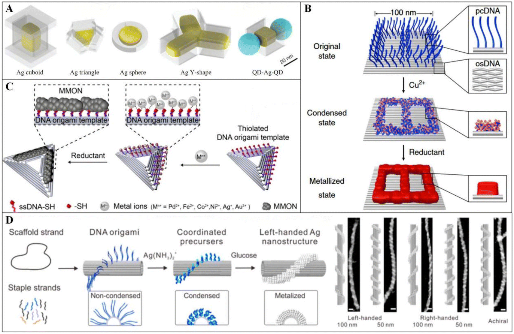 | ||
| Fig. 5 Various metallization strategies based on DNA origami. [A] Casting NPs with form-fitting “box-lid” DNA origami molds. Reproduced from ref. 78 with permission from AAAS, copyright 2014. [B] High-resolution metal patterning on a single-origami breadboard by strong coordination of Cu2+ or Ag+ to pcDNA. Reproduced from ref. 81 with permission from Springer Nature, copyright 2019. [C] Synthesis of metal and metal oxide nanoclusters at specific sites on DNA origami triangles. Reproduced from ref. 82 with permission from ACS, copyright 2019. [D] Fabrication of chiral helical silver patterns and their assembly on DNA origami tubes. Reproduced from ref. 83 with permission from ACS, copyright 2021. | ||
Jia et al. introduced a method that does not require exogenous metallic nanoparticles, instead inducing the formation of various metallic patterns based on the differing affinities of low-valent metal ions, such as Cu2+ and Ag+, for protruding clustered DNA (pcDNA) and DNA origami substrates (Fig. 5B). This approach enabled the fabrication of nanoscale printed circuit boards with single and double layers.81 Li and co-workers devised a patterning strategy relying on sulfhydryl-specific sites on DNA origami triangles, promoting the formation of proto-metal and metal–oxide nanoclusters (Fig. 5C).82 In a similar vein, Zhang et al. employed DNA origami nanotubes, where diamine–silver complexes were locally enriched on specific single-stranded protruding clusters of DNA, resulting in chiral helix–silver patterns extending up to one micrometer in length (Fig. 5D).83
DNA nanostructures serve as highly adaptable templates for chemical vapor deposition (CVD) and ALD, enabling the fabrication of DNA origami–metal oxide complexes.84 CVD, a widely used chemical technique, facilitates the production of monolayer or multilayer 2D materials by inducing gas-phase chemical reactions between precursors at elevated temperatures.85 This method is particularly effective for creating large-scale, high-quality 2D materials, including graphene monolayers86 and hexagonal boron nitride.87 Surwade et al. achieved precise SiO2 and TiO2 deposition on DNA nanostructures with nanoscale resolution using a CVD method that regulated water adsorption at room temperature (Fig. 6A).88 By controlling water adsorption at the nanoscale, they successfully manipulated the display of positive and negative tone patterns on DNA templates.
 | ||
| Fig. 6 Chemistry combined with DNA origami templates for nanofabrication processes. [A] Schematic representation of customised inorganic oxide nanostructures on DNA origami templates by CVD. Reproduced from ref. 88 with permission from ACS, copyright 2013. [B] Schematic representation of the assembly of DNA origami diamond cubic lattice and surface ALD growth of high refractive index cover layer. Reproduced from ref. 93 with permission from AAAS, copyright 2024. [C] Schematic representation of region-selective ALD preparation of Pt nanostructures based on DNA metal templates. Reproduced from ref. 94 with permission from ACS, copyright 2022. | ||
ALD, a technique derived from CVD,89 relies on surface-specific chemical reactions for thin-film preparation. Compared with CVD, ALD offers advantages such as precise control over thin-film thickness, uniform coverage, and effective coating of complex structures at relatively low temperatures (<350 °C).90–92 Posnjak et al. used ALD to modulate the thickness of a high-refractive-index titanium dioxide cladding layer on DNA origami diamond structures, achieving a complete photonic band gap (Fig. 6B).93 Hui et al. employed ALD for regioselective Pt deposition mediated by DNA self-assembled structures, successfully fabricating various one- and two-component nanopatterns, including Pt, TiO2/Pt, and Al2O3/Pt (Fig. 6C).94
Etching, a top-down micro- and nano-fabrication method, can be enhanced by incorporating DNA origami to produce metal nanostructures with improved continuity and structural integrity. DNA origami patterns can be deposited onto silica substrates, serving as masks similar to those used in photolithography. By integrating bottom-up and top-down fabrication approaches, this technique enables highly precise nanoscale fabrication. After etching with hydrogen fluoride vapor, the areas covered by DNA origami remain intact, resulting in patterns on the silica substrate that closely replicate the template's structure and shape.95,96
Since Seeman established the DNA molecular framework in 1982,1 dsDNA and ssDNA have been extensively utilized as fundamental building units for self-assembled nanostructures, serving as fine material templates for region-selective metallization.97 Dai et al. introduced a deposition strategy integrating silica–metal heterojunctions by leveraging the density and length of protruding dsDNA strands in DNA origami templates. This method enabled precise and addressable incorporation of silica–gold and silica–silver heterostructures.98
As electronic components continue to miniaturize, the line widths of integrated devices are shrinking, reaching as small as 5 nm through photolithography, which enables rapid, parallel, large-area fine processing. DNA origami holds significant potential as a surface lithography pattern mask due to the unique properties of its building blocks. DNA nanofabrication achieves a minimum size of 2 nm, making it an ideal candidate for precise nanoscale patterning. Shen's team introduced a modular epitaxy method for designing 3D DNA masks with programmable size, shape, and spacing. In this approach, a flat DNA brick crystal serves as the foundational surface for further assembly or modification, onto which 3D DNA modules grow in a seed-mediated manner. The DNA brick unit consists of a short 32-nucleotide DNA strand, allowing for a high resolution of 7 nm lateral critical size and 2 nm vertical critical size, which is preserved during DNA pattern transfer. The assembled 3D DNA mask is stabilized by Ni2+ ions, preventing feature collapse after air drying on silicon substrates. This Ni2+-stabilized 3D DNA mask was then used to directly generate oversized Si patterns through a single run of reactive ion etching (RIE). The pitch and critical dimension (CD) of the Si patterns were reduced to 16.2 ± 0.6 nm and 7.2 ± 1.0 nm, respectively—a 50% reduction compared with current methods such as quadruple patterning or extreme ultraviolet (EUV) lithography.99 This approach introduces a new technique for high-resolution silicon patterning on a wafer scale.
3.2 Interdisciplinary with biology
Due to its precise spatial addressability and numerous manipulable sites on the surface, DNA origami offers a novel solution for nucleic acid detection. Choi's group utilized DNA origami structures instead of traditional single-stranded DNA to attach dye molecules, combining this approach with planar microbead assays (Fig. 7A).109 This method enables brighter and more sensitive quantitative measurement of target short oligonucleotides at lower concentrations. The new reporter DNA origami expands the dynamic detection range and lowers the detection limit for oligonucleotides.
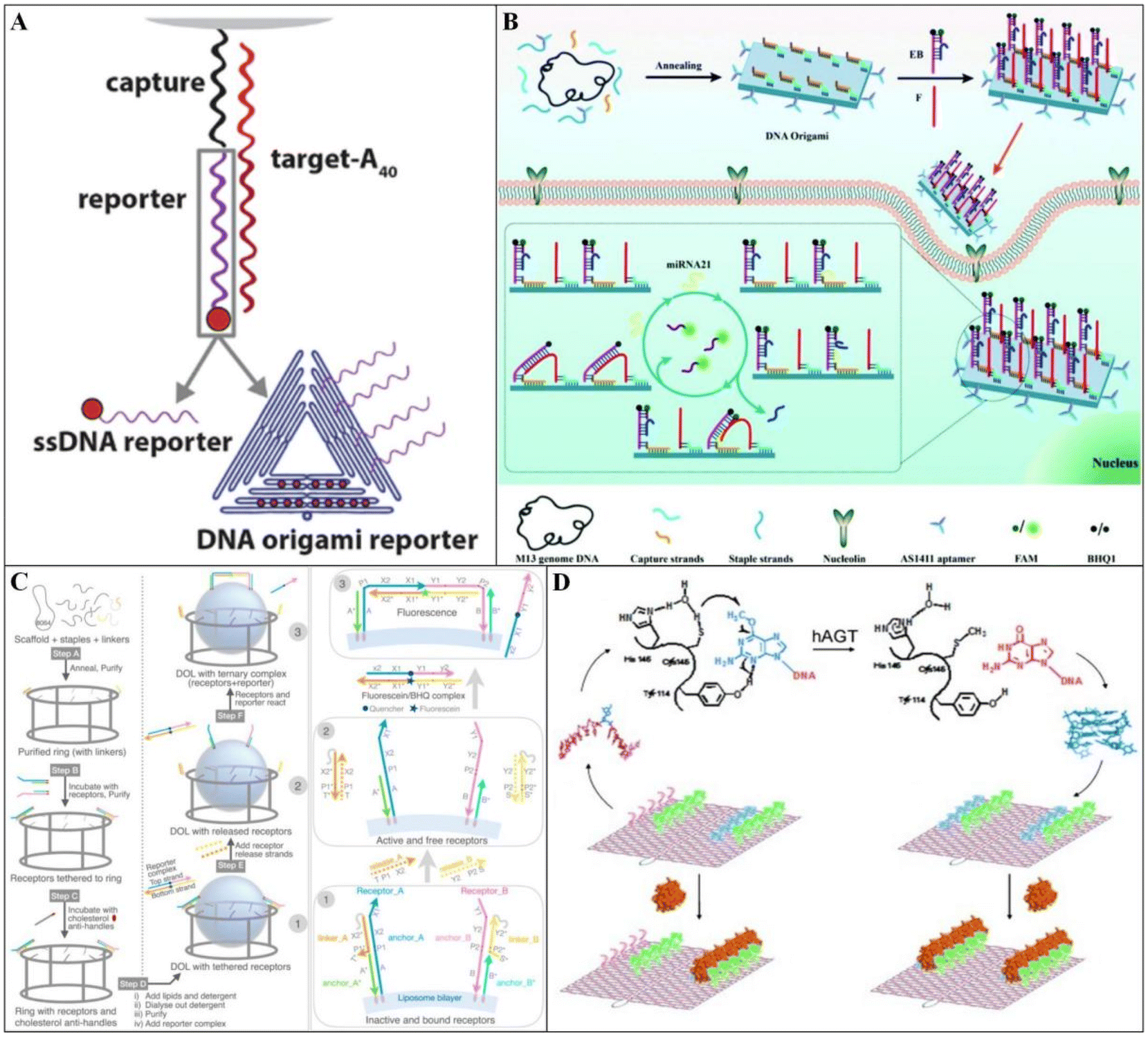 | ||
| Fig. 7 DNA origami biosensors. [A] Microbeads are functionalized with streptavidin to which biotinylated capture DNAs are immobilized. Reproduced from ref. 109 with permission from Springer Nature, copyright 2019. [B] Model diagram of DNA origami amplifier. Reproduced from ref. 111 with permission from RSC, copyright 2022. [C] Schematic diagram of DOL assembly synthesis. Reproduced from ref. 112 with permission from Springer Nature, copyright 2023. [D] Schematic representation of the process by which hAGT repairs the methyl-TBA aptamer to symmetrically bind DNA origami quadruplexes to α-thrombin. Reproduced from ref. 114 with permission from Wiley, copyright 2013. | ||
Dysregulated microRNA (miRNA) expression has been associated with cancer, and miRNA profiles have emerged as potential therapeutic targets.110 Xing et al. developed an aptamer-tethered DNA origami amplifier that enables stable and highly sensitive detection of miRNAs in living cells (Fig. 7B).111 Two substrate modules were assembled onto rectangular origami planes with edge-modified aptamers designed to target cancer cells. The presence of miRNA initiates sequential hybridization of the substrate modules, producing amplified and stable fluorescent signals via an intramolecular entropy-driven reaction.
Maingi et al. created a DNA origami ring-based nanoreactor that enables precise control of the connections between lipid-binding molecular receptors in three aspects: time, space, and stoichiometry.112 This DNA origami-templated liposome (DOL) digital nanoreactor not only templates the synthesis of liposomes but also provides tethering sites for DNA-based receptors, potentially offering a customized instrument for studying and dissecting complex membrane protein interactions (Fig. 7C).
The dimensions of typical complementary metal oxide semiconductor (CMOS) devices closely match those of many biological systems. However, significant challenges remain in transmitting cellular biochemical signals to biosensors, primarily due to the insulating properties of cell membranes, which often result in considerable signal attenuation in extracellular measurements. To address this, Livernois et al. combined field-effect transistors (FETs) with DNA origami nanopores to create an ion detection device that can be seamlessly integrated into living cells.113 The DNA nanopore forms an overlapping electrical double layer (EDL) that acts as a gate to the FET, achieving higher sensitivity than standard MOSFETs for single-ion detection.
Tintoré et al. developed the first biosensor to visualize hAGT activity on an origami-based platform.114 Their work applied single-molecule characterization of DNA origami for DNA repair assays by combining α-thrombin with TBA to determine the repair effect of hAGT on methylated TBA-origami (Fig. 7D). This platform lays the foundation for creating innovative biosensors that can detect the activity of various complexes and other proteins.
Effective vaccine delivery with targeted transport and on-demand release remains a major challenge in cancer treatment. This has led to the development of delivery platforms based on programmable molecular systems, offering promising pathways for improvement. Liu's team designed a tubular DNA nanostructure to enable the precise assembly of two types of molecular adjuvants and antigenic peptides within its lumen, forming a vaccine (Fig. 8A).124 A pH-sensitive DNA “locking strand” on the exterior of the tube allows the vaccine to open inside the lysosomes of antigen-presenting cells, effectively exposing the adjuvant and antigen to trigger a potent immune response.
 | ||
| Fig. 8 DNA origami drug delivery devices. [A] Schematic illustration of the tumor antigen peptide/CpG loop/double-stranded RNA-encapsulated DNA origami robot. Scale bars: 200 nm. Reproduced from ref. 124 with permission from Springer Nature, copyright 2021. [B] Schematic design of the DNA carrier–drug complex. Reproduced from ref. 125 with permission from ACS, copyright 2014. [C] Schematic diagram of DNA origami nano-capsules. Reproduced from ref. 127 with permission from ACS, copyright 2019. [D] Hexagonal antiparallel dimer network modelling of CD95 receptor resting state. Reproduced from ref. 129 with permission from Springer Nature, copyright 2024. [E] Schematic of DNA origami nanoplatforms delivering DNAzyme and antibiotics against infection. Reproduced from ref. 117 with permission from Wiley, copyright 2023. | ||
The anthracycline doxorubicin (DOX) is a widely used anticancer drug known for its ability to intercalate into the DNA double helix, thereby inhibiting DNA replication and RNA synthesis, ultimately preventing cancer cell division. Zhang et al. non-covalently loaded DOX onto triangular origami structures (Fig. 8B). This DOX/origami therapy demonstrated enhanced anticancer efficacy in mammary tumors of BALB/c mice without causing systemic toxicity.125
Additionally, rapid advancements in technology have led to significant progress in the design of dynamic DNA origami devices, including nanorobots and nanoboxes with openable lids.126 Ijäs and colleagues reported a pH-regulated dynamic DNA origami nanocapsule for studying enzyme-catalyzed reactions.127 This capsule was equipped with multiple reversible “pH locks” for controlled switching and featured a functional cavity for anchoring molecules, effectively shielding the loaded cargo (Fig. 8C). Similarly, Douglas et al. designed dynamic nanodevices that can be activated in response to specific proteins on the cell surface, enabling the release of signaling molecules and the delivery of payloads such as Au NPs, antibodies, and more.128
Beyond cancer treatment, DNA origami has been explored for other diseases, including autoimmune disorders and bacterial infections. Li et al. developed a pH-responsive, dynamically reversible DNA origami structure that transitions from a closed to an open state, revealing a hexagonal array of CD95 ligands with molecular spacing of approximately 10 nm—matching the spatial configuration of CD95 receptor clusters on immune cell surfaces.129 This design selectively activates CD95 death-inducing signals in immune cells within inflamed synovial tissue while sparing healthy hepatocytes in the liver, which express lower levels of CD95 receptors, thereby reducing hepatotoxicity. Wu and collaborators reported a bactericidal approach using DNA origami technology, in which DNA enzymes (G4/hemin) are precisely arranged on DNA origami to generate reactive oxygen species (ROS) in a controlled manner.117 When combined with the broad-spectrum antibiotic levofloxacin, this method achieves highly effective bacterial killing. Additionally, the DNA origami-based bactericide enables targeted antimicrobial therapy through DNA aptamers that selectively bind to bacterial peptidoglycan, accelerating infected wound healing and presenting a novel approach for treating infectious diseases in living organisms.
3.3 Interdisciplinary with optics
In nano-optics, the precise control of various NPs in 3D space and time is essential for fabricating nanoscale optical components and presents a significant challenge in the field.130 Metal NP cluster structures can manipulate light at the nanoscale, and such supramolecules, with spatially coupled atoms, have applications as potential metamaterials in nanocircuits, plasmonic sensors, subwavelength waveguides, and more. Fang et al. anchored AuNPs of varying sizes to rhombic origami templates to create supramolecular tetramers (Fig. 9A).131 They found that Fano-like resonances in symmetric tetramers, identified using the dye SYBR Green I, resulted in a significant increase in electric intensity for SERS. The enhancement factor was further quantified using ROX as a Raman dye. Bald and collaborators investigated the effect of nanoparticle composition on the SERS performance of DNA origami nano-antennas based on DNA origami nanoforks.132,133 By combining various shapes of Au or Ag NPs with DNA origami nanoforks, they successfully fabricated five different equilibrated excitonic nanoparticle dimers and systematically characterized their SERS intensities and enhancement factors at the single-dimer level. The study found that anisotropic nanoparticles featuring sharp edges, such as nanoflowers, significantly improved SERS performance and were particularly suitable for single-molecule SERS detection. | ||
| Fig. 9 The application of DNA origami and nanoparticle conjugates in the field of optics. [A] The process of immobilization of Au NPs and dyes by DNA hybridization on a rhombus-shaped origami. Reproduced from ref. 131 with permission from AAAS, copyright 2019. [B] Multistep transformation paths for fluorescent resonance energy transfer networks. Reproduced from ref. 135 with permission from Springer Nature, copyright 2022. [C] Schematic illustration of precision-located single molecule inside plasmonic cavity, with DNA origami as spacer. Reproduced from ref. 139 with permission from Springer Nature, copyright 2019. [D] Schematic of DNA-origami-assembled plasmonic helical superstructure composed of anisotropic AuNRs. Reproduced from ref. 140 with permission from ACS, copyright 2017. [E] Van Gogh's The Starry Night obtained by super-resolution mapping of origami placed in PCC. Reproduced from ref. 142 with permission from Springer Nature, copyright 2016. [F] 2D polarimeter composed of “small moon” shaped DNA origami labelled with fluorescent dyes. Reproduced from ref. 143 with permission from AAAS, copyright 2021. | ||
Semiconductor QDs are ideal materials for displays, lasers, transistors, photodetectors, and fiber optic communications due to their ability to emit tunable pure-colored light in the visible and near-infrared spectra, depending on their size. However, device applications require quantum rods (QRs) to be aligned along their long axis with nanoscale precision to achieve outcoupling efficiencies of up to 40% in LEDs.134 DNA origami structures, with their excellent spatial addressability and programmability, offer a novel tool and method for the bottom-up assembly of nanophotonic systems. Chen et al. embedded monovalent QDs of varying sizes into wireframe origami structures, allowing accurate manipulation of the spatial arrangements between QDs and dyes (Fig. 9B).135 By creating energy transfer circuits and colloidal molecules, they achieved control over the geometry and spatial addressing of QDs. This approach also integrates other materials, including dyes, to create nanophotonic devices, resulting in functional hybrid materials. Tinnefeld and co-workers pioneered fluorescence enhancement in DNA origami nanoantennas for signal amplification in single-molecule detection.136 The main challenge in fluorescence-based diagnostics is distinguishing the signal from the background. While DNA origami nanoantennas enhance the photon budget of organic fluorophores, they do not overcome intrinsic limitations such as dark-state formation and photoinduced isomerization.137 To address this, Yaadav et al. introduced QDs as an alternative, successfully capturing them in a hotspot formed by two 100 nm silver nanoparticles.138 This setup resulted in broadband enhancement of QD fluorescence. Notably, QDs of different sizes exhibited significant fluorescence enhancement in the hotspots, with the smallest QDs showing more than a 100-fold fluorescence enhancement and a shortened fluorescence lifetime. This breakthrough offers a new approach for developing advanced equipartition excitation devices and customized bioassays.
The goal of quantum optics is to achieve strong coupling between cavities and single emitters. Among these, it is crucial to manufacture single-molecule nanocavities that operate at room temperature. Baumberg and co-workers used a single molecule of Atto647, aligned with DNA origami, to interact with a plasmonic nanocavity, exhibiting cooperative states (Fig. 9C).139
Metal nanostructures and their plasmon resonances are extensively utilized as optical transducers in sensing applications. When metal particles are closely positioned, they can couple to form a plasmon that supports collective electron oscillations. Circular dichroism (CD) spectroscopy, which is sensitive to minute conformational changes in the optical region in situ, can effectively reflect the optical response of plasmonic nanostructures comprising small metal particles. Zhou et al. proposed a plasmonic crossover structure capable of nanoscale conformational changes, forming a photostimulus-controlled system that switches between chiral locked and achiral relaxed states. To enhance the CD response, anisotropic Au nanorods (NRs) were used to fabricate chiral plasmonic crossover and helical superstructures (Fig. 9D).140 Liedl and colleagues investigated both long-range and short-range chiral interactions in plasmonic nanoclusters constructed using DNA origami techniques, achieving CD signal amplification through the equi-isolated motif coupling of structural elements.141 The team designed and synthesized a composite DNA origami structure incorporating two gold nanorods, with a third gold nanorod positioned between them to enable chiral signal transmission over long distances (up to approximately 100 nm). They experimentally verified the optical chiral properties of the structure, supported by numerical simulations. This work demonstrates the structure's potential applications in chiral sensors for biomolecules and chiral spin-locking in optical circuits.
Gopinath and collaborators proposed a precise placement technique using DNA origami to couple molecular emitters with photonic crystal cavities, enabling exact control and mapping of nanocavity emission.142 The study demonstrated how DNA origami can serve as a modular adapter to position emitters with high precision at various locations within a photonic crystal cavity, thus controlling both the emission intensity and the cavity mode. Furthermore, this technology enables the independent programming of thousands of photonic crystal cavities through large-scale integration, showcasing its vast potential for the rapid prototyping of nanophotonic devices.
Gopinath and co-workers reported an asymmetric “small moon”-shaped DNA origami with offset holes, achieved by stretching 20T ssDNA.143 By exploiting the matching relationship between the overall shape of the DNA origami and photolithographically patterned binding sites, thousands of origami molecules were precisely oriented on a SiO2 substrate. This orientation was accomplished both in an absolute manner, where all degrees of freedom were precisely defined, and in an arbitrary manner, where the orientation of each molecule was independently specified with an accuracy of ±3.2° in orientation. By integrating 3456 small crescent-shaped origami molecules labeled with TOTO-3 dye, they constructed a miniature fluorescence polarizer capable of distinguishing six different polarization directions. Furthermore, coupling these crescent-shaped origami molecules to a photonic crystal cavity (PCC) optimized the emission intensity by controlling the orientation of the origami, resulting in a 4.5-fold enhancement of the emission intensity.
3.4 Interdisciplinary with robotics
DNA origami structures rely primarily on reversible noncovalent interactions, specifically Watson–Crick base pairing and base stacking.144 This reversibility has been exploited to design nanoscale robots, enabling precise mechanical manipulation through chain substitution reactions,145 light irradiation,146 thermal actuation,147 pH-driven processes,148 and other techniques.To replace costly top-down nanomanipulation techniques, Benson et al. integrated three independently operated DNA origami linear actuators to construct a 2D-printed nanorobot using a bottom-up mechanical design strategy.145 This device consists of a 2D origami canvas, a frame (comprising two base beams and a sliding rail), and a functional sleeve that selectively modifies pixels on the canvas. The sleeve introduces a signaling nucleotide that triggers a strand displacement reaction, displacing ink strands onto the canvas with high precision (Fig. 10A). Wang et al. developed a nanoscale DNA force spectrometer (nDFS) capable of reversibly switching hinge states with tunable mechanical and dynamic properties using DNA origami.149 This system enables precise control at the nanoscale by applying tensile forces and pressures to individual molecules (Fig. 10B). Similarly, Mills and colleagues designed a DNA origami-based molecular actuator, the “nanowinch”, which facilitates the parallel manipulation of multiple mechanoreceptors.150 By leveraging adjustable single- and double-stranded DNA bonds, the nanowinch applies finely tuned low-piconewton forces in both autonomous and remote activation modes (Fig. 10C). This innovative method offers an instrument-free approach for studying cellular mechanical processes and exploring mechanotransduction circuits in living cells.
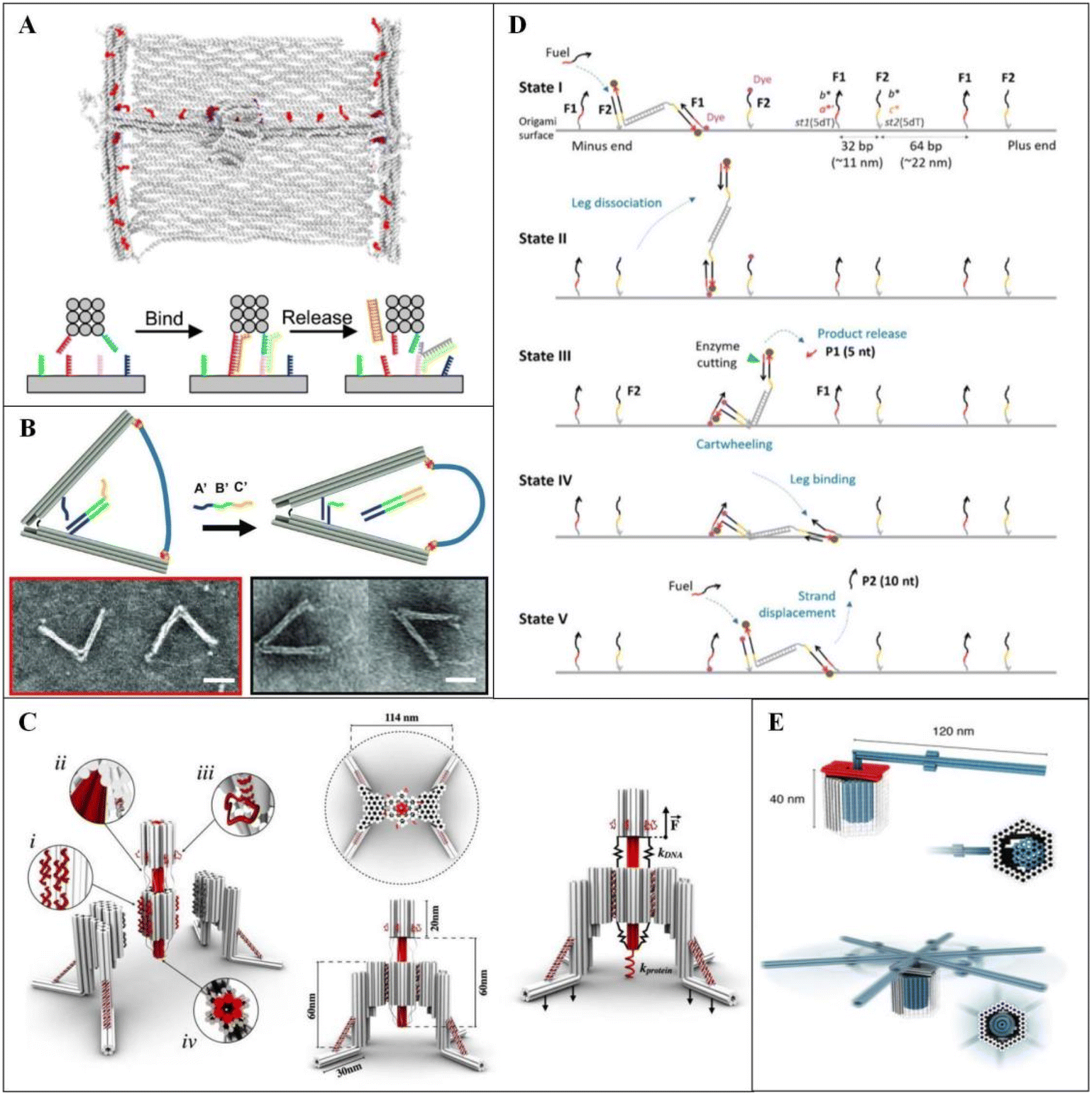 | ||
| Fig. 10 Design of various DNA origami machines. [A] Schematic of the assembled 2D printed nanorobot and the controlled 2D printing process. Reproduced from ref. 145 with permission from AAAS, copyright 2022. [B] Schematic diagram of the working mechanism of an nDFS. Scale bars: 50 nm. Reproduced from ref. 149 with permission from Oxford University Press, copyright 2021. [C] A nano-winch assembled from honeycomb lattice DNA origami. Reproduced from ref. 150 with permission from Springer Nature, copyright 2022. [D] Schematic diagram of DNA bipedal motor operation. Reproduced from ref. 155 with permission from AAAS, copyright 2023. [E] A rotary DNA nanodevice composed of precisely interlocking 3D DNA components. Reproduced from ref. 156 with permission from AAAS, copyright 2016. | ||
Integrating DNA origami platforms with molecular motors enables the creation of robots with virtually unlimited shapes and extensive functionalization capabilities. These nanomechanical systems perform various functions, including assembly,151 sorting,152 environmental response,153 and directional transport154 at the microscopic scale. However, to achieve fully autonomous and sustainable directional manipulation beyond externally driven motions, DNA-based autonomous molecular motors are necessary. Addressing this challenge, Siti et al. introduced a DNA molecular motor that drives a DNA walker along an origami surface, executing hybrid translational-rotational movements with high directional fidelity and remarkable speed (Fig. 10D).155 Additionally, Ketterer et al. used multilayer DNA origami self-assembly to design a rotational mechanism that mimics biological rotating motors, undergoing conformational transitions without requiring an external energy source.156 This mechanism enables random walks on docking points (Fig. 10E).
Zhou et al. created a DNA origami cube corner with DNA sticky-end edges, functioning as a robot controllable via ultraviolet light and temperature.157 By modifying each face with AuNRs, the robot exhibited chiral and optical activity. Furthermore, it could self-assemble into complete cubes through a two-by-two assembly process and replicate itself within two cycles, enabling exponential growth in yield.
Wang and collaborators developed a DNA origami robotic switch that responds to the tumor microenvironment.148 This switch autonomously displays cytotoxic ligand patterns that induce apoptosis in cancer cells within the acidic tumor environment while remaining inactive under normal physiological conditions. It demonstrated effective apoptosis induction in breast cancer cells in vitro and significantly inhibited tumor growth in a mouse model, presenting a promising strategy for precise tumor-targeted therapy.
Kopperger et al. reported an electrically driven nanoscale robotic arm, constructed via DNA self-assembly, that can reposition itself on a platform with millisecond precision.158 This system enables rapid, electric field-driven transport of molecules or nanoparticles within milliseconds. Compared with conventional DNA hybridization reaction-driven systems, the electric field drive significantly enhances speed and efficiency, offering promising applications in molecular synthesis, assembly, photonics, and plasmonic process control.
3.5 Interdisciplinary with computer and information
Over the past few decades, computing technologies have evolved from specialized systems to general-purpose architectures, encompassing silicon-based computers, carbon nanotube-based computers, and quantum computers. The key to achieving general-purpose computing lies in programmability and scalability.159 DNA-integrated circuits (DICs) are liquid-phase biocomputing systems that leverage biomolecular interactions to perform computations based on neuromorphic structures at the molecular level. These circuits have achieved breakthroughs in applications such as machine learning,160 logic circuits,161 decision-making machines162 and neural networks.163 Due to their programmability, immense parallel computing power, and compatibility with biological systems, DICs hold significant potential for broader applications and further generalization.DNA-integrated circuits based on machine learning can accurately classify complex biological data, such as genomic, transcriptomic, and proteomic information, significantly improving the efficiency and effectiveness of medical diagnoses. This advancement has greatly enhanced the early detection of different cancer subtypes and stages.164 Yang et al. accelerated cancer diagnosis by utilizing spatially constrained DNA origami frames (Fig. 11A). This approach prevents mutual interference between probes in different regions, reducing diagnosis time to as little as three hours—much faster than traditional diffusion-based systems.165
 | ||
| Fig. 11 [A] Schematic of the classification process of cancer diagnosis by utilizing miRNAs as input signals. Reproduced from ref. 165 with permission from Springer Nature, copyright 2024. [B] Robot emulates several logic gates in living animals. Reproduced from ref. 166 with permission from Springer Nature, copyright 2014. [C] Schematic diagram of the DOC security protocol. Reproduced from ref. 167 with permission from Springer Nature, copyright 2019. | ||
Fig. 11B illustrates dynamically interacting nanoscale robots constructed using DNA origami. These robots can switch states in response to specific protein cues, undergoing significant conformational changes to reveal internal payloads when gate chains are separated. By incorporating “positive regulators” (P) and “negative regulators” (N) as DNA keys, these robots can achieve mutual activation. Various configurations, designed by combining P and N robots with effector (E) robots in specific ratios, enable the simulation of a wide range of logic gate operations, including AND, OR, XOR, NAND, NOT, CNOT, and half-adders.166 This innovative biocomputing platform is particularly suited for the controlled delivery of therapeutic molecules within living organisms, paving the way for computational regulation of human therapeutic processes and other biological functions.
With the growing importance of information security, the advent of quantum computers presents new challenges to cryptographic systems. DNA origami offers unique structural potential, expanding encryption protocols beyond traditional computational problems like factorization to include spatial structure folding. In 2019, Zhang et al. introduced a DNA origami cryptography (DOC) technique that utilizes self-assembled nanoscale Braille-like patterns for secure communication.167 This method generates over 700-bit encryption keys and enhances confidentiality through protein-bound steganography while ensuring message integrity and differential access control. The system supports a diverse range of data formats, including text, music notation, and images, demonstrating its versatility for secure communication across multiple mediums (Fig. 11C). In 2020, Fan et al. proposed an information encoding strategy based on reconfigurable DNA origami domino arrays.168 This method enables conformational shifts within the arrays upon the introduction of a specific key, achieved through dynamic DNA units that rearrange intrinsic patterns for steganographic encryption while preserving overall array structure. Additionally, the strategy integrates encryption algorithms such as ASCII encoding to enhance security. Furthermore, a cryptographic anti-counterfeiting method, utilizing conformational transition-mediated chain substitution reactions, was introduced to ensure the complexity and unforgeability of encoded information. In 2024, Jiang et al. developed a subtraction-based encryption technique for DNA origami, where structural defects within the DNA origami framework are intentionally designed to store encrypted information.169 Specific “hook” strands selectively remove target strands from a pool of intact staple strands, creating encrypted defective sites. These sites are then biotinylated with intact biotin molecules and incubated with a pool of biotinylated strands to form an encoded biotin pattern. Decryption is achieved through atomic force microscopy, which identifies the biotin markers. This approach demonstrates significant potential in biomolecular encryption and ensures a high level of message recovery accuracy.
4 Conclusions and future perspectives
Since the invention of DNA origami technology in 2006, researchers have explored this innovative approach to nanomaterial construction in diverse ways. Due to its high assembly precision, a wide variety of origami structures have been created for practical applications, establishing DNA origami as a key player across multiple fields. The phenomenon of interdisciplinary communication and integration occurs as DNA origami nanostructures serve as a bridge between numerous disciplines.144 This includes not only the fields mentioned above but also emerging domains. Static and dynamic structures created with DNA origami have inspired advancements in photolithography and optical engineering, while DNA biocomputing systems have proved highly efficient for biochemical testing. Additionally, the fusion of DNA crystal frameworks with colloidal chemistry has led to the development of complex, porous structures.Despite significant progress in creating DNA assemblies with programmable functionality, several challenges remain. Although DNA molecules are inherently biocompatible, the biotoxicity and potential antigenicity of DNA origami-based nano-assemblies must be carefully considered. The scaffold, derived from phage amplification, can retain lipopolysaccharide endotoxins that may trigger immunostimulatory effects in mammalian organisms. Furthermore, residual toxic chemicals such as agarose gel and ethidium bromide from the purification process must be addressed before DNA origami can be utilized as a drug delivery platform. When employed as a drug carrier, DNA origami faces challenges such as short blood residence time and limited ability to accumulate in target tissues due to elimination by cells and organs. In the realm of large-scale production and widespread application, DNA origami encounters challenges related to raw material costs, stringent environmental requirements, complex multi-step processes, and low structural yield.170 Advances in biotechnology have reduced the cost of long-strand scaffolds to approximately $200 per gram and short-strand staples to under $300 per gram, making laboratory-scale preparation more feasible.171,172 However, there remains a significant gap between this and the cost required for commercial production. Additionally, the stable existence of DNA origami structures depends on buffer salt solutions containing specific cations (e.g., magnesium and sodium ions)101 and maintaining a certain pH range.173 When the solution deviates from these conditions, DNA origami structures may undergo unwanted hydrolysis, deformation, or denaturation.174,175 Moreover, unstable ionic interactions and competition from secondary ions can disrupt the solution state of DNA origami monomers, leading to disordered aggregation and precipitation.176–178 To address the technical challenges surrounding DNA structure strength and stability, various methods have been proposed, including silicification, metallization, protein cage encapsulation,179 polyethylene glycolated oligo-lysine encapsulation,180 co-assembly of protein polymers,181 and UV spot welding for internal cross-linking.182 These solutions could play a critical role in expanding the application of DNA origami nanostructures in the future.
In the coming decades, researchers are expected to push DNA origami technology into even more fields of study. For example, DNA origami could be constructed on a chip to integrate dynamic arrays of Au NPs, enabling rapid prototyping and the creation of nano-factories where individual components function similarly to living cells. DNA–NP conjugates can be assembled into 2D/3D patterns, liquid nanohydrogels, dynamic containers, bionic structures, and even avant-garde artwork, providing a new canvas for those with both scientific expertise and artistic creativity. The flexible joints, abundant binding sites, and negatively charged phosphate backbone of DNA nano-assemblies offer natural advantages for their use as nanoscale carriers. These carriers are capable of effectively delivering aptamers (e.g., AS1411, MUC1, HApt),183,184 antitumor drugs (e.g., DOX, PTX, 5-FU),185 immune factors (e.g., CpG),186 and fluorescent markers (e.g., FAM), making them advanced nanotools for biosensing, bioimaging, and logic gate operations. As DNA origami technology merges with other advanced processing methods, nanostructures will increasingly influence macroscopic systems, enabling the creation of intricate constructs that were once deemed unfeasible. Furthermore, due to its inherent flexibility, DNA origami is expected to play a pivotal role in the development of innovative smart wearable devices. In summary, since its invention, DNA origami technology has served as an ideal tool for fostering interdisciplinary connections. Through the integration of diverse concepts, theories, and methods, it has opened many previously unimaginable application pathways in DNA nanotechnology. We believe that, as interdisciplinary collaboration continues to deepen, DNA origami technology will lead to innovative applications across even more fields in the future.
Data availability
No primary research results, software or code have been included and no new data were generated or analysed as part of this review.Conflicts of interest
The authors declare no conflict of interest.Acknowledgements
This work is supported by the Fundamental Research Funds for the Central Universities (2024300319) and by the Program B for Outstanding PhD Candidates of Nanjing University (202402B15). The ToC was generated with permission from the American Chemical Society (copyright 2017, 2019, and 2021), The American Association for the Advancement of Science (copyright 2018), and John Wiley and Sons (copyright 2020).References
- N. C. Seeman, J. Theor. Biol., 1982, 99, 237–247 CAS
.
- A. Shaw, I. T. Hoffecker, I. Smyrlaki, J. Rosa, A. Grevys, D. Bratlie, I. Sandlie, T. E. Michaelsen, J. T. Andersen and B. Högberg, Nat. Nanotechnol., 2019, 14, 184–190 CAS
- S. Li, Q. Jiang, S. Liu, Y. Zhang, Y. Tian, C. Song, J. Wang, Y. Zou, G. J. Anderson, J.-Y. Han, Y. Chang, Y. Liu, C. Zhang, L. Chen, G. Zhou, G. Nie, H. Yan, B. Ding and Y. Zhao, Nat. Biotechnol., 2018, 36, 258–264 CAS
- F. Lu, H. Xin, W. Xia, M. Liu, Y. Zhang, W. Cai and O. Gang, ACS Cent. Sci., 2018, 4, 1742–1750 CAS
- Y. Chen, G. Ke, Y. Ma, Z. Zhu, M. Liu, Y. Liu, H. Yan and C. J. Yang, J. Am. Chem. Soc., 2018, 140, 8990–8996 CAS
- J. Wang, L. Yue, Z. Li, J. Zhang, H. Tian and I. Willner, Nat. Commun., 2019, 10, 4963 Search PubMed
- M. Wang, L. Dai, J. Duan, Z. Ding, P. Wang, Z. Li, H. Xing and Y. Tian, Angew. Chem., Int. Ed., 2020, 59, 6389–6396 CAS
- P. W. K. Rothemund, Nature, 2006, 440, 297–302 CAS
- M. Madsen and K. V. Gothelf, Chem. Rev., 2019, 119, 6384–6458 CAS
- Y. Wang, X. Yan, Z. Zhou, N. Ma and Y. Tian, Angew. Chem., Int. Ed., 2022, 61, e202208290 CAS
- X. Yan, Y. Wang, N. Ma, Y. Yu, L. Dai and Y. Tian, J. Am. Chem. Soc., 2023, 145, 3978–3986 CAS
- L. Dai, X. Hu, M. Ji, N. Ma, H. Xing, J.-J. Zhu, Q. Min and Y. Tian, Proc. Natl. Acad. Sci. U. S. A., 2023, 120, e2302142120 CAS
- L. Qian, Y. Wang, Z. Zhang, J. Zhao, D. Pan, Y. Zhang, Q. Liu, C. Fan, J. Hu and L. He, Chin. Sci. Bull., 2006, 51, 2973–2976 CAS
- E. S. Andersen, M. Dong, M. M. Nielsen, K. Jahn, A. Lind-Thomsen, W. Mamdouh, K. V. Gothelf, F. Besenbacher and J. Kjems, ACS Nano, 2008, 2, 1213–1218 CAS
- T. Gerling, K. F. Wagenbauer, A. M. Neuner and H. Dietz, Science, 2015, 347, 1446–1452 CAS
- E. Benson, A. Mohammed, J. Gardell, S. Masich, E. Czeizler, P. Orponen and B. Högberg, Nature, 2015, 523, 441–444 CAS
- F. Zhang, S. Jiang, S. Wu, Y. Li, C. Mao, Y. Liu and H. Yan, Nat. Nanotechnol., 2015, 10, 779–784 CAS
- R. M. Zadegan, M. D. E. Jepsen, K. E. Thomsen, A. H. Okholm, D. H. Schaffert, E. S. Andersen, V. Birkedal and J. Kjems, ACS Nano, 2012, 6, 10050–10053 CrossRef CAS PubMed
- D. Han, X. Qi, C. Myhrvold, B. Wang, M. Dai, S. Jiang, M. Bates, Y. Liu, B. An, F. Zhang, H. Yan and P. Yin, Science, 2017, 358, eaao2648 CrossRef PubMed
- H. Jun, F. Zhang, T. Shepherd, S. Ratanalert, X. Qi, H. Yan and M. Bathe, Sci. Adv., 2019, 5, eaav0655 Search PubMed
- X. Wang, S. Li, H. Jun, T. John, K. Zhang, H. Fowler, J. P. K. Doye, W. Chiu and M. Bathe, Sci. Adv., 2022, 8, eabn0039 CrossRef CAS PubMed
- S. M. Douglas, H. Dietz, T. Liedl, B. Högberg, F. Graf and W. M. Shih, Nature, 2009, 459, 414–418 CAS
- S. M. Douglas, A. H. Marblestone, S. Teerapittayanon, A. Vazquez, G. M. Church and W. M. Shih, Nucleic Acids Res., 2009, 37, 5001–5006 CAS
- H. Dietz, S. M. Douglas and W. M. Shih, Science, 2009, 325, 725–730 CAS
- D. Han, S. Pal, J. Nangreave, Z. Deng, Y. Liu and H. Yan, Science, 2011, 332, 342–346 CAS
- Y. Yang, D. Han, J. Nangreave, Y. Liu and H. Yan, ACS Nano, 2012, 6, 8209–8215 CAS
- Z. Zhao, Y. Liu and H. Yan, Nano Lett., 2011, 11, 2997–3002 CAS
- A. N. Marchi, I. Saaem, B. N. Vogen, S. Brown and T. H. LaBean, Nano Lett., 2014, 14, 5740–5747 CAS
- E. Pound, J. R. Ashton, H. A. Becerril and A. T. Woolley, Nano Lett., 2009, 9, 4302–4305 CAS
- H. Zhang, J. Chao, D. Pan, H. Liu, Q. Huang and C. Fan, Chem. Commun., 2012, 48, 6405–6407 CAS
- X. Chen, Q. Wang, J. Peng, Q. Long, H. Yu and Z. Li, ACS Appl. Mater. Interfaces, 2018, 10, 24344–24348 CAS
- W. Liu, H. Zhong, R. Wang and N. C. Seeman, Angew. Chem., Int. Ed., 2011, 50, 264–267 CAS
- T. Zhang, C. Hartl, K. Frank, A. Heuer-Jungemann, S. Fischer, P. C. Nickels, B. Nickel and T. Liedl, Adv. Mater., 2018, 30, 1800273 Search PubMed
- X. Wang, H. Jun and M. Bathe, J. Am. Chem. Soc., 2022, 144, 4403–4409 CAS
- P. Wang, S. Gaitanaros, S. Lee, M. Bathe, W. M. Shih and Y. Ke, J. Am. Chem. Soc., 2016, 138, 7733–7740 CAS
- Y. Liu, Z. Dai, X. Xie, B. Li, S. Jia, Q. Li, M. Li, C. Fan and X. Liu, J. Am. Chem. Soc., 2024, 146, 5461–5469 CAS
- Y. Tian, J. R. Lhermitte, L. Bai, T. Vo, H. L. Xin, H. Li, R. Li, M. Fukuto, K. G. Yager, J. S. Kahn, Y. Xiong, B. Minevich, S. K. Kumar and O. Gang, Nat. Mater., 2020, 19, 789–796 CAS
- M. Ji, Z. Zhou, W. Cao, N. Ma, W. Xu and Y. Tian, Sci. Adv., 2022, 8, eadc9755 CAS
- Z. Lin, H. Emamy, B. Minevich, Y. Xiong, S. Xiang, S. Kumar, Y. Ke and O. Gang, J. Am. Chem. Soc., 2020, 142, 17531–17542 CrossRef CAS PubMed
- T. Bayrak, S. Helmi, J. Ye, D. Kauert, J. Kelling, T. Schönherr, R. Weichelt, A. Erbe and R. Seidel, Nano Lett., 2018, 18, 2116–2123 CrossRef CAS PubMed
- J. F. Berengut, J. C. Berengut, J. P. K. Doye, D. Prešern, A. Kawamoto, J. Ruan, M. J. Wainwright and L. K. Lee, Nucleic Acids Res., 2019, 47, 11963–11975 CAS
- S. F. J. Wickham, A. Auer, J. Min, N. Ponnuswamy, J. B. Woehrstein, F. Schueder, M. T. Strauss, J. Schnitzbauer, B. Nathwani, Z. Zhao, S. D. Perrault, J. Hahn, S. Lee, M. M. Bastings, S. W. Helmig, A. L. Kodal, P. Yin, R. Jungmann and W. M. Shih, Nat. Commun., 2020, 11, 5768 CrossRef CAS PubMed
- Y. Wang, L. Dai, Z. Ding, M. Ji, J. Liu, H. Xing, X. Liu, Y. Ke, C. Fan, P. Wang and Y. Tian, Nat. Commun., 2021, 12, 3011 CrossRef PubMed
- G. Yao, F. Zhang, F. Wang, T. Peng, H. Liu, E. Poppleton, P. Šulc, S. Jiang, L. Liu, C. Gong, X. Jing, X. Liu, L. Wang, Y. Liu, C. Fan and H. Yan, Nat. Chem., 2020, 12, 1067–1075 CAS
- A. Aghebat Rafat, T. Pirzer, M. B. Scheible, A. Kostina and F. C. Simmel, Angew. Chem., Int. Ed., 2014, 53, 7665–7668 CrossRef CAS PubMed
- S. Woo and P. W. K. Rothemund, Nat. Commun., 2014, 5, 4889 CrossRef CAS PubMed
- Y. Yonamine, K. Cervantes-Salguero, K. Minami, I. Kawamata, W. Nakanishi, J. P. Hill, S. Murata and K. Ariga, Phys. Chem. Chem. Phys., 2016, 18, 12576–12581 RSC
- Y. Suzuki, M. Endo and H. Sugiyama, Nat. Commun., 2015, 6, 8052 CrossRef CAS PubMed
- Y. Xin, B. Shen, M. A. Kostiainen, G. Grundmeier, M. Castro, V. Linko and A. Keller, Chem. – Eur. J., 2021, 27, 8564–8571 CrossRef CAS PubMed
- S. Ramakrishnan, S. Subramaniam, A. F. Stewart, G. Grundmeier and A. Keller, ACS Appl. Mater. Interfaces, 2016, 8, 31239–31247 CrossRef CAS PubMed
- G. Bellot, M. A. McClintock, C. Lin and W. M. Shih, Nat. Methods, 2011, 8, 192–194 CrossRef CAS PubMed
- E. Stahl, T. G. Martin, F. Praetorius and H. Dietz, Angew. Chem., Int. Ed., 2014, 53, 12735–12740 CrossRef CAS PubMed
- A. Shaw, E. Benson and B. Högberg, ACS Nano, 2015, 9, 4968–4975 CrossRef CAS PubMed
- A. Buchberger, C. R. Simmons, N. E. Fahmi, R. Freeman and N. Stephanopoulos, J. Am. Chem. Soc., 2020, 142, 1406–1416 CrossRef CAS PubMed
- J. Ye, J. Teske, U. Kemper and R. Seidel, Small, 2021, 17, e2007218 CrossRef PubMed
- M. K. Masukawa, Y. Sato, F. Yu, K. Tsumoto, K. Yoshikawa and M. Takinoue, ChemBioChem, 2022, 23, e202200240 CrossRef CAS PubMed
- C. Jin, L. Han and S. Che, Angew. Chem., Int. Ed., 2009, 48, 9268–9272 Search PubMed
- S. Che, A. E. Garcia-Bennett, T. Yokoi, K. Sakamoto, H. Kunieda, O. Terasaki and T. Tatsumi, Nat. Mater., 2003, 2, 801–805 CrossRef CAS PubMed
- E. Auyeung, R. J. Macfarlane, C. H. J. Choi, J. I. Cutler and C. A. Mirkin, Adv. Mater., 2012, 24, 5181–5186 CrossRef CAS PubMed
- B. Liu, L. Han and S. Che, Angew. Chem., Int. Ed., 2012, 51, 923–927 CrossRef CAS PubMed
- B. Liu, Y. Yao and S. Che, Angew. Chem., Int. Ed., 2013, 52, 14186–14190 CrossRef CAS PubMed
- Y. Cao, K. Kao, C. Mou, L. Han and S. Che, Angew. Chem., Int. Ed., 2016, 55, 2037–2041 CrossRef CAS PubMed
- X. Liu, F. Zhang, X. Jing, M. Pan, P. Liu, W. Li, B. Zhu, J. Li, H. Chen, L. Wang, J. Lin, Y. Liu, D. Zhao, H. Yan and C. Fan, Nature, 2018, 559, 593–598 CrossRef CAS PubMed
- L. Nguyen, M. Döblinger, T. Liedl and A. Heuer-Jungemann, Angew. Chem., Int. Ed., 2019, 58, 912–916 CrossRef CAS PubMed
- X. Jing, F. Zhang, M. Pan, X. Dai, J. Li, L. Wang, X. Liu, H. Yan and C. Fan, Nat. Protoc., 2019, 14, 2416–2436 CrossRef CAS PubMed
- Y. Shang, N. Li, S. Liu, L. Wang, Z.-G. Wang, Z. Zhang and B. Ding, Adv. Mater., 2020, 32, 2000294 Search PubMed
- L. M. Wassermann, M. Scheckenbach, A. V. Baptist, V. Glembockyte and A. Heuer-Jungemann, Adv. Mater., 2023, 35, 2212024 Search PubMed
- S. Wang, P.-A. Lin, M. DeLuca, S. Zauscher, G. Arya and Y. Ke, J. Am. Chem. Soc., 2024, 146, 358–367 CrossRef CAS PubMed
- H. Wang, Z. Li, X. Liu, S. Jia, Y. Gao and M. Li, ACS Appl. Bio Mater., 2024, 7, 2511–2518 CrossRef CAS PubMed
- S. K. Vittala and D. Han, ACS Appl. Bio Mater., 2020, 3, 2702–2722 CrossRef CAS PubMed
- B. R. Aryal, T. R. Westover, D. R. Ranasinghe, D. G. Calvopiña, B. Uprety, J. N. Harb, R. C. Davis and A. T. Woolley, Langmuir, 2018, 34, 15069–15077 CrossRef CAS PubMed
- S. K. Vittala, Y. Zhao and D. Han, ChemPlusChem, 2022, 87, e202100478 CAS
- J. Richter, M. Mertig, W. Pompe, I. Mönch and H. K. Schackert, Appl. Phys. Lett., 2001, 78, 536–538 Search PubMed
- R. Seidel, L. Colombi Ciacchi, M. Weigel, W. Pompe and M. Mertig, J. Phys. Chem. B, 2004, 108, 10801–10811 CAS
- C. F. Monson and A. T. Woolley, Nano Lett., 2003, 3, 359–363 CAS
- J. Liu, B. Uprety, S. Gyawali, A. T. Woolley, N. V. Myung and J. N. Harb, Langmuir, 2013, 29, 11176–11184 CAS
- Q. Gu and D. T. Haynie, Mater. Lett., 2008, 62, 3047–3050 Search PubMed
- W. Sun, E. Boulais, Y. Hakobyan, W. L. Wang, A. Guan, M. Bathe and P. Yin, Science, 2014, 346, 1258361 Search PubMed
- T. Bayrak, S. Helmi, J. Ye, D. Kauert, J. Kelling, T. Schönherr, R. Weichelt, A. Erbe and R. Seidel, Nano Lett., 2018, 18, 2116–2123 CAS
- S. Helmi, C. Ziegler, D. J. Kauert and R. Seidel, Nano Lett., 2014, 14, 6693–6698 CAS
- S. Jia, J. Wang, M. Xie, J. Sun, H. Liu, Y. Zhang, J. Chao, J. Li, L. Wang, J. Lin, K. V. Gothelf and C. Fan, Nat. Commun., 2019, 10, 5597 CAS
- N. Li, Y. Shang, R. Xu, Q. Jiang, J. Liu, L. Wang, Z. Cheng and B. Ding, J. Am. Chem. Soc., 2019, 141, 17968–17972 CAS
- Y. Zhang, Z. Qu, C. Jiang, Y. Liu, R. Pradeep Narayanan, D. Williams, X. Zuo, L. Wang, H. Yan, H. Liu and C. Fan, J. Am. Chem. Soc., 2021, 143, 8639–8646 CAS
- Y. Chen, F. Fang and N. Zhang, npj 2D Mater. Appl., 2024, 8, 1–23 Search PubMed
- Z. Cai, B. Liu, X. Zou and H.-M. Cheng, Chem. Rev., 2018, 118, 6091–6133 CAS
- X. Gao, L. Zheng, F. Luo, J. Qian, J. Wang, M. Yan, W. Wang, Q. Wu, J. Tang, Y. Cao, C. Tan, J. Tang, M. Zhu, Y. Wang, Y. Li, L. Sun, G. Gao, J. Yin, L. Lin, Z. Liu, S. Qin and H. Peng, Nat. Commun., 2022, 13, 5410 CAS
- S. M. Kim, A. Hsu, M. H. Park, S. H. Chae, S. J. Yun, J. S. Lee, D.-H. Cho, W. Fang, C. Lee, T. Palacios, M. Dresselhaus, K. K. Kim, Y. H. Lee and J. Kong, Nat. Commun., 2015, 6, 8662 CAS
- S. P. Surwade, F. Zhou, B. Wei, W. Sun, A. Powell, C. O'Donnell, P. Yin and H. Liu, J. Am. Chem. Soc., 2013, 135, 6778–6781 CAS
- J. E. Crowell, J. Vac. Sci. Technol., A, 2003, 21, S88–S95 CAS
- G. N. Parsons and R. D. Clark, Chem. Mater., 2020, 32, 4920–4953 CAS
- R. W. Johnson, A. Hultqvist and S. F. Bent, Mater. Today, 2014, 17, 236–246 CAS
- S. M. George, Chem. Rev., 2010, 110, 111–131 CrossRef CAS PubMed
- G. Posnjak, X. Yin, P. Butler, O. Bienek, M. Dass, S. Lee, I. D. Sharp and T. Liedl, Science, 2024, 384, 781–785 CrossRef CAS PubMed
- L. Hui, C. Chen, M. A. Kim and H. Liu, ACS Appl. Mater. Interfaces, 2022, 14, 16538–16545 CrossRef CAS PubMed
- S. P. Surwade, S. Zhao and H. Liu, J. Am. Chem. Soc., 2011, 133, 11868–11871 CrossRef CAS PubMed
- C. T. Diagne, C. Brun, D. Gasparutto, X. Baillin and R. Tiron, ACS Nano, 2016, 10, 6458–6463 CrossRef CAS PubMed
- J. Liu, Y. Geng, E. Pound, S. Gyawali, J. R. Ashton, J. Hickey, A. T. Woolley and J. N. Harb, ACS Nano, 2011, 5, 2240–2247 CrossRef CAS PubMed
- X. Dai, X. Chen, X. Jing, Y. Zhang, M. Pan, M. Li, Q. Li, P. Liu, C. Fan and X. Liu, Angew. Chem., Int. Ed., 2022, 61, e202114190 CrossRef CAS PubMed
- J. Shen, W. Sun, D. Liu, T. Schaus and P. Yin, Nat. Mater., 2021, 20, 683–690 CrossRef CAS PubMed
- R. L. Siegel, K. D. Miller, N. S. Wagle and A. Jemal, Ca-Cancer J. Clin., 2023, 73, 17–48 CrossRef PubMed
- C. E. Castro, F. Kilchherr, D.-N. Kim, E. L. Shiao, T. Wauer, P. Wortmann, M. Bathe and H. Dietz, Nat. Methods, 2011, 8, 221–229 CrossRef CAS PubMed
- D. Ye, X. Zuo and C. Fan, Annu. Rev. Anal. Chem., 2018, 11, 171–195 CrossRef CAS PubMed
- J. Dai, C. Xing, Y. Lin, Y. Huang, Y. Yang, Z. Chen, C. Lu and H. Yang, Sens. Actuators, B, 2021, 344, 130195 CrossRef CAS
- Y. Ke, S. Lindsay, Y. Chang, Y. Liu and H. Yan, Science, 2008, 319, 180–183 CrossRef CAS PubMed
- E. Weinhold and B. Chakraborty, Nanoscale, 2021, 13, 2465–2471 RSC
- M. Scherf, F. Scheffler, C. Maffeo, U. Kemper, J. Ye, A. Aksimentiev, R. Seidel and U. Reibetanz, Nanoscale, 2022, 14, 18041–18050 RSC
- M. J. Urban, P. K. Dutta, P. Wang, X. Duan, X. Shen, B. Ding, Y. Ke and N. Liu, J. Am. Chem. Soc., 2016, 138, 5495–5498 CrossRef CAS PubMed
- C. Wen, E. Bertosin, X. Shi, C. Dekker and S. Schmid, Nano Lett., 2023, 23, 788–794 CAS
- Y. Choi, C. Schmidt, P. Tinnefeld, I. Bald and S. Rödiger, Sci. Rep., 2019, 9, 4769 Search PubMed
- J. J. Rossi, Cell, 2009, 137, 990–992 CAS
- C. Xing, S. Chen, Q. Lin, Y. Lin, M. Wang, J. Wang and C. Lu, Nanoscale, 2022, 14, 1327–1332 CAS
- V. Maingi, Z. Zhang, C. Thachuk, N. Sarraf, E. R. Chapman and P. W. K. Rothemund, Nat. Commun., 2023, 14, 1532 CAS
- W. Livernois, P. (Simon) Cao, S. Saha, Q. Ding, A. Gopinath and M. P. Anantram, Nanotechnology, 2024, 35, 325202 CAS
- M. Tintoré, I. Gállego, B. Manning, R. Eritja and C. Fàbrega, Angew. Chem., Int. Ed., 2013, 52, 7747–7750 Search PubMed
- J. A. Kretzmann, A. Liedl, A. Monferrer, V. Mykhailiuk, S. Beerkens and H. Dietz, Nat. Commun., 2023, 14, 1017 CAS
- J. Wang, Y. Li and G. Nie, Nat. Rev. Mater., 2021, 6, 766–783 CAS
- T. Wu, H. Wang, R. Tian, S. Guo, Y. Liao, J. Liu and B. Ding, Angew. Chem., Int. Ed., 2023, 62, e202311698 CAS
- W. Engelen, C. Sigl, K. Kadletz, E. M. Willner and H. Dietz, J. Am. Chem. Soc., 2021, 143, 21630–21636 CAS
- A. Comberlato, M. M. Koga, S. Nüssing, I. A. Parish and M. M. C. Bastings, Nano Lett., 2022, 22, 2506–2513 CAS
- J. Liu, B. Ding, X. Wu, C. Yang, H. Wang, X. Lu, Y. Shang, Q. Liu and J. Fan, J. Am. Chem. Soc., 2023, 145, 9343–9353 Search PubMed
- T. Masubuchi, M. Endo, R. Iizuka, A. Iguchi, D. H. Yoon, T. Sekiguchi, H. Qi, R. Iinuma, Y. Miyazono, S. Shoji, T. Funatsu, H. Sugiyama, Y. Harada, T. Ueda and H. Tadakuma, Nat. Nanotechnol., 2018, 13, 933–940 CAS
- J. Hahn, L. Y. T. Chou, R. S. Sørensen, R. M. Guerra and W. M. Shih, ACS Nano, 2020, 14, 1550–1559 CAS
- S. Julin, Nonappa, B. Shen, V. Linko and M. A. Kostiainen, Angew. Chem., Int. Ed., 2021, 60, 827–833 CAS
- S. Liu, Q. Jiang, X. Zhao, R. Zhao, Y. Wang, Y. Wang, J. Liu, Y. Shang, S. Zhao, T. Wu, Y. Zhang, G. Nie and B. Ding, Nat. Mater., 2021, 20, 421–430 CrossRef CAS PubMed
- Q. Zhang, Q. Jiang, N. Li, L. Dai, Q. Liu, L. Song, J. Wang, Y. Li, J. Tian, B. Ding and Y. Du, ACS Nano, 2014, 8, 6633–6643 CrossRef CAS PubMed
- Q. Jiang, Y. Shang, Y. Xie and B. Ding, Adv. Mater., 2024, 36, e2301035 CrossRef PubMed
- H. Ijäs, I. Hakaste, B. Shen, M. A. Kostiainen and V. Linko, ACS Nano, 2019, 13, 5959–5967 CrossRef PubMed
- S. M. Douglas, I. Bachelet and G. M. Church, Science, 2012, 335, 831–834 CrossRef CAS PubMed
- L. Li, J. Yin, W. Ma, L. Tang, J. Zou, L. Yang, T. Du, Y. Zhao, L. Wang, Z. Yang, C. Fan, J. Chao and X. Chen, Nat. Mater., 2024, 23, 993–1001 CrossRef CAS PubMed
- L. Xin, C. Zhou, X. Duan and N. Liu, Nat. Commun., 2019, 10, 5394 CrossRef PubMed
- W. Fang, S. Jia, J. Chao, L. Wang, X. Duan, H. Liu, Q. Li, X. Zuo, L. Wang, L. Wang, N. Liu and C. Fan, Sci. Adv., 2019, 5, eaau4506 CrossRef PubMed
- K. Tapio, A. Mostafa, Y. Kanehira, A. Suma, A. Dutta and I. Bald, ACS Nano, 2021, 15, 7065–7077 CrossRef CAS PubMed
- Y. Kanehira, K. Tapio, G. Wegner, S. Kogikoski Jr., S. Rüstig, C. Prietzel, K. Busch and I. Bald, ACS Nano, 2023, 17, 21227–21239 Search PubMed
- S. Nam, N. Oh, Y. Zhai and M. Shim, ACS Nano, 2015, 9, 878–885 CrossRef CAS PubMed
- C. Chen, X. Wei, M. F. Parsons, J. Guo, J. L. Banal, Y. Zhao, M. N. Scott, G. S. Schlau-Cohen, R. Hernandez and M. Bathe, Nat. Commun., 2022, 13, 4935 CAS
- V. Glembockyte, L. Grabenhorst, K. Trofymchuk and P. Tinnefeld, Acc. Chem. Res., 2021, 54, 3338–3348 CrossRef CAS PubMed
- L. Grabenhorst, K. Trofymchuk, F. Steiner, V. Glembockyte and P. Tinnefeld, Methods Appl. Fluoresc., 2020, 8, 024003 CAS
- R. Yaadav, K. Trofymchuk, F. Gong, X. Ji, F. Steiner, P. Tinnefeld and Z. He, J. Phys. Chem. C, 2024, 128, 9154–9160 CAS
- O. S. Ojambati, R. Chikkaraddy, W. D. Deacon, M. Horton, D. Kos, V. A. Turek, U. F. Keyser and J. J. Baumberg, Nat. Commun., 2019, 10, 1049 Search PubMed
- C. Zhou, X. Duan and N. Liu, Acc. Chem. Res., 2017, 50, 2906–2914 CrossRef CAS PubMed
- K. Martens, F. Binkowski, L. Nguyen, L. Hu, A. O. Govorov, S. Burger and T. Liedl, Nat. Commun., 2021, 12, 2025 CrossRef CAS PubMed
- A. Gopinath, E. Miyazono, A. Faraon and P. W. K. Rothemund, Nature, 2016, 535, 401–405 CrossRef CAS PubMed
- A. Gopinath, C. Thachuk, A. Mitskovets, H. A. Atwater, D. Kirkpatrick and P. W. K. Rothemund, Science, 2021, 371, eabd6179 CAS
- S. Dey, C. Fan, K. V. Gothelf, J. Li, C. Lin, L. Liu, N. Liu, M. A. D. Nijenhuis, B. Saccà, F. C. Simmel, H. Yan and P. Zhan, Nat. Rev. Methods Primers, 2021, 1, 1–24 Search PubMed
- E. Benson, R. C. Marzo, J. Bath and A. J. Turberfield, Sci. Rob., 2022, 7, eabn5459 CrossRef PubMed
- X. R. Liu, I. Y. Loh, W. Siti, H. L. Too, T. Anderson and Z. Wang, Nanoscale Horiz., 2023, 8, 827–841 CAS
- J. List, M. Weber and F. C. Simmel, Angew. Chem., Int. Ed., 2014, 53, 4236–4239 Search PubMed
- Y. Wang, I. Baars, I. Berzina, I. Rocamonde-Lago, B. Shen, Y. Yang, M. Lolaico, J. Waldvogel, I. Smyrlaki, K. Zhu, R. A. Harris and B. Högberg, Nat. Nanotechnol., 2024, 19, 1366–1374 Search PubMed
- Y. Wang, J. V. Le, K. Crocker, M. A. Darcy, P. D. Halley, D. Zhao, N. Andrioff, C. Croy, M. G. Poirier, R. Bundschuh and C. E. Castro, Nucleic Acids Res., 2021, 49, 8987–8999 CrossRef CAS PubMed
- A. Mills, N. Aissaoui, D. Maurel, J. Elezgaray, F. Morvan, J. J. Vasseur, E. Margeat, R. B. Quast, J. Lai Kee-Him, N. Saint, C. Benistant, A. Nord, F. Pedaci and G. Bellot, Nat. Commun., 2022, 13, 3182 CAS
- H. Gu, J. Chao, S.-J. Xiao and N. C. Seeman, Nature, 2010, 465, 202–205 CAS
- A. J. Thubagere, W. Li, R. F. Johnson, Z. Chen, S. Doroudi, Y. L. Lee, G. Izatt, S. Wittman, N. Srinivas, D. Woods, E. Winfree and L. Qian, Science, 2017, 357, eaan6558 Search PubMed
- K. Lund, A. J. Manzo, N. Dabby, N. Michelotti, A. Johnson-Buck, J. Nangreave, S. Taylor, R. Pei, M. N. Stojanovic, N. G. Walter, E. Winfree and H. Yan, Nature, 2010, 465, 206–210 CAS
- C. Zhou, X. Duan and N. Liu, Nat. Commun., 2015, 6, 8102 CAS
- W. Siti, H. L. Too, T. Anderson, X. R. Liu, I. Y. Loh and Z. Wang, Sci. Adv., 2023, 9, eadi8444 CAS
- P. Ketterer, E. M. Willner and H. Dietz, Sci. Adv., 2016, 2, e1501209 Search PubMed
- F. Zhou, H. Ni, G. Zhu, L. Bershadsky, R. Sha, N. C. Seeman and P. M. Chaikin, Sci. Rob., 2023, 8, eadf1274 Search PubMed
- E. Kopperger, J. List, S. Madhira, F. Rothfischer, D. C. Lamb and F. C. Simmel, Science, 2018, 359, 296–301 CAS
- H. Lv, N. Xie, M. Li, M. Dong, C. Sun, Q. Zhang, L. Zhao, J. Li, X. Zuo, H. Chen, F. Wang and C. Fan, Nature, 2023, 622, 292–300 CAS
- C. Truong-Quoc, J. Y. Lee, K. S. Kim and D.-N. Kim, Nat. Mater., 2024, 1–9 Search PubMed
- Z. Tang, Z.-X. Yin, J.-Z. Cui, J. Yang and R.-S. Wang, J. Nanoelectron. Optoelectron., 2021, 16, 534–545 CAS
- Y. Liu, X. Hu, T. Fu, R. Wang and W. Tan, Sci. China: Chem., 2019, 62, 407–408 CAS
- C. Truong-Quoc, J. Y. Lee, K. S. Kim and D.-N. Kim, Nat. Mater., 2024, 23, 984–992 CAS
- M. C. Liu, G. R. Oxnard, E. A. Klein, C. Swanton, M. V. Seiden and CCGA Consortium, Ann. Oncol., 2020, 31, 745–759 CAS
- L. Yang, Q. Tang, M. Zhang, Y. Tian, X. Chen, R. Xu, Q. Ma, P. Guo, C. Zhang and D. Han, Nat. Commun., 2024, 15, 4583 CAS
- Y. Amir, E. Ben-Ishay, D. Levner, S. Ittah, A. Abu-Horowitz and I. Bachelet, Nat. Nanotechnol., 2014, 9, 353–357 CrossRef CAS PubMed
- Y. Zhang, F. Wang, J. Chao, M. Xie, H. Liu, M. Pan, E. Kopperger, X. Liu, Q. Li, J. Shi, L. Wang, J. Hu, L. Wang, F. C. Simmel and C. Fan, Nat. Commun., 2019, 10, 5469 CrossRef PubMed
- S. Fan, D. Wang, J. Cheng, Y. Liu, T. Luo, D. Cui, Y. Ke and J. Song, Angew. Chem., Int. Ed., 2020, 59, 12991–12997 CrossRef CAS PubMed
- C. Jiang, R. Tan, W. Li, Y. Zhang and H. Liu, Small, 2024, 20, 2406470 CrossRef CAS PubMed
- W. Jiang, J. Li, Z. Lin, J. Guo, J. Ma, Z. Wang, M. Zhang and Y. Wu, Small Struct., 2023, 4, 2200376 CrossRef CAS
- F. Praetorius, B. Kick, K. L. Behler, M. N. Honemann, D. Weuster-Botz and H. Dietz, Nature, 2017, 552, 84–87 CrossRef CAS PubMed
- Y. Jia, L. Chen, J. Liu, W. Li and H. Gu, Chem, 2021, 7, 959–981 CAS
- A. Bednarz, S. M. Sønderskov, M. Dong and V. Birkedal, Nanoscale, 2023, 15, 1317–1326 RSC
- S. Ramakrishnan, G. Krainer, G. Grundmeier, M. Schlierf and A. Keller, Small, 2017, 13, 1702100 CrossRef PubMed
- V. Linko and A. Keller, Small, 2023, 19, 2301935 CAS
- F. Matter, A. L. Luna and M. Niederberger, Nano Today, 2020, 30, 100827 CrossRef
- Y. Xin, S. Martinez Rivadeneira, G. Grundmeier, M. Castro and A. Keller, Nano Res., 2020, 13, 3142–3150 CrossRef CAS
- M. Hanke, N. Hansen, R. Chen, G. Grundmeier, K. Fahmy and A. Keller, Int. J. Mol. Sci., 2022, 23, 2817 CrossRef CAS PubMed
-
V. Linko, J. Mikkilä and M. A. Kostiainen, in Virus-Derived Nanoparticles for Advanced Technologies: Methods and Protocols, ed. C. Wege and G. P. Lomonossoff, Springer, New York, NY, 2018, pp. 267–277 Search PubMed
- F. M. Anastassacos, Z. Zhao, Y. Zeng and W. M. Shih, J. Am. Chem. Soc., 2020, 142, 3311–3315 CrossRef CAS PubMed
- N. A. Estrich, A. Hernandez-Garcia, R. de Vries and T. H. LaBean, ACS Nano, 2017, 11, 831–842 CrossRef CAS PubMed
- J. A. Kretzmann, A. Liedl, A. Monferrer, V. Mykhailiuk, S. Beerkens and H. Dietz, Nat. Commun., 2023, 14, 1017 CrossRef CAS PubMed
- O. Kovecses, F. E. Mercier and M. McKeague, Leukemia, 2024, 38, 1441–1454 CrossRef CAS PubMed
- B. R. Janakaloti Narayanareddy, N. R. Allipeta, J. Allard and S. P. Gross, Commun. Biol., 2024, 7, 1–10 Search PubMed
- B. Wang, S. Hu, Y. Teng, J. Chen, H. Wang, Y. Xu, K. Wang, J. Xu, Y. Cheng and X. Gao, Signal Transduction Targeted Ther., 2024, 9, 1–65 CrossRef PubMed
- Y. C. Zeng, O. J. Young, C. M. Wintersinger, F. M. Anastassacos, J. I. MacDonald, G. Isinelli, M. O. Dellacherie, M. Sobral, H. Bai, A. R. Graveline, A. Vernet, M. Sanchez, K. Mulligan, Y. Choi, T. C. Ferrante, D. B. Keskin, G. G. Fell, D. Neuberg, C. J. Wu, D. J. Mooney, I. C. Kwon, J. H. Ryu and W. M. Shih, Nat. Nanotechnol., 2024, 19, 1055–1065 CrossRef CAS PubMed
| This journal is © The Royal Society of Chemistry 2025 |