
Enhancing photodynamic therapy for cancer: a two-photon excited approach with a novel mitochondrial-targeted photosensitizer†
Wenhao
Du‡
,
Wenzhao
Shang‡
,
Wei
Wen‡
,
Xiepeng
Deng
,
Dalu
Xie
,
Yuhao
Zhang
,
Huifang
Su
 * and
Hongjian
Liu
*
* and
Hongjian
Liu
*
Department of Orthopaedic Surgery, The First Affiliated Hospital of Zhengzhou University, Zhengzhou, Henan 450052, P. R. China. E-mail: suhuif@mail2.sysu.edu.cn; hongjianmd@126.com
First published on 4th February 2025
Abstract
Photodynamic therapy (PDT) has emerged as a non-invasive and safe cancer treatment owing to its excellent control, high selectivity, minimal systemic toxicity, and low drug resistance, contrasting sharply with conventional treatments such as chemotherapy, radiotherapy, and surgery. Two-photon PDT has garnered significant interest in the biomedical field for its ability to activate photosensitizers by simultaneously absorbing two near-infrared (NIR) photons. Distinguished from one-photon excited PDT, two-photon excited PDT (TPE-PDT) utilizes two NIR photons for excitation, offering increased tissue penetration, improved spatial resolution, reduced background fluorescence, and decreased photodamage and photobleaching. These advantages make it highly beneficial for tumor treatment. Developing novel TPE-PDT strategies that feature long emission wavelengths, robust light stability, and subcellular organelle targeting is challenging. This study introduces a mitochondrial targeting TPE-PDT scheme based on aggregation-induced emission properties. The nanoparticles demonstrated highly specific mitochondrial targeting. These nanoparticles efficiently generated reactive oxygen species upon exposure to an 830-nm femtosecond laser. This activates the GSDME pyroptosis pathway, leading to effective tumor cell eradication. Hence, this study presents an innovative therapy approach that significantly enhances the efficacy of PDT, making it a possible contender for cancer therapy in translational nanomedicine.
1. Introduction
Osteosarcoma, the primary bone tumor found in children and adolescents, exhibits a high propensity for early dissemination of adjacent bones and blood vessels.1,2 Although the survival rate of patients with non-metastatic osteosarcoma has improved following neoadjuvant chemotherapy, the 5-year survival rate of those with recurrence or metastasis remains below 30%. Furthermore, conventional chemotherapy often results in serious side effects.3–5 Notably, no significant improvement was observed in the long-term survival of patients with osteosarcoma over the past 20 years.6 Osteosarcoma is a highly aggressive and destructive disease, with significant efforts over recent decades focused on developing effective treatments to improve therapeutic outcomes and minimize adverse effects.Photodynamic therapy (PDT) is a medically sanctioned, non-intrusive method for combating tumors that offers precise spatio-temporal control, minimal invasion, fewer side effects, low resistance, and reduced systemic toxicity, making it an increasingly compelling option for cancer treatment.7–9 PDT comprises three essential elements: specific wavelengths of light, photosensitizers (PSs), and oxygen. It leverages PSs and light to produce cytotoxic reactive oxygen species (ROS), especially singlet oxygen (1O2), which, in the presence of oxygen, destroys tumor cells.10 PDT is classified into two modes of action according to its photochemical reaction pathways: type I and type II.11 Type II PDT primarily depends on generating highly toxic 1O2 to eliminate cancer cells and is the predominant method in contemporary PDT.12 However, the efficacy of type II PDT is significantly affected by the oxygen levels at the tumor site, which often contradicts the inherently hypoxic nature of tumors, leading to unsatisfactory treatment outcomes.
In contrast, the type I mechanism generates superoxides (O2˙−) and hydroxyl radicals (˙OH) by utilizing excited photosensitizers. This method is considered a superior therapeutic option because it consumes less oxygen.13 However, only a limited number of straightforward and universally applicable approaches for designing type I photosensitizers are available. Under light excitation, type I PSs engage in type I and/or type II photochemical reactions to generate ROS, inducing oxidative stress in tumor cells, ultimately leading to their apoptosis or necrosis.14–16 Most conventional PSs exert tumor-killing activity by producing toxic 1O2 through highly O2-dependent photochemical processes.17,18 The therapeutic efficacy of type II PSs on hypoxic tumors is significantly compromised due to the oxygen shortage caused by the inadequate circulatory system of tumor tissues and limited tissue penetration capabilities of these PSs.19–21 Researchers have enhanced the effectiveness of type II PDT by augmenting intratumoral oxygen levels through increased O2 supply and improved blood circulation. However, this tumor-killing strategy still presents challenges such as a low oxygenation degree, rapid O2 outflow, susceptibility to damage of endogenous ROS, and complex operation, making its effectiveness far from satisfactory.22–24
The clinical application of PDT is still unideal, with harmful side effects on healthy tissues due to the excessive heating consequence facilitated by intense laser irradiation, inherent phototoxicity, and low selectivity.25–27 Conventional one-photon excited PDT (OPE-PDT) cannot be performed in biological tissues due to the significant scattering impact of ultraviolet (UV)-visible excitation light. To address this issue, we introduced two-photon excited PDT (TPE-PDT), which is a major advancement in PDT. TPE-PDT has a greater penetration depth due to the longer excitation wavelengths of light, which have lower energy and cause less photodamage to the surrounding normal tissues. PDT guided by two-photon imaging has higher spatial selectivity, more precise imaging, and higher treatment precision. It also enables visualization of the treatment process, making the evaluation of the treatment effect more intuitive and more rapid, an advantage that underscores the integration of diagnosis and treatment.28 To activate PSs, TPE-PDT uses simultaneous absorption of near infrared (NIR) photons, which has garnered significant interest in the biomedical domain.29,30 While TPE-PDT is a potent stereotactic technique for targeting diseased regions, ideal PSs remain scarce. The tissue transparency and limited excitation volume enhanced by NIR light allow TPE-PDT to achieve a significantly increased response depth and precise processing accuracy, successfully overcoming the limitations of traditional PDT and expanding the range of available treatment options.31–33 Creating PSs with a reduced oxygen requirement represents a simple and effective strategy for efficient cancer treatment.34
Mitochondria, crucial organelles in eukaryotic cells, are the primary energy generators.35 Additionally, they are involved in signal transmission, cell growth and death, and cell differentiation, significantly impacting the generation of new neurons and facilitating cellular apoptosis.36–38 Consequently, it is anticipated that PSs capable of targeting mitochondria could induce programmed cell death by producing substantial ROS levels within these organelles.39–41 Most mitochondrial-targeted PSs identified to date can only be activated by a one-photon laser within the visible spectrum. However, few aggregation-induced emission luminogens (AIEgens) currently utilize two-photon stimulated fluorescence imaging for mitochondria.42–44 Therefore, there is a need to develop novel PSs targeting mitochondria through two-photon activation, which should have a simple synthesis route and significantly enhance the bright fluorescence emission of 1O2 generation and aggregation state.
We have rationally designed and successfully developed a mitochondrial targeting TPE-PDT strategy based on AIEgens. These AIEgens exhibit high ROS production efficiency and target mitochondria without requiring any further chemical modification or nanoparticle treatment. The astute integration of all necessary attributes within a single molecular probe makes it optimal for cancer therapy. Both in vitro and in vivo experiments have proven the novel AIEgen's effectiveness in cancer therapy.
2. Results and discussion
2.1 Photophysical and photodynamic properties
MeTTPy's optical characteristics were examined with the use of photoluminescence (PL) and UV-vis spectroscopies. As depicted in Fig. 1(A) and (B), the maximum absorption peak of MeTTPy in dimethylsulfoxide (DMSO) occurred at approximately 480 nm, and its maximum emission peak in DMSO was approximately at 700 nm. This indicated a large Stokes shift (∼220 nm), likely due to its extensive intramolecular conjugation. MeTTPy was highly soluble in DMSO and nearly insoluble in water; hence, a DMSO/water mixture with varying water fractions was used to examine the AIE characteristics of MeTTPy. As shown in Fig. 1(B), at a water fraction of 99%, MeTTPy exhibited a significantly higher fluorescence emission intensity than in pure DMSO, demonstrating excellent AIE properties. This enhancement is attributed to the formation of aggregates, which restricts the intramolecular rotational motion. | ||
| Fig. 1 (A) Absorption spectra of MeTTPy in dimethyl sulfoxide (DMSO) solution (10 μM). (B) Photoluminescence (PL) spectra of MeTTPy in DMSO/water mixtures with different water fractions. (C) Plots of the relative PL intensity (I/I0 − 1) of DCFH (10 μM) in the presence of 10 μM MeTTPy in phosphate buffered saline (PBS), upon white light irradiation with 20 mW cm−2 for different periods. (D) Plots of relative decomposition rates (A/A0) of ABDA (50 μM) in the presence of 10 μM MeTTPy in PBS, upon white light irradiation with 20 mW cm−2 for different periods. | ||
Considering its significant absorption characteristics within the white light spectrum, the ROS production efficiency of MeTTPy in phosphate-buffered saline (PBS) solution was evaluated using white light as the light source. The total ROS production was initially assayed using DCFH as indicators, which are activated by ROS. As shown in Fig. 1(C), post-irradiation of the PBS solution containing MeTTPy with 20 mW cm−2 white light, the degree to which DCFH fluoresces gradually increased, ultimately increasing 16-fold within 150 s. In contrast, the control group without MeTTPy exhibited a slightly increased fluorescence intensity. Dihydrorhodamine123 (DHR123), a commercial probe for detecting superoxide radicals (O2˙−), was used to measure O2˙− generation. As shown in ESI,† Fig. S1, after irradiating the PBS solution of DHR123 containing MeTTPy with 20 mW cm−2 white light, the fluorescence intensity of DHR123 gradually increased approximately 9-fold within 300 s, compared to a minor increase in the control group. Furthermore, 9,10-ABDA, a commercial probe for detecting 1O2, was used to measure 1O2 generation. As depicted in Fig. 1(D), after 150 s of light exposure, 9,10-anthracenediyl-bis (methylene)dimalonic acid (ABDA) was almost completely depleted in the presence of MeTTPy, indicating robust 1O2 production. In contrast, ABDA absorbance remained relatively stable in the absence of MeTTPy, further confirming the potent 1O2 production capability of MeTTPy.
2.2 Bioimaging experiments
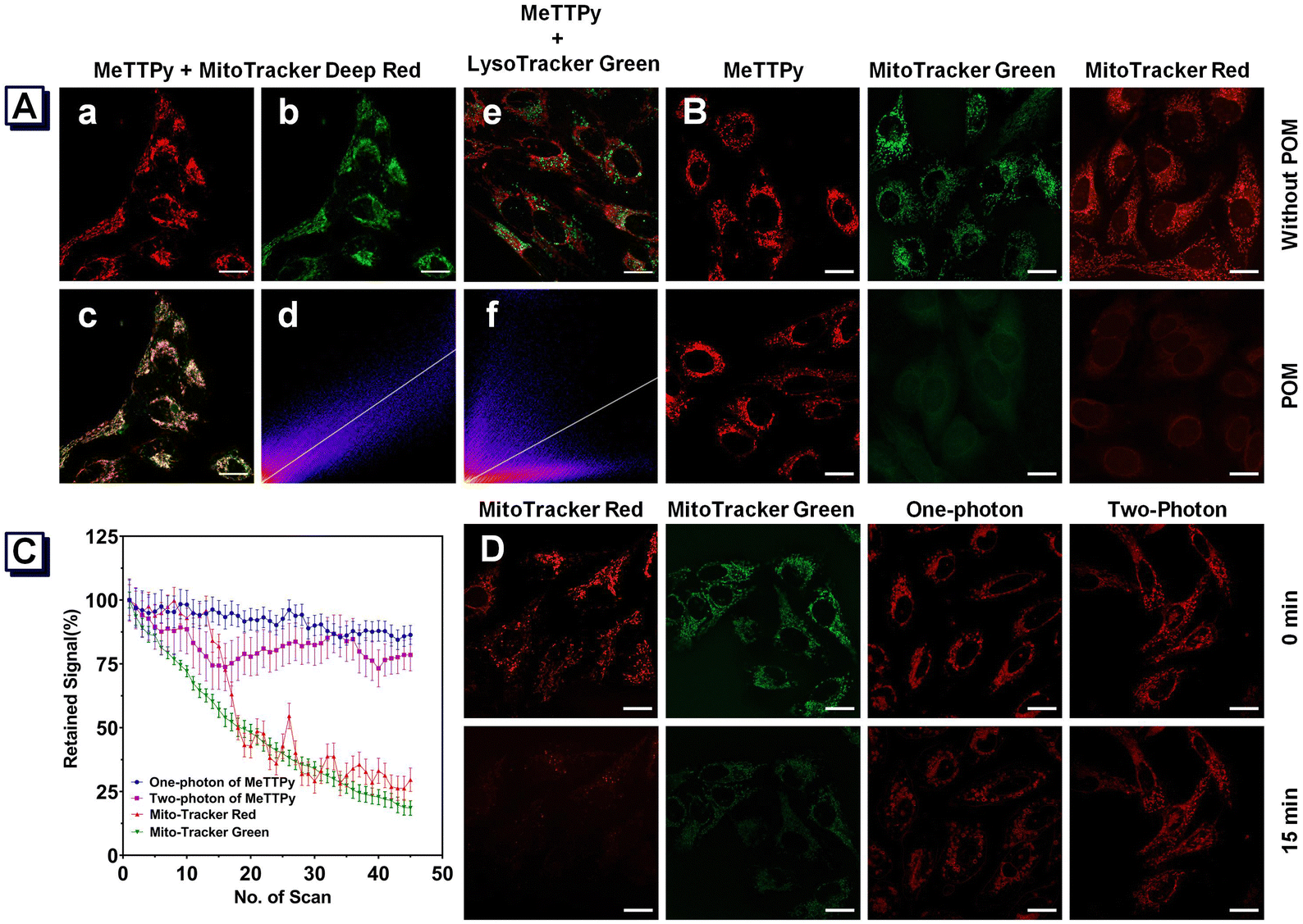 | ||
| Fig. 2 (A) Co-localization imaging of U2OS cells stained with Mito-Tracker Deep Red FM and MeTTPy. Confocal images of U2OS cells stained with (a) Mito-Tracker Deep Red FM and (b) MeTTPy. (c) Merged images of panels (a) and (b). (d) A scatter plot indicating a correction coefficient between panels (a) and (b). (e) Merged images of panels MeTTPy and Lyso-Tracker Green. (f) A scatter plot indicating a correction coefficient between panels MeTTPy and Lyso-Tracker Green. Scale bar: 20 μm. (B) Confocal images of U2OS cells with or without paraformaldehyde fixation after incubation with MeTTPy, Mito-Tracker Green, and Mito-Tracker Red CMXRos. Scale bar: 20 μm. (C) Loss in the fluorescence of U2OS cells stained with MeTTPy Mito-Tracker Green and Mito-Tracker Red CMXRos, with an increasing number of scans of laser irradiation. (D) Confocal images of U2OS cells stained with Mito-Tracker Red CMXRos and Mito-Tracker Green pre- and post-laser irradiation for 15 min, and one-photon and two-photon confocal images of U2OS cells incubated with MeTTPy pre- and post-laser irradiation for 15 min. Scale bar: 20 μm. | ||
We conducted comparative experiments using commercially available mitochondrial probes as controls to further validate the targeting efficiency of MeTTPy in both viable and non-viable cells. We incubated live U2OS cells with MeTTPy, Mito-Tracker Green, and Mito-Tracker Red CMXRos (Chloromethyl-X-rosamine) and performed CLSM imaging. As depicted in Fig. 2(B), the images obtained from MeTTPy, Mito-Tracker Green, and Mito-Tracker Red CMXRos exhibited clear textures, indicating effective mitochondrial localization. The same probes were then used to stain U2OS cells fixed with 4% paraformaldehyde. Notably, only the mitochondria of the fixed cells stained with MeTTPy retained a clear, detailed structure, demonstrating the exceptional targeting ability of MeTTPy. This suggests that MeTTPy functions as an imaging agent in living cells and retains its imaging capabilities in cells fixed with paraformaldehyde, highlighting its potential for broader applications in cellular imaging.
 | ||
| Fig. 3 (A) Reactive oxygen species (ROS) generation in the in situ test: one-photon CLSM image of U2OS cells after incubation with 1.0 μM MeTTPy and 2 μM DCFH-DA for 30 min. One-photon CLSM image of U2OS cells after incubation with 2 μM DCFH-DA for 30 min. Scale bar: 20 μm. (B) The U2OS and K7M2 cell viabilities with MeTTPy and with or without white light irradiation at 28 mW cm−2. (C) Confocal fluorescence images of U2OS cells with MeTTPy, Calcein-AM, and PI under laser irradiation at 488 nm or in the dark. Scale bar:100 μm. CLSM images of U2OS cells stained with a Calcein AM/PI detection kit under different conditions. (D) U2OS cells were incubated with MeTTPy (1 μM, λex = 488 nm, and λem = 490–731 nm), and confocal fluorescence images were obtained. Only U2OS cells were observed in the confocal fluorescence images (λex = 488 nm and λem = 490–731 nm). Scale bar: 20 μm. (E) Representative transmission electron microscopy (TEM) images of mitochondrial apparatus after different treatments. | ||
Owing to the great efficiency of ROS generation facilitated by MeTTPy, we further explored its application in OPE-PDT in living cells, including U2OS, K7M2 (Fig. 3B), and 4T1 cells (Fig. S3, ESI†). All three cell lines were quantitatively evaluated in vitro using the conventional Cell Counting Kit-8 method (CCK-8). Generally, after incubating with MeTTPy under dark conditions for 24 h at varying concentrations, cell viabilities were analyzed by CCK-8. Despite an increase in the MeTTPy concentration to 2.5 μM, the cell viability remained over 90% for U2OS (90%), K7M2 (91%), and 4T1 (93%) cells, demonstrating the excellent biocompatibility of MeTTPy under dark conditions. However, all three cell lines consistently exhibited considerable dose-dependent toxicity under white light irradiation (28 mW cm−2, 30 min). For instance, the viability of U2OS cells dropped to 3% at a MeTTPy concentration of 2.5 μM. K7M2 and 4T1 cells were 2% and 4%, respectively, under the same conditions. These results demonstrate an efficient photodynamic therapeutic effect on all three cell lines under one-photon excitation with white light.
To further explore the one-photon PDT effects of MeTTPy on osteosarcoma tumor cells, we utilized the live/dead cell staining method (Calcein-AM/PI) for U2OS and K7M2 cells. Calcein-AM produces a fluorescence signal in green color when used in living cells, whereas propidium iodide (PI) produces a fluorescence signal in red color specifically in the nuclei of dead cells. Initially, the cells were incubated with MeTTPy (1 μM) for 30 min, followed by irradiation using continuous scanning with a one-photon laser (ZEISS LSM 980, one-photon mode with 488 nm laser excitation). As shown clearly in Fig. 3(C) and Fig. S4 (ESI†), the proportion of cells and the intensity of red fluorescence signals in the light-irradiated MeTTPy group (MeTTPy + Light) increased as the irradiation periods grew longer. Notably, after 100 min of scanning, the nuclei of U2OS cells exhibited bright red fluorescence, with no green fluorescence signal detected. A similar phenomenon occurred in K7M2 cells after only 50 min of scanning. Therefore, MeTTPy exhibits significant phototoxicity to cancer cells under one-photon irradiation. The varying disappearance times of the green fluorescence signal between U2OS and K7M2 cells may relate to variations in the killing efficiency of MeTTPy on different tumor cells under one-photon laser excitation. Under dark conditions, as shown in Fig. 3(C) and Fig. S4 (ESI†), the persistent green fluorescence signal from Calcein-AM suggested the minimal toxicity of MeTTPy.
To further investigate the morphological changes in cancer cells and PDT effects of MeTTPy under one-photon excitation, we irradiated MeTTPy-stained U2OS cells (Fig. 3D) and K7M2 cells (Fig. S5, ESI†) using 488 nm one-photon laser scanning. With increased scanning, cells displayed progressive morphological changes, culminating in the formation of bubbles within cells, eventually leading to the rupture of various cell membrane structures.
We performed transmission electron microscopy (TEM) analysis on U2OS cells to evaluate the ultrastructural changes and observe internal mitochondrial alterations with or without exposure to white light. As depicted in Fig. 3(E), following incubation with MeTTPy and subsequent white light irradiation, TEM images revealed severe mitochondrial damage characterized by swelling, enlargement, rounding, ridge breakage or disappearance, membrane damage, matrix overflow, and vacuolation. In contrast, U2OS cells without MeTTPy incubation with white light irradiation exhibited well-preserved mitochondria with intact membranes and ridges. These data underscore that MeTTPy targets mitochondria and induces cell death by compromising mitochondrial integrity, further elucidating the role of mitochondria-targeted photosensitizers in tumor cell eradication under microscopic observation.
 | ||
| Fig. 4 (A) One-photon confocal images of U2OS cells incubated with MeTTPy (λem = 490–730 nm and λex = 488 nm). Two-photon confocal images of U2OS cells incubated with MeTTPy (λem = 490–730 nm and λex = 830 nm). Scale bar: 20 μm. (B) ROS generation in the in situ test: the two-photon CLSM image of U2OS cells post-incubation with 1.0 μM MeTTPy and 2 μM DCFH-DA for 30 min; two-photon CLSM image of U2OS cells incubated with 2 μM DCFH-DA for 30 min. DCFH-DA: λem = 500–571 nm and λex = 488 nm. MeTTPy: λem = 571–728 nm and λex = 830 nm. Scale bar: 20 μm. (C) Confocal fluorescence images of Ca/PI and MeTTPy (1 μM) under 830 nm femtosecond laser irradiation. Scale bar: 100 μm. (D) MeTTPy (1 μM, λex: 830 nm and λem: 490–731 nm) was incubated with U2OS cells, and the confocal fluorescence images were obtained. Only U2OS cells were observed in the confocal fluorescence images (λex: 830 nm and λem: 490–731 nm). Scale bar: 20 μm. | ||
Considering the substantial PDT effects under one-photon excitation, we hypothesized that MeTTPy might also efficiently generate ROS under two-photon laser irradiation. Subsequently, MeTTPy performance was examined using two-photon fluorescence microscopy. MeTTPy exhibited a δ2PA of 41 GM (GM: Goeppert–Mayer units) at 830 nm (Fig. S6, ESI†), suggesting that it could effectively transfer normal oxygen to cytotoxic ROS upon excitation at 830 nm. To test this, we first assessed ROS production in U2OS and K7M2 cells using both DCFH-DA and MeTTPy, compared to controls incubated solely with DCFH-DA under two-photon irradiation. As illustrated in Fig. 4(B) and Fig. S7 and S8 (ESI†), the cells exposed to DCFH-DA and MeTTPy exhibited significantly more green fluorescence, indicating effective ROS generation under 830 nm laser irradiation. In contrast, no noticeable increase in fluorescence was observed in the absence of MeTTPy.
Further evaluations involved TPE-PDT of MeTTPy using live/dead cell staining (Calcein AM/PI method). U2OS cells stained with Calcein AM and PI for 30 min exhibited red fluorescence dots (PI) after 32 min of 830 nm laser irradiation, as shown in Fig. 4(C). The quantity of red dots served as a direct measure of the therapeutic efficacy of TPE-PDT. After 40 min of laser exposure, nearly all U2OS cells within the irradiated area succumbed to TPE-PDT effects. Similar tests on K7M2 cells (Fig. S9, ESI†) revealed green fluorescence pre-irradiation, indicating low cytotoxicity and good biocompatibility of MeTTPy in the absence of light. Post-irradiation with the 830 nm laser, red fluorescence dots appeared at 15 min, and by 25 min, most K7M2 cells in the irradiated area had died.
These findings confirm the TPE-PDT action of MeTTPy on U2OS and K7M2 cells under 830 nm laser irradiation and highlight the differential response times, potentially reflecting variations in TPE-PDT efficacy across different tumor cell types.
To further investigate the cell morphology and ultrastructure changes following TPE-PDT with MeTTPy, U2OS cells were co-stained with MeTTPy (1 μM) and then subjected to continuous femtosecond laser irradiation at 830 nm. As depicted in Fig. 4(D), the U2OS cells exhibited notable morphological alterations, such as the development and expansion of intracellular bubbles, a phenomenon frequently associated with cellular death.28,45,46 These changes intensified with a prolonged two-photon irradiation period. In contrast, U2OS cells not treated with MeTTPy did not exhibit bubble formation under the same irradiation conditions. We performed the same experiment with different fields of view, yielding the same results (Fig. S10, ESI†). Similar effects were observed in K7M2 cells (Fig. S11, ESI†).
2.3 In vivo fluorescence imaging of the mice model
Fluorescent materials are pivotal for in vivo fluorescence imaging as they directly visualize biological processes and their alterations. To evaluate the long-term imaging capabilities of MeTTPy, it was administered directly to the tumor sites in NOD-scid mice bearing K7M2 tumors. Gradually, the dye diffused from the injection site, eventually encompassing the entire tumor tissue. As demonstrated in Fig. 5, the fluorescence emission intensity of MeTTPy achieved its highest point 24 h after injection, with detectable signals persisting up to 96 h. These observations suggest that MeTTPy is an ideal probe for live animal imaging, offering sustained visibility and effective tissue penetration. | ||
| Fig. 5 Biodistribution of MeTTPy in K7M2-tumor-bearing mice at various time points following intratumoral injection of MeTTPy (4 × 10⁻³ M, 100 μL/100 mm3 tumor). | ||
2.4 In vivo PDT and biosafety evaluation
Considering the vital significance of biosafety in biomedical applications and upcoming clinical deployment, we thoroughly evaluated the possible systemic toxicity of MeTTPy in murine models. Initially, the in vivo PDT efficacy of MeTTPy was evaluated on K7M2 tumor-bearing mice through intratumoral injection. For the experimental setup, the treatment group received intratumoral injections of MeTTPy (4 × 10⁻³ M, 100 μL/100 mm3 tumor), whereas the control group received an equal amount of normal saline solution. After a 30-min interval, the mice were subjected to laser treatment at a power of 140 mW for 15 min. The control group received identical radiation treatment. The weight and tumor volume were assessed and noted bi-daily. The tumor volume was calculated using the formula: volume = (tumor length × [tumor width]2)/2. On the 14th d, the tumors were surgically excised and documented photographically.A comparison of the experimental and control groups' body weights pre- and post- PDT revealed no statistically significant differences (Fig. 6A), indicating that the systemic influence was low. However, tumor volumes diverged significantly between the groups after 14 d of PDT treatment (Fig. 6(B) and (C)). In the treatment group, where MeTTPy was not combined with simultaneous laser irradiation (140 mW, 15 min), the tumor volume increased rapidly from day 2, whereas the experimental group exhibited substantial tumor suppression starting from day 3. By the end of the 14 d, the tumor size in the control group had expanded 12-fold relative to day 2. In contrast, the treatment group exhibited a tumor size suppression rate exceeding 90%. These findings underscore the significant potential of MeTTPy for PDT in mice bearing K7M2 tumors, highlighting its effectiveness and minimal systemic toxicity.
 | ||
| Fig. 6 Assessment of the safety and effectiveness of MeTTPy by the in vivo photodynamic treatment of healthy BALB/c mice. (A) Changes in the mice weight after 14 d of various treatments. (B) Growth trajectories of tumors in mice treated with different interventions over time (n = 4, ***p < 0.001). (C) Images of separated tumors. (D) Average tumor weights at day 14 post-treatment (mean + SD; n = 4, ***p <0.001). No significant alteration was observed at day 14 post-injection of MeTTPy in comparison to the control group regarding biochemical blood biomarkers, including ALB (E) and TP (F). (G) The cleavage of GSDME and caspase-3 was observed by western blotting; cleavage of pro-caspase-8 under different conditions was observed by western blotting; and the ratio of mGSDME-N to mGSDME-F was statistically analyzed; statistical analysis of the ratio of cleaved-caspase-3 to pre-caspase-3; statistical analysis of pro-caspase-8 to the internal reference ratio. Error bar: deviation of more than three replicates (SD). The p-values were calculated using the t-test (**p < 0.01 or ***p < 0.001). | ||
Serum biochemical analysis (Fig. 6D–F and Fig. S12, ESI†) demonstrated that MeTTPy had minimal interference with the metabolic functions of primary organs. After a 14-d treatment period, tissue sections were subjected to histological examination. A pathologist comprehensively evaluated each pathological section (Fig. S13, ESI†), noting the absence of lymphocyte infiltration in all organs. No abnormal changes were observed in the morphology or structure of cardiac muscle cells. Similarly, no evidence of fibrosis was observed in the lung and liver samples. The white and red pulp structure in splenic tissue sections appeared normal, and the kidney sections showed no aberrant glomerular or tubular features. Furthermore, no evidence of necrosis was discovered in any of the histological specimens. These data indicate that MeTTPy did not cause immediate harm to BALB/c mice during the evaluation period, which supports its prospective use in clinical settings.
2.5 Analysis of the pyroptosis mechanism induced by MeTTPy
To understand the process of MeTTPy-induced cell death, we first assessed the expression levels of GSDME, caspase-3, and caspase-8 in treated U2OS cells via western blotting. As depicted in Fig. 6(G), no significant changes in the expression of GSDME, caspase-3, and caspase-8 were determined in cells not treated with MeTTPy and those incubated under dark conditions after MeTTPy treatment. However, the levels of GSDME-N, cleaved caspase-3, and pro-caspase-8 changed significantly in cells treated with MeTTPy and exposed to light. These findings indicate that MeTTPy promotes GSDME cleavage and the activation of caspase-3 and caspase-8 following PDT treatment. According to previous literature, PDT could cause cell death via the apoptosis pathway.47 MeTTPy generates a large amount of ROS in the cell under light conditions. Initially, ROS initiate the activation of BAK and BAX and oligomerization in the mitochondrial outer membrane, which results in mitochondrial outer membrane permeabilization and the release of cytochrome C.48 Subsequently, downstream enzyme caspase-3 is activated, facilitating the caspase-3-mediated cleavage of GSDME, releasing the N-terminal fragment, which forms a pore in the cell membrane, leading to the rupture of the cell membrane and release of cellular contents, thus triggering pyroptosis.49–52 In addition, ROS can induce mitochondria to release the second mitochondria-derived apoptotic factor (Smac/DIABLO), which promotes the activation of caspase-8, further promoting the activation of caspase-3, which cleaves the GSDME and further promotes pyroptosis.533. Conclusion
Our studies explored the capabilities of the two-photon aggregation-induced luminescence probe MeTTPy. Demonstrating exceptional targeting accuracy, minimal cellular harm, and superior photostability under two-photon excitation, MeTTPy proved effective in cellular imaging experiments. These findings confirmed its specific mitochondrial targeting ability, making it a potent photosensitizer for two-photon stimulated PDT. Notably, MeTTPy facilitated tumor cell pyroptosis under illumination by activating the GSDME pyroptosis pathway.Moreover, in vivo, fluorescence imaging of MeTTPy in tumor-bearing allogeneic nude mice at various times highlighted its potential as a valuable mitochondrial probe. Considering these attributes, we believe that MeTTPy is an excellent candidate for in vivo and in vitro mitochondrial investigation, employing both one-photon and two-photon imaging techniques.
Author contributions
Huifang Su and Hongjian Liu conceptualized the project. Wenhao Du, Wenzhao Shang, Wei Wen, Xiepeng Deng, and Dalu Xie conducted the experiments. Wenhao Du, Wenzhao Shang, and Wei Wen analyzed the experimental results. Wenhao Du, Wenzhao Shang, and Wei Wen prepared the initial draft. Huifang Su and Hongjian Liu revised the manuscript. Huifang Su and Hongjian Liu were involved in fund acquisition. All authors have read and approved the manuscript.Data availability
The authors confirm that the data supporting the findings of this study are available within the article and/or its ESI.†Conflicts of interest
The authors declare that they have no conflict of interest.Acknowledgements
We gratefully acknowledge the National Natural Science Foundation of China (No. 81972523 and 82172484), Natural Science Foundation of Henan Province (232300421053), the Henan Province Young and Middle-Aged Health Sciences and Technology Innovation Talent Project (No. YXKC2022032), and the Program for Science & Technology Innovation Talents in Universities of Henan Province (24HASTIT066). The procedures for all the animal experiments were approved by the Ethics Review Board of Zhengzhou University. All the animal experiments were strictly performed in compliance with the 1964 Declaration of Helsinki. We thank the Translational Medical Center, First Affiliated Hospital of Zhengzhou University for technical support.References
- J. A. Park and N.-K. V. Cheung, GD2 or HER2 targeting T cell engaging bispecific antibodies to treat osteosarcoma, J. Hematol. Oncol., 2020, 13(1), 172 CrossRef CAS PubMed.
- W. G. Ward, K. Mikaelian, F. Dorey, J. M. Mirra, A. Sassoon, E. C. Holmes, F. R. Eilber and J. J. Eckardt, Pulmonary metastases of stage IIB extremity osteosarcoma and subsequent pulmonary metastases, J. Clin. Oncol., 1994, 12(9), 1849–1858 CrossRef CAS PubMed.
- A. B. Shaikh, F. Li, M. Li, B. He, X. He, G. Chen, B. Guo, D. Li, F. Jiang, L. Dang, S. Zheng, C. Liang, J. Liu, C. Lu, B. Liu, J. Lu, L. Wang, A. Lu and G. Zhang, Present Advances and Future Perspectives of Molecular Targeted Therapy for Osteosarcoma, Int. J. Mol. Sci., 2016, 17(4), 506 CrossRef PubMed.
- J. Gill and R. Gorlick, Advancing therapy for osteosarcoma, Nat. Rev. Clin. Oncol., 2021, 18(10), 609–624 CrossRef PubMed.
- J. Niu, T. Yan, W. Guo, W. Wang, T. Ren, Y. Huang, Z. Zhao, Y. Yu, C. Chen, Q. Huang, J. Lou and L. Guo, The COPS3-FOXO3 positive feedback loop regulates autophagy to promote cisplatin resistance in osteosarcoma, Autophagy, 2023, 19(6), 1693–1710 CrossRef CAS PubMed.
- P. S. Meltzer and L. J. Helman, New Horizons in the Treatment of Osteosarcoma, N. Engl. J. Med., 2021, 385(22), 2066–2076 CrossRef CAS PubMed.
- P. Cen, J. Huang, C. Jin, J. Wang, Y. Wei, H. Zhang and M. Tian, Aggregation-induced emission luminogens for in vivo molecular imaging and theranostics in cancer, Aggregate, 2023, 4(5), e352 CrossRef CAS.
- H. Shi and P. J. Sadler, How promising is phototherapy for cancer?, Br. J. Cancer, 2020, 123(6), 871–873 CrossRef PubMed.
- H. Zhang, C. He, L. Shen, W. Tao, J. Zhu, J. Song, Z. Li and J. Yin, Membrane-targeting amphiphilic AIE photosensitizer for broad-spectrum bacteria imaging and photodynamic killing of bacteria, Chin. Chem. Lett., 2023, 34(9), 108160 CrossRef CAS.
- M. Akbari, T. B. L. Kirkwood and V. A. Bohr, Mitochondria in the signaling pathways that control longevity and health span, Ageing Res. Rev., 2019, 54, 100940 CrossRef CAS PubMed.
- J. J. Hu, W. Jiang, L. Yuan, C. Duan, Q. Yuan, Z. Long, X. Lou and F. Xia, Recent advances in stimuli-responsive theranostic systems with aggregation-induced emission characteristics, Aggregate, 2021, 2(1), 48–65 CrossRef CAS.
- M. Kang, Z. Zhang, N. Song, M. Li, P. Sun, X. Chen, D. Wang and B. Z. Tang, Aggregation-enhanced theranostics: AIE sparkles in biomedical field, Aggregate, 2020, 1(1), 80–106 CrossRef.
- H.-F. Su, Q.-C. Peng, Y. U. Liu, T. Xie, P.-P. Liu, Y.-C. Cai, W. Wen, Y.-H. Yu, K. Li and S.-Q. Zang, A near-infrared AIE probe and its applications for specific in vitro and in vivo two-photon imaging of lipid droplets, Biomaterials, 2022, 288, 121691 CrossRef CAS PubMed.
- H. Li, H. Kim, J. Han, V. N. Nguyen, X. Peng and J. Yoon, Activity-based smart AIEgens for detection, bioimaging, and therapeutics: Recent progress and outlook, Aggregate, 2021, 2(4), e51 CrossRef CAS.
- D. Wang, M. M. S. Lee, G. G. Shan, R. T. K. Kwok, J. W. Y. Lam, H. F. Su, Y. C. Cai and B. Z. Tang, Highly Efficient Photosensitizers with Far-Red/Near-Infrared Aggregation-Induced Emission for In Vitro and In Vivo Cancer Theranostics, Adv. Mater., 2018, 30(39), e1802105 Search PubMed.
- C. Gui, E. G. Zhao, R. T. K. Kwok, A. C. S. Leung, J. W. Y. Lam, M. J. Jiang, H. Q. Deng, Y. J. Cai, W. J. Zhang, H. F. Su and B. Z. Tang, AIE-active theranostic system: selective staining and killing of cancer cells, Chem. Sci., 2017, 8(3), 1822–1830 Search PubMed.
- H. Wang, C. Qiao, Q. Guan, M. Wei and Z. Li, Nanoparticle-mediated synergistic anticancer effect of ferroptosis and photodynamic therapy: Novel insights and perspectives, Asian J. Pharm. Sci., 2023, 18(4), 100829 Search PubMed.
- D. Wang, H. F. Su, R. T. K. Kwok, X. L. Hu, H. Zou, Q. X. Luo, M. M. S. Lee, W. H. Xu, J. W. Y. Lam and B. Tang, Rational design of a water-soluble NIR AIEgen, and its application in ultrafast wash-free cellular imaging and photodynamic cancer cell ablation, Chem. Sci., 2018, 9(15), 3685–3693 RSC.
- B. Ji, M. Wei and B. Yang, Recent advances in nanomedicines for photodynamic therapy (PDT)-driven cancer immunotherapy, Theranostics, 2022, 12(1), 434–458 CrossRef CAS PubMed.
- R. Alzeibak, T. A. Mishchenko, N. Y. Shilyagina, I. V. Balalaeva, M. V. Vedunova and D. V. Krysko, Targeting immunogenic cancer cell death by photodynamic therapy: past, present and future, J. Immunother. Cancer, 2021, 9(1), e001926 CrossRef PubMed.
- H. Su, T. Xie, Y. U. Liu, Y. Cui, W. Wen, B. Z. Tang and W. Qin, Facile synthesis of ultrabright luminogens with specific lipid droplets targeting feature for in vivo two-photon fluorescence retina imaging, Chin. Chem. Lett., 2023, 34(6), 107949 CrossRef CAS.
- Z. Liu, Z. Xie, W. Li, X. Wu, X. Jiang, G. Li, L. Cao, D. Zhang, Q. Wang, P. Xue and H. Zhang, Photodynamic immunotherapy of cancers based on nanotechnology: recent advances and future challenges, J. Nanobiotechnol., 2021, 19(1), 160 CrossRef CAS PubMed.
- Q. Peng and Z. Shuai, Molecular mechanism of aggregation-induced emission, Aggregate, 2021, 2(5), e91 CrossRef CAS.
- T. C. Pham, V.-N. Nguyen, Y. Choi, S. Lee and J. Yoon, Recent Strategies to Develop Innovative Photosensitizers for Enhanced Photodynamic Therapy, Chem. Rev., 2021, 121(21), 13454–13619 Search PubMed.
- J. Qi, H. Ou, Q. Liu and D. Ding, Gathering brings strength: How organic aggregates boost disease phototheranostics, Aggregate, 2021, 2(1), 95–113 CrossRef CAS.
- H. Su, Z. Deng, Y. Liu, Y. Zhao, H. Liu, Z. Zhao and B. Z. Tang, A brightly red emissive AIEgen and its antibody conjugated nanoparticles for cancer cell targeting imaging, Mater. Chem. Front., 2022, 6(10), 1317–1323 Search PubMed.
- H. Zhao, N. Li, C. Ma, Z. Wei, Q. Zeng, K. Zhang, N. Zhao and B. Z. Tang, An AIE probe for long-term plasma membrane imaging and membrane-targeted photodynamic therapy, Chin. Chem. Lett., 2023, 34(4), 107699 CrossRef CAS.
- X. He, B. Situ, M. Gao, S. Guan, B. He, X. Ge, S. Li, M. Tao, H. Zou, B. Z. Tang and L. Zheng, Stereotactic Photodynamic Therapy Using a Two-Photon AIE Photosensitizer, Small, 2019, 15(50), 1905080 CrossRef CAS PubMed.
- S. Wang, W. Wu, P. Manghnani, S. Xu, Y. Wang, C. C. Goh, L. G. Ng and B. Liu, Polymerization-Enhanced Two-Photon Photosensitization for Precise Photodynamic Therapy, ACS Nano, 2019, 13(3), 3095–3105 CrossRef CAS PubMed.
- J. G. Croissant, J. I. Zink, L. Raehm and J.-O. Durand, Two-Photon-Excited Silica and Organosilica Nanoparticles for Spatiotemporal Cancer Treatment, Adv. Healthcare Mater., 2018, 7(7), e1701248 CrossRef PubMed.
- H. Dai, Q. Shen, J. Shao, W. Wang, F. Gao and X. Dong, Small Molecular NIR-II Fluorophores for Cancer Phototheranostics, Innovation, 2021, 2(1), 100082 CAS.
- S. Wang, Y. Liao, Z. Wu, Y. Peng, Y. Liu, Y. Chen, L. Shao, Z. Zeng and Y. Liu, A lysosomes and mitochondria dual-targeting AIE-active NIR photosensitizer: Constructing amphiphilic structure for enhanced antitumor activity and two-photon imaging, Mater. Today Bio, 2023, 21, 100721 CrossRef CAS PubMed.
- J. Sun and X. He, AIE-based drug/gene delivery system: Evolution from fluorescence monitoring alone to augmented therapeutics, Aggregate, 2022, 3(6), e282 CrossRef CAS.
- R. Han, M. Zhao, Z. Wang, H. Liu, S. Zhu, L. Huang, Y. Wang, L. Wang, Y. Hong, Y. Sha and Y. Jiang, Super-efficient in Vivo Two-Photon Photodynamic Therapy with a Gold Nanocluster as a Type I Photosensitizer, ACS Nano, 2020, 14(8), 9532–9544 CrossRef CAS PubMed.
- X. Guo, N. Yang, W. Ji, H. Zhang, X. Dong, Z. Zhou, L. Li, H.-M. Shen, S. Q. Yao and W. Huang, Mito-Bomb: Targeting Mitochondria for Cancer Therapy, Adv. Mater., 2021, 33(43), e2007778 CrossRef PubMed.
- L. Sainero-Alcolado, J. Liaño-Pons, M. V. Ruiz-Pérez and M. Arsenian-Henriksson, Targeting mitochondrial metabolism for precision medicine in cancer, Cell Death Differ., 2022, 29(7), 1304–1317 CrossRef CAS PubMed.
- J. S. Harrington, S. W. Ryter, M. Plataki, D. R. Price and A. M. K. Choi, Mitochondria in health, disease, and aging, Physiol. Rev., 2023, 103(4), 2349–2422 CrossRef CAS PubMed.
- E. N. Main, T. M. Cruz and G. L. Bowlin, Mitochondria as a therapeutic: a potential new frontier in driving the shift from tissue repair to regeneration, Regener. Biomater., 2023, 10, rbad070 CrossRef CAS PubMed.
- S. Lu, X. Guo, F. Zhang, X. Li, M. Zou and L.-L. Li, Bioactivated in vivo assembly (BIVA) peptide-tetraphenylethylene (TPE) probe with controllable assembled nanostructure for cell imaging, Chin. Chem. Lett., 2021, 32(6), 1947–1952 CrossRef CAS.
- Z. Zeng, Y. Luo, X. Xu, T. Shan, M. Chen, Z. Huang, Y. Huang and C. Zhao, A mitochondria-targeting ROS-activated nanoprodrug for self-augmented antitumor oxidation therapy, J. Controlled Release, 2023, 359, 415–427 CrossRef CAS PubMed.
- D. B. Zorov, M. Juhaszova and S. J. Sollott, Mitochondrial reactive oxygen species (ROS) and ROS-induced ROS release, Physiol. Rev., 2014, 94(3), 909–950 CrossRef CAS PubMed.
- S. Kuang, L. Sun, X. Zhang, X. Liao, T. W. Rees, L. Zeng, Y. Chen, X. Zhang, L. Ji and H. Chao, A Mitochondrion-Localized Two-Photon Photosensitizer Generating Carbon Radicals Against Hypoxic Tumors, Angew. Chem., Int. Ed., 2020, 59(46), 20697–20703 CrossRef CAS PubMed.
- Y. Ni, H. Zhang, C. Chai, B. Peng, A. Zhao, J. Zhang, L. Li, C. Zhang, B. Ma, H. Bai, K.-L. Lim and W. Huang, Mitochondria-Targeted Two-Photon Fluorescent Photosensitizers for Cancer Cell Apoptosis via Spatial Selectability, Adv. Healthcare Mater., 2019, 8(14), e1900212 CrossRef PubMed.
- Z. He, Y. Gao, H. Zhang, Y. Xue, F. Meng and L. Luo, Mitochondrion-Anchored Photosensitizer with Near Infrared-I Aggregation-Induced Emission for Near Infrared-II Two-Photon Photodynamic Therapy, Adv. Healthcare Mater., 2021, 10(24), e2101056 CrossRef PubMed.
- Y. Shen, A. J. Shuhendler, D. Ye, J.-J. Xu and H.-Y. Chen, Two-photon excitation nanoparticles for photodynamic therapy, Chem. Soc. Rev., 2016, 45(24), 6725–6741 RSC.
- Z. Zheng, H. Liu, S. Zhai, H. Zhang, G. Shan, R. T. K. Kwok, C. Ma, H. H. Y. Sung, I. D. Williams, J. W. Y. Lam, K. S. Wong, X. Hu and B. Z. Tang, Highly efficient singlet oxygen generation, two-photon photodynamic therapy, and melanoma ablation by rationally designed mitochondria-specific near-infrared AIEgens, Chem. Sci., 2020, 11(9), 2494–2503 RSC.
- T. Zhang, Y. Li, Z. Zheng, R. Ye, Y. Zhang, R. T. K. Kwok, J. W. Y. Lam and B. Z. Tang, In Situ Monitoring Apoptosis Process by a Self-Reporting Photosensitizer, J. Am. Chem. Soc., 2019, 141(14), 5612–5616 CrossRef CAS PubMed.
- S.-K. Hsu, C.-Y. Li, I. L. Lin, W.-J. Syue, Y.-F. Chen, K.-C. Cheng, Y.-N. Teng, Y.-H. Lin, C.-H. Yen and C.-C. Chiu, Inflammation-related pyroptosis, a novel programmed cell death pathway, and its crosstalk with immune therapy in cancer treatment, Theranostics, 2021, 11(18), 8813–8835 CrossRef CAS PubMed.
- H. Kalkavan and D. R. Green, MOMP, cell suicide as a BCL-2 family business, Cell Death Differ., 2017, 25(1), 46–55 CrossRef PubMed.
- X. Chen, W.-t He, L. Hu, J. Li, Y. Fang, X. Wang, X. Xu, Z. Wang, K. Huang and J. Han, Pyroptosis is driven by non-selective gasdermin-D pore and its morphology is different from MLKL channel-mediated necroptosis, Cell Res., 2016, 26(9), 1007–1020 CrossRef CAS PubMed.
- L. Hu, M. Chen, X. Chen, C. Zhao, Z. Fang, H. Wang and H. Dai, Chemotherapy-induced pyroptosis is mediated by BAK/BAX-caspase-3-GSDME pathway and inhibited by 2-bromopalmitate, Cell Death Dis., 2020, 11(4), 281 Search PubMed.
- V. Gogvadze, E. Norberg, S. Orrenius and B. Zhivotovsky, Involvement of Ca2+ and ROS in α-tocopheryl succinate-induced mitochondrial permeabilization, Int. J. Cancer, 2010, 127(8), 1823–1832 CrossRef CAS PubMed.
- Z. Rao, Y. Zhu, P. Yang, Z. Chen, Y. Xia, C. Qiao, W. Liu, H. Deng, J. Li, P. Ning and Z. Wang, Pyroptosis in inflammatory diseases and cancer, Theranostics, 2022, 12(9), 4310–4329 CrossRef CAS PubMed.
Footnotes |
| † Electronic supplementary information (ESI) available. See DOI: https://doi.org/10.1039/d4qm00870g |
| ‡ Wenhao Du, Wenzhao Shang, and Wei Wen contributed equally to this paper. |
| This journal is © the Partner Organisations 2025 |