
The application and challenges of antimicrobial drug-loaded scaffold materials for the treatment of bone infections
Shenghua Wu,
Fu Qin and
Yongbiao Meng*
Department of Orthopedics, The First People's Hospital of Hangzhou Linping Distric, Hangzhou, 311100, China. E-mail: mengyb2010@126.com
First published on 24th June 2025
Abstract
Bone infections are a complex clinical issue, typically caused by bacterial infections, leading to damage and dysfunction of bone tissue. Traditional treatments primarily rely on antibiotics (ABs); however, they have limitations such as low permeability and the development of resistance. In recent times, the progress in tissue engineering has introduced novel approaches to tackle bone infections. Biocompatible scaffolds not only serve as supportive structures for bone defects but also function as drug delivery systems, enabling targeted release of antimicrobial drugs to enhance anti-inflammatory and antibacterial effects. The drug release rate from drug-loaded scaffolds can be controlled, which helps to manage infections while promoting bone healing and enhancing the synergistic effects of antibacterial action and bone repair. This review discusses the latest advancements in scaffold materials for the release of antimicrobial drugs, analyzes the current challenges in research, and outlines future directions for development, providing a basis for clinical applications.
1. Introduction
Bone infection refers to a series of musculoskeletal system diseases caused by microorganisms through exogenous or hematogenous routes.1 These infections primarily include implant-associated osteomyelitis, fracture-related infections, acute hematogenous osteomyelitis, and infectious bone defects.2,3 Bone infections are common complications following orthopedic surgery, which can lead to joint deformities, limb amputation, or even death, imposing a significant economic burden on society. Despite employing top-tier medical care, osteomyelitis continues to be difficult to treat, with failure rates of treatment ranging between 10% and 40%.4,5 The typical causative agents of osteomyelitis often encompass Staphylococcus aureus (S. aureus),6 Streptococcus pyogenes,7 Streptococcus pneumoniae,8 Escherichia coli (E. coli),9 and Pseudomonas aeruginosa.10 Among these, S. aureus exhibits strong invasive abilities, colonization, and proliferation of bone cells, accounting for over 70% of the pathogen spectrum in osteomyelitis.11 Necrotic bone is often caused by and infection forms a microenvironment characterized by acidity, immune suppression, and poor antibiotic penetration due to its association with a limited vascular system.12,13 In the majority of instances, this leads to the formation of persistent biofilms, resulting in chronic infections that require surgical intervention.14 The long-term presence of intracellular bacteria can exacerbate recurrent chronic bone infections; therefore, selecting an antibiotic that effectively eliminates intracellular bacteria will be a significant milestone in the management of bone infections.The main mechanisms by which antibiotics (ABs) inhibit bacteria associated with bone infections include four types:15 blocking bacterial cell wall formation, disrupting the cell membrane, hindering protein synthesis, and inhibiting nucleic acid replication and transcription. The current commonly employed approaches for managing bone infections primarily involve systemic antibiotic administration combined with local drug delivery treatment. For cases with bone necrosis, surgical debridement to remove the dead bone is also necessary.16 However, due to the persistent presence of bacteria deep within the bone, these traditional treatment methods often struggle to effectively cure bone infections. Systemic use of ABs also has some drawbacks, such as insufficient local drug concentrations to combat the infection, vulnerability to resistance and systemic adverse effects.17,18 Additionally, when bone infections occur, they are often accompanied by bone loss. Therefore, effective management of bone infections demands not only the efficient elimination of pathogens but also the rapid regeneration of bone in regions with defects.19 Prompt surgical debridement in conjunction with appropriate ABs can effectively treat acute osteomyelitis. However, traditional treatment strategies often yield poor results in chronic infections associated with bone defects. Repair of bone defects can only commence once the infection is under control. Although techniques such as bone grafting, bone transport, osteoinductive membrane formation, and the implantation of antibiotic-loaded slow-release carriers are widely used in clinical practice for bone infections, challenges remain, including prolonged treatment durations, insufficient donor bone availability, and complications at the donor site.20 Therefore, the development of antibacterial bioactive bone fillers represents a significant clinical advancement in treating these conditions.
In recent years, with the rapid development of bone tissue engineering (BTE), bone repair scaffolds have become an indispensable core component in this field. Other therapeutic functions rely on scaffolds to effectively perform their roles.21,22 The creation of bone infection repair scaffolds with superior mechanical strength, strong bone-forming capacity, and localized antimicrobial properties has emerged as a key area of research.23,24 Ongoing studies on anti-infection bone scaffolds mainly concentrate on the choice of materials, scaffold fabrication techniques, drug delivery mechanisms, and non-antibiotic biomaterials. For irregular open bone defects in clinical settings, it is important to further load traditional bone scaffolds with antibacterial agents to promote bone reconstruction.25 The selection of these antibacterial agents significantly impacts both infection healing and the quality of bone reconstruction. The advantages and disadvantages of various antibacterial agents, along with their target bacterial species, are presented in Table 1. Tissue engineering involves the integration and synergistic interaction of cells, bioactive factors, and scaffold biomaterials. This approach aims to enhance the microenvironment at the site of local infected bone defects, thereby promoting bone healing and regeneration.26,27 After effectively filling bone defect sites, composite biological scaffolds can rapidly release antibacterial components to inhibit pathogen proliferation and prevent excessive early inflammation. Research on anti-infection bone scaffolds requires collaborative innovation across material science, pharmacology, and tissue engineering. By optimizing scaffold materials, structures, drug loading, and integrating with biologic therapies, tissue engineering methods offer potential solutions for tackling the dual challenges of infection management and bone restoration in infected bone defects. This review innovatively integrates the latest advancements in the field of bone infection treatment, covering both traditional and emerging antimicrobial strategies. First, it provides an in-depth analysis of common antimicrobial agents, including the application and challenges of ABs, the mechanisms and potential of antimicrobial peptides (AMPs), and the types of antimicrobial nanomaterials and their precision antimicrobial technologies. Second, the review focuses on BTE scaffolds, systematically summarizing the characteristics and prospects of various drug-loading methods (such as direct loading, coating, and composite preparation), which provide theoretical support for drug release, long-term antimicrobial effects, and bone repair. It also explores the clinical applications of antimicrobial bone scaffolds (Fig. 1). This interdisciplinary integrative perspective offers new insights for the study of bone infection treatment and promotes innovative development in this field.
| Antimicrobial agents | Advantage | Disadvantage | Species of action |
|---|---|---|---|
| Antibiotic | Broad-spectrum antibacterial, easy to prepare and inexpensive | Resistance is readily developed and systemic use has low toxicity | Gram-positive bacteria and most Gram-negative bacteria |
| Antibacterial peptide | Broad-spectrum antibacterial, no drug resistance, good biocompatibility | Extraction is difficult and costly | S. aureus, E. coli and other Gram-positive bacteria, and a small number of Gram-negative bacteria |
| Nano metal particles | It has strong antibacterial ability and low synthesis difficulty | It is difficult to control the concentration dilution and has certain cytotoxicity | S. aureus, E. coli, Bacillus subtilis, etc. |
 | ||
| Fig. 1 Graphical abstract. | ||
2. Type of antimicrobial agent
2.1 Antibiotics (ABs)
Bone infection is a disease of bone tissue inflammation caused by bacterial or fungal infection, and common pathogenic bacteria such as S. aureus, Staphylococcus epidermid,28 etc. ABs are commonly used antibacterial agents with a variety of types, mainly including: broad-spectrum ABs, such as penicillins, cephalosporins, etc., which can extensively eliminate a variety of bacteria by inhibiting bacterial cell wall synthesis and disrupting the cell wall structure.29,30The effect of antibiotics is obvious. Aldhaeefi et al. assessed the effectiveness of penicillin targeting S. aureus, demonstrating that penicillin use was both safe and effective.31 ABs against Gram-positive bacteria, such as vancomycin (Van)32 and teicoplanin,33 mainly target S. aureus and can cause lysis and death of bacterial cells by impeding the creation and reconstruction of bacterial cell wall, destroying the structure of bacterial cell wall, interfering with bacterial protein synthesis, and inducing changes in bacterial cell membrane permeability, respectively. Namdar et al. coloaded lysozyme (Lys) and Van onto mesoporous silica nanoparticles (MSN) to form Lys–VAN–MSN. The minimal concentration to inhibit of the nanoparticles (NPs) against S. aureus was determined, and it was observed that the NPs possessed notable antibacterial efficacy.34 Watanabe et al. evaluated the efficacy of teicoplanin against S. aureus in a murine model of thigh infection, demonstrating robust bactericidal activity against this pathogen.35 Anti-Gram-negative ABs such as aztreonam and piperacillin/tazobactam, which mainly target Pseudomonas aeruginosa, can achieve antibacterial effect by interfering with protein synthesis and destroying bacterial cell membrane, through inhibiting bacterial cell wall synthesis and mitigating drug resistance.36,37 In the study by Gahane et al. Fmoc-F was conjugated with the Gram-negative specific antibiotic aztreonam to form a novel combination. In a murine wound infection model, this formulation exhibited potent antibacterial activity against Gram-negative bacteria, effectively diminishing bacterial count. Notably, it demonstrated enhanced efficacy against Pseudomonas aeruginosa compared to other Gram-negative pathogens.38 As the most classical and widely used antibacterial drugs, ABs are broadly applied in the therapy of conventional bone infections. As reported by Ran et al. found that patients with an infection of the left tibia treated with ABs had rapid tissue healing, and the patients did not have to amputate their leg and recovered well.39 Fan et al. evaluated the antibacterial efficacy of antibiotic-eluting materials using a rat model. They simulated joint replacement by inserting polyethylene (PE) plugs into the femur and titanium (Ti) screws into the tibia, and inoculated methicillin-susceptible S. aureus into the medullary canal and joint space. The experimental group received daqingmycin-eluting PE materials, which effectively treated periprosthetic joint infection.40
In addition, drug release is critical in the treatment of bone infection, some ABs, such as penicillins and cephalosporins, can effectively penetrate bone tissues and reach the infection sites to exert their effects. This represents the cornerstone of antibiotic therapy for bone infections.41,42 Torkington et al. collected bone samples from patients undergoing total hip arthroplasty (THA) and total knee arthroplasty (TKA) to assess the bone permeability of flucloxacillin and gentamicin during intravenous antibiotic prophylaxis for hip and knee arthroplasty. Antibiotic concentrations in the samples were analyzed using HPLC. Results indicated that both flucloxacillin and gentamicin exhibited good bone permeability and antibacterial efficacy.42 The bone penetration of ABs is an important indicator to evaluate their ability to treat bone infection and prevent postoperative infection, which has a key guiding significance for the selection of clinical drugs and the formulation of treatment plans.
However, as ABs travel through various organs and tissue layers en route to the site of infection following oral or intravenous administration, they may become diluted or cause toxicity. Yamada et al. studied 43 patients receiving 2 g of intravenous cefazolin before THA and TKA. Results showed that cefazolin achieved the MIC for S. aureus in serum and bone but not for cefazolin-resistant coagulase-negative Staphylococcus in bone, posing a potential risk of deep surgical site infections in these procedures.43 To enhance therapeutic efficacy and minimize adverse effects, drug delivery carriers have been employed to encapsulate and deliver ABs, addressing the aforementioned issues.44 Composed of at least one phospholipid bilayer and containing an internal aqueous compartment, liposomes are self-assembled lipid vesicles that serve as excellent carriers for delivering ABs.45 On the other hand, outer membrane vesicles (OMVs) are nanoscale spherical carriers released into the surrounding environment by Gram-negative bacteria throughout their growth, which can also serve as carriers for antibiotic delivery.46 In a research study conducted by Nguyen et al. ampicillin-loaded chitosan (CS)/starch nanocomposites were prepared through the combination of diverse combinations of CS and starch ratios. The findings indicated that these nanocomposites exhibited an antibacterial effect of 95.41% against E. coli, which was higher than the 91.40% observed with the equivalent concentration of 5.0 μg mL−1 ampicillin following a 24-hour treatment duration. These research findings further confirm the high-efficiency application potential of CS/starch nanocomposites in the field of antibiotic drug delivery.47
In summary, ABs serve as a primary treatment method for bone infections, effectively targeting pathogens, penetrating bone tissue, and working in conjunction with other therapeutic measures to ultimately control infections and repair bone integrity. However, several issues still require attention in the future, such as drug resistance, targeting strategies, and administration methods. The sole use of ABs has become insufficient to meet the complex needs of bone infections. Future approaches may involve more targeted comprehensive treatment plans, integrating other therapeutic methods, and enhancing prevention and early diagnosis to better control and treat bone infections.
2.2 Antimicrobial peptides (AMPs)
During the process of bone defect repair, traditional ABs often fail to effectively prevent or treat bone infections due to issues of drug resistance and recurrent infections. As a result, there is a pressing demand in BTE for new antibacterial agents that minimize resistance.48 AMPs are naturally occurring amphipathic small molecular peptides that exhibit broad-spectrum antimicrobial, antiviral, and antitumor activities. Characterized by their cationic, hydrophobic, and amphipathic properties, AMPs are ubiquitous across all forms of life. Given these attributes, they hold great promise as potential candidates for the development of next-generation ABs. They rarely induce resistance and exhibit strong antibacterial efficacy,49 while also possessing various biological activities such as anti-inflammatory effects,50 angiogenesis,51 and bone formation.52 Recently, they have received widespread attention. The combination of these important biological activities with the unique antibacterial mechanisms of AMPs has led to their increasingly significant role in bone regeneration.Studies have demonstrated that AMPs, due to their bactericidal, anti-biofilm formation, immune-regulatory, and regenerative functions,53–55 hold great promise in the field of BTE.48 Bactericidal activity is a key function of AMPs, and the antimicrobial mechanisms involved are diverse. These mechanisms include disrupting membrane integrity or interfering with cell wall biosynthesis, protein synthesis, DNA replication, RNA transcription, and fatty acid biosynthesis.56–58 The structural types of AMPs can generally be classified into four categories, the antimicrobial mechanisms of AMPs with different structures are not the same. (1) α-Helical peptides,59 typically composed of amphipathic amino acids with cationic properties, can interact with the negatively charged lipids of bacterial cell membranes, thereby disrupting membrane integrity. Their amphipathic nature enables them to insert into bacterial cell membranes, forming pores that lead to the leakage of cellular contents and subsequent cell death;60 (2) β-sheet peptides, which typically contain multiple cysteine residues, form stable β-sheet structures through disulfide bonds. This structural configuration facilitates the binding of AMP to bacterial cell membranes and enables them to exert their antibacterial effects by disrupting membrane integrity;61 (3) linear peptides, some AMPs lack a well-defined three-dimensional structure and exist in a linear extended state. They typically exert their effects by adsorbing onto the surface of bacterial cell membranes. Upon reaching a certain concentration, they induce membrane rupture. For instance, indolicidin (ILPWKWPWWPWRR) is an AMP rich in tryptophan and proline, which demonstrates high efficacy against a broad spectrum of microorganisms, including bacteria, fungi, and viruses;62 (4) peptides including both α-helix and β-sheet structure, some AMPs possess both α-helix and β-sheet structures. This complex structural configuration enables them to exert their effects through multiple mechanisms. For example, melimine (TLISWIKNKRKQRPRVSRRRRRRGGRRRR) is a synthetic AMP which combines functional regions of mellitin (an α-helical AMP with 26 residues obtained from the venom of European honey bee) and protamine, which shows broad spectrum antibacterial properties.63
Moreover, the structure of AMPs also influences their activity. The secondary structure is a fundamental element for the interaction between AMPs and bacterial cell membranes. Most AMPs exhibit disordered structures in aqueous solutions, but they form stable secondary structures, such as α-helices and β-sheets, upon interacting with phospholipids in the membrane environment. The formation of secondary structures facilitates the insertion of AMPs into bacterial membranes, thereby exerting their antimicrobial effects.64 In addition to the impact of secondary structure on antimicrobial activity, other structural features of AMPs, such as net charge, hydrophobicity, and amphipathicity, also significantly influence their antimicrobial activity.65 AMPs are rich in basic amino acid residues, such as arginine, lysine, and histidine, which confer a positive charge on them. The quantity and distribution of positive charges can affect the electrostatic binding affinity of AMPs to the negatively charged phospholipids in bacterial cell membranes, as well as the antimicrobial concentration of AMPs; generally, an increase in net charge can enhance antimicrobial activity. For instance, in the study by Wang et al. a stable cationic peptide HJH-1 was selected as the parent peptide. By replacing histidine and alanine residues in HJH-1 with the cationic amino acid arginine to increase the net charge, different HJH peptides were designed. The results showed that the designed HJH peptides with increased net charge exhibited enhanced antimicrobial efficacy.66 Hydrophobicity is one of the key factors for AMPs to exert their antimicrobial activity. Hydrophobic amino acid residues facilitate the interaction between AMPs and the lipid components of bacterial cell membranes, thereby disrupting membrane integrity. Le et al. compared three AMP: mastoparan C, mastoparan-AF, and mastoparan L. Their hydrophobicity varies, but other structural features are nearly identical. Mastoparan C is the most hydrophobic, with the highest helical content and hemolytic activity. Mastoparan-AF has slightly lower hydrophobicity and better selectivity. Mastoparan L is the least hydrophobic, showing the weakest antimicrobial potency and lowest hemolytic activity, but the least self-assembly. The study suggests that optimal hydrophobicity, rather than the highest, enhances antimicrobial efficacy while minimizing hemolytic activity.67 Amphipathicity (the presence of both hydrophobic and hydrophilic regions) is a crucial structural feature of AMPs that facilitates their interaction with bacterial cell membranes. This characteristic enables AMPs to selectively bind to bacterial membranes, disrupt their integrity, and exert antimicrobial effects without significantly damaging eukaryotic cell membranes. Qi et al. successfully synthesized deoxycholic acid-modified CS (CS–DA) by grafting deoxycholic acid (DA) onto chitosan C2-NH2, followed by grafting succinic anhydride, to prepare a novel amphiphilic CS (CS–DA–SA). Results indicate that CS–DA–SA exerts antibacterial effects by disrupting cell membranes or forming a membrane on the cell surface, with superior antibacterial efficacy.68 However, it should be noted that a perfect amphipathic structure often leads to a simultaneous increase in both bactericidal activity and cytotoxicity, whereas an imperfect distribution of amphipathic residues is more conducive to optimizing cell selectivity.
The effectiveness of microbial inhibitorsagainst infecting bacteria is crucial for the management of bone infections.69 Despite the identification of many AMPs, only a few are clinically available. AMP activity depends on factors like the stable substrate, spacer uniqueness, surface concentration, exposure duration, orientation, and peptide resilience. To enhance the robustness and bioactivity of AMPs while reducing their cytotoxicity in vivo, researchers modify materials by focusing on attributes like porosity, hydrophobicity, and electrostatic forces. This results in varying encapsulation efficiencies and drug release characteristics. Machado et al. developed an amphiphilic self-assembled micelle material by covalently synthesizing dextrin with succinylated vitamin D3 and succinic anhydride. This micelle formulation effectively loaded the antimicrobial peptide LL18, significantly enhancing its antibacterial activity against S. aureus while completely eliminating its severe hemolytic activity.70 Lin et al. developed a luminescent antimicrobial peptide-modified dressing for treating bacterial infections by grafting crosslinked AMP onto functionalized sodium alginate (NaAlg) and silk fibroin (SF) sponges. The dressing combines hyaluronic acid (HA), SF, and Alg. This dressing demonstrated effective detection and elimination of bacteria.71 Zhang et al. focused on creating a duplex coating for orthopedic implants, comprising a MgF2 primer layer and phenolic-amine grafted AMPs on the AZ31 surface, which is referred to as AZ31-FE/AMPs. This innovative coating is intended to provide initial antibacterial properties, promote osteogenesis in the later stages, and enhance corrosion resistance.72
In summary, AMPs, as a class of emerging bioactive substances, demonstrate significant potential for treating bone infections. Moving forward, they may be used in conjunction with conventional ABs and other treatment methods, or as an alternative, providing new therapeutic options for clinical use. Nonetheless, further extensive research is necessary to explore their mechanisms of action and confirm their safety.
2.3 Nano materials
In the current era, the increasing prevalence antibiotic resistance and the growing incidence of drug-resistant bacterial infections introduce major issues to public health management. The rate of new antibiotic development has failed to match the swift evolution of antimicrobial resistance,73,74 the efficacy of traditional ABs is greatly limited, and the development of novel antibacterial strategies is urgently needed. Nanomaterials, due to their unique physicochemical properties and antibacterial mechanisms, have shown great potential as alternatives to ABs.74,75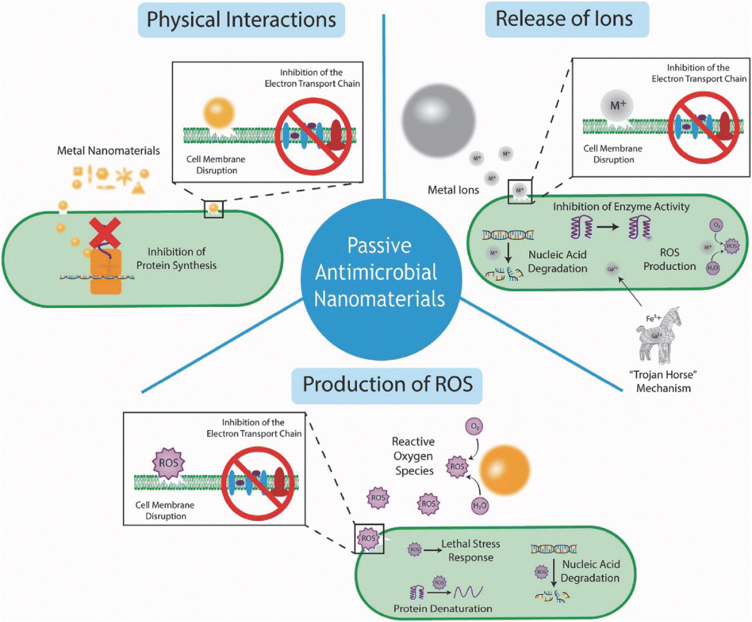 | ||
| Fig. 2 A schematic illustration depicting the various passive antimicrobial mechanisms of metal nanomaterials (not to scale), including physical interactions, ion release, and ROS production.80 Copyright Cheeseman et al. 2020 Springer Wiley. | ||
In terms of antibacterial properties, nanosheets also exhibit unique advantages.100 In the study by Feng et al. Ti carbide nanosheets were combined with graphitic carbon nitride nanosheets to prepare nanocomposites via electrostatic self-assembly. These nanocomposites were then incorporated into a PLLA matrix to fabricate bone scaffolds using selective laser sintering (SLS). The results indicated that the incorporation of the nanosheet composites into the scaffold material via 3D printing endowed the bone scaffolds with good antibacterial properties.101 Zeolite imidazolate frameworks (ZIFs) are a class of crystalline porous materials, in which metal ions and organic ligands are connected by coordination bonds to form a periodic network structure.102 Two-dimensional (2D) ZIF nanosheets have attracted increasing attention due to their ultrathin thickness, large surface area, abundant active sites, and excellent chemical and mechanical stability. In the study by Zhu et al. a novel copper-functionalized two-dimensional zeolitic imidazolate framework-7 nanocomposites ZIF-7@Cu2O was successfully fabricated by in situ growth methodology, the antibacterial kinetics and bacterial adhesion properties were investigated. The results demonstrated that the material possesses excellent reactive ROS generation capacity, inhibits biofilm formation, and exhibits superior antibacterial efficacy.103 Additionally, nanosheets are also capable of generating reactive ROS to kill bacteria. Some nanosheets exhibit photothermal and photodynamic therapeutic effects. Under light irradiation, they can produce ROS or localized high temperatures, thereby achieving photothermal antibacterial and photodynamic antibacterial effects, in the study by Li et al. palladium nanosheets (PdNS) were synthesized. The PdNS exhibit intrinsic nanosword (mechanical cutting) effects and peroxidase-like activity. Under near-infrared (NIR) light irradiation, PdNS demonstrate photothermal effects by generating localized heat and photodynamic effects by producing singlet oxygen (1O2). Ultimately, these synergistic mechanisms induce metabolic energy disruption, elevated cellular reactive ROS levels, and excessive oxidative stress, while inhibiting nucleic acid synthesis within bacterial cells, thereby achieving highly efficient antibacterial efficacy,104 In summary, nanosheets hold numerous advantages for antibacterial applications. Their high specific surface area and good biocompatibility enable efficient loading of antibacterial agents and precise controlled release, thereby significantly enhancing antibacterial efficacy and reducing the risk of drug resistance. These features highlight the broad application prospects of nanosheets in the field of antimicrobial therapy.
CNTs have been demonstrated to possess broad-spectrum antibacterial activity against pathogenic microorganisms.108,109 The antibacterial mechanisms of CNTs are influenced by their intrinsic properties (such as structure, modifications, surface area, chemical nature, morphology, and solubility) as well as the type of bacteria and the interaction environment,110 Single-walled carbon nanotubes (SWCNTs) exhibit stronger antibacterial activity due to their higher toxicity to microorganisms. However, multi-walled carbon nanotubes (MWCNTs) are more suitable for biomedical synthesis applications. This is because MWCNTs have higher purity, lower toxicity to the human body, and a lower probability of defects occurring during functionalization. Venkatesan et al. synthesized chitosan-MWCNTs hydrogels via freeze-drying and found that with the increase of MWCNTs concentration, higher antibacterial activity was exhibited against S. aureus, E. coli, and Candida tropicalis.111
Ti and Ti alloys are frequently used in orthopedic surgical interventions.112 They possess several desirable properties, including low density, excellent corrosion resistance, high mechanical strength, and the ability to bond with bone and other tissues without cytotoxic effects. These characteristics make Ti-based materials highly suitable for orthopedic implants.113 As unique micro/nanostructures, TiO2 nanotubes possess a large specific surface area. TiO2 nanotubes with different diameters exhibit varying antibacterial properties. Heat-treated TiO2 nanotubes with a diameter of 80 nm demonstrate the most potent antibacterial effect.114 Similarly, TiO2 nanotubes also serve as excellent drug carriers. Yao et al. fabricated silver-loaded TiO2 nanotubes (TiO2 NT) via anodic oxidation and evaluated their cytocompatibility and antibacterial properties. The experimental results demonstrated no cytotoxicity, sustained antibacterial characteristics, and enhanced osteogenic potential.115 Feng et al. prepared TiO2 nanotubes via electrochemical anodic oxidation and adsorbed gentamicin sulfate (GS) onto them, followed by coating with a mixture film of GS and CS (referred to as GSCH). The study revealed that the GS-loaded TiO2 nanotubes coated with GSCH hold great potential as a biomaterial, exhibiting strong antibacterial properties and the ability to prevent periprosthetic infections.116
In summary, antimicrobial nano materials offer significant potential for various applications, particularly in infection prevention and the treatment of bone infections. Their excellent antibacterial properties, targeting capabilities, and controlled release mechanisms make them highly promising. These nano materials are expected to become one of the emerging strategies for orthopedic infection control in the future. These different kinds of antibacterial nano materials have broad application prospects in medical, food, and daily chemical fields. The appropriate nanomaterials should be selected according to the actual needs for specific use.
3. Type of scaffolds
Oral and injectable ABs are currently used for the prevention and bone infection treatment protocols117 and implantation of infected or injured bone tissue and bone grafts.118 However, due to the challenge of antibiotic resistance, long-term treatment and adverse reactions, the creation of novel therapeutic approaches is of urgent importance. Biocompatible antimicrobial carriers offer a potential alternative for the management and prevention of bone infections,119 enabling controlled release, targeted delivery, and the possibility of personalized therapy.Regenerative therapies and tissue engineering are creating new opportunities for creating scaffold biomaterials to replace bone grafts. Adding diverse bactericidal compounds, such as ABs, metal particles, metal NPs, and essential microbial inhibitors, can enhance antibacterial activity while the scaffold facilitates osteogenesis.120 Therefore, the combination of synthetic scaffolds with antibacterial agents to prevent bacterial infection is a potential solution for both bone regeneration and the control of bone infections.121 This article reviews the preparation of scaffolds and the drug loading methods of scaffolds.
3.1 Requirements of bone scaffold
In recent years, drug-loaded material technology has demonstrated significant promise in treating bone infections. By delivering antimicrobial agents directly to the site of infection and achieving high local drug concentrations, this system enhances therapeutic efficacy while minimizing the toxic side effects linked to systemic drug administration. Common drug-loaded materials like bone cement and biodegradable biomaterials can act as scaffolds or fillers to slowly release drugs and effectively control infections. Key factors for an ideal scaffold include material composition, biomimetic design, antibacterial performance, bone regeneration promotion, and their interplay. An ideal system should combine infection treatment and bone defect regeneration by using diverse matrices and suitable fabrication methods. This integrated approach avoids multiple surgeries and overcomes traditional challenges in bone infection and defect repair.An ideal scaffold for bone infection should possess the following integrated characteristics (Fig. 3):122 firstly, good mechanical properties are the foundation, providing stable support during the bone healing process.123 Secondly, biocompatibility is crucial, as natural biologically derived materials such as CS and hydroxyapatite (HAP) possess excellent biocompatibility and can effectively promote the growth and integration of bone tissue.124 In addition, the degradation rate of the scaffold should match the rate of bone regeneration to avoid adverse effects on the healing process due to premature or delayed degradation. A porous structure is essential for promoting cell infiltration, nutrient diffusion, and waste removal. Studies have shown that 3D-printed hierarchical porous scaffolds can significantly enhance cell infiltration and proliferation rates.125 Moreover, the customizability of the scaffold is essential to meet the specific needs of individual patients. The advent of 3D printing technology has enabled the precise fabrication of scaffolds tailored to the shape and size of bone defects.126 Additionally, the scaffold material should minimize cell stress to promote cell attachment and proliferation.127 Furthermore, an ideal bone infection scaffold should be capable of modulating the inflammatory response. This can be achieved by loading anti-inflammatory drugs or modifying the surface properties of the scaffold to effectively control inflammation, thereby facilitating bone healing.128
 | ||
| Fig. 3 Construction of a multifunctional filling scaffold for bone infection.122 (a) Mechanical properties. (b) Biocompatibility. (c) Biodegradation. (d) Multiple pore structure. (e) Customizability. (f) Cell stress. (g) Antibacterial. (h) Pro-inflammatory response. Copyright Qin et al. 2024 Springer Nature. | ||
In summary, the construction of an ideal bone infection scaffold requires a comprehensive consideration of its mechanical properties, biocompatibility, biodegradability, porous structure, customizability, cell stress, antibacterial properties, and the ability to modulate inflammatory responses. Through interdisciplinary research involving materials science, biomedical engineering, and clinical medicine, it is anticipated that more efficient and safer scaffolds for the treatment of bone infections can be developed.
3.2 Preparation of scaffolds
In tissue engineering applications, many techniques for the production of porous materials have been developed to be used as scaffolds, including electrospinning,129 phase separation,130 gas foaming,131 pore-forming agent methods,132 polymerized fiber mesh coating,133 self-assembly,134 membrane lamination,135 freeze-drying,136 3D printing,137 and bioprinting.138 The creation of antibacterial scaffolds in these approaches typically relies on the introduction of fillers with inherent antibacterial attributes.139 In this section, we will discuss techniques for making BTE scaffolds, including traditional methods and additive manufacturing like 3D and 4D printing. Some application scenarios are listed (Fig. 4 and Table 2). The types and methods of scaffold preparation were as follows: | ||
| Fig. 4 Antibacterial scaffold fabrication methods:139 (a) electrospinning; (b) phase separation; (c) gas foaming; (d) porogen leaching; (e) solution polymerization; (f) self-assembly; (g) 3D printing; (h) freeze drying. Copyright Serrano-Aroca et al. 2022 Elsevier. | ||
| Fabrication methods | Materials | Processing methods | Pore size | Mechanical strength | Culture cell | Type of bone repair | Ref. |
|---|---|---|---|---|---|---|---|
| Fused deposition modeling (FDM) | PLA/nano-hydroxyapatite (nHAP) | Hybrid printing | 50 and 80 nm | — | BMSCs | Bone defect | 161 |
| Selective laser sintering (SLS) | Poly(vinyl alcohol) (PVA)/HAP | Hybrid printing | Porosity was 68.3% | — | MC3T3-E1 | Bone defect | 162 |
| Digital light processing (DLP) | Polydopamine (PDA)/biphasic calcium phosphate (BCP) | Self-assembly Ca-P/PDA nanocoating | Pore size of 570 μm | Compressive strength (∼3.6 MPa) | BMSC | Bone defect | 163 |
| Electrostatic spinning | β-Tricalcium phosphate (β-TCP)/Mxene/poly(lactic acid-carbonate) (PDT) | Hybrid printing | PDT with an average diameter of 1.2 μm | Tensile strength (14.4 MPa) | BMSCs, HUVECs | Bone defect | 164 |
| Freeze drying | Gallium-based metal–organic framework (GaMOF)/Gelatin methacrylate (GelMA)/HA | Quaternized CS (QCS) coated | Average particle sizes were 907.3 nm | ∼350 kPa | RBMSCs | Bone infection | 165 |
| Gas foaming | Polycaprolactone (PCL)/strontium NPs (3DS-Sr) | Drug loading | The diameter was 527 ± 148 nm. The pore size was 58 ± 19 μm2 | — | HUVECs | Bone infection | 166 |
| Hot sintering | Poly(L-lactic acid)/bioactive glass matrix/PDA | Hybrid printing | Average particle sizes were 226.52 nm | Compressive strength and modulus were 28.40 ± 0.48 MPa and 0.8 ± 0.02 GPa | HOS cells | Osteosarcoma | 167 |
| Solvent casting/particulate leaching | HAP/PLGA | Hybrid printing | The average particle sizes were 232 μm, 323 μm, and 464 μm | MC3T3-E1, C2C12 | Bone defect | 168 | |
| Low temperature injection | PLGA/PLA/HA/CS | Nano-spraydrying technique | — | — | — | Bone infection | 169 |
| 3D printing | Alg/GOx/CaP@CAT | Cross-links | 800 μm | — | rBMSCs,HUVECs, and RAW 264.7 macrophages | Diabetic bone regeneration | 170 |
| 4D printing | TCP/P(DLLA-TMC) | Photothermal effect | 360 ± 40 μm | 2.2 ± 0.2 to 2.4 ± 0.2 MPa (20 °C), 1.5 ± 0.2 MPa (37 °C), 0.4 ± 0.05 MPa (45 °C) | BMSCs | Critical size bone defect | 171 |
(a) Electrospinning technology: electrospinning technology involves placing a polymer solution into a jetting device and generating an electric field at the nozzle using a high-voltage power supply.140 As the electric field strength increases, the liquid droplet is stretched to form a “Taylor cone” When the electrostatic force overcomes the surface tension, a jet is ejected from the Taylor cone. This jet undergoes oscillation and stretching under the influence of the electric field, and the solvent evaporates to form nanoscale fibers. These fibers are then randomly deposited onto a collector to form a nonwoven mat. Characterized by a large surface area, the substrate serves as an optimal environment for cell expansion and subsequent tissue regrowth. For instance, an antimicrobial scaffold can be created using polymers like CS that exhibit inherent antimicrobial properties.141
(b) Phase separation: phase separation is a method that induces phase separation to form porous structures by mixing polymer solutions with non-solvents.142 The principle involves the phase separation of polymer solutions under specific conditions (such as changes in temperature or concentration), resulting in the formation of polymer-rich and polymer-poor phases. By controlling the phase separation process, scaffolds with different pore structures can be obtained. The key lies in selecting appropriate polymer and solvent systems, as well as precisely controlling the operating conditions to achieve the desired pore structure. Combining phase separation techniques with other methods allows for constructing 3D scaffolds with precise pore configurations. This approach is extensively applied in the production of polymeric scaffold constructs, such as polylactic acid (PLA) scaffolds, for application in the realm of regenerative medicine.143,144
(c) Gas foaming: gas foaming is a method that forms porous structures by introducing gases into polymers.145 The principle involves dissolving gas into a polymer melt or solution, and then expanding the gas to form bubbles by reducing pressure or increasing temperature. Depending on the type of blowing agent used, gas foaming can be categorized into physical foaming and chemical foaming. Physical foaming generates gas through physical means, such as injecting high-pressure gas or evaporating low-boiling-point liquids. In contrast, chemical foaming releases gas through chemical reactions, such as the decomposition of blowing agents. The key strength of gas foam scaffold fabrication is that it avoids the use of chemicals and high-temperature conditions, avoiding these factors to damage cells, tissues, and microenvironment.146
(d) Porogen leaching: the porogen leaching method involves adding soluble porogens (such as salts, sugars, polymer microspheres, etc.) into the polymer matrix, followed by dissolving and removing these porogens using a solvent to create a porous structure. The principle is based on the distribution of the porogens within the polymer matrix, which forms temporary pore structures. Once the porogens are dissolved by the solvent, the spaces left behind become the pores of the scaffold. This method allows for precise control over the size and distribution of the pores and is suitable for the fabrication of scaffolds with specific pore structures.147,148 This technique is frequently integrated with melt molding to fabricate biodegradable polymer scaffolds,148 thereby establishing a foundation for numerous advancements in antibacterial scaffolds.
(e) Solution polymerization: solution polymerization is a method for fabricating scaffolds through polymerization reactions in a solvent.149 The principle involves dissolving monomers and initiators in a solvent, where they undergo chemical reactions to form polymers. During the polymerization process, the molecular weight and structure of the polymer can be regulated by controlling reaction conditions such as temperature and concentration.150 Additionally, solution polymerization can impart antibacterial properties to the scaffold by incorporating antibacterial nanomaterials, such as graphene oxide.151
(f) Self-assembly: self-assembly technology is based on the spontaneous organization of molecules in a specific medium to form ordered structures with specific functions. The principle involves the use of non-covalent interactions between molecules (such as hydrogen bonds, van der Waals forces, and electrostatic interactions) to enable the molecules to spontaneously aggregate in solution, forming nanofibers or other ordered structures. For example, amphiphilic peptides can self-assemble into three-dimensional nanofiber networks in aqueous solutions, which are used in tissue engineering.152,153
(g) 3D printing: 3D printing is an additive manufacturing technology controlled by computer-aided design (CAD) models, capable of precisely fabricating porous scaffolds with complex structures. The principle involves the layer-by-layer deposition or solidification of polymeric materials (such as filaments, powders, or liquids) to construct the scaffold according to the predefined three-dimensional model.154 Commonly used 3D printing techniques include fused deposition modeling (FDM), SLS, and stereolithography (SLA). Through 3D printing technology, patient-specific scaffolds can be precisely manufactured according to the shape and size of the bone defect. For instance, using FDM, a fusion peptide-modified poly(lactic-co-glycolic acid)(PLGA)/HA composite scaffold has been developed to effectively support bone defect regeneration and repair. Characterized by excellent antibacterial and cytocompatibility outcomes in vitro, these scaffolds also showed promising performance in an in vivo rabbit model, underlining their potential for bone regeneration owing to their compatibility, antibacterial efficacy, and mechanical attributes.155
(h) Freeze-drying: utilizing the principle of sublimation, freeze-drying technology is a go-to method for generating porous scaffolds in tissue engineering applications.156 First, the solvent was used to dissolve the polymer in order to construct a scaffold with high porosity. Subsequently, the mixture was frozen and solvent removed by sublimation, and the scaffold structure was finally obtained. This method has the advantage of simple operation and makes it possible to fabricate highly porous scaffolds with controllable pore size, a key requirement for tissue engineering. The highly porous structure of the scaffold can promote cell adhesion, proliferation, and differentiation by providing a suitable microenvironment for cell growth,157 thereby improving the functionality of the regenerated tissue. Therefore, freeze-drying technology has shown important application value and potential in the field of tissue engineering.156
In addition, using precise deposition techniques controlled by computers, bioprinting constructs 3D structures of tissues or organs by placing cells and biological materials accurately.158 Commonly used in tissue engineering, regenerative medicine, and drug screening, bioprinting techniques encompass laser-induced forward transfer, inkjet printing, and robotic dispensing.159 Through bioprinting, tissue structures similar to real organs can be created for studying disease mechanisms, testing drug effects, and functional organ transplantation such as kidney and liver in the future. Compared with traditional organ culture technology, bioprinting can significantly shorten the research and development time, improve the success rate of organ transplantation, and bring new opportunities for future medical research and clinical application.160
3.3 Loading scaffolds with antimicrobial agents
In the field of tissue engineering and regenerative medicine, the antibacterial properties of scaffold materials are of vital importance.172 Bacterial infection is a common challenge in bone repair processes and often leads to implant failure. However, the extensive use of ABs has limited local antibacterial effects at the site of infection and is prone to side effects. Therefore, the development of scaffolds loaded with antibacterial agents has become a research hotspot. These scaffolds, by incorporating antibacterial components into the material, can effectively inhibit bacterial growth, reduce inflammatory responses, and provide a safer and more conducive microenvironment for tissue regeneration.173,174 Moreover, scaffolds loaded with antibacterial agents can prolong the effective duration of drug action, reduce systemic toxicity, and enhance therapeutic efficacy.175 These scaffolds, characterized by their multifunctionality, biocompatibility, and customizability, exhibit broad application prospects and have become an important research direction in tissue engineering and regenerative medicine.Biodegradable and alternative natural and synthetic vectors, developed in recent years, now offer a means to deliver ABs and drugs locally to the site of infection, thanks to advancements in biomaterials.176 Combining scaffold implantation with local antibiotic delivery at the injury site may enhance bone regeneration while preventing infections. In this study, Iglesias-Mejuto et al. developed a novel approach combining 3D-printing of polysaccharide-based inks with supercritical (sc) CO2 drying to create methylcellulose aerogels containing nHAP for BTE. These aerogels were loaded with Van for treating bone infections. Biocompatibility tests and antibacterial experiments demonstrated that the scaffolds exhibited good biocompatibility, excellent bactericidal ability, and sustained antibiotic release, the drug-loaded aerogel has the advantages of high safety, low irritation and obvious mineralization ability (Fig. 5).177 It has been found that bisphosphonates, an anti-catabolic drug, exhibit potent suppression of bone resorptive activity through strong interaction with HAP crystals. Qayoom et al. used a composite of semi-hydrated calcium sulfate (CSH) and nHAP to deliver anti-tuberculosis drugs rifampin (RFP) and isoniazid (INH). Analysis revealed that the addition of drugs did not significantly alter the nanocement microstructure. The setting time of the composite increased with drug incorporation, rising with higher drug concentrations. Drug release studies revealed rapid release of INH and slower release of RFP, indicating different interactions between the ABs and nHAP. The biphasic ceramic-based delivery system effectively released antibacterial and anti-biofilm medications, showing potential for treating skeletal and joint tuberculosis.178
 | ||
| Fig. 5 Biological evaluation of 3D-printed aerogel scaffolds:177 (a) cell viability of NIH/3T3 cells after 24 and 72 hours of contact with aerogels, assessed using the MTT assay. (b) Mortality rate of brine shrimp after 24 hours of contact with aerogels; controls include DMSO (positive) and artificial seawater (negative). (c) HET-CAM test results for aerogels, with 0.9% NaCl (negative control) and 0.1 N NaOH (positive control). (d) Aerogels cultured for 24 hours on TSA plates and in TSB tubes inoculated with S. aureus. (e) Mineralization values of aerogel formulations at 14, 21, and 28 days, measured by Alizarin Red staining. (f) Alizarin Red staining images of MC3T3 cells in contact with aerogels (wet and dry forms) after 28 days (scale bar: 200 μm). Copyright Iglesias-Mejuto et al. 2024 Elsevier. | ||
Nanotechnology-based methods are gaining prominence as effective solutions for treating a range of bone diseases, such as infections, osteoporosis, and cancer. For instance, NPs made from mesoporous bioactive glass (MBG) exhibit superior structural and textural characteristics. MBG is a novel biomaterial primarily composed of oxides of silicon, calcium, and phosphorus. It features a large number of nanoscale pores within its structure, which endow it with a greater specific surface area, enhanced biocompatibility, and improved bioactivity, it is a good scaffold material.179 Incorporating bioactive materials and therapeutic ions into scaffolds significantly enhances their properties and biological functionality. Sandra developed a Zn2+-rich, curcumin-loaded mesoporous SiO2–CaO–P2O5 glass nanoparticle and evaluated its bone regeneration, antibacterial properties, and biocompatibility. The NPs effectively degraded bacterial biofilms, inhibited bacterial growth, and prevented biofilm formation while promoting osteoblast establishment. The combination of Zn2+ and curcumin synergistically improved antibacterial efficacy and bone regeneration, achieving dual benefits of facilitating bone healing and managing infections.180 Chen et al. Obtained polycaprolactone (PCL)/polylactic acid (PLA) core–shell porous drug-carrying nanofibers by using coaxial electrospinning technology and the nonsolvent-induced phase separation method. Roxithromycin (ROX), a kind of antibacterial agent, was encapsulated in the core layer. The further antimicrobial activity demonstrated that the inhibition zone diameter of the coaxial nanofibers with two different pore sizes was 1.70 ± 0.10 cm and 1.73 ± 0.23 cm, exhibiting a good antibacterial effect against S. aureus. Therefore, the prepared nanofibers with the coaxial porous structures could serve as promising drug delivery systems.181
Tissue engineering in bone regeneration focuses on the development of bioactive implants with bone-conducting and bone-inducing properties. The utilization of 3D printing to customize the implant structure to create a suitable bone scaffold to load and deliver antibacterial NPs. Yang et al. used 3D printing to create in situ scaffolds made of PCL containing ampicillin (Amp) and Mg microspheres. The 3D printing ensured uniform distribution of Mg and Amp within the PCL matrix, as confirmed by imaging and analysis. The scaffold's slow degradation enabled sustained drug release at the defect site. In vitro and in vivo experiments showed that the composite scaffold effectively inhibited E. coli and S. aureus growth, enhanced cell adhesion and proliferation, and exhibited strong osteoinductive properties. This dual-functional scaffold, combining antibacterial and osteogenic capabilities, offers a promising solution for infected bone defects.182 Qian et al. prepared a novel Ta/GelMA/PLGA/Van composite scaffold by encapsulating Van in PLGA microspheres by double emulsion method, and loading it into the additive manufacturing porous tantalum (AM-Ta) scaffold by GelMA hydrogel. It is used to repair infected skeletal defects. The physicochemical characteristics of the scaffold suggested that the release of Van was maintained for over 2 weeks. The outcomes of the biological experiments demonstrated that the composite scaffold exhibited excellent biocompatibility, significant antibacterial properties and osseointegration ability, indicating its great potential in clinical application.183
In conclusion, high loading and long-term slow release of antibacterial drugs can be achieved by using scaffolds to load antibacterial drugs. The drug is separated from the scaffold material and has no bearing on the scaffold's mechanical properties. The preparation is relatively simple and flexible, but there may still be an initial mass release problem of drug on the scaffold surface. In summary, this treatment modality combines the advantages of local targeted drug delivery, sustained release, and biocompatibility, and is a promising new method for the treatment of bone infection.
3.4 Antibacterial coatings
Orthopedic implants possess outstanding biocompatibility, biodegradability, porosity, and mechanical strength, yet they lack antibacterial properties. Therefore, various methods have been developed to achieve antimicrobial activity, and surface coating is a promising strategy that has been extensively employed to simultaneously address the poor mechanical properties of scaffolds while enhancing their degradation rate and biological characteristics.173,184Inhibiting bacterial growth and killing invading bacteria through various mechanisms are the ways in which both metal particles and metal NPs typically display their antimicrobial activity.185 Coatings made from antibacterial materials, like metal ions or metal NPs such as Ag, Cu, Zn, and Mg, capable of effectively curbing the proliferation of Gram-positive and Gram-negative bacteria. They are commonly employed as coatings or incorporated within orthopedic implants to strengthen their antimicrobial effects and mitigate infection risks. For instance, additively fabricated ZN–1Mg porous scaffolds, in conjunction with composite coatings, can modify biodegradability (Fig. 6). Van is anchored by polydopamine (PDA) coatings to enhance antibacterial properties, while bone morphogenetic protein 2 (BMP2) growth factors are loaded into HAP coatings to further promote osteogenic activity. This combination effectively supports bone recovery and enhances antibacterial functions.186 Rodríguez-Contreras et al. prepared a calcium Ag titanate coating on the Ti surface by thermochemical treatment to boost the bioactivity and antibacterial performance of 3D-printed porous Ti constructs. The antimicrobial efficacy of the coating was evaluated using Gram-negative bacteria, such as Pseudomonas aeruginosa and E. coli, in addition to Gram-positive bacteria, including S. aureus and Staphylococcus epidermidis. Incorporating Ag ions into the calcium titanate layer was found to produce AgNPs on the 3D Ti surface. These NPs displayed significant antibacterial efficacy, reducing bacterial adhesion and growth.187 To confer antibacterial properties, other metals like Zn, Mg, and Cu have been incorporated into implants. For example, Mg alloys coated with Zn can inhibit bacterial growth in vitro but exhibit poor osteogenic capacity. In the design of bone implants, both antibacterial and osteogenic abilities are crucial.79 Owing to their outstanding biocompatibility, Ti materials are extensively utilized in orthopedic repair, mechanical properties, and osteointegration, which facilitate the restoration and reconstruction of human functions. 3D-printed porous Ti scaffolds reduce the elastic modulus of Ti alloys and enhance osteointegration. However, the bioinertness of Ti alloys weakens the implant-bone tissue interface bonding. Cheng et al. found that doping calcium silicate coatings with multiple metal ions can endow Ti alloys with various biological properties. A CuO and SrO-doped calcium silicate coating was applied to the exterior of 3D-printed porous Ti alloy scaffolds using atmospheric plasma spraying. In vivo bone integration was evaluated using a rabbit bone defect model, showing that 2% CuO–10% SrO-doped calcium silicate modified scaffolds improved osteointegration. The in vitro antibacterial properties of the scaffolds were assessed through bacterial platform coating, co-culture liquid absorbance detection, and crystal violet staining, demonstrating good antibacterial performance. In summary, the composite scaffold combines osteointegration and antibacterial properties, holding great clinical potential.188
 | ||
| Fig. 6 Additively fabricated Zn–1Mg porous scaffold:193 (a) the structure and elemental distribution of Zn–1Mg powder, as well as the designed porous scaffold model. The designed scaffolds had a pore size of 600 μm, a pore interval of 450 μm and the porosity of 63.55%. The size distribution of powders was 15–53 μm. The content of the Mg element in powders was 1.001 ± 0.004 wt%. (b) Schematic diagram of the preparation process. The Zn–1Mg samples were preprocessed and activated with NaOH. After the first PDA coating deposition, they were washed and dried to form PV/Zn–1Mg. Then, HA coating deposition and BMP2 treatment were conducted. Finally, the samples were dried to obtain HB/PV/Zn–1Mg. (c) Overview images of the samples. Copyright Zhang et al. 2023 Elsevier. | ||
Beyond metallic materials, polymer coatings are highly promising because of their biocompatibility, biodegradability, structural resemblance to the extracellular matrix, and superior cell affinity.189 The antibacterial and mechanical properties of bioactive glass scaffolds can be enhanced by Alg coatings, without causing any significant changes to their interconnected porosity.190 For BTE, biomimetic nHAP/polymer composite scaffolds hold great promise due to their excellent mechanical properties and ideal bioactivity. A concentrated alginate/gelatin scaffold with a nHAP coating was created by combining 3D printing and in situ mineralization. The thickness of the coating was adjusted by manipulating the phosphate ion concentration in the printing ink. Compared to uncoated scaffolds, the scaffold with the nHAP coating had a Young's modulus that was doubled. Additionally, the coating not only significantly enhanced the proliferation and osteogenic differentiation of rat bone marrow stem cells but also elevated protein adsorption on the scaffold surface. These features make the biomimetic nHAP/hydrogel composite scaffold a strong contender for BTE applications.191 CS, as a natural polysaccharide material, has a structure close to the natural extracellular matrix, and has good biocompatibility. The positive charge carried by CS can interact with the negative charge on the bacterial cell membrane to destroy the bacterial cell membrane structure, thereby playing an antibacterial and killing role. Qian et al. developed an antibacterial biocomposite scaffold based on polyelectrolyte modification. Using 3D printing, strontium-doped hydroxyapatite (SrHA) was embedded into a PCL scaffold, which was then coated with carboxymethyl chitosan (CCS) and hyperbranched polylysine (HBPL) to adsorb SLIT3 protein. This design allows for controlled release of SLIT3 and Sr2+. The scaffold exhibits good biocompatibility, promotes cell proliferation and migration, and stimulates angiogenesis and osteogenesis. It also enhances these processes indirectly by promoting macrophage M2 polarization and increasing anti-inflammatory factors, rendering it a promising option for bone defect repair.192
In addressing bone infections, the preparation of antibacterial coating on the surface of tissue engineering scaffolds is a cutting-edge technology, which can significantly lower the risk of infection. By selecting appropriate antibacterial materials, not only can the growth of bacteria be inhibited, but also improve the adhesion and proliferation of bone cells to facilitate the process of bone remodeling, so as to achieve the dual effects of antibacterial and regeneration. In the future, with the progress of material science and biotechnology, the preparation of antibacterial coatings on the surface of tissue engineering scaffolds will be developed in the direction of more efficient and safer.
3.5 Mixing antimicrobial agents during scaffold fabrication
Bone regeneration is a clinical challenge that requires multiple approaches sometimes, it also includes the development of osteogenic and antibacterial biomaterials to treat the emergence of infectious processes that may be caused by surgery. The preparation of antibacterial agents and scaffold materials with bone repair can achieve uniform dispersed mixing of drugs and scaffold materials,194 avoid a large amount of initial release on the surface, and the preparation process is simple and one-time, which will streamline the production process and minimize the need for subsequent treatment. This integrated manufacturing process can reduce costs, improve production efficiency, and is a good means of drug delivery.195It has been found that local sustained release of antimicrobial agents can be attained via mixing antibacterial agents directly with the scaffold material. This approach is able to provide a higher antimicrobial concentration at a specific site of the scaffold or in areas where bacteria accumulate, thus effectively preventing the occurrence of infection.196 The antibacterial agent mixed with the scaffold material can also significantly improve the antibacterial ability of the scaffold. Flexible nanoporous filaments were created by Ahangar et al. using blends of thermoplastic polyurethane and polyvinyl alcohol (LAY-FOMM and LAY-FELT) through 3D printing, aimed at delivering ABs in orthopedic applications. These materials, LAY-FOMM and LAY-FELT, can be shaped into drug delivery scaffolds that release tetracycline sustainably over a period of three days. When tested with primary human fibroblasts, they displayed excellent biocompatibility and no signs of cytotoxicity, suggesting that they are promising candidates for localized antibiotic delivery and effective options for preventing infections in orthopedic reconstruction surgeries.196 In the study by Shuai et al. oxygen vacancy (OV) defects Fe-doped TiO2 (OV-FeTiO2) NPs with antibacterial properties were synthesized using nano TiO2 and Fe3O4 as precursors. These NPs were subsequently incorporated into a polycaprolactone/polyglycolic acid (PCLGA) biodegradable polymer matrix, and a composite bone scaffold was constructed via selective laser sintering. The results demonstrated that the composite scaffold exhibited broad-spectrum antibacterial characteristics. Moreover, it possessed mechanical properties that matched those of human cancellous bone and exhibited excellent biocompatibility, which is conducive to cell adhesion, proliferation, and osteogenic differentiation.197
Hybrid printing can not only endowing scaffolds with antibacterial properties, but also introduce components that promote bone and cell adhesion on demand, so that scaffolds can prevent infection, promote cell growth and tissue repair, and achieve “therapeutic integration”. HAP is a kind of bioceramics commonly used in orthopedics. It integrates well with human tissues, does not cause immune rejection, has good osteogenic activity, and is a good drug-loaded scaffold. Qayoom et al. integrated the wide-range antibiotic rifampicin (RFP) into a biphasic nHAP/calcium sulfate ceramic support scaffold (NC) to treat osteomyelitis (Fig. 7). The results indicate that in vivo experiments showed that RFP-loaded NC could completely eradicate pathogens in bone lesions, and trabecular bone was formed at the debridement site when implanted with NC + RFP, and the defect healed completely.198 Beenken et al. developed an osteogenic bone-filling scaffold made of degradable polyurethane (dPU), nHAP, and decellularized bovine bone particles (DBP). The scaffold serves as a local antibiotic carrier to mitigate the risk of infections in bone defects contaminated by S. aureus. Results showed a significant reduction in live bacteria and maintained good osteogenic and bone repair properties even with high Van concentrations.199
 | ||
| Fig. 7 A broad-spectrum antibiotic, RFP, was incorporated into a biphasic nHAP/calcium sulfate ceramic carrier (NC) system, and its in vitro biocompatibility and degradation were investigated:198 (a) scanning electron microscopy (SEM) images of the NC and NC + RFP scaffolds, with magnified views shown in the insets, revealed that incorporating RFP into the scaffolds did not significantly alter their microarchitecture. (b) and (c) SEM and confocal laser scanning microscopy (CLSM) images of MC3T3E1 cells cultured on the surfaces of NC and NC + RFP for 7 days, demonstrating uniform cell distribution. (d) MTT assay results for MC3T3E1 cells on NC and NC + RFP surfaces, monitored over a 1-week period. The results showed that the cells continued to grow and proliferate, and all had good biocompatibility. (e) In vitro degradation profiles of NC and NC + RFP. After 5 weeks, the percentage degradation of NC was 39.6 ± 0.98% and that of NC + RFP was higher, around 47.53 ± 0.94%, showing enhanced degradation of NC upon incorporation of RFP. Copyright Qayoom et al. 2020 American Chemical Society. | ||
Recent advances in regenerative medicine have provided many reliable strategies for bone-related treatments. Hydrogel scaffolds, notable for their biocompatibility and ability to mimic the extracellular matrix of the surrounding environment, offer excellent properties for biomedical applications, have been successfully used as materials for graft scaffolds, is a good hybrid printing material. Ramprasad and his colleagues fabricated a 3D Ti scaffold incorporating gelatin hydrogel enriched with strontium-doped Ag NPs (Sr–Ag NPs) was fabricated using metal laser sintering technology. The composite scaffold was evaluated for cell compatibility and osteogenic potential. It was found that the scaffold promoted Runx2 expression and matrix mineralization, while also demonstrating excellent antibacterial properties. These characteristics point to its potential effectiveness in addressing bone infections.200 Photothermal therapy (PTT) is a promising antibacterial method, which has been focused on for dealing with bone infections and tissue engineering. This is because the photothermal agents used in PTT can generate heat NIR laser irradiation to kill bacteria and eliminate biofilms, with the advantages of minimal side effects, strong tissue penetration, and remarkable therapeutic effects. GelMA is a gelatin engineered with methacrylate (MA) groups through photo-polymerization. In addition to demonstrat the physical crosslinking behavior induced by temperature changes, as seen in gelatin, it allows for permanent photo-crosslinking under mild conditions during bioprinting. Nie et al. developed a composite hydrogel scaffold composed of GelMA, β-TCP, NaAlg (Sr2+), and MXene (Ti3C2) (GTAM) via 3D printing technology. The photothermal effect of MXene enables the GTAM scaffold to effectively kill both Gram-positive and Gram-negative bacteria NIR irradiation. Rat bone marrow mesenchymal stem cells were mixed into the GTAM bioink for 3D bioprinting. The resulting cell–laden GTAM scaffold exhibited good biocompatibility and osteogenic capacity under the action of MXene, cross-linked Sr2+, and β-TCP. It accelerated the reparation of infections and bone remodeling, while exerting a dual synergistic effect on antibacterial and osteogenic performance.201
In summary, combining antibacterial agents with 3D-printed scaffolds enables the precise delivery of these agents straight to the infected area, thereby significantly increasing local drug concentration while effectively reducing the side effects associated with systemic administration. Moreover, this combination allows for the slow and sustained release of antibacterial agents, maintaining effective concentrations over an extended period and further enhancing therapeutic outcomes. By integrating antibacterial agents directly into the scaffold material, the scaffolds themselves acquire sustained antibacterial properties, which can effectively prevent infections following implantation. With the aid of 3D printing technology, personalized treatment plans can be adjusted to fit the particular needs of each patient. Overall, this type of antibacterial 3D-printed scaffold integrates multiple advantages such as targeted delivery, controlled release, and inherent antibacterial properties, offering a new solution for preventing and treating bone infections and holding great promise for clinical applications.
4. Application of drug-loaded scaffolds in bone infections
Bone infection is a serious clinical problem that causes great suffering to patients. The aim of treating bone infection is to completely eliminate the infectious focus, ensure soft tissue coverage, promote bone end healing, and preserve limb length and function. After complete removal of the infected lesion, a bone defect is often present, which is the site of recurrence of the infection. The histological and biomechanical repair these defects is a time-consuming process and has emerged as a focal point in osteomyelitis treatment research. At present, several challenges remain in the treatment of bone infections,202–205 such as, multi-drug resistant bacteria reduce the effect of ABs; biofilm formation can occur by bacteria in bone tissue and attach to the surface of sequestrum or implants to resist immune and antibiotic attacks; the persistence and recurrence of infection can be attributed to infected phagocytes, such as macrophages, which act as “Trojan horses” protecting bacteria from antibiotic action.Hydrogels are soft, water-rich materials that can encapsulate drugs to deliver antibacterial agents to infected sites, reducing infection risk. Their injectable nature allows them to fill bone defects, providing scaffolding for new bone growth and promoting regeneration. They can also carry bioactive molecules like ABs and growth factors. By adjusting their properties and structure, these agents' release rate and dosage can be controlled to prevent bacterial growth and enhance bone repair and regeneration, ultimately alleviating bone infections. Bastami et al. developed a 3D-printed hydrogel using alginate, gelatin, and bone NPs. This hydrogel promoted the adhesion, growth, and bone-forming ability of rat stem cells. Results showed that it supported bone regeneration comparable to traditional methods, with staining and protein expression confirming its effectiveness. The hydrogel has potential to enhance bone repair.206 In the study by Tang et al. an injectable gelatin hydrogel was created via wet spinning. Optimized for injectability, it was evaluated in a mouse cranial defect model, where it enhanced cell survival, sped up bone regeneration, and demonstrated favorable degradation and biocompatibility. It shows promise as a biomaterial for stem cell delivery and tissue regeneration.207 Dutta employed highly oxidized 2D carbon nitride (Ox-gCN) to enhance alginate/gelatin (ALG) hydrogels. These hydrogels exhibit anti-inflammatory properties, aid in bone healing, and possess hemostatic capabilities. Oxidized g-CN promotes stem cell adhesion and growth. The ALG/CN scaffold has antibacterial activity, supports favorable immune responses, and facilitates bone formation. In rat studies, it promoted new bone formation without inducing inflammation, demonstrating potential for bone repair applications.208 Therefore, injectable hydrogels are favored for small defects due to their advantages of simple manipulation, minimal trauma, and good filling effect.
Osteomyelitis is a chronic condition that demands prolonged treatment, and surgical debridement often results in larger bone defects. These defects require filling materials that can withstand stress. Solid scaffolds, known for their stability, complex design, and multifunctionality, can be optimized to match the mechanical properties of bone tissue. As a result, they are commonly used to fill and treat infectious bone defects. This mainly includes using 3D-printed solid scaffolds for bone infections and combining them with active factors, stem cells, and drugs. Liu et al. combined 2D nanostructures (graphene oxide and black phosphorus) with IL-4 into 3D scaffolds. These scaffolds improved cell adhesion, released growth-promoting ions, and created a healing immune environment. In rats, they enhanced bone regeneration and angiogenesis. The study shows this approach is effective for treating bone infections and promoting bone repair.209 Wu et al. created NPs with magnetic and ultrasound properties, loaded with curcumin and stem cell membranes. These were combined with CS and tricalcium phosphate to form a 3D-printed scaffold. The scaffold could disrupt bacterial biofilms and activate immune and bone-forming responses using ultrasound. In vivo tests showed it healed infected bone defects effectively without damaging nearby tissues.210
In addition, a reliable animal model is essential for preclinical research and should closely mimic the disease. Sheep possess bone micro- and macrostructures as well as biomechanical properties that are similar to those of humans, and their use in skeletal research has been well established. Boot et al. introduced a clinically relevant large-animal model of chronic methicillin-resistant staphylococcus aureus (MRSA) infection. In this model, an injectable, thermoresponsive, HA-based hydrogel scaffold loaded with gentamicin and Van outperformed current clinical practices, eradicating bacteria in all animals. Local antibiotic delivery using the injectable hydrogel scaffold significantly enhanced the success rate of treatment.211 Local antimicrobial therapy is a crucial component in managing orthopedic device-related infection (ODRI), traditionally delivered through polymethyl-methacrylate (PMMA) bone cement. However, PMMA has limitations due to its suboptimal antibiotic release profile and lack of biodegradability. In a recent study, researchers compared the efficacy of PMMA versus an antibiotic-loaded hydrogel in a single-stage revision for chronic MRSA ODRI in sheep, a bone infection model was established using 13 Swiss Alpine sheep, which were treated with either antibiotic-loaded bone cement rods or hydrogels. The results demonstrated that the antibiotic-loaded hydrogel scaffold was effective in the single-stage exchange for MRSA ORDI in sheep. It exhibited good biodegradability and favorable drug release characteristics, with broad potential applications. However, further research is needed to facilitate the translation of this preclinical approach to clinical trials.212
As research continues to advance, antimicrobial BTE materials will further develop new high-performance antimicrobial materials, such as stimulus-responsive smart drug delivery systems, to enhance antimicrobial effectiveness. Additionally, by integrating advanced manufacturing technologies, we can achieve personalized design and customized production of antimicrobial materials. We will also investigate the mechanisms by which antimicrobial materials influence the behavior of bone cells to optimize their bioactivity. Furthermore, we will conduct long-term animal experiments and clinical trials to verify the safety and efficacy of antimicrobial BTE materials. This will facilitate the industrialization of these materials to meet clinical needs and benefit more patients. In summary, the research on antimicrobial BTE materials is thriving and is expected to provide new solutions for clinical bone repair and regenerative therapies, ultimately improving the treatment experience for patients.
5. Conclusion
Bone infection, like osteomyelitis, is a serious skeletal disorder caused by bacterial infection, leading to bone destruction, functional loss, and potential systemic infection. Treatment is difficult due to bacterial biofilms, poor antibiotic penetration, and limited local blood supply. Traditional methods include long-term ABs and surgery, but they often result in high recurrence rates, long treatment times, and significant side effects. In recent years, antibiotic-resistant bacteria have made bone infection treatment more challenging, driving the need for better strategies. Tissue engineering scaffolds with antimicrobial agents are a new approach that combines tissue engineering and drug delivery. These scaffolds are made from biocompatible materials, loaded with antimicrobial agents, and implanted at the infection site. This review summarizes antimicrobial agents and their incorporation into drug-loaded scaffolds. Scaffold materials enable sustained release of antimicrobial agents at the infection site, increasing local drug concentration while reducing antibiotic dosage, minimizing resistance risk, and lowering systemic side effects. Some scaffold materials like HAP and PLA have antimicrobial properties and support osteoblast growth to promote bone regeneration. Scaffolds can also load multiple drugs (e.g., ABs and anti-inflammatory agents) to achieve synergistic effects, combining antibacterial and bone repair functions. Drug loading methods include direct incorporation, coating, or mixing antimicrobial agents during scaffold fabrication. However, there are still some limitations, such as difficulties in achieving precise control, issues related to biocompatibility, mismatch between degradation rates of scaffolds and bone regeneration rates, limited drug activity, and challenges in clinical translation.Although drug-loading methods in tissue engineering scaffolds have limitations, they hold great promise for treating bone infections. Future research can focus on several key areas: optimizing drug release systems to create smart, responsive scaffolds; developing new biocompatible materials to improve mechanical properties and controllable degradation rates; integrating physical modalities like PTT and electrical stimulation to create multifunctional scaffolds for synergistic effects; strengthening the link between basic research and clinical needs; optimizing manufacturing processes for large-scale scaffold production and clinical application; and leveraging 3D printing technology for precise customization and rapid fabrication of scaffolds. We believe that future advancements in optimizing drug release systems, enhancing scaffold materials, designing multifunctional scaffolds, and promoting clinical translation will lead to significant breakthroughs in treating bone infections, offering patients more efficient and safer options. Additionally, by incorporating precision medicine and personalized treatment concepts, drug-loading methods in tissue engineering scaffolds will become increasingly important in this area. With ongoing research, we expect to see even more breakthroughs in the treatment of bone infections.
Conflicts of interest
The authors declare that they have no known competing financial interests or personal relationships that could have appeared to influence the work reported in this paper.Data availability
Data sharing is not applicable to this article as no new data were created or analyzed in this study.Acknowledgements
This work is financially supported by Medical and Health Science and Technology Program of Zhejiang Province (no. 2024KY1458).References
- L. Huang, Z. Guo, X. Yang, Y. Zhang, Y. Liang, X. Chen, X. Qiu and X. Chen, Advancements in GelMA bioactive hydrogels: strategies for infection control and bone tissue regeneration, Theranostics, 2025, 15(2), 460–493 CrossRef.
- A. R. Zelmer, R. Nelson, K. Richter and G. J. Atkins, Can intracellular Staphylococcus aureus in osteomyelitis be treated using current antibiotics? A systematic review and narrative synthesis, Bone Res., 2022, 10(1), 53 CrossRef PubMed.
- F. S. Fu, H. H. Chen, Y. Chen, Y. Yuan, Y. Zhao, A. Yu and X. Z. Zhang, Engineered bacillus subtilis enhances bone regeneration via immunoregulation and anti-Infection, Bioact. Mater., 2025, 46, 503–515 Search PubMed.
- T. H. Leta, S. A. Lie, A. M. Fenstad, S. H. L. Lygre, M. Lindberg-Larsen, A. B. Pedersen, W. Annette, O. Rolfson, E. Bülow and L. N. Van Steenbergen, Periprosthetic Joint Infection After Total Knee Arthroplasty With or Without Antibiotic Bone Cement, JAMA Netw. Open, 2024, 7(5), e2412898 CrossRef.
- J. B. Doub, A. Kang, C. Lee, D. Dyalram, P. Shih, W. S. Twaddell and J. E. Lubek, Risk Factors for Infection Recurrence After Surgical Resection of Advanced Stage Osteonecrosis of the Mandible, J. Oral Maxillofac. Surg., 2024, 82(3), 332–340 CrossRef.
- Z. Chen, Q. Jin, J. Zhong, Z. Xie, Q. Chen, L. Li, J. Li, C. Zhao, J. Wang and X. Tang, Staphylococcus aureus blocks host autophagy through circSyk/miR-5106/Sik3 axis to promote progression of bone infection, PLoS Pathog., 2025, 21(1), e1012896 CrossRef.
- J. Kaur, M. Jagannathan, T. Jordan, D. Kinsey, M. Truppiano, A. Shallal and G. Suleyman, P-1843. Genomic surveillance of invasive Streptococcus pyogenes in Metropolitan Detroit, Open Forum Infect. Dis., 2025, 12(Suppl 1), ofae631.2004 CrossRef.
- S. Y. Chong, S. Q. Lew, T. Alam, C. A. Gaulke and G. W. Lau, Comparative analysis of the Streptococcus pneumoniae competence development in vitro versus in vivo during pneumonia-derived sepsis, Front. Microbiol., 2025, 16, 1540511 CrossRef.
- Z. Wang, M. Li, W. Li, L. He, L. Wang, K. Cai, X. Zhao, Y. Chen and D. Li, Hybrid Outer Membrane Vesicles with Genetically Engineering for Treatment of Implant-Associated Infections and Relapse Prevention Through Host Immunomodulation, Advanced, Science, 2025, 2415379 Search PubMed.
- A. Q. M. Taher, R. Aqel, M. Alnajjar, C. Walker, E. Repetto, M. Raad, F. G. Gomez, I. Nyaruhirira, J. Michel, P. Herard, R. Kanapathipillai, K. Moussally and M. Khalife, Posttraumatic Pseudomonas aeruginosa Osteomyelitis in Mosul and Gaza: A Retrospective Cohort Study, 2018–2022, Open Forum Infect. Dis., 2024, 11(10), ofae579 CrossRef.
- S. Hunter, E. Brown, H. Crawford and C. J. I. Grant, Epidemiology of childhood bone and joint disease during the COVID-19 pandemic in New Zealand, Infection, 2024, 1–6 Search PubMed.
- J. S. Gogia, J. P. Meehan, P. E. Di Cesare and A. A. Jamali, Local antibiotic therapy in osteomyelitis, Seminars in plastic surgery, Thieme Medical Publishers, 2009, pp. 100–107 Search PubMed.
- J. A. Wright and S. P. Nair, Int. J. Med. Microbiol., 2010, 300(2–3), 193–204 CrossRef PubMed.
- J. Varin-Simon, M. Colin, F. Velard, M. Tang-Fichaux, X. Ohl, C. Mongaret, S. C. Gangloff and F. Reffuveille, Correction: cutibacterium acnes biofilm formation is influenced by bone microenvironment, implant surfaces and bacterial internalization, BMC Microbiol., 2025, 25(1), 48 CrossRef PubMed.
- E. M. Halawa, M. Fadel, M. W. Al-Rabia, A. Behairy, N. A. Nouh, M. Abdo, R. Olga, L. Fericean, A. M. Atwa and M. El-Nablaway, Antibiotic action and resistance: updated review of mechanisms, spread, influencing factors, and alternative approaches for combating resistance, Front. Pharmacol., 2024, 14, 1305294 CrossRef.
- M. Zegre, E. Poljańska, L. A. Caetano, L. Gonçalves and A. Bettencourt, Research progress on biodegradable polymeric platforms for targeting antibiotics to the bone, Int. J. Pharm., 2023, 648, 123584 CrossRef PubMed.
- P. Wang, H. Wang, S. Qi, W. Wang and H. Lu, Synergistic effects of quaternary ammonium compounds and antibiotics on the evolution of antibiotic resistance, Water Res., 2025, 275, 123206 CrossRef PubMed.
- N. Brustad, F. Buchvald, S. K. Jensen, J. N. Kyvsgaard, N. Vahman, J. Thorsen, A.-M. M. Schoos, U. Nygaard, N. Vissing, J. Stokholm, K. Bønnelykke and B. Chawes, Burden of Infections in Early Life and Risk of Infections and Systemic Antibiotics Use in Childhood, JAMA Network Open, 2025, 8(1), e2453284 CrossRef PubMed.
- W. Shuaishuai, Z. Tongtong, W. Dapeng, Z. Mingran, W. Xukai, Y. Yue, D. Hengliang, W. Guangzhi and Z. Minglei, Implantable biomedical materials for treatment of bone infection, Front. Bioeng. Biotechnol., 2023, 11, 1081446 CrossRef PubMed.
- S. Mistry, S. Roy, D. Bhowmick, S. Mistry, J. Chakraborty, R. Roy and I. Das, Investigation of Antibiotic-Releasing Biodegradable Composite Bone Cements for Treating Experimental Chronic Maxillofacial Bone Infection, J. Biomed. Mater. Res., Part A, 2025, 113(1), e37858 CrossRef.
- R. He, Y. Gu, J. Jia, F. Yang, P. Wu, P. Feng and C. Shuai, Semiconductor photocatalytic antibacterial materials and their application for bone infection treatment, Nanoscale Horiz., 2025 10.1039/d4nh00542b.
- Z. Wang, J. Xu, J. Zhu, H. Fang, W. Lei, X. Qu, Y. Y. Cheng, X. Li, Y. Guan and H. Wang, Osteochondral Tissue Engineering: Scaffold Materials, Fabrication Techniques and Applications, Biotechnol. J., 2025, 20(1), e202400699 CrossRef PubMed.
- D. Zhang, W. Liu, X. D. Wu, X. He, X. Lin, H. Wang, J. Li, J. Jiang and W. Huang, Efficacy of novel nano-hydroxyapatite/polyurethane composite scaffolds with silver phosphate particles in chronic osteomyelitis, J. Mater. Sci.: Mater. Med., 2019, 30, 1–12 CrossRef.
- C. M. Murphy, M. G. Haugh and F. J. O'brien, The effect of mean pore size on cell attachment, proliferation and migration in collagen–glycosaminoglycan scaffolds for bone tissue engineering, Biomaterials, 2010, 31(3), 461–466 CrossRef.
- J. M. Sadowska, R. N. Power, K. J. Genoud, A. Matheson, A. González-Vázquez, L. Costard, K. Eichholz, P. Pitacco, T. Hallegouet and G. Chen, A multifunctional scaffold for bone infection treatment by delivery of microRNA therapeutics combined with antimicrobial nanoparticles, Adv. Mater., 2024, 36(6), 2307639 CrossRef.
- M. P. Ferraz, An Overview on the Big Players in Bone Tissue Engineering: Biomaterials, Scaffolds and Cells, Int. J. Mol. Sci., 2024, 25(7), 3836 CrossRef.
- M. J. B. Boyetey, S. Torgbo and P. Sukyai, Bio-scaffold for bone tissue engineering with focus on bacterial cellulose, biological materials for hydroxyapatite synthesis and growth factors, Eur. Polym. J., 2023, 194, 112168 CrossRef.
- S. Wu, B. Wu, Y. Liu, S. Deng, L. Lei and H. Zhang, Mini review therapeutic strategies targeting for biofilm and bone infections, Front. Microbiol., 2022, 13, 936285 CrossRef PubMed.
- G. L. Hobby, the antibacterial action of penicillin against Gram negative organisms, Science, 1944, 100(2605), 500–501 CrossRef PubMed.
- J. Williams and F. J. D. Moosdeen, In vitro antibacterial effects of cephalosporins, Drugs, 1987, 34, 44–63 CrossRef PubMed.
- M. Aldhaeefi, J. Pearson, S. Kanjilal and B. Dionne, 301. Penicillin Versus Cefazolin or Anti-staphylococcal Penicillins for Penicillin-Susceptible Staphylococcus aureus Bacteremia, Open Forum Infect. Dis., 2020, S149 CrossRef.
- C. L. Wu, J. Y. Hsueh, B. S. Yip, Y. H. Chih, K. L. Peng and J. W. Cheng, Antimicrobial peptides display strong synergy with vancomycin against vancomycin-resistant E. faecium, S. aureus, and wild-type E. coli, Int. J. Mol. Sci., 2020, 21(13), 4578 CrossRef.
- P. Periti, E. Mini and G. Mosconi, Antimicrobial prophylaxis in orthopaedic surgery: the role of teicoplanin, J. Antimicrob. Chemother., 1998, 41(3), 329–340 CrossRef.
- N. Namdar, B. Nayeri Fasaei, P. Shariati, S. M. Joghataei and A. Arpanaei, Mesoporous silica nanoparticles co-loaded with lysozyme and vancomycin for synergistic antimicrobial action, Sci. Rep., 2024, 14(1), 29242 CrossRef PubMed.
- E. Watanabe, K. Matsumoto, K. Ikawa, Y. Yokoyama, A. Shigemi, Y. Enoki, Y. Umezaki, K. Nakamura, K. Ueno and H. J. J. Terazono, Pharmacokinetic/pharmacodynamic evaluation of teicoplanin against Staphylococcus aureus in a murine thigh infection model, J. Global Antimicrob. Resist., 2021, 24, 83–87 CrossRef PubMed.
- L. Zhao, X. Guo, Z. Zhang, X. Lu, Q. Zeng, T. Fan, X. Zhang, F. Chen, M. Xu and M. Yuan, Novel berberine derivatives as adjuvants in the battle against Acinetobacter baumannii: a promising strategy for combating multi-drug resistance, Chin. Chem. Lett., 2024, 109506 CrossRef.
- T. Goyal, P. Dhamija, G. Vardhan, P. Gupta and V. Trikha, In vitro study of elution kinetics and biological activity of piperacillin/tazobactam and gentamicin in acrylic bone cement, Orthop. Traumatol.: Surg. Res., 2022, 108(8), 103230 Search PubMed.
- A. Y. Gahane, V. Singh, A. Kumar and A. K. Thakur, Development of mechanism-based antibacterial synergy between Fmoc-phenylalanine hydrogel and aztreonam, Biomater. Sci., 2020, 8(7), 1996–2006 RSC.
- R. Nir-Paz, D. Gelman, A. Khouri, B. M. Sisson, J. Fackler, S. Alkalay-Oren, L. Khalifa, A. Rimon, O. Yerushalmy, R. Bader, S. Amit, S. Coppenhagen-Glazer, M. Henry, J. Quinones, F. Malagon, B. Biswas, A. E. Moses, G. Merril, R. T. Schooley, M. J. Brownstein, Y. A. Weil and R. Hazan, Successful Treatment of Antibiotic-resistant, Poly-microbial Bone Infection With Bacteriophages and Antibiotics Combination, Clin. Infect. Dis., 2019, 69(11), 2015–2018 CrossRef.
- Y. Fan, M. McCanne, J. Yuh, S. Lekkala1, C. P. Leape, S. Hugard, A. Thomson, J. E. Collins, O. K. Muratoglu, M. Randolph and E. Oral, The efficacy of antibiotic-eluting material in a two-stage model of periprosthetic joint infection, J. Orthop. Res., 2024, 42(2), 460–473 CrossRef.
- A. K. Thabit, D. F. Fatani, M. S. Bamakhrama, O. A. Barnawi, L. O. Basudan and S. F. Alhejaili, Antibiotic penetration into bone and joints: an updated review, Int. J. Infect. Dis., 2019, 81, 128–136 CrossRef CAS.
- M. S. Torkington, M. J. Davison, E. F. Wheelwright, P. J. Jenkins, I. Anthony, A. M. Lovering, M. Blyth and B. Jones, Bone penetration of intravenous flucloxacillin and gentamicin as antibiotic prophylaxis during total hip and knee arthroplasty, Bone Joint J., 2017, 99-B(3), 358–364 CrossRef CAS.
- K. Yamada, K. Matsumoto, F. Tokimura, H. Okazaki and S. Tanaka, Are bone and serum cefazolin concentrations adequate for antimicrobial prophylaxis?, Clin. Orthop. Relat. Res., 2011, 469(12), 3486–3494 CrossRef PubMed.
- N. Osman, N. Devnarain, C. A. Omolo, V. Fasiku, Y. Jaglal and T. Govender, Surface modification of nano-drug delivery systems for enhancing antibiotic delivery and activity, Wiley Interdiscip. Rev.: Nanomed. Nanobiotechnol., 2022, 14(1), e1758 CAS.
- A. Gonzalez Gomez and Z. Hosseinidoust, Liposomes for Antibiotic Encapsulation and Delivery, ACS Infect. Dis., 2020, 6(5), 896–908 CrossRef CAS PubMed.
- S. M. Collins and A. C. Brown, Bacterial Outer Membrane Vesicles as Antibiotic Delivery Vehicles, Front. Immunol., 2021, 12, 733064 CrossRef CAS.
- V. N. Nguyen, S. L. Wang, T. H. Nguyen, V. B. Nguyen, M. D. Doan and A. D. Nguyen, Preparation and Characterization of Chitosan/Starch Nanocomposites Loaded with Ampicillin to Enhance Antibacterial Activity against Escherichia coli, Polymers, 2024, 6(18), 2647 CrossRef.
- Z. Hao, R. Chen, C. Chai, Y. Wang, T. Chen, H. Li, Y. Hu, Q. Feng and J. Li, Antimicrobial peptides for bone tissue engineering: diversity, effects and applications, Front. Bioeng. Biotechnol., 2022, 10, 1030162 CrossRef.
- K. Zou, K. Yin, S. Ren, R. Zhang, L. Zhang, Y. Zhao and R. Li, Activity and mechanism of action of antimicrobial peptide ACPs against Candida albicans, Life Sci., 2024, 350, 122767 CrossRef CAS.
- H. Zhuo, X. Zhang, M. Li, Q. Zhang and Y. Wang, Antibacterial and Anti-Inflammatory Properties of a Novel Antimicrobial Peptide Derived from LL-37, Antibiotics, 2022, 11(6), 754 CrossRef CAS.
- Y. Umehara, M. Takahashi, H. Yue, J. V. Trujillo-Paez, G. Peng, H. L. Nguyen, K. Okumura, H. Ogawa and F. Niyonsaba, The Antimicrobial Peptides Human β-Defensins Induce the Secretion of Angiogenin in Human Dermal Fibroblasts, Int. J. Mol. Sci., 2022, 23(15), 8800 CrossRef CAS.
- J. Dai, N. G. Fischer, J. R. Rahimi, H. Wang, C. Hu, W. Chen, Y. Lin, T. Sang, H. P. Chew, L. Kong, C. Aparicio, Z. Ye and S. Huang, Interpenetrating nanofibrillar membrane of self-assembled collagen and antimicrobial peptides for enhanced bone regeneration, Int. J. Biol. Macromol., 2024, 267, 131480 CrossRef CAS.
- Y. Cheng, J. Qin, Y. Huang and T. Wang, The antimicrobial effects of PLGA microspheres containing the antimicrobial peptide OP-145 on clinically isolated pathogens in bone infections, Sci. Rep., 2022, 12(1), 14541 CrossRef CAS PubMed.
- W. Liang and J. Diana, The dual role of antimicrobial peptides in autoimmunity, Front. Immunol., 2020, 11, 2077 CrossRef CAS.
- Z. Wang, X. Chen, L. Yan, W. Wang, P. Zheng, A. Mohammadreza and Q. Liu, Antimicrobial peptides in bone regeneration: mechanism and potential, Expert Opin. Biol. Ther., 2024, 24(4), 285–304 CrossRef CAS PubMed.
- M. Wenzel, A. I. Chiriac, A. Otto, D. Zweytick, C. May, C. Schumacher, R. Gust, H. B. Albada, M. Penkova and U. Krämer, Small cationic antimicrobial peptides delocalize peripheral membrane proteins, Proc. Natl. Acad. Sci. U. S. A., 2014, 111(14), E1409–E1418 CrossRef CAS.
- K. Bush, Introduction to antimicrobial therapeutics reviews: the bacterial cell wall as an antimicrobial target, Ann. N. Y. Acad. Sci., 2013, 1277(1), v–vii Search PubMed.
- J. Medeiros-Silva, S. Jekhmane, E. Breukink and M. Weingarth, Towards the native binding modes of antibiotics that target lipid II, ChemBioChem, 2019, 20(14), 1731–1738 CrossRef CAS PubMed.
- Y. Liang, Y. Zhang, Y. Huang, C. Xu, J. Chen, X. Zhang, B. Huang, Z. Gan, X. Dong, S. Huang, C. Li, S. Jia, P. Zhang, Y. Yuan, H. Zhang, Y. Wang, B. Yuan, Y. Bao, S. Xiao and M. Xiong, Helicity-directed recognition of bacterial phospholipid via radially amphiphilic antimicrobial peptides, Sci. Adv., 2024, 10(35), eadn9435 CrossRef CAS.
- P. Zou, W. T. Chen, T. Sun, Y. Gao, L. L. Li and H. Wang, Recent advances: peptides and self-assembled peptide-nanosystems for antimicrobial therapy and diagnosis, Biomater. Sci., 2020, 8(18), 4975–4996 RSC.
- A. Bin Hafeez, X. Jiang, P. J. Bergen and Y. Zhu, Antimicrobial peptides: an update on classifications and databases, Int. J. Mol. Sci., 2021, 22(21), 11691 CrossRef CAS.
- J. Batista Araujo, G. Sastre de Souza and E. N. Lorenzon, Indolicidin revisited: biological activity, potential applications and perspectives of an antimicrobial peptide not yet fully explored, World J. Microbiol. Biotechnol., 2022, 38, 1–11 CrossRef.
- M. Yasir, D. Dutta, N. Kumar and M. D. P. Willcox, Interaction of the surface bound antimicrobial peptides melimine and Mel4 with Staphylococcus aureus, Biofouling, 2020, 36(9), 1019–1030 CAS.
- N. Nazeer, N. Kooner, A. Ghimire, J. K. Rainey, W. D. Lubell, D. Meneksedag-Erol and M. Ahmed, Secondary Structure Stabilization of Macrocyclic Antimicrobial Peptides via Cross-Link Swapping, J. Med. Chem., 2024, 67(11), 8693–8707 CrossRef CAS PubMed.
- P. Gagat, M. Ostrówka, A. Duda-Madej and P. Mackiewicz, Enhancing Antimicrobial Peptide Activity through Modifications of Charge, Hydrophobicity, and Structure, Int. J. Mol. Sci., 2024, 25(19), 10821 CrossRef CAS.
- Y. Wang, G. Zhu, W. Wang, Y. Zhang, Y. Zhu, J. Wang, M. Geng, H. Lu, Y. Chen, M. Zhou, J. Chen, F. Zhang, J. Yang and X. Cheng, Rational design of HJH antimicrobial peptides to improve antimicrobial activity, Bioorg. Med. Chem. Lett., 2023, 83, 129176 CrossRef CAS PubMed.
- B. Le Huy, H. B. T. Phuong, B. N. T. Thanh, Q. T. Van, H. V. Dinh and H. L. Xuan, Influence of hydrophobicity on the antimicrobial activity of helical antimicrobial peptides: a study focusing on three mastoparans, Mol. Diversity, 2024 DOI:10.1007/s11030-024-11046-w.
- Y. Qi, Q. Chen, X. Cai, L. Liu, Y. Jiang, X. Zhu, Z. Huang, K. Wu, H. Luo and Q. Ouyang, Self-Assembled Amphiphilic Chitosan Nanomicelles: Synthesis, Characterization and Antibacterial Activity, Biomolecules, 2023, 13(11), 1595 CrossRef CAS PubMed.
- Y. Sang, Z. Zhang, Q. Ren, J. Zhu and Y. He, Rapid Evaluation of Antimicrobial Potency Through Bacterial Collective Motion Analysis, ACS Appl. Bio Mater., 2024, 8(1), 519–526 CrossRef PubMed.
- A. Machado, M. Gama and J. A. Martins, Development of a dextrin–vitamin D3 micelle nanocarrier for the antimicrobial peptide LLKKK18 as a potential therapeutic agent for bone infections, J. Mater. Chem. B, 2024, 12(44), 11464–11476 RSC.
- Y. Lin, Y. Tan, R. Deng, L. Gong, X. Feng, Z. Cai, Y. He, L. Feng, B. Cheng and Y. Chen, Antibacterial Antimicrobial Peptide Grafted HA/SF/Alg Wound Dressing Containing AIEgens for Infected Wound Treating, ACS Omega, 2024, 9(22), 23499–23511 CrossRef CAS PubMed.
- H. Zhang, P. Zhang, X. Shen, J. Han, H. Wang, H. Qin, B. Wang, J. Qian, A. Udduttula, R. Luo, K. Zhao, Y. Wang and Y. Chen, A cross-linked coating loaded with antimicrobial peptides for corrosion control, early antibacterial, and sequential osteogenic promotion on a magnesium alloy as orthopedic implants, Acta Biomater., 2025, 193, 604–622 CrossRef CAS.
- S. Du, X. Liu, X. Hu and P. Zhan, Targeting novel sites represents an effective strategy for combating drug resistance, CCL, 2024, 110378 Search PubMed.
- H. T. Draviana, I. Fitriannisa, M. Khafid, D. I. Krisnawati, Widodo, C. H. Lai, Y. J. Fan and T. R. Kuo, Size and charge effects of metal nanoclusters on antibacterial mechanisms, J. Nanobiotechnol., 2023, 21(1), 428 CrossRef CAS PubMed.
- Y. Zheng, M. Wei, H. Wu, F. Li and D. Ling, Antibacterial metal nanoclusters, J. Nanobiotechnol., 2022, 20(1), 328 CrossRef PubMed.
- L. Wang, C. Hu and L. Shao, The antimicrobial activity of nanoparticles: present situation and prospects for the future, Int. J. Nanomed., 2017, 1227–1249 CrossRef.
- S. Shaikh, N. Nazam, S. M. D. Rizvi, K. Ahmad, M. H. Baig, E. J. Lee and I. Choi, Mechanistic Insights into the Antimicrobial Actions of Metallic Nanoparticles and Their Implications for Multidrug Resistance, Int. J. Mol. Sci., 2019, 20(10), 2468 CrossRef.
- J. Sahoo and M. De, Gram-selective antibacterial activity of mixed-charge 2D-MoS2, J. Mater. Chem. B, 2022, 10(24), 4588–4594 RSC.
- Z. Y. Chen, S. Gao, Y. W. Zhang, R. B. Zhou and F. Zhou, Antibacterial biomaterials in bone tissue engineering, J. Mater. Chem. B, 2021, 9(11), 2594–2612 RSC.
- S. Cheeseman, A. J. Christofferson, R. Kariuki, D. Cozzolino, T. Daeneke, R. J. Crawford, V. K. Truong, J. Chapman and A. Elbourne, Antimicrobial Metal Nanomaterials: From Passive to Stimuli-Activated Applications, Adv. Sci., 2020, 7(10), 1902913 CrossRef CAS PubMed.
- D. Li, L. Chen and X. Qiu, Rapid synthesis of ferulic acid-derived lignin coated silver nanoparticles with low cytotoxicity and high antibacterial activity, Int. J. Biol. Macromol., 2024, 277, 134471 CrossRef CAS PubMed.
- R. M. El-Sharkawy, I. A. Ahmed and T. G. Kharboush, Fusarium oxysporum assisted green synthesis of small-sized silver nanoparticles for high antibacterial, and photocatalytic decolorization performances, BMC Microbiol., 2025, 25(1), 4 CrossRef CAS.
- P. He, W. Wang and W. Jian, Antibacterial activity against pathogenic Vibrio and cytotoxicity on human hepatocyte of nano-silver prepared by polysaccharide-protein complexes, Front. Microbiol., 2024, 15, 1416844 CrossRef PubMed.
- F. Lin, Q. Qi, J. Zhang, W. Zhou, J. Zhang, P. Fu, X. Zhang, X. Qiao, M. Liu, X. Pang and Z. Cui, From Unimolecular Template to Silver Nanocrystal Clusters: An Effective Strategy to Balance Antibacterial Activity and Cytotoxicity, ACS Appl. Mater. Interfaces, 2021, 13(33), 39806–39818 CrossRef CAS.
- Y. Guo, W. Niu, Y. Xiao, J. Wen, H. Wang, X. Gao, B. Niu and W. Li, Synthesis and antimicrobial properties of ACS with AgNPs@MMT, Materials Today, Chemistry, 2025, 43, 102507 CAS.
- D. E. Burmistrov, A. V. Simakin, V. V. Smirnova, O. V. Uvarov, P. I. Ivashkin, R. N. Kucherov, V. E. Ivanov, V. I. Bruskov, M. A. Sevostyanov, A. S. Baikin, V. A. Kozlov, M. B. Rebezov, A. A. Semenova, A. B. Lisitsyn, M. V. Vedunova and S. V. Gudkov, Bacteriostatic and Cytotoxic Properties of Composite Material Based on ZnO Nanoparticles in PLGA Obtained by Low Temperature Method, Polymers, 2021, 14(1), 49 CrossRef.
- J. Ahmed, Farhana, G. A. Manzoor, A. R. Kaleri, B. Zulfiqar, A. Waheed, R. Iqbal and M. Rahimi, In vitro antifungal potential of myco versus bacteria synthesized ZnO NPs against chickpea and apricot pathogen, Sci. Rep., 2025, 15(1), 148 CrossRef CAS.
- L. Motelica, B. S. Vasile, A. Ficai, A. V. Surdu, D. Ficai, O. C. Oprea, E. Andronescu, G. Mustatea, E. L. Ungureanu and A. A. Dobre, Antibacterial Activity of Zinc Oxide Nanoparticles Loaded with Essential Oils, Pharmaceutics, 2023, 15(10), 2470 CrossRef CAS.
- K. J. Orie and B. I. Okocha, Biosynthesis and antimicrobial studies of zinc oxide nanoparticles of Vernonia amygdalina Leaf with varying concentration of zinc oxide, Inorg. Chem. Commun., 2024, 162, 112178 CrossRef CAS.
- P. Feng, H. Tian, F. Yang, S. Peng, H. Pan and C. Shuai, Reduced graphene oxide-mediated electron-hole separation using titanium dioxide increases the photocatalytic antibacterial activity of bone scaffolds, Bio-Des. Manuf., 2025, 8(1), 100–115 CrossRef.
- M. Shahriarinour, F. Divsar, A. Mehdipour, L. Youseftabar-Miri and V. Barkhordri, Antibacterial Properties of Cobalt Ferrite Magnetic Nanoparticles Loaded on Date Palm Pollen Against Multidrug-Resistant Bacteria, Arabian J. Sci. Eng., 2023, 48(6), 7315–7322 CrossRef CAS.
- S. J. Lam, E. H. H. Wong, C. Boyer and G. G. Qiao, Antimicrobial polymeric nanoparticles, Prog. Polym. Sci., 2018, 76, 40–64 CrossRef CAS.
- Q. Xin, H. Shah, A. Nawaz, W. Xie, M. Z. Akram, A. Batool, L. Tian, S. U. Jan, R. Boddula, B. Guo, Q. Liu and J. R. Gong, Antibacterial Carbon-Based Nanomaterials, Adv. Mater., 2019, 31(45), 1804838 CrossRef CAS.
- Y. Zhao, T. Fan, J. Chen, J. Su, X. Zhi, P. Pan, L. Zou and Q. Zhang, Magnetic bioinspired micro/nanostructured composite scaffold for bone regeneration, Colloids Surf., B, 2019, 174, 70–79 CrossRef CAS.
- V. Susithra, S. S. Kavi, A. F. A. El-Rehim and E. R. Kumar, Citrus sinensis assisted biogenic synthesis and physicochemical properties of Fe3O4 nanoparticles for antibacterial activity, Ceram. Int., 2024, 50(7, Part A), 10225–10231 CrossRef.
- S. L. Bee, Y. Bustami, A. Ul-Hamid, K. Lim and Z. A. Abdul Hamid, Synthesis of silver nanoparticle-decorated hydroxyapatite nanocomposite with combined bioactivity and antibacterial properties, J. Mater. Sci.: Mater. Med., 2021, 32(9), 106 CrossRef CAS PubMed.
- T. Taniguchi, L. Nurdiwijayanto, R. Ma and T. Sasaki, Chemically exfoliated inorganic nanosheets for nanoelectronics, Appl. Phys. Rev., 2022, 9(2), 021313 CAS.
- Y. Tao, W. Ji, X. Ding and B. H. Han, Exfoliated covalent organic framework nanosheets, J. Mater. Chem. A, 2021, 9(12), 7336–7365 RSC.
- Y. A. Fesseha, K. K. Kuchayita, W. N. Su, C. W. Chiu and C. C. Cheng, Organic–inorganic conjugated polymer-exfoliated tungsten disulfide nanosheet P–N heterojunction composites for efficient and stable hydrogen evolution reactions, Chem. Eng. J., 2025, 509, 161501 CrossRef CAS.
- X. Liu, Z. Yang, Y. Yang and H. Li, Construction of carbon quantum dots sensitized porous carbon nitride/titanium dioxide nanosheets for enhancing visible light photocatalytic degradation of tetracycline, J. Environ. Chem. Eng., 2022, 10(4), 108083 CrossRef CAS.
- P. Feng, R. He, F. Yang, Y. Lin, L. Fan, H. Pan and C. Shuai, Photocatalytic antibacterial scaffold for inhibiting bacterial infection: Carriers separation, ROS generation and antibacterial action, Sustainable Mater. Technol., 2025, 43, e01205 CrossRef CAS.
- R. Ahmad, U. A. Khan, N. Iqbal and T. Noor, Zeolitic imidazolate framework (ZIF)-derived porous carbon materials for supercapacitors: an overview, RSC Adv., 2020, 10(71), 43733–43750 RSC.
- G. Zhu, F. Kang, Q. Tan, H. Li, G. Zhang, M. Wu, Y. Tan and F. Q. Liu, Construction of copper-functionalized two-dimensional zeolitic imidazole framework-7 (ZIF-7) nanosheets for enhanced antibacterial and antibiofilm performance, Prog. Org. Coat., 2024, 190, 108383 CrossRef CAS.
- S. Li, M. Lu, C. Dai, B. Xu, N. Wu, L. Wang, C. Liu, F. Chen, H. Yang and Z. Huang, Advanced Palladium Nanosheet-Enhanced Phototherapy for Treating Wound Infection Caused by Multidrug-Resistant Bacteria, Small, 2025, 21(6), 2407180 CrossRef CAS.
- T. Mocan, C. T. Matea, T. Pop, O. Mosteanu, A. D. Buzoianu, S. Suciu, C. Puia, C. Zdrehus, C. Iancu and L. Mocan, Carbon nanotubes as anti-bacterial agents, Cell. Mol. Life Sci., 2017, 74, 3467–3479 CrossRef CAS.
- L. Ju, Y. Dai, W. Wei, M. Li, Y. Liang and B. Huang, One-dimensional cadmium sulphide nanotubes for photocatalytic water splitting, Phys. Chem. Chem. Phys., 2018, 20(3), 1904–1913 RSC.
- Z. Akram, A. Kausar and M. Siddiq, Engineering, Scrutinization of polystyrene microsphere-grafted multiwalled carbon nanotube and silver nanoparticle-based hybrids: morphology, thermal properties, and antibacterial activity, Polym.-Plast. Technol. Mater., 2017, 56(2), 202–215 CrossRef CAS.
- N. Jayaprakash, K. Elumalai, S. Manickam, G. Bakthavatchalam and P. Tamilselvan, Carbon nanomaterials: Revolutionizing biomedical applications with promising potential, Nano Mater. Sci., 2024 DOI:10.1016/j.nanoms.2024.11.004.
- M. Azizi-Lalabadi, H. Hashemi, J. Feng and S. M. Jafari, Carbon nanomaterials against pathogens; the antimicrobial activity of carbon nanotubes, graphene/graphene oxide, fullerenes, and their nanocomposites, Adv. Colloid Interface Sci., 2020, 284, 102250 CrossRef CAS.
- K. Nishshankage, A. B. Fernandez, S. Pallewatta, P. K. C. Buddhinie and M. Vithanage, Current trends in antimicrobial activities of carbon nanostructures: potentiality and status of nanobiochar in comparison to carbon dots, Biochar, 2024, 6(1), 2 CrossRef.
- J. Venkatesan, R. Jayakumar, A. Mohandas, I. Bhatnagar and S. K. Kim, Antimicrobial Activity of Chitosan-Carbon Nanotube Hydrogels, Materials, 2014, 7(5), 3946–3955 CrossRef CAS PubMed.
- K. Huo, X. Zhang, H. Wang, L. Zhao, X. Liu and P. K. Chu, Osteogenic activity and antibacterial effects on titanium surfaces modified with Zn-incorporated nanotube arrays, Biomaterials, 2013, 34(13), 3467–3478 CrossRef CAS.
- S. Mei, H. Wang, W. Wang, L. Tong, H. Pan, C. Ruan, Q. Ma, M. Liu, H. Yang, L. Zhang, Y. Cheng, Y. Zhang, L. Zhao and P. K. Chu, Antibacterial effects and biocompatibility of titanium surfaces with graded silver incorporation in titania nanotubes, Biomaterials, 2014, 35(14), 4255–4265 CrossRef CAS.
- B. Ercan, E. Taylor, E. Alpaslan and T. J. Webster, Diameter of titanium nanotubes influences anti-bacterial efficacy, Nanotechnology, 2011, 22(29), 295102 CrossRef PubMed.
- Y. Yao, P. Lin, D. Ye, H. Miao, L. Cao, P. Zhang, J. Xu and L. Dai, Enhanced Long-Term Antibacterial and Osteogenic Properties of Silver-Loaded Titanium Dioxide Nanotube Arrays for Implant Applications, Int. J. Nanomed., 2025, 3749–3764 CrossRef.
- W. Feng, Z. Geng, Z. Li, Z. Cui, S. Zhu, Y. Liang, Y. Liu, R. Wang and X. Yang, Controlled release behaviour and antibacterial effects of antibiotic-loaded titania nanotubes, Mater. Sci. Eng., C, 2016, 62, 105–112 CrossRef CAS PubMed.
- X. Wang, L. Fang, S. Wang, Y. Chen, H. Ma, H. Zhao and Z. Xie, Antibiotic treatment regimens for bone infection after debridement: a study of 902 cases, BMC Musculoskeletal Disord., 2020, 21, 1–6 CrossRef.
- A. A. Nasser, P. Fenton and D. Bose, Single stage versus two-stage orthoplastic management of bone infection, Injury, 2022, 53(3), 984–991 CrossRef.
- C. Wang, P. Xu, X. Li, Y. Zheng and Z. Song, Research progress of stimulus-responsive antibacterial materials for bone infection, Front. Bioeng. Biotechnol., 2022, 10, 1069932 CrossRef PubMed.
- K. Hayashi, M. Shimabukuro, C. Zhang, A. N. T. Alashkar, R. Kishida, A. Tsuchiya and K. Ishikawa, Silver phosphate-modified carbonate apatite honeycomb scaffolds for anti-infective and pigmentation-free bone tissue engineering, Mater. Today Bio, 2024, 27, 101161 CrossRef CAS PubMed.
- G. L. G. e Silva, M. S. D. S. de Bustamante, M. L. Dias, A. M. Costa, A. M. Rossi, A. P. dos Santos Matos, R. Santos-Oliveira and E. Ricci-Junior, Antibiotics-loaded nanofibers fabricated by electrospinning for the treatment of bone infections, Arabian J. Chem., 2023, 16(1), 104392 CrossRef.
- L. Qin, S. Yang, C. Zhao, J. Yang, F. Li, Z. Xu, Y. Yang, H. Zhou, K. Li, C. Xiong, W. Huang, N. Hu and X. Hu, Prospects and challenges for the application of tissue engineering technologies in the treatment of bone infections, Bone Res., 2024, 12(1), 28 CrossRef CAS.
- A. H. Foroughi and M. J. Razavi, Multi-objective Shape Optimization of Bone Scaffolds: Enhancement of Mechanical Properties and Permeability, Acta Biomater., 2022, 146, 317–340 CrossRef CAS.
- M. Ramezani Farani, I. Zare, M. Mirshafiei, A. Gholami, M. Zhang, E. Pishbin, J. E. Ahn, A. Mohammadi, M. Imani, M. Lak, J. Shen, H. Kang and Y. S. Huh, Graphene oxide-engineered chitosan nanoparticles: Synthesis, properties, and antibacterial activity for tissue engineering and regenerative medicine, Chem. Eng. J., 2025, 509, 160852 CrossRef CAS.
- L. Ruiz-Cantu, A. Gleadall, C. Faris, J. Segal, K. Shakesheff and J. Yang, Characterisation of the surface structure of 3D printed scaffolds for cell infiltration and surgical suturing, Biofabrication, 2016, 8(1), 015016 CrossRef PubMed.
- N. Sabahi, I. Roohani, C. H. Wang and X. Li, Material extrusion 3D printing of bioactive smart scaffolds for bone tissue engineering, Addit. Manuf., 2025, 98, 104636 CAS.
- G. Li, Q. Xiao, L. Zhang, Y. Zhao and Y. Yang, Nerve growth factor loaded heparin/chitosan scaffolds for accelerating peripheral nerve regeneration, Carbohydr. Polym., 2017, 171, 39–49 CrossRef CAS.
- A. Wang, X. Ma, Y. Yang, G. Shi, L. Han, X. Hu, R. Shi, J. Yan, Q. Guo and Y. Zhao, Biophysical-driven piezoelectric and aligned nanofibrous scaffold promotes bone regeneration by re-establishing physiological electrical microenvironment, Nano Res., 2024, 17(8), 7376–7393 CrossRef CAS.
- M. Rahmati, D. K. Mills, A. M. Urbanska, M. R. Saeb, J. R. Venugopal, S. Ramakrishna and M. Mozafari, Electrospinning for tissue engineering applications, Prog. Mater. Sci., 2021, 117, 100721 CrossRef CAS.
- N. Sheng, W. Lin, J. Lin, Y. Feng, Y. Wang, X. He, Y. He, R. Liang, Z. Li and J. Li, Cross-linking manipulation of waterborne biodegradable polyurethane for constructing mechanically adaptable tissue engineering scaffolds, Regener. Biomater., 2024, 11, rbae111 CrossRef CAS.
- S. A. Poursamar, A. N. Lehner, M. Azami, S. Ebrahimi-Barough, A. Samadikuchaksaraei and A. P. M. Antunes, The effects of crosslinkers on physical, mechanical, and cytotoxic properties of gelatin sponge prepared via in situ gas foaming method as a tissue engineering scaffold, Biomater. Adv., 2016, 63, 1–9 CAS.
- T. H. Thai, T. M. Nguyen, M. D. Truong, T. T. T. Phan, D. T. Le, H. N. Doan and T.-H. Nguyen, Fabrication of 3D PCL/PVP scaffolds using monosodium glutamate as porogen by solvent casting/particulate leaching method for oral and maxillofacial bone tissue engineering, Biomed. Mater., 2024, 19(5), 055030 CrossRef CAS PubMed.
- A. Naderi and G. Palardy, Comparison of microstructure and tensile behavior of hydroxyapatite-coated PEEK meshes and cellulose-based fabrics and mats, J. Ind. Text., 2022, 51(1_suppl), 1392S–1421S CrossRef CAS.
- T. Strunk, A. Joshi, M. Moeinkhah, T. Renzelmann, L. Dierker, D. Grotheer, N. Graupner, J. Müssig and D. Brüggemann, Structure, Properties and Degradation of Self-Assembled Fibrinogen Nanofiber Scaffolds, ACS Appl. Bio Mater., 2024, 7(9), 6186–6200 CrossRef CAS PubMed.
- L. Lu and A. G. Mikos, The importance of new processing techniques in tissue engineering, MRS Bull., 1996, 21(11), 28–32 CrossRef CAS.
- K. Joy, S. S. David, A. Shanmugavadivu, N. Selvamurugan and P. Mani, Polymer Edition, Three-dimensional porous polycaprolactone/chitosan/bioactive glass scaffold for bone tissue engineering, Polym. Ed., 2024, 35(18), 2829–2844 CAS.
- C. Wang, W. Huang, Y. Zhou, L. He, Z. He, Z. Chen, X. He, S. Tian, J. Liao and B. Lu, 3D printing of bone tissue engineering scaffolds, Bioact. Mater., 2020, 5(1), 82–91 Search PubMed.
- P. Rider, Ž. P. Kačarević, S. Alkildani, S. Retnasingh and M. Barbeck, Bioprinting of tissue engineering scaffolds, J. Tissue Eng., 2018, 9, 2041731418802090 CrossRef.
- Á. Serrano-Aroca, A. Cano-Vicent, R. Sabater i Serra, M. El-Tanani, A. Aljabali, M. M. Tambuwala and Y. K. Mishra, Scaffolds in the microbial resistant era: fabrication, materials, properties and tissue engineering applications, Mater. Today Bio, 2022, 16, 100412 CrossRef PubMed.
- T. Huang, Y. Zeng, C. Li, Z. Zhou, J. Xu, L. Wang, D. G. Yu and K. Wang, Application and Development of Electrospun Nanofiber Scaffolds for Bone Tissue Engineering, ACS Biomater. Sci. Eng., 2024, 10(7), 4114–4144 CrossRef CAS.
- S. Salehi, H. Ghomi, S. Hassanzadeh-Tabrizi, N. Koupaei and M. Khodaei, Antibacterial and osteogenic properties of chitosan-polyethylene glycol nanofibre-coated 3D printed scaffold with vancomycin and insulin-like growth factor-1 release for bone repair, Int. J. Biol. Macromol., 2025, 139883 CrossRef CAS PubMed.
- N. Munir and A. Callanan, Novel phase separated polycaprolactone/collagen scaffolds for cartilage tissue engineering, Biomed. Mater., 2018, 13(5), 051001 CrossRef CAS PubMed.
- W. Lan, M. Wang, Z. Lv, J. Li, F. Chen, Z. Liang, D. Huang, X. Wei and W. Chen, Carbon nanotubes-reinforced polylactic acid/hydroxyapatite porous scaffolds for bone tissue engineering, Front. Mater. Sci., 2024, 18(1), 240675 CrossRef CAS.
- S. Farzamfar, M. Naseri-Nosar, H. Sahrapeyma, A. Ehterami, A. Goodarzi, M. Rahmati, G. Ahmadi Lakalayeh, S. Ghorbani, A. Vaez and M. Salehi, Tetracycline hydrochloride-containing poly(ε-caprolactone)/poly lactic acid scaffold for bone tissue engineering application: in vitro and in vivo study, Int. J. Polym. Mater. Polym. Biomater., 2019, 68(8), 472–479 CrossRef CAS.
- C. Zhou, K. Yang, K. Wang, X. Pei, Z. Dong, Y. Hong and X. Zhang, Combination of fused deposition modeling and gas foaming technique to fabricated hierarchical macro/microporous polymer scaffolds, Mater. Des., 2016, 109, 415–424 CrossRef CAS.
- E. Bianchi, M. Ruggeri, B. Vigani, C. T. Fila, A. I. Cornaglia, C. Boselli, C. Viseras, S. Rossi and G. Sandri, Gas foamed scaffolds as smart 3D structures in skin tissue engineering, J. Drug Delivery Sci. Technol., 2024, 95, 105541 CrossRef CAS.
- C. Ruiz-Aguilar, U. Olivares-Pinto, R. Drew, E. Aguilar-Reyes and I. Alfonso, Porogen effect on structural and physical properties of β-TCP scaffolds for bone tissue regeneration, IRBM, 2021, 42(5), 302–312 CrossRef.
- M. Spedicati, A. Zoso, L. Mortati, V. Chiono, E. Marcello and I. Carmagnola, Three-Dimensional Microfibrous Scaffold with Aligned Topography Produced via a Combination of Melt-Extrusion Additive Manufacturing and Porogen Leaching for In Vitro Skeletal Muscle Modeling, Bioengineering, 2024, 11(4), 332 CrossRef CAS PubMed.
- B. Safari and A. Aghanejad, Porous gelatin-based phosphorylated scaffold: microstructure, cell response and osteogenic differentiation of human adipose-derived mesenchymal stem cells, J. Drug Delivery Sci. Technol., 2024, 99, 106008 CrossRef CAS.
- E. Hermosillo-Ochoa and N. A. Cortez-Lemus, End-group controlling aqueous solution properties in star-shaped poly(2-hydroxyethyl acrylate) and poly(2-hydroxyethyl acrylate)-b-poly(N-isopropylacrylamide) polymers, Polym. Sci., 2023, 61(9), 794–805 CrossRef CAS.
- A. Gautam, H. Dabral, A. Singh, S. Tyagi, N. Tyagi, D. Srivastava, H. R. Kushwaha and A. Singh, Graphene-based metal/metal oxide nanocomposites as potential antibacterial agents: a mini-review, Biomater. Sci., 2024, 12, 4630–4649 RSC.
- E. Utoiu, V. S. Manoiu, E. I. Oprita and O. Craciunescu, Bacterial Cellulose: A Sustainable Source for Hydrogels and 3D-Printed Scaffolds for Tissue Engineering, Gels, 2024, 10(6), 387 CrossRef CAS.
- N. Abie, C. Ünlü, A. R. Pinho, M. C. Gomes, T. Remmler, M. Herb, D. Grumme, E. Tabesh, M. A. Shahbazi and S. Mathur, Designing of a Multifunctional 3D-Printed Biomimetic Theragenerative Aerogel Scaffold via Mussel-Inspired Chemistry: Bioactive Glass Nanofiber-Incorporated Self-Assembled Silk Fibroin with Antibacterial, Antiosteosarcoma, and Osteoinductive Properties, ACS Appl. Mater. Interfaces, 2024, 16(18), 22809–22827 CrossRef CAS.
- S. C. Ligon, R. Liska, J. Stampfl, M. Gurr and R. Mülhaupt, Polymers for 3D Printing and Customized Additive Manufacturing, Chem. Rev., 2017, 117(15), 10212–10290 CrossRef CAS PubMed.
- Z. Liu, G. Tian, L. Liu, Y. Li, S. Xu, Y. Du, M. Li, W. Jing, P. Wei, B. Zhao, S. Ma and J. Deng, A 3D-printed PLGA/HA composite scaffold modified with fusion peptides to enhance its antibacterial, osteogenic and angiogenic properties in bone defect repair, J. Mater. Res. Technol., 2024, 30, 5804–5819 CrossRef CAS.
- M. Qamash, S. M. Imani, M. Omidi, C. Glancy and L. Tayebi, Investigation of the Effect of Preparation Parameters on the Structural and Mechanical Properties of Gelatin/Elastin/Sodium Hyaluronate Scaffolds Fabricated by the Combined Foaming and Freeze-Drying Techniques, J. Compos. Sci., 2024, 8(10), 408 CrossRef CAS.
- H. Yuan, X. Zhao, W. Zhao, Y. Zhang, Q. Ye, S. Liu, B. Yu, S. Ma and F. Zhou, Crystalline-Constrain Enables Preparation of Hierarchical Porous Hydrogels with High Porosity and Good Mechanics, Adv. Funct. Mater., 2024, 2416898 Search PubMed.
- P. Datta, A. Barui, Y. Wu, V. Ozbolat, K. K. Moncal and I. T. Ozbolat, Essential steps in bioprinting: From pre-to post-bioprinting, Biotechnol. Adv., 2018, 36(5), 1481–1504 CrossRef CAS PubMed.
- P. Jain, H. Kathuria, S. Ramakrishna, S. Parab, M. M. Pandey and N. Dubey, In Situ Bioprinting: Process, Bioinks, and Applications, ACS Appl. Bio Mater., 2024, 7(12), 7987–8007 CrossRef CAS PubMed.
- H. G. Yi, H. Kim, J. Kwon, Y. J. Choi, J. Jang and D. W. Cho, Application of 3D bioprinting in the prevention and the therapy for human diseases, Signal Transduction Targeted Ther., 2021, 6(1), 177 CrossRef CAS.
- W. Wang, B. Zhang, M. Li, J. Li, C. Zhang, Y. Han, L. Wang, K. Wang, C. Zhou, L. Liu, Y. Fan and X. Zhang, 3D printing of PLA/n-HA composite scaffolds with customized mechanical properties and biological functions for bone tissue engineering, Composites, Part B, 2021, 224, 109192 CrossRef CAS.
- T. Li, Z. Peng, Q. Lv, L. Li, C. Zhang, L. Pang, C. Zhang, Y. Li, Y. Chen and X. Tang, SLS 3D Printing To Fabricate Poly(vinyl alcohol)/Hydroxyapatite Bioactive Composite Porous Scaffolds and Their Bone Defect Repair Property, ACS Biomater. Sci. Eng., 2023, 9(12), 6734–6744 CrossRef CAS PubMed.
- Z. Yang, L. Xie, B. Zhang, G. Zhang, F. Huo, C. Zhou, X. Liang, Y. Fan, W. Tian and Y. Tan, Preparation of BMP-2/PDA-BCP Bioceramic Scaffold by DLP 3D Printing and its Ability for Inducing Continuous Bone Formation, Front. Bioeng. Biotechnol., 2022, 10, 854693 CrossRef.
- S. Zhang, L. Huang, M. Bian, L. Xiao, D. Zhou, Z. Tao, Z. Zhao, J. Zhang, L. B. Jiang and Y. Li, Multifunctional Bone Regeneration Membrane with Flexibility, Electric. Stimul. Activity Osteoinductive Activity, 2024, 20(47), 2405311 CAS.
- Y. Liu, K. Yuan, Y. Lin, Y. Yang, W. Kong, J. Shan, H. Niu, W. Kong, F. Li, X. Yue, Y. Du, Y. Liang, H. Chang, Z. Yu, J. Wang, G. Yang, L. Cao, K. Huang, S. Yang and T. Tang, Directional Freeze-Casting Cryogel Loaded with Quaternized Chitosan Modified Gallium Metal–Organic Frameworks to Capture and Eradicate the Resistant Bacteria for Guided Regeneration in Infected Bone Defects, Adv. Mater., 2025, 2414437 CrossRef CAS.
- Y. Chen, Y. Li, X. Wang, X. Mo, Y. Chen, Z. Deng, X. Ye and J. Yu, One-Step Gas Foaming Strategy for Constructing Strontium Nanoparticle Decorated 3D Scaffolds: a New Platform for Repairing Critical Bone Defects, ACS Appl. Mater. Interfaces, 2024, 16(45), 61664–61678 CrossRef CAS.
- Z. Yan, Y. Deng, L. Huang, J. Zeng, D. Wang, Z. Tong, Q. Fan, W. Tan, J. Yan, X. Zang and S. Chen, Biopolymer-based bone scaffold for controlled Pt(IV) prodrug release and synergistic photothermal-chemotherapy and immunotherapy in osteosarcoma, J. Nanobiotechnol., 2025, 23(1), 286 CrossRef CAS PubMed.
- C. Wang, R. Zhang, K. J. Jeong, W. Xiong, Z. Liu, Z. Xie, L. Hou, J. Gong, Z. Lian, M. Zu, K. Fan and T. Ji, Fabrication of a Whitlockite/PLGA Scaffold with Hierarchical Porosity for Bone Repair, Nano Lett., 2025, 25(11), 4386–4392 CrossRef CAS.
- R. K. Wassif, S. A. Elkheshen, R. N. Shamma, M. S. Amer, R. Elhelw and M. El-kayal, Injectable systems of chitosan in situ forming composite gel incorporating linezolid-loaded biodegradable nanoparticles for long-term treatment of bone infections, Drug Delivery Transl. Res., 2024, 14(1), 80–102 CrossRef CAS PubMed.
- C. Yang, Z. Zheng, M. R. Younis, C. Dong, Y. Chen, S. Lei, D. Y. Zhang, J. Wu, X. Wu, J. Lin, X. Wang and P. Huang, 3D Printed Enzyme-Functionalized Scaffold Facilitates Diabetic Bone Regeneration, Adv. Funct. Mater., 2021, 31(20), 2101372 CrossRef CAS.
- C. Wang, H. Yue, J. Liu, Q. Zhao, Z. He, K. Li, B. Lu, W. Huang, Y. Wei and Y. Tang, Advanced reconfigurable scaffolds fabricated by 4D printing for treating critical-size bone defects of irregular shapes, Biofabrication, 2020, 12(4), 045025 CrossRef CAS.
- A. Dubey, H. Vahabi and V. Kumaravel, Antimicrobial and Biodegradable 3D Printed Scaffolds for Orthopedic Infections, ACS Biomater. Sci. Eng., 2023, 9(7), 4020–4044 CrossRef CAS.
- M. Yekani, S. M. Dizaj, S. Sharifi, H. Sedaghat, M. Saffari and M. Y. Memar, Nano-scaffolds as drug delivery systems of antimicrobial agents for osteomyelitis treatment; a narrative review, Heliyon, 2024, 10(21), e38392 CrossRef CAS PubMed.
- P. Feng, R. He, Y. Gu, F. Yang, H. Pan and C. Shuai, Construction of antibacterial bone implants and their application in bone regeneration, Mater. Horiz., 2024, 11(3), 590–625 RSC.
- G. Pandey, P. Pandey, D. K. Arya, S. Kanaujiya, D. Deepak Kapoor, R. K. Gupta, S. Ranjan, K. Chidambaram, B. Manickam and P. S. Rajinikanth, Multilayered nanofibrous scaffold of Polyvinyl alcohol/gelatin/poly(lactic-co-glycolic acid) enriched with hemostatic/antibacterial agents for rapid acute hemostatic wound healing, Int. J. Pharm., 2023, 638, 122918 CrossRef CAS.
- E. M. Pritchard, T. Valentin, B. Panilaitis, F. Omenetto and D. L. Kaplan, Antibiotic-Releasing Silk Biomaterials for Infection Prevention and Treatment, Adv. Funct. Mater., 2013, 23(7), 854–861 CrossRef CAS.
- A. Iglesias-Mejuto, B. Magariños, T. Ferreira-Gonçalves, R. Starbird-Pérez, C. Álvarez-Lorenzo, C. P. Reis, I. Ardao and C. A. García-González, Vancomycin-loaded methylcellulose aerogel scaffolds for advanced bone tissue engineering, Carbohydr. Polym., 2024, 324, 121536 CrossRef CAS.
- I. Qayoom, R. Verma, P. A. Murugan, D. B. Raina, A. K. Teotia, S. Matheshwaran, N. N. Nair, M. Tägil, L. Lidgren and A. Kumar, A biphasic nanohydroxyapatite/calcium sulphate carrier containing Rifampicin and Isoniazid for local delivery gives sustained and effective antibiotic release and prevents biofilm formation, Sci. Rep., 2020, 10(1), 14128 CrossRef CAS PubMed.
- C. Heras, J. Jiménez-Holguín, A. L. Doadrio, M. Vallet-Regí, S. Sánchez-Salcedo and A. J. Salinas, Multifunctional antibiotic- and zinc-containing mesoporous bioactive glass scaffolds to fight bone infection, Acta Biomater., 2020, 114, 395–406 CrossRef CAS PubMed.
- S. Sánchez-Salcedo, C. Heras, D. Lozano, M. Vallet-Regí and A. J. Salinas, Nanodevices based on mesoporous glass nanoparticles enhanced with zinc and curcumin to fight infection and regenerate bone, Acta Biomater., 2023, 166, 655–669 CrossRef.
- X. Chen, H. Li, W. Lu and Y. Guo, Antibacterial Porous Coaxial Drug-Carrying Nanofibers for Sustained Drug-Releasing Applications, Nanomaterials, 2021, 11, 1316 CrossRef CAS PubMed.
- C. Yang, L. Zhou, X. Geng, H. Zhang, B. Wang and B. Ning, New dual-function in situ bone repair scaffolds promote osteogenesis and reduce infection, J. Biol. Eng., 2022, 16(1), 23 CrossRef CAS.
- H. Qian, T. Lei, L. Hua, Y. Zhang, D. Wang, J. Nan, W. Liu, Y. Sun, Y. Hu and P. Lei, Fabrication, bacteriostasis and osteointegration properties researches of the additively-manufactured porous tantalum scaffolds loading vancomycin, Bioact. Mater., 2023, 24, 450–462 CAS.
- K. You, B. Gao, M. Wang, X. Wang, K. C. Okoro, A. Rakhimbekzoda and Y. Feng, Versatile polymer-based strategies for antibacterial drug delivery systems and antibacterial coatings, J. Mater. Chem. B, 2022, 10(7), 1005–1018 RSC.
- G. D. Sahin, F. Sahin, F. B. Barlas, M. S. Onses and S. Acar, Antimicrobial, anti-biofouling, antioxidant, and biocompatible fabrics with high durability via green growth of trimetallic nanoparticles, Mater. Today Commun., 2024, 41, 110807 CrossRef.
- Z. Zhang, A. Liu, J. Fan, M. Wang, J. Dai, X. Jin, H. Deng, X. Wang, Y. Liang and H. Li, A drug-loaded composite coating to improve osteogenic and antibacterial properties of Zn–1Mg porous scaffolds as biodegradable bone implants, Bioact. Mater., 2023, 27, 488–504 CAS.
- A. Rodríguez-Contreras, D. Torres, B. Rafik, M. Ortiz-Hernandez, M. P. Ginebra, J. A. Calero, J. M. Manero and E. Ruperez, Bioactivity and antibacterial properties of calcium- and silver-doped coatings on 3D printed titanium scaffolds, Surf. Coat. Technol., 2021, 421, 127476 CrossRef.
- X. Q. Cheng, W. Xu, L. H. Shao, H. Q. Shen and H. W. Liu, Enhanced osseointegration and antimicrobial properties of 3D-Printed porous titanium alloys with copper-strontium doped calcium silicate coatings, J. Biomater. Appl., 2024, 08853282241287916 Search PubMed.
- R. Sreena, G. Raman, G. Manivasagam and A. J. Nathanael, Bioactive glass-polymer nanocomposites: a comprehensive review on unveiling their biomedical applications, J. Mater. Chem. B, 2024, 12(44), 11278–11301 RSC.
- B. Guagnini, B. Medagli, B. Zumbo, V. Cannillo, G. Turco, D. Porrelli and D. Bellucci, Alginate-Sr/mg containing bioactive glass scaffolds: the characterization of a new 3D composite for bone tissue engineering, J. Funct. Biomater., 2024, 15(7), 183 CrossRef CAS PubMed.
- Y. Luo, Y. Li, X. Qin and Q. Wa, 3D printing of concentrated alginate/gelatin scaffolds with homogeneous nano apatite coating for bone tissue engineering, Mater. Des., 2018, 146, 12–19 CrossRef CAS.
- Y. Qian, C. Li, Q. Feng, X. Mao, G. Yang, S. Chen, T. Li, X. Zhou and C. He, Antibacterial bioadaptive scaffold promotes vascularized bone regeneration by synergistical action of intrinsic stimulation and immunomodulatory activity, Chem. Eng. J., 2024, 501, 157682 CrossRef CAS.
- Z. Zhang, A. Liu, J. Fan, M. Wang, J. Dai, X. Jin, H. Deng, X. Wang, Y. Liang, H. Li, Y. Zhao, P. Wen and Y. Li, A drug-loaded composite coating to improve osteogenic and antibacterial properties of Zn–1Mg porous scaffolds as biodegradable bone implants, Bioact. Mater., 2023, 27, 488–504 CAS.
- K. Kanimozhi, S. KhaleelBasha, V. SuganthaKumari and K. Kaviyarasu, Development and characterization of sodium alginate/poly(vinyl alcohol) blend scaffold with ciprofloxacin loaded in controlled drug delivery system, J. Nanosci. Nanotechnol., 2019, 19(5), 2493–2500 CrossRef CAS.
- M. Stepanova, I. Averianov, I. Gofman, N. Shevchenko, A. Rubinstein, T. Egorova, A. Trulioff, Y. Nashchekina, I. Kudryavtsev and E. Demyanova, Drug loaded 3D-printed poly(ε-caprolactone) scaffolds for local antibacterial or anti-inflammatory treatment in bone regeneration, Polymers, 2023, 15(19), 3957 CrossRef CAS PubMed.
- P. Ahangar, J. Li, L. S. Nkindi, Z. Mohammadrezaee, M. E. Cooke, P. A. Martineau, M. H. Weber, E. Saade, N. Nateghi and D. H. Rosenzweig, A Nanoporous 3D-Printed Scaffold for Local Antibiotic Delivery, Micromachines, 2023, 15(1), 83 CrossRef.
- C. Shuai, X. Shi, F. Yang, H. Tian and P. Feng, Oxygen vacancy boosting Fenton reaction in bone scaffold towards fighting bacterial infection, Int. J. Extreme Manuf., 2024, 6(1), 015101 CrossRef CAS.
- I. Qayoom, A. K. Teotia, A. Panjla, S. Verma and A. Kumar, Local and Sustained Delivery of Rifampicin from a Bioactive Ceramic Carrier Treats Bone Infection in Rat Tibia, ACS Infect. Dis., 2020, 6(11), 2938–2949 CrossRef CAS.
- K. E. Beenken, M. J. Campbell, A. M. Ramirez, K. Alghazali, C. M. Walker, B. Jackson, C. Griffin, W. King, S. E. Bourdo and R. Rifkin, Evaluation of a polymeric composite bone filler scaffold for local antibiotic delivery to prevent Staphylococcus aureus infection in a contaminated bone defect, Microbiology, 2020, 12(21), 423753 Search PubMed.
- R. Anushikaa, S. S. Ganesh, V. S. S. Victoria, A. Shanmugavadivu, K. Lavanya, S. Lekhavadhani and N. Selvamurugan, 3D-printed titanium scaffolds loaded with gelatin hydrogel containing strontium-doped silver nanoparticles promote osteoblast differentiation and antibacterial activity for bone tissue engineering, Biotechnol. J., 2024, 19(8), 2400288 CrossRef CAS.
- R. Nie, Y. Sun, H. Lv, M. Lu, H. Huangfu, Y. Li, Y. Zhang, D. Wang, L. Wang and Y. Zhou, 3D printing of MXene composite hydrogel scaffolds for photothermal antibacterial activity and bone regeneration in infected bone defect models, Nanoscale, 2022, 14(22), 8112–8129 RSC.
- E. A. Masters, R. P. Trombetta, K. L. de Mesy Bentley, B. F. Boyce, A. L. Gill, S. R. Gill, K. Nishitani, M. Ishikawa, Y. Morita, H. Ito, S. N. Bello-Irizarry, M. Ninomiya, J. D. Brodell, C. C. Lee, S. P. Hao, I. Oh, C. Xie, H. A. Awad, J. L. Daiss, J. R. Owen, S. L. Kates, E. M. Schwarz and G. Muthukrishnan, Evolving concepts in bone infection: redefining “biofilm”, “acute vs. chronic osteomyelitis”, “the immune proteome” and “local antibiotic therapy”, Bone Res., 2019, 7(1), 20 CrossRef PubMed.
- D. H. Libraty, C. Patkar and B. Torres, Staphylococcus aureus reactivation osteomyelitis after 75 years, N. Engl. J. Med., 2012, 366(5), 481–482 CrossRef CAS PubMed.
- J. L. Krauss, P. M. Roper, A. Ballard, C. C. Shih, J. A. Fitzpatrick, J. E. Cassat, P. Y. Ng, N. J. Pavlos and D. J. Veis, Staphylococcus aureus infects osteoclasts and replicates intracellularly, mBio, 2019, 10.5, 10–1128 Search PubMed.
- G. A. Govaert, R. Kuehl, B. L. Atkins, A. Trampuz, M. Morgenstern, W. T. Obremskey, M. H. Verhofstad, M. A. McNally and W. Metsemakers, Diagnosing fracture-related infection: current concepts and recommendations, J. Orthop. Trauma, 2020, 34(1), 8–17 CrossRef.
- F. Bastami, S. M. Safavi, S. Seifi, N. Nadjmi and A. J. M. B. Khojasteh, Addition of Bone-Marrow Mesenchymal Stem Cells to 3D-Printed Alginate/Gelatin Hydrogel Containing Freeze-Dried Bone Nanoparticles Accelerates Regeneration of Critical Size Bone Defects, J. Orthop. Trauma, 2020, 34(1), 8–17 CrossRef.
- Y. Tang, X. Tong, B. Conrad and F. J. T. Yang, Injectable and in situ crosslinkable gelatin microribbon hydrogels for stem cell delivery and bone regeneration in vivo, Theranostics, 2020, 10(13), 6035 CrossRef CAS PubMed.
- S. D. Dutta, J. Hexiu, M. Moniruzzaman, T. V. Patil, R. Acharya, J. S. Kim and K. T. Lim, Tailoring osteoimmunity and hemostasis using 3D-Printed nano-photocatalytic bactericidal scaffold for augmented bone regeneration, Biomaterials, 2025, 316, 122991 CrossRef CAS.
- X. Liu, B. Gaihre, S. Park, L. Li, B. Dashtdar, M. D. Astudillo Potes, A. Terzic, B. D. Elder and L. Lu, 3D-printed scaffolds with 2D hetero-nanostructures and immunomodulatory cytokines provide pro-healing microenvironment for enhanced bone regeneration, Bioact. Mater., 2023, 27, 216–230 CAS.
- H. Wu, C. Chen, J. Li, D. Yu, X. Wu, H. Huang, Z. Tang, Q. Wu, S. Yan, N. Wang, M. Wang, F. Wei, Y. Yu, D. Wang, M. Shi, X. Yue, P. Cao, Z. Zheng, X. Li, B. Guo, L. Shi and Z. Guo, Engineered Magneto-Piezoelectric Nanoparticles-Enhanced Scaffolds Disrupt Biofilms and Activate Oxidative Phosphorylation in Icam1 + Macrophages for Infectious Bone Defect Regeneration, Bioact. Mater., 2023, 27, 216–230 Search PubMed.
- W. Boot, T. Schmid, M. D’Este, O. Guillaume, A. Foster, L. Decosterd, R. G. Richards, D. Eglin, S. Zeiter and T. F. Moriarty, A Hyaluronic Acid Hydrogel Loaded with Gentamicin and Vancomycin Successfully Eradicates Chronic Methicillin-Resistant Staphylococcus aureus Orthopedic Infection in a Sheep Model, Antimicrob. Agents Chemother., 2021, 65(4), 10–1128 CrossRef PubMed.
- A. L. Foster, W. Boot, V. Stenger, M. D'Este, A. Jaiprakash, D. Eglin, S. Zeiter, R. G. Richards and T. F. Moriarty, Single-stage revision of MRSA orthopedic device-related infection in sheep with an antibiotic-loaded hydrogel, J. Orthop. Res., 2021, 39(2), 438–448 CrossRef CAS PubMed.
| This journal is © The Royal Society of Chemistry 2025 |