Template-free synthesis of uniform magnetic mesoporous TiO2 nanospindles for highly selective enrichment of phosphopeptides†
Wei
Li
a,
Minbo
Liu
b,
Shanshan
Feng
a,
Xiaomin
Li
a,
Jinxiu
Wang
a,
Dengke
Shen
a,
Yuhui
Li
a,
Zhenkun
Sun
a,
Ahmed A.
Elzatahry
cd,
Haojie
Lu
*b and
Dongyuan
Zhao
*a
aDepartment of Chemistry, Laboratory of Advanced Materials, Shanghai Key Lab of Molecular Catalysis and Innovative Materials, and State Key Laboratory of Molecular Engineering of Polymers, Fudan University, Shanghai 200433, P. R. China
bDepartment of Chemistry and Institutes of Biomedical Sciences, Fudan University, Shanghai 200032, P. R. China
cDepartment of Chemistry-College of Science, King Saud University, Riyadh 11451, Saudi Arabia
dPolymer Materials Research Department, Advanced Technology and New Materials Research Institute, City of Scientific Research and Technology Applications, New Borg El-Arab City, Alexandria 21934, Egypt. E-mail: dyzhao@fudan.edu.cn; luhaojie@fudan.edu.cn
First published on 8th April 2014
Abstract
A novel, simple and template-free strategy was developed for the synthesis of uniform magnetic mesoporous TiO2 nanospindles. Unlike previous synthetic approaches which mainly involve the strict control of sol–gel processes, templates, or capping agents, our strategy is based on an additional post-hydrolysis route, giving rise to mesoporous TiO2 with a high surface area (366 m2 g−1). After crystallization, the mesoporous TiO2 nanospindles obtained a high surface area (∼117 m2 g−1) and a large pore size (∼5.4 nm), and exhibited a high magnetic susceptibility (∼18.0 emu g−1), as well as showed a remarkable performance for highly selective and rapid enrichment of phosphopeptides with an unprecedented enrichment capacity of ∼300 mg g−1. These understandings open up a new way for the preparation of functional mesoporous TiO2-based materials with high surface areas in a template-free manner, which is of significant importance for applications from both environmental and industrial points of view.
Titanium dioxide (TiO2) has received increasing attention over the past decades because of its potential applications in diverse fields, including photocatalysis, dye-sensitized solar cells, lithium-ion batteries, catalyst supports, as well as selective separation and adsorption of biomolecules.1–6 Most of these applications are strongly dependent on the crystal structure, particle size, crystallinity, and specific surface area of TiO2 materials.7–11 It is known that mesoporous materials possess high surface areas, large pore volumes and uniform mesopore channels that can not only extraordinarily increase the density of accessible active sites, but also greatly facilitate mass diffusion within frameworks.12,13 As a result, much work has been done to synthesize mesoporous TiO2 materials by using various strategies such as the soft-templating route, which relies on the synergistic self-assembly of surfactant templates and inorganic precursors.14–16 Since it has been well established to produce mesoporous silica, there is a great tendency to extend this methodology to mesoporous TiO2 analogues.17 However, it is difficult to control the reactivity of titanium precursors compared to silicates, because they are highly sensitive to water or even moisture and tend to hydrolyze fast and form dense precipitates prior to assembly with the surfactant templates.18 To overcome this problem, a nanocasting method based on mesoporous silica or carbon as a hard template has been applied to synthesize various mesoporous titanias.19,20 However, the nanocasting method is costly, time-consuming, and laborious. Moreover, it is noted that most of the present strategies for the synthesis of mesoporous TiO2 involve the use of templates, which should be removed after the synthesis by calcination or etching to make the pores accessible.21 In addition, the resultant mesoporous TiO2 powders are difficult to separate from the catalytic or adsorption system because of their small sizes.22,23 These challenges greatly prevent their practical and widespread applications. Therefore, an efficient, facile, general and environmentally friendly method for the synthesis of mesoporous TiO2 with a high surface area and magnetic recyclable properties is greatly desirable.
Protein phosphorylation, one of the most important post-translational modifications, plays a vital role in regulating many cellular processes such as growth, division, signaling transduction, and so on.24,25 It is therefore of great importance to identify the phosphorylation sites and quantify their dynamic changes for the fundamental understanding of biological processes. Recently, mass spectrometry (MS) has been presented as a reliable and powerful tool for the analysis of protein phosphorylation owing to its ultrahigh sensitivity, wide dynamic range, and rapid sequence mapping. However, the identification and characterization of phosphopeptides remain challenging due to their low dynamic stoichiometry and the signal suppression of nonphosphorylated peptides. Consequently, robust and highly selective phosphorylated protein/peptide enrichment methods are in great demand for MS-based phosphoproteome analysis.
Unlike previous approaches mentioned above which mainly involve the strict control of sol–gel processes, templates, or capping agents, we report herein a novel strategy for the synthesis of mesoporous TiO2 nanomaterials. We term this new concept as “post-hydrolysis”. In a practical design, the amorphous TiO2 derived from the Stöber sol–gel method before calcination is subjected to an additional ultrasonic treatment in water, which greatly promotes further hydrolysis of unhydrolyzed alkoxy moieties and the condensation of the amorphous TiO2 framework, giving rise to mesoporous TiO2 with a high surface area (366 m2 g−1). It is featured with simplicity, reproducibility and template-free. After crystallization and reduction, the resultant mesoporous TiO2 nanospindles obtained a high surface area (∼117 m2 g−1) and large pore size (∼5.4 nm), and showed a remarkable performance for highly selective and rapid enrichment of phosphopeptides, as well as exhibited a fast response to an external magnetic field. We found that without the additional post-hydrolysis process, the end products could be conglutinated bulky materials with a low surface area (∼21 m2 g−1), exhibiting a poor performance for selective enrichment of phosphopeptides. In light of these findings, functional mesoporous TiO2-based materials with high surface areas can be facilely and widely synthesized in a template-free manner, which is of significant importance for applications from both environmental and industrial points of view.
The synthetic process for the uniform magnetic mesoporous TiO2 nanospindles (denoted as Fe3O4@mTiO2) is illustrated in Fig. 1. In the first step, a compact amorphous TiO2 layer was deposited on the surface of α-Fe2O3 nanospindles via a Stöber coating method,26,27 resulting in the α-Fe2O3@TiO2 core–shell nanospindles. In the second step, the resultant products were immersed into water and then subjected to the additional post-hydrolysis process (for details, please see ESI†), which led to the formation of mesoporous nanospindles (denoted as α-Fe2O3@mTiO2). After a thermal treatment and a reduction with H2, uniform magnetic mesoporous Fe3O4@mTiO2 nanospindles were obtained with a highly crystalline anatase shell. In a control experiment, the α-Fe2O3@TiO2 nanospindles were directly annealed without the additional post-hydrolysis process, which resulted in bulky materials (denoted as Fe3O4@TiO2).
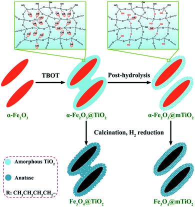 | ||
| Fig. 1 The template-free strategy towards the synthesis of uniform core–shell structured magnetic mesoporous TiO2 nanospindles (Fe3O4@mTiO2) based on a new post-hydrolysis process. | ||
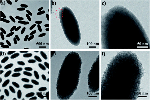
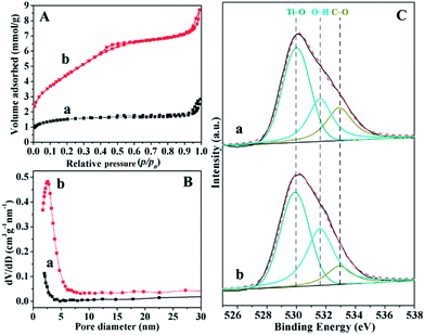
Uniform α-Fe2O3 nanospindles with a size of ∼100 nm in diameter and ∼480 nm in length were synthesized as cores via a hydrothermal method (Fig. S1†). In the facile Stöber system, titanium species from the hydrolysis and condensation of tetrabutyl titanate (TBOT) can deposit on the α-Fe2O3 nanospindles, leading to the formation of α-Fe2O3@TiO2 core–shell nanospindles with a diameter of ∼220 nm and a length of ∼600 nm (Fig. 2a and S2a†). Field-emission scanning electron microscopy (FESEM) and transmission electron microcopy (TEM) images of the α-Fe2O3@TiO2 nanospindles (Fig. S2b and S3a†) clearly reveal the well-defined core–shell structure and the presence of cross-linking between adjacent nanospindles (marked with arrows). The TEM image of a single nanospindle (Fig. 2b) shows the obvious tearing junctions (marked with a circle) on the surface of the TiO2 shells, which may be induced by external forces. It suggests that the linkage is not a physical aggregation but a continuous TiO2 network. In addition, the TiO2 shells appear to be a compact matrix with a thickness of ∼60 nm (Fig. 2c). After the post-hydrolysis process, uniform mesoporous α-Fe2O3@mTiO2 core–shell nanospindles are obtained (Fig. 2d and S2c†). FESEM and TEM images clearly demonstrate that the α-Fe2O3@mTiO2 nanospindles are individually separated without linkages, and the previously smooth surface of the TiO2 shells is converted into a rough one (Fig. S2d and S3b†). More importantly, the TiO2 shells exhibit a highly mesoporous structure, which is derived from the voids between the aggregated TiO2 oligomers (Fig. 2e and f). N2 sorption isotherms (Fig. 3A) of the α-Fe2O3@mTiO2 nanospindles show typical type IV curves and a distinct hysteresis loop close to the H1 type, with an increase in the adsorption branch at a relative pressure (P/P0) of 0.2–0.5, indicating that the TiO2 shells contain uniform mesopores. The Brunauer–Emmett–Teller (BET) surface area and pore volume of the α-Fe2O3@mTiO2 nanospindles are calculated to be ∼366 m2 g−1 and 0.3 cm3 g−1, respectively, which are much higher than those of the α-Fe2O3@TiO2 nanospindles (∼119 m2 g−1, 0.1 cm3 g−1). Moreover, the corresponding pore size distribution curve (Fig. 3B) of the α-Fe2O3@mTiO2 spindles derived from the adsorption branch of the isotherms by using the Barrett–Joyner–Halenda (BJH) method clearly shows a uniform pore size of ∼2.6 nm, larger than that of the α-Fe2O3@TiO2 spindles (<1.7 nm). The physical data are summarized in Table S1.† These imply that the post-hydrolysis process can greatly improve the monodispersity and mesoporosity of the TiO2 shells.
X-ray photoelectron spectroscopy (XPS) was used to investigate the surface molecular states of the TiO2 shells before and after the post-hydrolysis process. The survey spectra (Fig. S4†) of both the α-Fe2O3@TiO2 and α-Fe2O3@mTiO2 nanospindles show three well-resolved peaks of C 1s, Ti 2p, and O 1s without the typical one of Fe element, further indicating that the α-Fe2O3 core is uniformly coated by a thick TiO2 layer. The high content of carbon in the α-Fe2O3@TiO2 nanospindles clearly demonstrates the presence of plentiful alkoxy moieties (R) in the matrix of the TiO2 shells, suggesting the incompletely hydrolyzed nature of TBOT under the low content of ammonia in this system. After the post-hydrolysis process, the carbon content decreases from 52.9 to 39.0%, but the ratio of Ti/O is almost unchanged (Table S2†), indicating that the alkoxy moieties can be partly removed in water under ultrasound. The O 1s high-resolution XPS spectra clearly demonstrate the variation in the chemical states of the oxygen atom (Fig. 3C). The spectra can be deconvoluted into three single peaks corresponding to Ti–O (530.0 eV), O–H (531.6 eV), and C–O (533.0 eV) functional groups,28 respectively; the relative ratios of these peaks are listed in Table S1.† It can be clearly observed that the content of the C–O group decreases from 22.9 to 11.5%; in contrast, that of the OH group increases from 26.9 to 39.0%. In addition, the concentration of Ti–O species remains almost unchanged. All the results suggest that the Stöber-derived TiO2 shells contain a large number of alkoxy moieties; and the corresponding Ti–OR moieties can be hydrolyzed into Ti–OH groups in water under ultrasound. FTIR spectroscopy (Fig. S5†) was also used to characterize the structural change of the TiO2 shells upon the reaction with H2O. The peaks in the range of 3100–3600 cm−1 and at 1626 cm−1 can be assigned to the –OH stretching and deformation vibrations.29 Bands at 2956 and 2922 cm−1 can be attributed to the –CH2– and/or –CH3 stretching vibration, whereas the peaks in the region of 1400–1600 cm−1 correspond to the deformation vibrations, further revealing the presence of alkoxy moieties in the TiO2 matrix. The variations of the relative intensities of the hydroxyl and alkyl groups further confirm that post-hydrolysis occurs during the ultrasonic treatment.
Notably, when directly annealed at 500 °C in air without the post-hydrolysis process, the nanospindles appear to be fused together (denoted as α-Fe2O3@TiO2-500) and the TiO2 shells are composed of large merged nanoparticles with a size of ∼15 nm, as revealed by FESEM images (Fig. 4a and S6a†). The TEM image further demonstrates that the α-Fe2O3@TiO2-500 samples are conglutinated bulky materials (Fig. 4b). In contrast, discrete and uniform nanospindles are well retained when crystallizing the α-Fe2O3@mTiO2 ones (Fig. S6b†). The XPS spectrum and TG analysis of the α-Fe2O3@mTiO2-500 nanospindles indicate that the left alkoxy moieties can be effectively removed during the annealing process at 500 °C in air (Fig. S7†). FESEM and TEM images of the α-Fe2O3@mTiO2-500 nanospindles (Fig. 4c and d and S8a†) clearly show that the TiO2 shells are constructed of numerous aggregated nanoparticles. Moreover, the HRTEM image (Fig. S8b†) shows that the TiO2 nanoparticles are well crystallized with a size of ∼6 nm and a d-spacing of 0.35 nm, well-matched to the d101 of anatase,30 and packed into the highly mesoporous structures. XRD patterns (Fig. S9†) of the resultant core–shell spindles reveal that the amorphous TiO2 shells are well crystallized into the anatase phase.30 Interestingly, it can be identified that the anatase characteristic peaks of the α-Fe2O3@mTiO2-500 nanospindles are much broader than those of the α-Fe2O3@TiO2-500 samples; calculated from the Scherrer formula, the average crystal sizes of TiO2 are estimated to be ∼5 and 15 nm, respectively, which are in good agreement with the FESEM results. N2 sorption isotherms of the resultant materials show type IV curves with a distinct hysteresis loop close to the H1 type (Fig. S10A†). The BET surface area and pore volume of the α-Fe2O3@mTiO2-500 nanospindles are calculated to be ∼117 m2 g−1 and 0.2 cm3 g−1, respectively, which are much higher than those of the α-Fe2O3@TiO2-500 samples (∼21 m2 g−1, 0.1 cm3 g−1). In contrast, the corresponding pore size of the α-Fe2O3@mTiO2-500 spindles (∼5.4 nm) is smaller than that of the α-Fe2O3@TiO2-500 samples (∼7.3 nm) (Fig. S10B†). This further confirms that the TiO2 shells of the α-Fe2O3@mTiO2-500 spindles are made of small nanocrystals, resulting in higher porosity and smaller voids, whereas those of the α-Fe2O3@TiO2-500 samples are different, as illustrated in the inset of Fig. S10B†.
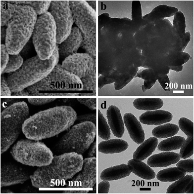 | ||
| Fig. 4 FESEM and TEM images of the bulk α-Fe2O3@TiO2-500 samples (a and b), and the uniform mesoporous α-Fe2O3@mTiO2-500 core–shell nanospindles (c and d). | ||
In this case, since the low content of ammonia is used as a catalyst, the hydrolysis and condensation of TBOT are directed toward the middle structures rather than the ends of completely cross-linked chains based on the positively charged values (δ) of the Ti atom in the partially hydrolyzed species (Fig. S11a†),31 which leads to a continuous and highly branched polymer matrix possessing a large number of unhydrolyzed alkoxy groups (Fig. 1) in the as-made TiO2 shell. When being subjected to an additional ultrasonic water treatment process, the Ti–OR moieties easily undergo further hydrolysis to form Ti–OH groups with the elimination of ROH molecules (Fig. 1 and S11b†) due to the high sensitivity to moisture. Meanwhile, the overall continuous and low-polymerized matrix would be further condensed and transformed into an aggregate of small TiO2 grains (Fig. 1 and S12†). Such synergistic effects leave mesopores in the amorphous TiO2 frameworks. In addition, the little linkages of the polymer networks between the α-Fe2O3@TiO2 nanospindles can be easily broken down owing to the high reactivity of water molecules under ultrasound, leading to the formation of discrete nanospindles. When annealed at high temperatures, it is known that the grain growth of TiO2 experiences a localized crystallization process into the well-defined nanocrystals.32 Since the disconnected boundary nature of the parent α-Fe2O3@mTiO2 nanospindles require an additional high activation barrier for reconstruction and mass transport, the resultant α-Fe2O3@mTiO2-500 nanospindles can retain the discrete and uniform core–shell structure during the thermal treatment at 500 °C. Meanwhile, the small internal packing TiO2 grains would grow large into anatase nanocrystals, which leads to relatively large packing pore sizes (Fig. S12†). In contrast, conglutinated bulky materials are obtained with large merged nanocrystals as the initial α-Fe2O3@TiO2 nanospindles approach one another and continuous boundaries exist in the adjacent TiO2 shell matrix (Fig. 1 and S12†). With these understandings, uniform functional mesoporous TiO2 shelled core–shell nanoparticles with high surface areas can be facilely synthesized via a new route without using templates, for example, SiO2@mTiO2 and Fe3O4@mTiO2 nanoparticles (Fig. S13†).
It has been well demonstrated that hematite can be easily converted to magnetite by the reduction with H2.33 Here, we explore this conversion reaction to introduce magnetic functionality into the resultant mesoporous α-Fe2O3@mTiO2-500 nanospindles under a continuous flow of H2–air at 400 °C. XRD patterns (Fig. S14A†) clearly reveal that α-Fe2O3 cores are well reduced to Fe3O4, and the TiO2 shells show no apparent change in the crystal phase and size. However, the average crystal size of Fe3O4 nanoparticles is estimated by using the Scherrer formula to be ∼13 nm, much smaller than that of α-Fe2O3 ones (∼50 nm) owing to the crystal reconstruction during the conversion reaction.33 Notably, the resultant uniform mesoporous Fe3O4@mTiO2 core–shell nanospindles show a superparamagnetic behavior with little detectable remanence or coercivity (Fig. S14B†). The saturation magnetization value is measured to be ∼18.0 emu g−1. As expected, the mesoporous Fe3O4@mTiO2 core–shell nanospindles can be conveniently separated upon application of an external magnetic field (Fig. S14B,† inset). This feature is greatly desirable as it can be employed to separate TiO2 nanoparticles in a reaction medium. In addition, TEM images of mesoporous Fe3O4@mTiO2 nanospindles (Fig. S14C†) further demonstrate that the uniform core–shell nanostructures are well maintained during the H2 reduction process.
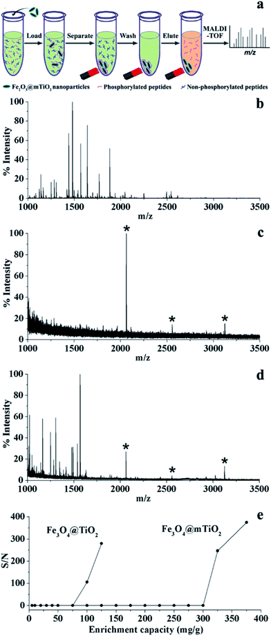
Recently, TiO2 has been widely applied for the enrichment of phosphopeptides, where the phosphate functional group can bind to the surface of TiO2 particles through bridging bidentate bonds.34,35 In this respect, the resultant mesoporous Fe3O4@mTiO2 core–shell nanospindles are used to enrich phosphopeptides from a complex mixture and separated by an external magnetic field, as illustrated in Fig. 5a. As a basic test, the mesoporous Fe3O4@mTiO2 nanospindles are first used to isolate phosphopeptides from tryptic digests of the standard protein β-casein. A direct analysis of the β-casein digest by MALDI-TOF MS indicates that only three phosphopeptides are detected with weak intensities (Fig. S15a†). In contrast, after being enriched by the mesoporous Fe3O4@mTiO2 nanospindles (Fig. S15b†), almost all the peaks in the mass spectrum are related to phosphopeptides, and no non-phosphorylated peptides are observed. Additionally, the signal-to-noise (S/N) ratio for phosphopeptides is significantly improved, indicating that the mesoporous Fe3O4@mTiO2 core–shell nanospindles are promising for the selective enrichment of phosphopeptides.
The selectivity of enrichment is further evaluated by capturing phosphopeptides from a more complex mixture of tryptic β-casein and BSA digests with a molar ratio of 1![[thin space (1/6-em)]](https://www.rsc.org/images/entities/char_2009.gif) :
:![[thin space (1/6-em)]](https://www.rsc.org/images/entities/char_2009.gif) 500. It can be clearly identified that no phosphopeptides are detected from the initial mixture due to the presence of a high-abundance of non-phosphopeptides from BSA (Fig. 5b). By contrast, after the enrichment by the mesoporous Fe3O4@mTiO2 nanospindles, the signals related to phosphopeptides are highly enhanced and dominate the mass spectrum with a very clean background (Fig. 5c), which is much better than that for the TiO2 nanoparticles reported previously.36–39 In a control experiment, the bulk Fe3O4@TiO2 samples are also employed to enrich phosphopeptides from the complex mixture. Fig. 5d shows that the mass spectrum is dominated by nonphosphorylated peptides' signals and three phosphopeptides with weak intensities can be detected. This is because large merged TiO2 nanoparticles with a low surface area are produced during thermal annealing without the post-hydrolysis process, which leads to lower active sites for the adsorption of phosphopeptides. To study the enrichment capacity of phosphopeptides with the resultant products, various concentrations of tryptic β-casein digests are prepared and then incubated with an equivalent amount of products. After magnetically separating the products, the supernatants are collected and subjected to MALDI-MS analysis. The products would reach the maximum enrichment capacity once the signal of phosphopeptides could be detected in the supernatants. As a result, the enrichment capacity (Fig. 5e) of the mesoporous Fe3O4@mTiO2 nanospindles is estimated to be ∼300 mg g−1, which is much higher than that of the bulk Fe3O4@TiO2 samples (∼75 mg g−1). This can be attributed to the small particle size and high surface area of the TiO2 shells in the mesoporous Fe3O4@mTiO2 nanospindles, which possess more active sites for phosphopeptide loading.
500. It can be clearly identified that no phosphopeptides are detected from the initial mixture due to the presence of a high-abundance of non-phosphopeptides from BSA (Fig. 5b). By contrast, after the enrichment by the mesoporous Fe3O4@mTiO2 nanospindles, the signals related to phosphopeptides are highly enhanced and dominate the mass spectrum with a very clean background (Fig. 5c), which is much better than that for the TiO2 nanoparticles reported previously.36–39 In a control experiment, the bulk Fe3O4@TiO2 samples are also employed to enrich phosphopeptides from the complex mixture. Fig. 5d shows that the mass spectrum is dominated by nonphosphorylated peptides' signals and three phosphopeptides with weak intensities can be detected. This is because large merged TiO2 nanoparticles with a low surface area are produced during thermal annealing without the post-hydrolysis process, which leads to lower active sites for the adsorption of phosphopeptides. To study the enrichment capacity of phosphopeptides with the resultant products, various concentrations of tryptic β-casein digests are prepared and then incubated with an equivalent amount of products. After magnetically separating the products, the supernatants are collected and subjected to MALDI-MS analysis. The products would reach the maximum enrichment capacity once the signal of phosphopeptides could be detected in the supernatants. As a result, the enrichment capacity (Fig. 5e) of the mesoporous Fe3O4@mTiO2 nanospindles is estimated to be ∼300 mg g−1, which is much higher than that of the bulk Fe3O4@TiO2 samples (∼75 mg g−1). This can be attributed to the small particle size and high surface area of the TiO2 shells in the mesoporous Fe3O4@mTiO2 nanospindles, which possess more active sites for phosphopeptide loading.
In summary, we developed a novel, simple and template-free strategy, which is based on a new post-hydrolysis concept, for the synthesis of uniform mesoporous TiO2 nanospindles in a facile Stöber system. We have demonstrated that the TiO2 shells derived from the Stöber system are incompletely hydrolyzed networks, and the unhydrolyzed alkoxy groups can be easily removed by water, yielding mesoporous TiO2 with a high surface area. Benefitting from the well-defined structural features of the parent α-Fe2O3@mTiO2 nanospindles including the disconnected boundary nature and the internal packing of TiO2 grains, uniform mesoporous magnetic TiO2 nanospindles are obtained with a high surface area (∼117 m2 g−1) and large pore size (∼5.4 nm) without the use of templates or capping agents. Most importantly, the resultant Fe3O4@mTiO2 nanospindles exhibit a superparamagnetic behavior with a high magnetic susceptibility of ∼18.0 emu g−1, and a remarkable performance for highly selective and rapid enrichment of phosphopeptides with an unprecedented enrichment capacity of ∼300 mg g−1. We believe that these new understandings can be exploited to develop versatile and template-free approaches for fabricating functional mesoporous TiO2-based materials with exceptionally high surface areas.
Acknowledgements
This work was supported by the State Key Basic Research Program of the PRC (2012CB224805, 2012CB910602, 2013CB934104), NSFC of China (Grant no. 21210004, 21025519, 21335002) and King Saud University research project (RGP-VPP-036).References
- X. Chen and S. S. Mao, Chem. Rev., 2007, 107, 2891 CrossRef CAS PubMed.
- G. Liu, L. Wang, H. G. Yang, H. M. Cheng and G. Q. Lu, J. Mater. Chem., 2010, 20, 831 RSC.
- L. Zhao, X. F. Chen, X. C. Wang, Y. J. Zhang, W. Wei, Y. H. Sun, M. Antonietti and M. M. Titirici, Adv. Mater., 2010, 22, 3317 CrossRef CAS PubMed.
- W. F. Ma, Y. Zhang, L. L. Li, L. J. You, P. Zhang, Y. T. Zhang, J. M. Li, M. Yu, J. Guo, H. J. Lu and C. C. Wang, ACS Nano, 2012, 6, 3179 CrossRef CAS PubMed.
- S. H. Liu, Z. Y. Wang, C. Yu, H. B. Wu, G. Wang, Q. Dong, J. S. Qiu, A. Eychmuller and X. W. Lou, Adv. Mater., 2013, 25, 3462 CrossRef CAS PubMed.
- D. H. Chen and R. A. Caruso, Adv. Funct. Mater., 2013, 23, 1356 CrossRef CAS.
- J. S. Chen, Y. L. Tan, C. M. Li, Y. L. Cheah, D. Luan, S. Madhavi, F. Y. C. Boey, L. A. Archer and X. W. Lou, J. Am. Chem. Soc., 2010, 132, 6124 CrossRef CAS PubMed.
- J. B. Joo, Q. Zhang, I. Lee, M. Dahl, F. Zaera and Y. D. Yin, Adv. Funct. Mater., 2012, 22, 166 CrossRef CAS.
- W. Li, Y. Deng, Z. Wu, X. Qian, J. Yang, Y. Wang, D. Gu, F. Zhang, B. Tu and D. Y. Zhao, J. Am. Chem. Soc., 2011, 133, 15830 CrossRef CAS PubMed.
- R. Zhang, B. Tu and D. Zhao, Chem. – Eur. J., 2010, 16, 9977 CrossRef CAS PubMed.
- D. H. Chen, F. Z. Huang, Y. B. Cheng and R. A. Caruso, Adv. Mater., 2009, 21, 2206 CrossRef CAS.
- W. Li, Q. Yue, Y. Deng and D. Zhao, Adv. Mater., 2013, 25, 5129 CrossRef CAS PubMed.
- W. Li and D. Y. Zhao, Chem. Commun., 2013, 49, 943 RSC.
- D. M. Antonelli and J. Y. Ying, Angew. Chem., Int. Ed., 1995, 34, 2014 CrossRef CAS.
- P. Yang, D. Zhao, D. I. Margolese, B. F. Chmelka and G. D. Stucky, Nature, 1998, 396, 152 CrossRef CAS PubMed.
- D. Chen, L. Cao, F. Huang, P. Imperia, Y.-B. Cheng and R. A. Caruso, J. Am. Chem. Soc., 2010, 132, 4438 CrossRef CAS PubMed.
- D. Gu and F. Schüth, Chem. Soc. Rev., 2014, 43, 313 RSC.
- W. Li, Z. Wu, J. Wang, A. A. Elzatahry and D. Zhao, Chem. Mater., 2014, 26, 287 CrossRef CAS.
- W. Yue, C. Randorn, P. S. Attidekou, Z. Su, J. T. S. Irvine and W. Zhou, Adv. Funct. Mater., 2009, 19, 2826 CrossRef CAS.
- Z. Zhang, F. Zuo and P. Feng, J. Mater. Chem., 2010, 20, 2206 RSC.
- R. Y. Zhang, A. A. Elzatahry, S. S. Al-Deyab and D. Y. Zhao, Nano Today, 2012, 7, 344 CrossRef CAS PubMed.
- J. S. Chen, C. Chen, J. Liu, R. Xu, S. Z. Qiao and X. W. Lou, Chem. Commun., 2011, 47, 2631 RSC.
- M. Ye, Q. Zhang, Y. Hu, J. Ge, Z. Lu, L. He, Z. Chen and Y. D. Yin, Chem. – Eur. J., 2010, 16, 6243 CrossRef CAS PubMed.
- J. V. Olsen, B. Blagoev, F. Gnad, B. Macek, C. Kumar, P. Mortensen and M. Mann, Cell, 2006, 127, 635 CrossRef CAS PubMed.
- E. L. Huttlin, M. P. Jedrychowski, J. E. Elias, T. Goswami, R. Rad, S. A. Beausoleil, J. Villen, W. Haas, M. E. Sowa and S. P. Gygi, Cell, 2010, 143, 1174 CrossRef CAS PubMed.
- W. Li, J. Yang, Z. Wu, J. Wang, B. Li, S. Feng, Y. Deng, F. Zhang and D. Zhao, J. Am. Chem. Soc., 2012, 134, 11864 CrossRef CAS PubMed.
- W. Li and D. Zhao, Adv. Mater., 2013, 25, 142 CrossRef CAS.
- D. Li, X. Lu, H. Lin, F. Ren and Y. Leng, J. Mater. Sci.: Mater. Med., 2013, 24, 489 CrossRef CAS PubMed.
- Y. D. Wang, C. L. Ma, X. D. Sun and H. D. Li, J. Non-Cryst. Solids, 2003, 319, 109 CrossRef CAS.
- H. B. Wu, H. H. Hng and X. W. Lou, Adv. Mater., 2012, 24, 2567 CrossRef CAS PubMed.
- J. Livage, M. Henry and C. Sanchez, Prog. Solid State Chem., 1988, 18, 259 CrossRef CAS.
- W. Li, F. Wang, S. S. Feng, J. X. Wang, Z. K. Sun, B. Li, Y. H. Li, J. P. Yang, A. A. Elzatahry, Y. Y. Xia and D. Y. Zhao, J. Am. Chem. Soc., 2013, 135, 18300 CrossRef CAS PubMed.
- X. W. Lou and L. A. Archer, Adv. Mater., 2008, 20, 1853 CrossRef CAS.
- Z. D. Lu, M. M. Ye, N. Li, W. W. Zhong and Y. D. Yin, Angew. Chem., Int. Ed., 2010, 49, 1862 CrossRef CAS PubMed.
- Z. Lu, J. Duan, L. He, Y. Hu and Y. D. Yin, Anal. Chem., 2010, 82, 7249 CrossRef CAS PubMed.
- Y. Li, J. S. Wu, D. W. Qi, X. Q. Xu, C. H. Deng, P. Y. Yang and X. M. Zhang, Chem. Commun., 2008, 564 RSC.
- Y. Li, X. Q. Xu, D. W. Qi, C. H. Deng, P. Y. Yang and X. M. Zhang, J. Proteome Res., 2008, 7, 2526 CrossRef CAS PubMed.
- C. T. Chen and Y. C. Chen, J. Biomed. Nanotechnol., 2008, 4, 73 CAS.
- J. H. Wu, X. S. Li, Y. Zhao, Q. Gao, L. Guo and Y. Q. Feng, Chem. Commun., 2010, 46, 9031 RSC.
Footnote |
| † Electronic supplementary information (ESI) available. See DOI: 10.1039/c4mh00030g |
| This journal is © The Royal Society of Chemistry 2014 |

