 Open Access Article
Creative Commons Attribution 3.0 Unported Licence
Phthalocyanine blue in aqueous solutions†
Mark
C. Staniford
a,
Marina M.
Lezhnina
ab and
Ulrich H.
Kynast
*a
aMuenster University of Applied Sciences, Institute for Optical Technologies, Münster/Steinfurt, Stegerwald-Str. 39, 48565 Steinfurt, Germany. E-mail: uk@fh-muenster.de
bOn Leave from Volga State University of Technology, Department of Physics, Lenin-pl.3, Yoshkar-Ola 424000, Russia
First published on 5th December 2014
Abstract
Using laponite nano-clay carriers, a facile method for the solubilisation of natively insoluble phthalocyanines into aqueous solution is described. Copper(II) phthalocyanine, technologically a most relevant pigment (C.I. Pigment Blue 15), thus yields hitherto unknown clear and stable aqueous dispersions of either colloidal α-CuPc or monomeric CuPc, depending on details of the preparation.
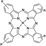
Copper(II) phthalocyanine (CuPc, Fig. 1) is one of the most frequently used pigments in painting and coating industries, with an annual production of approximately 60
![[thin space (1/6-em)]](https://www.rsc.org/images/entities/char_2009.gif) 000 t per year. The blue colorant possesses a very high extinction coefficient due to the intense π–π* transitions within the ligand electronic system. Its insolubility in common solvents is obviously of advantage, if the CuPc is intended for pigment use in coatings. However, for applications requiring solubility, substantial effort has been devoted to the synthesis of substituted phthalocyanines to grant solubility,1 in aqueous ambience in particular, e.g. the widely used examples phthalocyanine-3,4′,4′′,4′′′-tetrasulfonic acid or Alcian Blue (“Ingrain blue 1”). Obviously, the preparative effort involved will go to the price of elevated costs. Readily soluble phthalocyanines (also including other ions than Cu(II)) hold the potential to impact mere coating applications just as much as more filigrane uses in homogeneous (photo-) catalysis,2 solar cells,3 LEDs (both as hole injection aids4 and emitters5), or even photodynamic therapies (PDT).6,7
000 t per year. The blue colorant possesses a very high extinction coefficient due to the intense π–π* transitions within the ligand electronic system. Its insolubility in common solvents is obviously of advantage, if the CuPc is intended for pigment use in coatings. However, for applications requiring solubility, substantial effort has been devoted to the synthesis of substituted phthalocyanines to grant solubility,1 in aqueous ambience in particular, e.g. the widely used examples phthalocyanine-3,4′,4′′,4′′′-tetrasulfonic acid or Alcian Blue (“Ingrain blue 1”). Obviously, the preparative effort involved will go to the price of elevated costs. Readily soluble phthalocyanines (also including other ions than Cu(II)) hold the potential to impact mere coating applications just as much as more filigrane uses in homogeneous (photo-) catalysis,2 solar cells,3 LEDs (both as hole injection aids4 and emitters5), or even photodynamic therapies (PDT).6,7
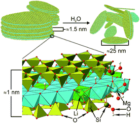
As an illustrative example for phthalocyanine solubilisation, we here report on a straight forward method to solubilize CuPc into water in concentrations, this far only known for organic solvents, by the aid of the nano-clay shuttles. The commercial clay used most successfully for this purpose, “LAPONITE RD”‡, Na0.7(H2O)n{(Li0.3Mg5.5)[Si8O20(OH)4]}, can be viewed as a nano-scaled hectorite derivative8 or a charged offspring of talcum, primarily consisting of disk-shaped platelets of 25 nm in diameter and 1 nm in height, which completely exfoliate to form clear dispersions in water (see also Fig. 2 and the corresponding caption).9
 | ||
| Fig. 2 Nano-clay (“laponite”) used in this investigation. Like the parent mineral talcum, structurally each individual disk or platelet consists of a tetrahedral–octahedral–tetrahedral arrangement (“TOT”). Chemically, ionisable Si–O–H and Mg–O–H groups protrude from the rims of the platelets and contribute to the laponite's unusual rheological properties; if Li+ (green) is introduced to replace Mg2+ cations (blue) within the octahedra, additional cations (e.g. Na+, not shown, but see Fig. 4) are accommodated between the aligned disks as indicated in the top left of the sketch. As opposed to talcum, these very charges now enable complete exfoliation on exposure to aqueous media, leaving individual platelets with negatively charged faces behind. | ||
The resulting negatively charged platelets have been shown to adsorb numerous cationic dye species, among them Methylene Blue10 or luminescent Rhodamine dyes,11 which can be attracted via polar and coulomb forces. However, very recently, the neutral and nonpolar dyes Indigo12 and Nile Red,13 both completely insoluble in water, have also been found to interact strong enough with nano-clays to be solubilised, the luminescent Nile Red surprisingly even retaining its emissive character in water. While the capability of laponite to mobilise these dyes may be ascribed to polar interactions and hydrogen bonding, the precise nature of the bonding in laponite–CuPc hybrids remains yet to be clarified. Despite considerable analytical adversaries encountered in their analysis, we find their solution behavior so unique that it seems worth to be reported on: not only can the CuPc be solubilised, but the presence of monomeric species in aqueous solutions and the corresponding impact for other Pc materials, such as singlet oxygen generators, struck us as especially remarkable.
Among the approaches to establish an efficient adsorption pathway for CuPc on the laponite surface, refluxing the laponite and CuPc materials for 48 h in water–solvent mixtures (acetonitrile, methanol) in a 1![[thin space (1/6-em)]](https://www.rsc.org/images/entities/char_2009.gif) :
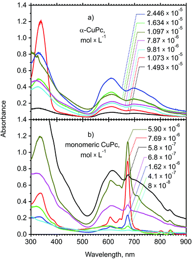
:![[thin space (1/6-em)]](https://www.rsc.org/images/entities/char_2009.gif) 1 ratio at 1 wt% laponite-dispersion yielded the best samples with respect to homogeneity and (re-) dispersability; no noteworthy difference was found between the use of CH3CN–H2O and CH3OH–H2O as dispersants. The CuPc adsorption could further be enhanced by mortaring the materials very thoroughly prior to loading. Other pathways (reactions via the gas phase, i. e. sublimation of the Pc, in situ formation of the CuPc from Cu2+–phthalocyanine mixtures in various solvents) proved to be less suited due to non-re-dispersability of the powders. The preparation in water proved to be possible also, but necessitated several steps of filtration and centrifugation, respectively, in order to obtain transparent solutions and was only used in efforts to obtain extinction coefficients (see below). After filtration of the CH3CN–H2O dispersion to separate non-dispersible or insoluble residues, absorption spectra of the mother liquors, referred to as solutions (1), were recorded (Fig. 3a). The spectra were taken from dispersions at a constant 1 wt% with respect to laponite, which, using data of the supplier (ρ = 2.53 g cm−3, disk diameter of 25 nm)14 and a value of 1.5 nm for the disk thickness accounting for the inclusion of interlayer water,13 can be recalculated to correspond to 5.2 × 10¹⁸ disks L−1.
1 ratio at 1 wt% laponite-dispersion yielded the best samples with respect to homogeneity and (re-) dispersability; no noteworthy difference was found between the use of CH3CN–H2O and CH3OH–H2O as dispersants. The CuPc adsorption could further be enhanced by mortaring the materials very thoroughly prior to loading. Other pathways (reactions via the gas phase, i. e. sublimation of the Pc, in situ formation of the CuPc from Cu2+–phthalocyanine mixtures in various solvents) proved to be less suited due to non-re-dispersability of the powders. The preparation in water proved to be possible also, but necessitated several steps of filtration and centrifugation, respectively, in order to obtain transparent solutions and was only used in efforts to obtain extinction coefficients (see below). After filtration of the CH3CN–H2O dispersion to separate non-dispersible or insoluble residues, absorption spectra of the mother liquors, referred to as solutions (1), were recorded (Fig. 3a). The spectra were taken from dispersions at a constant 1 wt% with respect to laponite, which, using data of the supplier (ρ = 2.53 g cm−3, disk diameter of 25 nm)14 and a value of 1.5 nm for the disk thickness accounting for the inclusion of interlayer water,13 can be recalculated to correspond to 5.2 × 10¹⁸ disks L−1.
 | ||
| Fig. 3 Absorption spectra of laponite hybrids with α-CuPc (a) in saturated CH3CN–H2O solutions (solutions (1), (a)) and of monomeric CuPc species in water (solutions (2), (b)) at a concentration of 1 wt% of laponite. The data reproduced in the plots were recalculated from the extinction coefficient of monomeric CuPc from the sample containing 7.68 × 10⁻⁶ mol × L−1 ((b), red curve). Original concentrations, extinction coefficients and corresponding amounts of CuPc-molecules per disk (“mpd”, see ESI†) are also listed in Table S1.† | ||
The volume of the first filtrates (solutions (1)), containing only α-CuPc, was subsequently reduced with a rotary evaporator to a thick gel and dried in a vacuum drying chamber for 24 h at 60 °C. The resulting, blue colored powders, showed diffraction peaks of the laponite only and no signs of CuPc crystallinity in XRD (see (ESI), Fig. S2 and S4† additionally shows the FTIR spectra of a hybrid in comparison to pure α-CuPc), although a trend towards an increased order of the platelets with increasing CuPc contents may be depicted, as indicated by the [001] and [003] reflexes at 2Θ = 6.6° and 28.1°. The α-CuPc aggregates could be re-dispersed completely in pure H2O to obtain 1 wt% – laponite dispersions (solutions (2)).
Absorption spectra of the first filtrates (solutions (1), Fig. 3a) closely resemble the spectra obtained from α-CuPc,15,16 and unambiguously suggest the exclusive presence of dimers or higher associates, of the CuPc in the aqueous solution as well, as evident from the split Q-bands at 616 and 697 nm,15,17–19 also known as Davydov splitting,15,20i.e. the broadening due to a variety of interactions with neighboring molecules. Accounts on the electronic structure of α-CuPc have been elaborated using absorption spectroscopy, electron microscopies, AFM, XRD21,22 and UPS;16 a recent review exhaustively discusses the nature of the electronic transitions in monomeric CuPc as well.23
The true concentrations of α-CuPc in the first filtrate (solution (1), CH3CN–H2O, Fig. 3a and 4), could not be analyzed with sufficient precision: carbon analyses of the samples carried out after evaporation of the solvent, revealed amounts well above the expected values for pure CuPc–laponites, as all samples still contained co-adsorbed CH3CN in appreciable and, unfortunately, non-reproducible amounts (up to a fivefold excess in carbon over the theory for the pure CuPc–laponites). Despite appreciable effort (e.g. vacuum drying at various elevated temperatures of up to 250 °C, freeze drying), we were not able to completely remove the organic solvent without carbonisation. Carbon determinations are feasible for the laponite systems in principle, as we found from vacuum-sublimed hybrids, where we found a very good fit between the experimental data and theoretical values, however, as mentioned, such samples could not be re-dispersed. Therefore all elemental analyses of samples prepared from solutions containing organic matter, including copper and nitrogen determinations contained too large an error to be useful, a situation, which is yet more complicated by the relatively small theoretical overall carbon content (e.g. 0.5 wt% at 10 molecules per disk, “mpd” see also eqn (S1),† and lower, taking the true loading efficiencies into account) and a continuous release of water extending into a range of 700 °C (see DTG, Fig. S3†). An estimate of the extinction coefficient εα-CuPc (approximately 2.1 × 10⁴ L × mol−1 × cm−1) was thus possible only indirectly via solutions containing monomeric CuPc, as described below.
Absorption spectra of the water – re-dispersed samples (solution (2)), reproduced in Fig. 3a, now clearly demonstrate the presence of monomeric species as evidenced by the emergence of the Q-band at 673 nm, which is very close to the 671 and 678 nm observed for CuPc in solution (THF and 1-chloronaphthalene, respectively24,25), while the absorption in the vapor phase is reported to be blue-shifted to 656 nm.26,27 For loadings below a threshold of approximately 7.69 × 10⁻⁶ mol × L−1 (see also below), corresponding to 0.89 mpd, the monomer is by far the dominating guest species, above that level, dimers or higher associates are formed. For samples of lower CuPc contents, the absorption spectra suggest monomeric CuPc to be the dominating species by far in the re-dispersed aqueous solution (see the blue and red curves in Fig. 3b for example). At this stage, we also need to mention that in water, monomeric CuPc is slightly prone to protonation via Brønsted acid sites characteristic of the laponite clay, if exposed to temperatures higher than approximately 60 °C. Minute absorptions at 810 nm and above are indicative of this protonation.
As pointed out above, we encountered substantial difficulties in the determination of the laponite loading with CuPc. We have therefore chosen to estimate the apparent concentrations of the monomeric CuPc on laponite from the absorption spectra. Due to its very low solubility, data on the absorption coefficient of CuPc are sparse,19 although data have been obtained from solutions of 1-chloronapthalene25,28,29 and THF,30 in the gas phase,26 in thin films,31 glasses and polymers. Obviously, the low solubility of CuPc causes large uncertainties in the reported extinction coefficients. However, a recent diligent treatise24 suggests an extinction coefficient ε673 for protonated CuPc of (1.4 ± 0.7) × 105 L × mol−1 × cm−1 in trifluoroacetic acid and (1.6 ± 0.16) × 105 L × mol−1 × cm−1 in concentrated sulfuric acid. Thus, we prepared a calibration curve of pure CuPc in H2SO4 (see Fig. S1†), from which an extinction coefficient of 157![[thin space (1/6-em)]](https://www.rsc.org/images/entities/char_2009.gif) 762 L × mol−1 × cm−1 in close proximity to the above value could be extracted, which served as the basis of our subsequent concentration estimates in H2SO4. We preferred the use of H2SO4 due to the additional presence of the clay, for which weaker acids required a prolonged dissolution time.
762 L × mol−1 × cm−1 in close proximity to the above value could be extracted, which served as the basis of our subsequent concentration estimates in H2SO4. We preferred the use of H2SO4 due to the additional presence of the clay, for which weaker acids required a prolonged dissolution time.
Samples used to obtain the extinction coefficient of monomeric CuPc were prepared in pure water, in order to avoid obscuring side reactions of organic residues on the laponite, additionally, to avoid protonation, during all drying, re-dispersion and filtration steps, temperatures below 60 °C were maintained. The absorbances of clear monomeric CuPc solutions, free of α-CuPc, were measured, evaporated to almost dryness, and redissolved in H2SO4. Using the calibration curve, the CuPc content could be determined. As this content corresponds to the amount of CuPc in the preceding solution, an extinction coefficient εmono,673 of 102![[thin space (1/6-em)]](https://www.rsc.org/images/entities/char_2009.gif) 777 L × mol−1 × cm−1 (±23%) for monomeric CuPc in aqueous solution could be calculated (for further details to the procedures and experimental errors refer to the ESI†).
777 L × mol−1 × cm−1 (±23%) for monomeric CuPc in aqueous solution could be calculated (for further details to the procedures and experimental errors refer to the ESI†).
This derived extinction coefficient εmono,673 in turn enabled us to re-estimate the true concentrations of the CuPc monomers at various loadings, and in a reverse manner, to recalculate the concentration and extinction coefficient of the (aggregated) α-CuPc solutions of the first CH3CN–H2O filtrate (solution (1)). The detailed procedure for these estimates is reported in the supplement to this paper. An extinction coefficient for α-CuPc in aqueous solution has not previously been reported for its obvious insolubility. Inspecting Fig. 3b of samples that were prepared and recorded in CH3CN–H2O after filtration (solutions (1)), the spectral position of the Q-band related absorptions at 616 and 679 nm is not affected by the nano-clay loading level. Also evident is the absence of any monomeric species, which would give rise to a strong absorption at 673 nm. The absorption spectra furthermore reveal an increasing shoulder of the Soret-band just above 400 nm, which is due to the increasing formation of scattering agglomerates; only for loadings below approximately 8 × 10⁻⁶ mol × L−1, agglomeration appears to be negligible. While we realise that the integrated absorption coefficient is a better value for the broad band absorptions of α-CuPc, we here use the extinction coefficient εα,616 of 2.1 × 10⁴ L × mol−1 × cm−1 for the purpose of comparison with monomeric CuPc.
The content of CuPc eventually found in the hybrids was in our series restricted to approximately 2.5 × 10⁻⁵ mol × L−1, corresponding to roughly 2.8 α-CuPc molecules per clay platelet (mpd). As opposed to that, the highest amount of monomeric CuPc (Fig. 3b), after careful removal of the CH3CN under the moderate conditions described above (vacuum drying chamber for 24 h at 60 °C) and re-dispersion in water, was found as 7.68 × 10⁻⁶ mol × L−1 only, i.e., 0.89 mpd of monomeric CuPc – now less than one molecule per platelet. Significantly, the loading efficiency with regard to the actual amounts of CuPc weighed out amounts to some 5% only, and drops dramatically to less than 1%, if concentrations in excess of 6 × 10⁻⁴ mol × L−1 CuPc are applied (see Fig. S5 and Table S1†).
The colloidal α-CuPc particles adhering to laponites are preferentially located at the rims rather than at the faces of the clay (see Fig. 4), since the intergallery distance between the platelets does not change, as evident from the unaltered basal reflex [001] (2Θ = 6.6°) in the XRDs of the solids (Fig. S2†). The formation of small colloidal particles is obviously promoted by the presence of stabilizing CH3CN. Consequently, on addition of CH3CN to aqueous dispersions containing only monomeric CuPc–laponite, the monomer band vanishes again to the cost of the two broad α-CuPc bands (Fig. S6†).
Conclusions
While the mobilization of unsubstituted CuPc into aqueous media, additionally accompanied by the formation of monomers, is a surprising observation in its own right, efforts to increase the pigment's solubility further are desirable for e.g. coating applications. Beyond the use as colorants, monomeric phthalocyanines are currently being investigated with regard to their (photo-) catalytic activity. As preliminary results indicate, Si(OH)2Pc and Al(OH)Pc are promising candidates for singlet oxygen generation in aqueous solution. Furthermore, established nano-clay functionalisation schemes via siloxane reagents readily enable the coupling to relevant bioassays, and pathways to targeted photodynamic therapy and anti-microbial uses of the Pc–laponit hybrids seem feasible.Notes and references
- F. Dumoulin, M. Durmuş, V. Ahsen and T. Nyokong, Coord. Chem. Rev., 2010, 254, 2792 CrossRef CAS PubMed.
- B. Sorokin, Chem. Rev., 2013, 113, 8152 CrossRef PubMed.
- M. Ince, A. Medina, J. H. Yum, A. Yella, C. G. Claessens, M. V. Martínez-Díaz, M. Grätzel, M. K. Nazeeruddin and T. Torres, Chem.–Eur. J., 2014, 20, 2016 CrossRef CAS PubMed.
- D. Hohnholz, S. Steinbrecher and M. Hanack, J. Mol. Struct., 2000, 521, 231 CrossRef CAS.
- X. Barker, S. Zeng, A. S. Bettington, M. R. Batsanov, Bryce and A. Beeby, Chem.–Eur. J., 2007, 13, 6710 CrossRef PubMed.
- J. F. Lovell, T. W. B. Liu, J. Chen and G. Zheng, Chem. Rev., 2010, 110, 2839 CrossRef CAS PubMed.
- T. Nyokong, Pure Appl. Chem., 2011, 833, 1763 Search PubMed.
- J. Breu, W. Seidl and A. Stoll, Z. Anorg. Allg. Chem., 2003, 629, 503 CrossRef CAS.
- W. Thompson and J. T. Butterworth, J. Colloid Interface Sci., 1992, 151, 236 CrossRef.
- J. C. a. R. A. Schoonheydt, Clays Clay Miner., 1988, 26, 214 Search PubMed.
- F. Lopez Arbeloa and V. Martinez Martinez, Chem. Mater., 2006, 18, 1407 CrossRef CAS.
- M. M. Lezhnina, T. Grewe, H. Stoehr and U. Kynast, Angew. Chem., Int. Ed., 2012, 51, 10652 CrossRef CAS PubMed.
- T. Felbeck, T. Behnke, K. Hoffmann, M. Grabolle, M. M. Lezhnina, U. H. Kynast and U. Resch-Genger, Langmuir, 2013, 29, 11489 CrossRef CAS PubMed.
- www.byk.com/fileadmin/byk/additives/product_groups/rheology/former_rockwood_additives/technical_brochures/BYK_B-RI21_LAPONITE_EN.pdf, 19.03.2014.
- E. A. Lucia and F. D. Verderame, J. Chem. Phys., 1968, 48, 2674 CrossRef CAS PubMed.
- L. Lozzi, S. Santucci, S. La Rosa, B. Delley and S. Picozzi, J. Chem. Phys., 2004, 121, 1883 CrossRef CAS PubMed.
- V. Plyashkevich, T. Basova, P. Semyannikov and A. Hassan, Thermochim. Acta, 2010, 501, 108 CrossRef CAS PubMed.
- M. J. Stillman and A. J. Thomson, J. Chem. Soc., Faraday Trans. 2, 1974, 70, 805 RSC.
- Y. A. Mikheev, L. N. Guseva and Y. A. Ershov, Russ. J. Phys. Chem. A, 2007, 81, 617 CrossRef CAS.
- A. S. Davidov, J. Exp. Theor. Phys., 1948, 18, 210 Search PubMed.
- S. Karan and B. Mallik, J. Phys. Chem. C, 2007, 111, 7352 CAS.
- S. Karan, D. Basak and B. Mallik, Chem. Phys. Lett., 2007, 434, 265 CrossRef CAS PubMed.
- K. Ishii, Coord. Chem. Rev., 2012, 256, 1556 CrossRef CAS PubMed.
- F. Ghani, J. Kristen and H. Riegler, J. Chem. Eng. Data, 2012, 57, 439 CrossRef CAS.
- F. H. Moser and A. L. Thomas, Phthalocyanine compounds, Reinhold, Chapman & Hall, London, 1963 Search PubMed.
- D. Eastwood, L. Edwards, M. Gouterman and J. Steinfeld, J. Mol. Spectrosc., 1966, 20, 381 CrossRef CAS.
- L. Edwards and M. Gouterman, J. Mol. Spectrosc., 1970, 33, 292 CrossRef CAS.
- J. S. Anderson, E. F. Bradbrook, A. H. Cook and R. P. Linstead, J. Chem. Soc., 1938, 1151 RSC.
- E. Ficken and R. P. Linstead, J. Chem. Soc., 1952, 4846 RSC.
- J. Gardener, J. H. G. Owen, K. Miki and S. Heutz, Surf. Sci., 2008, 602, 843 CrossRef CAS PubMed.
- M. Farag, Opt. Laser Technol., 2007, 39, 728 CrossRef PubMed.
Footnotes |
| † Electronic supplementary information (ESI) available: Experimental details and methods; Fig. S1–S6; Table S1. See DOI: 10.1039/c4ra11139g |
| ‡ LAPONITE, is a former trademark of Rockwood Additives Limited, now distributed by Altana/Byck. The term “laponite” is used synonymously in this text for Laponite RD. |
| This journal is © The Royal Society of Chemistry 2015 |


