 Open Access Article
Creative Commons Attribution 3.0 Unported Licence
Fluoride binding to an organoboron wire controls photoinduced electron transfer†
Jing
Chen
abc and
Oliver S.
Wenger
*a
aDepartment of Chemistry, University of Basel, St. Johanns-Ring 19, CH-4056 Basel, Switzerland. E-mail: oliver.wenger@unibas.ch
bXiamen Institute of Rare Earth Materials, Chinese Academy of Sciences, Xiamen 361021, People's Republic of China
cKey Laboratory of Design and Assembly of Functional Nanostructures, Fujian Provincial Key Laboratory of Nanomaterials, Fujian Institute of Research on the Structure of Matter, Chinese Academy of Sciences, People's Republic of China
First published on 1st May 2015
Abstract
We demonstrate that the rates for long-range electron transfer can be controlled actively by tight anion binding to a rigid rod-like molecular bridge. Electron transfer from a triarylamine donor to a photoexcited Ru(bpy)32+ acceptor (bpy = 2,2′-bipyridine) across a 2,5-diboryl-1,4-phenylene bridge occurs within less than 10 ns in CH2Cl2 at 22 °C. Fluoride anions bind with high affinity to the organoboron bridge due to strong Lewis base/Lewis acid interactions, and this alters the electronic structure of the bridge drastically. Consequently, a large tunneling barrier is imposed on photoinduced electron transfer from the triarylamine to the Ru(bpy)32+ complex and hence this process occurs more than two orders of magnitude more slowly, despite the fact that its driving force is essentially unaffected by fluoride addition. Electron transfer rates in proteins could potentially be regulated via a similar fundamental principle, because interactions between charged amino acid side chains and counter-ions can modulate electronic couplings between distant redox partners. In artificial donor-bridge-acceptor compounds, external stimuli have been employed frequently to control electron transfer rates, but the approach of exploiting strong Lewis acid/Lewis base interactions to regulate the tunneling barrier height imposed by a rigid rod-like molecular bridge is conceptually novel and broadly applicable, because it is largely independent of the donor and the acceptor, and because the effect is not based on a change of the driving-force for electron transfer. The principle demonstrated here can potentially be used to switch between conducting and insulating states of molecular wires between electrodes.
1. Introduction
The electronic structure of an intervening medium between a donor and an acceptor strongly affects the rate at which an electron can be transferred.1 Consequently, in artificial donor-bridge-acceptor compounds as well as in biological systems scientists have strived to optimize the electronic structure of molecular bridges in order to accelerate electron transfer over long distances (>10 Å).2 Similarly, efforts have been made to optimize electron transport across nanowires between two electrodes.3 In several cases it has been possible to modulate the electronic structure of a photoisomerizable molecular bridge or a nanowire with light as an external stimulus to such an extent that electron transfer rates or conductivities could be altered by several orders of magnitude.4 In this paper we demonstrate that the rate for long-range electron tunneling across a rigid rod-like organoboron bridge can be controlled by tight fluoride binding to the bridge, without affecting the donor or the acceptor, and without changing the driving-force for electron transfer.It is well known that fluoride binding alters the electronic structure of organoboron compounds substantially, as this forms the basis for F− or CN− detection in numerous dimesitylboron-substituted molecules and metal complexes.5 When using bulky dimesitylboryl groups, the electron-deficient boron atom is well protected from nucleophilic attack, and only small anions such as F− or CN− can bind efficiently. The disruption of pπ–π* conjugation between the vacant p orbital of boron and the adjacent π-conjugated framework upon anion binding to the boron center causes significant changes in HOMO/LUMO energies.6 While organoboron compounds received considerable attention for various optical, electronic, and sensory applications,7 they have, to the best of our knowledge, never been used as molecular bridges that mediate electron transfer between covalently attached donors and acceptors, or as well-defined nanowires between two electrodes.
There are many studies in which organoboron units acted as terminal electron acceptors, and upon F− binding electron transfer was suppressed due to a change in driving force.5a,c–e,j,7h,i,k,o,8 We investigated a fundamentally different aspect in that we explored how fluoride binding to an organoboron bridge influences electron transfer between distant donors and acceptors which themselves are unaffected by F− addition, i.e., without changing the driving force. In several other prior studies the addition of ions lead to an effect on the luminescence or absorption properties of an oligomer or polymer material, but the fundamental electron transfer properties across the oligomer/polymer material were not investigated.5n,6a,7s,t,9
The key compound of this study (TAA-B-Ru2+, Scheme 1a) is comprised of a triarylamine donor, a 2,5-diboryl-1,4-phenylene bridge (grey shaded unit), and a Ru(bpy)32+ acceptor (bpy = 2,2′-bipyridine). Electron transfer from the triarylamine to the photoexcited Ru(bpy)32+ complex occurs with a rate constant (kET) exceeding 108 s−1 in CH2Cl2 at 22 °C (Scheme 1a). When fluoride anions are bound to the organoboron bridge, the same electron transfer process occurs with a rate constant lower than 106 s−1 under identical conditions (Scheme 1b). A reference compound comprised of only the organoboron bridge and the Ru(bpy)32+ complex but lacking the triarylamine donor (B-Ru2+, Scheme 1c) was also investigated.
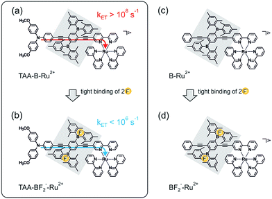 | ||
| Scheme 1 Chemical structure of the TAA-B-Ru2+ dyad (a) and the B–Ru2+ reference compound (c) investigated here, along with an illustration of the key finding from this study: intramolecular photoinduced electron transfer is rapid in TAA-B-Ru2+ in CH2Cl2 (a), but upon fluoride addition the TAA-BF2−-Ru2+ species is formed, and the rate constant for intramolecular electron transfer (kET) decreases by more than 2 orders of magnitude (b). All compounds in scheme were isolated as PF6− salts, see ESI.† | ||
Many prior studies have used chemical stimuli to control electron transfer rates, for example protons which are bound by amine donors,10 cations which are coordinated by aza crown ether donors,11 or Lewis acids which interact with quinone acceptors,12 just to name a few. However, in the vast majority of cases the chemical stimulus interacts directly with the donor or the acceptor, and control is achieved by changing the driving force for electron transfer. In a few cases alkali cations or ammonium ions were coordinated to molecular bridges bearing crown ether functions.13 However, cation binding in these cases is typically rather weak (with association constants on the order of 102–105 M−1), and the effects on electron transfer between the covalently attached donors and acceptor were comparatively small. Similarly, anion binding by molecular squares, calixarenes, thioureas, anion-π interactions, etc., often suffers from relatively low binding constants, and in most cases control of electron transfer is achieved by changing its driving-force.14 Many of the abovementioned anion-binding systems are structurally rather complex or flexible, but for a future molecular electronics technology anion-responsive wires made from rigid rod-like molecular units are highly desirable.
Our study introduces the concept of anion-controllable rigid rod-like molecular bridges and nanowires which should be broadly applicable because it is largely independent of what donors and acceptors are used and at what driving force the electron transfer operates. In some proteins, electron transfer rates are potentially regulated by a similar fundamental principle because the interaction of ions with charged amino acid side chains can modulate electronic couplings between distant redox partners.15
2. Results and discussion
Synthesis
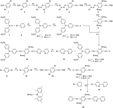
The synthesis of the TAA-B-Ru2+ and B-Ru2+ compounds from Scheme 1 involved 15 and 8 steps, respectively, as illustrated by Scheme 2. The key building block for the molecular bridge is the new compound 6 which in principle could be used for the modular (step-by-step) synthesis of mono-disperse oligomers of 2,5-diboryl-1,4-phenylene ethynylene following previously published synthetic protocols for other oligo-p-phenylene ethynylene (OPE) wires.16 For the synthesis of the B-Ru2+ reference compound, the known symmetrical building block 20 was used instead of compound 6.7s Complete synthetic details and product characterization data are given in the ESI.†Fluoride binding to the organoboron unit
The solid blue line in Fig. 1a is the optical absorption spectrum of TAA-B-Ru2+ in CH2Cl2 at 22 °C. The band maximizing at 468 nm is caused by MLCT transitions of the Ru(bpy)32+ unit, and the band at 290 nm is due to bpy-localized π–π* transitions. The absorption band centered at 357 nm is absent in the UV-Vis spectrum of Ru(bpy)32+ and is attributed to charge transfer from the triarylamino-group to the dimesitylboron-decorated bridging unit. The spectrum of reference compound B-Ru2+ in Fig. 1b (solid green line) exhibits the same MLCT and bpy-localized π–π* bands, but the N → B charge transfer band around 357 nm is missing because ligand 22 lacks the triarylamino-group.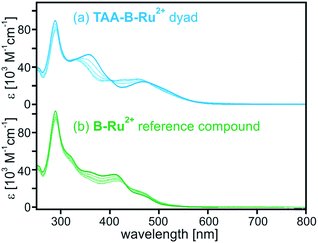 | ||
| Fig. 1 Optical absorption spectra of (a) TAA-B-Ru2+ and (b) B-Ru2+ in CH2Cl2 at 22 °C. Solid lines: in absence of TBAF; dashed lines: in presence of 1, 2, 3, and 4 equivalents of TBAF. | ||
For both compounds spectral changes are easily detectable upon addition of 1–4 equivalents of TBAF (tetra-n-butylammonium fluoride) (dashed lines in Fig. 1). The spectral changes observed for TAA-B-Ru2+ are somewhat more dramatic than those observed for B-Ru2+ because the abovementioned N → B charge transfer band around 357 nm disappears upon fluoride binding to TAA-B-Ru2+. This is a well-known phenomenon because it forms the basis for fluoride detection in many sensor materials.5a–n,6,7
In the ESI† we report titration curves displaying the absorbance of CH2Cl2 solutions with known TAA-B-Ru2+ (Fig. S1†) and B-Ru2+ concentrations (Fig. S2†) at selected wavelengths as a function F− concentration. The two data sets were analyzed in terms of a 1![[thin space (1/6-em)]](https://www.rsc.org/images/entities/char_2009.gif) :
:![[thin space (1/6-em)]](https://www.rsc.org/images/entities/char_2009.gif) 2 binding model using the Specfit software, i.e., it was assumed that two fluoride anions bind per TAA-B-Ru2+ or B-Ru2+ molecule because they both contain two dimesitylboron units. The obtained fits to the experimental titration curves are reasonably good (Fig. S1 and S2†), unlike what is obtained with a simpler 1
2 binding model using the Specfit software, i.e., it was assumed that two fluoride anions bind per TAA-B-Ru2+ or B-Ru2+ molecule because they both contain two dimesitylboron units. The obtained fits to the experimental titration curves are reasonably good (Fig. S1 and S2†), unlike what is obtained with a simpler 1![[thin space (1/6-em)]](https://www.rsc.org/images/entities/char_2009.gif) :
:![[thin space (1/6-em)]](https://www.rsc.org/images/entities/char_2009.gif) 1 binding model. So-called component spectra used to obtain the fits with the 1
1 binding model. So-called component spectra used to obtain the fits with the 1![[thin space (1/6-em)]](https://www.rsc.org/images/entities/char_2009.gif) :
:![[thin space (1/6-em)]](https://www.rsc.org/images/entities/char_2009.gif) 2 binding model are included in the ESI (Fig. S3 and S4†). The cumulative binding constants for the formation of 1
2 binding model are included in the ESI (Fig. S3 and S4†). The cumulative binding constants for the formation of 1![[thin space (1/6-em)]](https://www.rsc.org/images/entities/char_2009.gif) :
:![[thin space (1/6-em)]](https://www.rsc.org/images/entities/char_2009.gif) 1 (β1,1) and 1
1 (β1,1) and 1![[thin space (1/6-em)]](https://www.rsc.org/images/entities/char_2009.gif) :
:![[thin space (1/6-em)]](https://www.rsc.org/images/entities/char_2009.gif) 2 (β1,2) adducts obtained in this manner are summarized in Table 1. In both TAA-B-Ru2+ and B-Ru2+ the first fluoride anion binds with an association constant (KA) on the order of 107 M−1 while the second F− binds with KA = 105–106 M−1. These values are in line with fluoride binding constants reported earlier for related organoboron compounds in similarly apolar solution.5f,7b,o,8b,e,17
2 (β1,2) adducts obtained in this manner are summarized in Table 1. In both TAA-B-Ru2+ and B-Ru2+ the first fluoride anion binds with an association constant (KA) on the order of 107 M−1 while the second F− binds with KA = 105–106 M−1. These values are in line with fluoride binding constants reported earlier for related organoboron compounds in similarly apolar solution.5f,7b,o,8b,e,17
Addition of TBAF to CD2Cl2 solutions of TAA-B-Ru2+ and B-Ru2+ leads to the appearance of a resonance at −170 ppm in the 19F NMR spectrum (Fig. S5 and S6†), and when excess TBAF is present an additional resonance appears at −128 ppm. The former is characteristic for dimesitylboron-bound fluoride, the latter is due to free F−.7b,8b Our 19F NMR experiment is unable to distinguish between the two chemically slightly distinct fluoride binding sites present in both TAA-B-Ru2+ and B-Ru2+, but this is not uncommon.7b,8b With 11B NMR spectroscopy one observes the appearance of a resonance at 5 ppm and the disappearance of a resonance at 80 ppm upon fluoride addition (Fig. S7 and S8†), both indicative of F− binding to dimesitylboron.7b,8b,17c Thus it is clear that F− binding occurs at the B atoms, compatible with other recently explored organoboron systems containing Ru(bpy)32+ (or similar) complex moieties.5d,5e,17b,18 Furthermore it is clear that TBAF solution usually contains some residual water, and given the high hydration enthalpy of F− this is undesirable, and it will lead to lower (apparent) binding constants. However, use of TBAF solution is common practice,5a–n,7 and we can exclude that any of the effects reported in the following arise just from the addition of water. The hexafluorophosphate counter-anions of TAA-B-Ru2+ and B-Ru2+ are sterically too demanding in order to interact strongly with our organoboron units,5a–n,7 and hence even the use of 0.1 M TBAPF6 as a supporting electrolyte for cyclic voltammetry (see below) is unproblematic.17a,b,19
Photoinduced electron transfer and electrochemistry in absence of fluoride
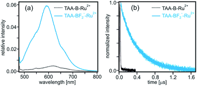
Compared to Ru(bpy)32+ or the B-Ru2+ reference compound the TAA-B-Ru2+ dyad is essentially non-luminescent in de-oxygenated CH2Cl2 at 22 °C (Fig. S9a†). The lowest-energetic 3MLCT excited state of the B–Ru2+ reference compound has a lifetime (τ) of 2040 ns under these conditions (Table 2), but in the TAA-B-Ru2+ dyad the respective excited state depopulates within less than 10 ns (Fig. S9b†). Evidently, the 3MLCT excited state of the TAA-B-Ru2+ dyad is quenched by an efficient nonradiative process.| 3MLCTa [ns] | 3ILb [ns] | |
|---|---|---|
| a Emissive state, measured by time-resolved luminescence and transient absorption. b Dark state, measured by transient absorption. Lifetimes extracted from the data in Fig. 6, S9b, S10, S12 and S14. Excitation occurred at 532 nm with laser pulses of ∼10 ns duration. Detection was at 620 nm for luminescence and at the wavelengths reported in the text or in the ESI for transient absorption. For more detailed discussion of these lifetimes see the ESI. All lifetime values are associated with an experimental uncertainty of ±5%. | ||
| Ru(bpy)32+ | 650 | |
| B-Ru2+ | 2040 | |
| BF2−-Ru2+ | 1120 | 7160 |
| TAA-B-Ru2+ | <10 | |
| TAA-BF2−-Ru2+ | 380 | 7350 |
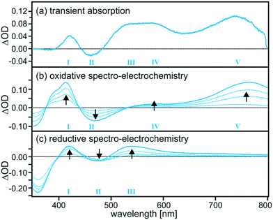
Based on the transient absorption and spectro-electrochemical data in Fig. 2 the efficient nonradiative excited-state quenching process in TAA-B-Ru2+ can be identified unambiguously as intramolecular electron transfer from the triarylamine unit to the photoexcited Ru(bpy)32+ moiety. The transient difference spectrum in Fig. 2a was recorded after excitation of 2 × 10⁻⁵ M TAA-B-Ru2+ in de-oxygenated CH2Cl2 at 22 °C using laser pulses of 532 nm wavelength and ∼10 ns duration. Selective excitation into the MLCT absorption band was followed by time-integration of the resulting transient difference spectrum over an interval of 200 ns using a iCCD camera. One observes a weak band at 430 nm, a bleach at 465 nm, a broad spectral feature which seems to be due to superposed bands with local maxima near 540 and 580 nm, and finally a well-separated band peaking at 740 nm. These five spectral features (numbered I–V in Fig. 2a) clearly indicate the formation of oxidized triarylamine and reduced ruthenium complex as will be demonstrated in the following.
The series of spectro-electrochemical data in Fig. 2b and c are difference spectra in which the UV-Vis spectrum of TAA-B-Ru2+ in CH2Cl2 (prior to applying any potential) was used as a baseline. For measurement of the spectra in Fig. 2b an electrochemical potential of 0.2 V vs. Fc+/Fc was applied with a Pt grid electrode. This is sufficient for oxidation of the triarylamine unit of TAA-B-Ru2+ (see below), but neither the Ru(bpy)32+ moiety nor the bridging organoboron unit will undergo any oxidation at this potential. Thus, the data in Fig. 2b show the spectral changes associated with oxidation of triarylamine to its monocationic form (oxidation to the dication requires more positive potentials, see below).20 The longer the potential is applied, the more readily detectable the spectral changes become (black arrows). For the spectra in Fig. 2c an electrochemical potential of −1.9 V vs. Fc+/Fc was applied, and this lead to one-electron reduction of the Ru(bpy)32+ moiety of TAA-B-Ru2+. Neither the triarylamine unit nor the organoboron bridge can be reduced at this potential (see below). Comparison of Fig. 2a with 2b and c clearly shows that all spectral features observed in the transient absorption spectrum can be explained by combined triarylamine oxidation and Ru(bpy)32+ reduction. This is conclusive evidence for intramolecular photoinduced electron transfer and the formation of a photoproduct that we abbreviate as TAA+-B-Ru+ in the following.
The transient absorption signals at 740 and 580 nm including the bleach at 460 nm (bands V, IV, and II in Fig. 2a) all exhibit the same temporal evolution (Fig. 3). Each signal reaches its maximum intensity immediately after the laser pulse and decays with a time constant of 95 ns in de-oxygenated CH2Cl2 at 22 °C. This indicates that photoinduced electron transfer occurs within ≤10 ns (rate constant kET ≥ 108 s−1 in Scheme 1a), and the reverse (thermal) electron transfer to reinstate the starting material in the electronic ground state takes place with a time constant of 95 ns (kbET = 1.05 × 10⁷ s−1).
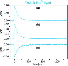 | ||
| Fig. 3 Decays of the transient absorption signals measured on a 2 × 10⁻⁵ M solution of TAA-B-Ru2+ in de-oxygenated CH2Cl2 after excitation at 532 nm with laser pulses of ∼10 ns duration. Detection occurred at (a) 740 nm (band V in Fig. 2a), (b) 580 nm (band IV in Fig. 2a), and (c) 460 nm (band II in Fig. 2a). | ||
The reaction free energies for photoinduced electron transfer (ΔG0ET) and for reverse (thermal) electron transfer (ΔG0bET) can be estimated on the basis of electrochemical potentials determined by cyclic voltammetry in dry CH2Cl2 in presence of 0.1 M TBAPF6. In the voltammogram of TAA-B-Ru2+ (Fig. 4a) oxidation of the triarylamine to its monocationic form manifests as a reversible wave at 0.2 V vs. Fc+/Fc.20a,c–e Oxidation to the triarylamine dication is seen as a quasi-reversible wave at 0.8 V,20a superposed on the (irreversible) oxidation of Ru(II) to Ru(III) with a peak potential of 1.1 V vs. Fc+/Fc. On the reductive side, the first (reversible) wave at −1.6 V is due to reduction of a bpy-ligand.20e,21 Comparison with the voltammogram of the B-Ru2+ reference compound (Fig. 4b) and with the voltammogram of organoboron bridging unit 20 (Fig. 4c; see Scheme 2 for chemical structure) supports our assignments of redox waves to the individual molecular components of TAA-B-Ru2+. The voltammogram of compound 20 shows that reduction of the organoboron bridging unit occurs at −2.1 V vs. Fc+/Fc, in line with previously reported reduction potentials for related species.5h,7k,8e,19b,22 All redox potentials are summarized in Table 3.
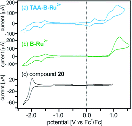 | ||
| Fig. 4 Cyclic voltammograms of (a) TAA-B-Ru2+, (b) B-Ru2+, and (c) bridge reference molecule 20 (molecular structure shown in Scheme 2) in CH2Cl2. The supporting electrolyte was 0.1 M TBAPF6. The potential scan rate was 0.1 V s−1 in all cases. The shoulder wave at 0.85 V in (a) is due to oxidation of TAA+ to TAA2+.20a All other redox potentials extracted from this data set are summarized in Table 3. | ||
| boryl0/− | bpy0/− | TAA+/0 | RuIII/II | |
|---|---|---|---|---|
| a The electrolyte was 0.1 M TBAPF6, the voltage scan rate was 0.1 V s−1. Determined from the data in Fig. 4 and S15. The chemical structure of compound 20 is shown in Scheme 2. Ru(bpy)32+ denotes the reference complex with three identical (un-substituted) bpy ligands. | ||||
| TAA-B-Ru2+ | −2.2 | −1.6 | 0.2 | 1.1 |
| B-Ru2+ | −2.2 | −1.6 | 1.2 | |
| Ru(bpy)32+ | −1.7 | 0.9 | ||
| Compound 20 | −2.1 | |||
| TAA-BF2−-Ru2+ | −1.5 | 0.2 | ||
Based on the electrochemical potential for oxidation of triarylamine to its monocationic form (0.2 V), the electrochemical potential for one-electron reduction of the Ru(bpy)32+ moiety (−1.6 V), the MLCT energy of the Ru(bpy)32+ unit (2.1 eV),21b and a donor–acceptor distance of ∼15 Å (geometrical triarylamine-N–Ru distance) one can use the Weller equation to estimate a reaction free energy of ΔG0ET ≈ −0.3 eV for photoinduced electron transfer in TAA-B-Ru2+ in CH2Cl2.23 Thermal electron transfer in the reverse direction then occurs with ΔG0bET ≈ −1.8 eV. As noted above, the photoinduced reaction occurs with kET ≥ 108 s−1 whereas for the thermal reverse reaction one finds kbET = 1.05 × 10⁷ s−1. Thus, the thermal reverse reaction is slower than photoinduced electron transfer despite a substantially higher driving force, indicating that the thermal process occurs in the so-called Marcus inverted region,24 as observed before for several other examples of simple donor–acceptor dyads with d6 metal diimine photosensitizers.20c–e,25 However, the most important result until now is that photoinduced electron transfer from the triarylamine to the Ru(bpy)32+ unit of TAA-B-Ru2+ in de-oxygenated CH2Cl2 at 22 °C occurs with a rate constant of kET ≥ 108 s−1 (Scheme 1a).
Photophysics and electrochemistry in presence of fluoride
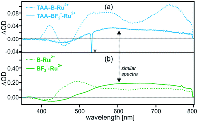
When adding 4 equivalents of TBAF to a 2 × 10⁻⁵ M solution of TAA-B-Ru2+ in dry CH2Cl2 one reaches conditions under which >99% of all dyads have two F− anions bound to the organoboron bridge, based on the association constants in Table 1. Thus, the resulting solution contains almost exclusively a species that we will refer to as TAA-BF2−-Ru2+ (Scheme 1b). This is much different from many previously explored systems in which ion binding to, e.g. crown ether donors or ureas, is often orders of magnitude weaker.11,13,14 In this regard the tight Lewis acid/Lewis base interaction between the organoboron unit and F− stands out.7b,26When performing the exact same pump-probe experiment with TAA-BF2−-Ru2+ as before with TAA-B-Ru2+, one obtains the transient difference spectrum shown as a solid blue trace in Fig. 5a. For reference, the spectrum of TAA-B-Ru2+ from Fig. 2a is reproduced in Fig. 5a as a dashed trace. Clearly the transient difference spectra of TAA-BF2−-Ru2+ and TAA-B-Ru2+ are very different. In order to understand the transient difference spectrum of TAA-BF2−-Ru2+, an analogous pump-probe experiment with the reference dyad B-Ru2+ (chemical structure shown in Scheme 1c) is most insightful. When adding 4 equivalents of TBAF to a 2 × 10⁻⁵ M solution of B-Ru2+ in dry CH2Cl2, two fluoride anions are tightly bound to the organoboron unit in >99% of all B-Ru2+ reference molecules present (Table 1), resulting in a species that we will refer to as BF2−-Ru2+ in the following. The transient difference spectrum of BF2−-Ru2+ shown as a solid green trace in Fig. 5b was obtained under the exact same conditions as that of TAA-BF2−-Ru2+ in Fig. 5a. The solid traces in Fig. 5a and b are virtually identical, indicating that the same photoproduct is formed in the dyad and in the reference compound when F− is bound. Since the reference compound lacks the triarylamine donor, this observation strongly suggests that intramolecular photoinduced electron transfer from the triarylamine to Ru(bpy)32+ does no longer occur to a significant extent in TAA-BF2−-Ru2+.
Moreover, whereas the TAA-B-Ru2+ dyad without any bound F− ions is essentially non-luminescent in de-oxygenated CH2Cl2 at 22 °C (τ < 10 ns, Table 2, black trace in Fig. 6b), the TAA-BF2−-Ru2+ compound exhibits 3MLCT luminescence with a lifetime (τ) of 380 ns under identical conditions (blue traces in Fig. 6 and Table 2). This lifetime corresponds to a 3MLCT decay rate constant of kobs = 2.6 × 10⁶ s−1, which must be equal to the sum of all rate constants for radiative and nonradiative relaxation processes occurring from the 3MLCT state. Thus it seems reasonable to conclude that kET < 106 s−1 for TAA-BF2−-Ru2+ (Scheme 1b).
The remaining open question then is: “What exactly is the photoproduct observed for TAA-BF2−-Ru2+ and BF2−-Ru2+ in Fig. 5?”. The observation of 3MLCT luminescence in presence of fluoride strongly suggests that the spectral signature of the 3MLCT excited state will contribute substantially to the transient difference spectra shown as solid traces in Fig. 5a and b. This is indeed the case, but the situation is slightly more complicated because there is a contribution from a dark (i.e., non-luminescent) photoproduct in both cases. This conclusion is reached on the basis of the temporal evolution of the transient absorption signals from Fig. 5a and b at various detection wavelengths (Fig. S10†). For TAA-BF2−-Ru2+ in de-oxygenated CH2Cl2, the transient absorption signals at 460, 580, and 740 nm all decay in a bi-exponential manner with a fast decay component that corresponds to the 3MLCT luminescence lifetime (380 ns) and a slow decay component of 7350 ns (Table 2 and Fig. S10a†). At all three detection wavelengths the intensity ratio of fast versus slow decay components is roughly 40%:60%. When performing the exact same experiments with BF2−-Ru2+, one also finds bi-exponential decays. The faster decay component (1120 ns, Table 2) again corresponds to the 3MLCT luminescence lifetime (Fig. S14d†) whereas the slower decay component (7160 ns, Fig. S10b and S14a–c†) is not observed in luminescence. The relative intensities of fast and slow decay components are again roughly 40% versus 60%, at all three detection wavelengths. Thus, the 3MLCT state is clearly detectable in transient absorption, and the fact that the resulting spectrum is substantially different from that of the 3MLCT state of isolated Ru(bpy)32+ is no surprise given the presence of a functionalized bpy ligand in both TAA-BF2−-Ru2+ and BF2−-Ru2+.27 The longer-lived dark state is attributed to a triplet state localized on the functionalized bpy ligands (molecules 16 and 22 in Scheme 2).28 Further details regarding possible population of this 3IL (IL = intraligand) dark state (including additional spectroscopic data for B-Ru2+) are given only in the ESI† because this issue is of no further interest for the present study.
Clearly the most important finding here is that fluoride binding to TAA-B-Ru2+ in CH2Cl2 slows down intramolecular electron transfer from the triarylamine to photoexcited Ru(bpy)32+ by more than two orders of magnitude, the rate constant kET decreases from >108 s−1 to <106 s−1.
Physical origin of the electron transfer rate decrease upon fluoride binding
As noted in the introduction, in the vast majority of prior studies in which a chemical stimulus was used to regulate electron transfer rates, this has occurred by direct interaction of the chemical stimulus with either the donor or the acceptor.10–12 Consequently, control of electron transfer occurred by modulating its driving force (ΔG0ET). In the TAA-B-Ru2+ dyad the chemical stimulus (F−) interacts with the bridge, and this is comparatively rare.13 Importantly, interaction of F− with the organoboron bridge does not affect the driving force for intramolecular electron transfer from triarlyamine to photoexcited Ru(bpy)32+ to a significant extent, because the donor and acceptor redox potentials stay largely unaffected. The cyclic voltammogram of TAA-BF2−-Ru2+ (Fig. S15†) is less clean than that of TAA-B-Ru2+ (Fig. 4a) due to the presence of fluoride, but one can readily extract electrochemical potentials of 0.2 V vs. Fc+/Fc for triarylamine oxidation and −1.5 V vs. Fc+/Fc for one-electron reduction of the Ru(bpy)32+ unit (Table 3). Consequently, the driving force for photoinduced electron transfer in TAA-BF2−-Ru2+ is ΔG0ET = −0.4 eV, very similar to what has been found above in absence of F− (ΔG0ET = −0.3 eV).Thus, in principle there is still sufficient driving force for photoinduced electron transfer in TAA-BF2−-Ru2+, yet this process is kinetically not competitive with other excited-state deactivation pathways. We attribute this to a decrease in electronic coupling strength (HDA) between the triarylamine donor and the Ru(bpy)32+ acceptor. According to superexchange theory, HDA depends strongly on the so-called tunneling energy gap,29i.e., a quantity which is strongly dependent on the HOMO/LUMO energies of the bridging units mediating long-range electron tunneling.1a,2g,4g,30 An alternative and equally valid view (discussed in more detail below) is that the barrier height associated with the tunneling process depends crucially on the redox potentials of the bridging units.1a,2g,31 As noted in the introduction, F− binding to organoboron units strongly affects their HOMO/LUMO energies due to disruption of pπ–π* conjugation between the boron atom and the π-system to which it is attached.7b,f,s Moreover, and perhaps even more importantly, the resulting organofluoroborate species is anionic, and consequently the LUMO is energetically raised compared to the organoboron unit before F− addition. The π-acceptor capacity of the organofluoroborate is substantially lower.
In free TAA-B-Ru2+ without fluoride, photoinduced charge transfer is likely to proceed via an electron (rather than hole) tunneling mechanism for which the LUMO energy of the organoboron bridge is relevant.2j,11c,32 In the electron tunneling picture, the energy of the (virtual) one-electron reduced state of the organoboron bridge determines the height of the tunneling barrier, hence it is the LUMO energy of the bridge which matters. For hole tunneling, the energy of the (virtual) one-electron oxidized state of the organoboron bridge would be relevant, and given the fact that the bridge is very electron-deficient prior to F− binding, this can be expected to lead to a substantially higher barrier than in the case of the electron tunneling model.
In the simplistic picture used in Scheme 3a the energy difference between the initial TAA-B-*Ru2+ state (the asterisk denotes the 3MLCT excited species) and the (virtual) intermediate TAA+-B−-Ru2+ (comprised of oxidized donor, reduced bridge, and terminal acceptor in its electronic ground state) defines the barrier height for electron tunneling from the triarylamine to the photoexcited ruthenium complex. Based on the electrochemical potentials in Table 3 and the Weller equation, the height of this barrier is ∼0.3 eV. Tunneling across such a barrier can readily occur on the sub-nanosecond timescale.1a,2c,g,i,30a,33 When two fluoride anions bind to the organoboron bridge, its reduction potential is expected to shift to significantly more negative values.17a,19a,34 We have not been able to measure the respective potential and were unable to find reduction potentials of comparable organofluoroborate species in the literature. However, based on electrochemical studies in chemically related systems it seems reasonable to expect a cathodic shift of at least ∼0.3 V per bound anionic charge,5h,34a,35 increasing the barrier height for photoinduced electron tunneling by at least ∼0.6 eV when two fluorides are bound (Scheme 3b). Such an increase in barrier height is expected to decrease the tunneling probability by several orders of magnitude and can explain why photoinduced electron transfer is no longer observed in TAA-BF2−-Ru2+.36 On the other hand, the electron-rich organofluoroborate bridge is expected to have a significantly lower oxidation potential than its electron-deficient organoboron counterpart without fluorides, and this might result in an increasingly efficient hole tunneling pathway.2j However, this effect seems to be less important, presumably because the barrier for hole tunneling remains relatively large. More theoretical assessments are certainly possible but beyond the scope of this paper.37
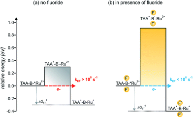 | ||
| Scheme 3 Barrier heights and reaction free energies (ΔG0ET) for intramolecular electron transfer from triarylamine to photoexcited Ru(bpy)32+ in the TAA-B−-Ru2+ dyad in absence (a) and in presence of bound F− anions (b). In presence of 4 eq. of F− (b), TAA-BF2−-Ru2+ is the predominant species in CH2Cl2 solution due to tight anion binding, see text. The asterisk (*) denotes the 3MLCT-excited Ru(bpy)32+ moiety. The reaction free energies (ΔG0ET) were estimated on the basis of the electrochemical potentials in Table 3 using the Weller equation as described in the text.23 The barrier heights were estimated in analogous manner because they correspond to the reaction free energies for charge injection from the triarylamine donor into the organoboron bridge out of the 3MLCT-excited dyad. The reduction potential of the organoboron bridge with 2 attached F− ions was estimated as described in the text. | ||
3. Conclusions
Organoboron compounds have received considerable attention in recent years and many scientists have exploited the change in electronic structure associated with fluoride or cyanide binding to such materials.5a–n,6,7 Even though the interest of this class of compounds for various optical, electronic, and sensory applications has been pointed out numerous times,7 curiously, there have been no fundamental studies of photoinduced electron transfer across organoboron bridges until now.9The key effect observed in this study is illustrated by Scheme 1a and b, and its physical origin is explained graphically by Scheme 3. In the TAA-B-Ru2+ dyad, electron transfer from the triarylamine to the photoexcited Ru(bpy)32+ complex across the 2,5-diboryl-1,4-phenylene spacer occurs within less than 10 ns in CH2Cl2 at 22 °C (kET ≥ 108 s−1). Under these conditions, two fluoride anions bind to the organoboron unit with association constants greater than 105 M−1 due to strong Lewis base/Lewis acid interactions.5 With two F− anions bound to the 2,5-diboryl-1,4-phenylene spacer, the abovementioned photoinduced electron transfer process occurs with a time constant longer than 1000 ns (kET < 106 s−1). The decrease of electron transfer rates by more than two orders of magnitude is caused by an increase of the tunneling barrier height upon fluoride binding (Scheme 3),31a leading to a decrease of the electronic interaction between the donor and the acceptor as the bridge LUMO is shifted to higher energy;1a,38 the organoboron bridging unit in its initial form is a strong π-acceptor, but the organofluoroborate species resulting from F− addition has a much weaker π-acceptor capacity.22a In a more simplistic view, the bound F− anions act as a Coulomb barrier for the transferring electron.39 Importantly, the driving force for electron transfer is essentially unaffected, unlike in the vast majority of previously explored examples in which chemical stimuli interact either with the donor or the acceptor to modulate the driving force for electron transfer (ΔG0ET).10–12 In our case the chemical stimulus (F−) interacts exclusively with the molecular bridge, and, to the best of our knowledge, our system is the first example of an anion-controllable rigid rod-like molecular wire.13 The use of strong Lewis acid/Lewis base interactions to regulate electron transfer across a rigid rod-like molecular wire (without affecting the driving force) is conceptually novel.14a–g
Our results are relevant in the greater context of a future molecular electronics technology because they demonstrate the basic principle of anion-responsive molecular wires which could become part of nanoscopic electrical circuits. Based on our electron transfer study, the anion-triggered switching between conducting and insulating states of such wires has become a realistic goal. For this purpose rigid rod-like molecular structures are highly desirable. In our study, the electron transfer occurs over a distance of ∼15 Å but in principle the functional 2,5-diboryl-1,4-phenylene element can also be incorporated into longer wires.
Finally we note that our findings are also relevant in a biochemical context. In some proteins, electron transfer between distant redox partners occurs along a pathway containing charged amino acid side chains.38,40 Interactions of the latter with counter-ions will affect the electron density at the respective amino acids and hence will influence the strength of the electronic coupling between the donor and the acceptor.15b Thus, it is conceivable that Nature uses ion-responsive structural elements acting in a similar fashion as the organoboron bridge investigated herein.15a Our study shows that this particular type of electron transfer rate regulation can be very effective.
Acknowledgements
This work was funded by the Swiss National Science Foundation through grant number 200021_146231/1 and by the Deutsche Forschungsgemeinschaft through grant number WE4815/3-1. Daniel Häussinger is acknowledged for help with 11B and 19F NMR spectroscopy, and Christian Reber is thanked for useful discussions.Notes and references
- (a) H. B. Gray and J. R. Winkler, Proc. Natl. Acad. Sci. U. S. A., 2005, 102, 3534 CrossRef CAS PubMed; (b) M. D. Newton, Chem. Rev., 1991, 91, 767 CrossRef CAS; (c) P. P. Edwards, H. B. Gray, M. T. J. Lodge and R. J. P. Williams, Angew. Chem., Int. Ed., 2008, 47, 6758 CrossRef CAS PubMed.
- (a) F. Giacalone, J. L. Segura, N. Martín and D. M. Guldi, J. Am. Chem. Soc., 2004, 126, 5340 CrossRef CAS PubMed; (b) W. B. Davis, W. A. Svec, M. A. Ratner and M. R. Wasielewski, Nature, 1998, 396, 60 CrossRef CAS; (c) E. A. Weiss, M. J. Ahrens, L. E. Sinks, A. V. Gusev, M. A. Ratner and M. R. Wasielewski, J. Am. Chem. Soc., 2004, 126, 5577 CrossRef CAS PubMed; (d) M. Cordes and B. Giese, Chem. Soc. Rev., 2009, 38, 892 RSC; (e) E. C. Minnihan, D. G. Nocera and J. Stubbe, Acc. Chem. Res., 2013, 46, 2524 CrossRef CAS PubMed; (f) F. D. Lewis, R. L. Letsinger and M. R. Wasielewski, Acc. Chem. Res., 2001, 34, 159 CrossRef CAS PubMed; (g) O. S. Wenger, Acc. Chem. Res., 2011, 44, 25 CrossRef CAS PubMed; (h) A. Harriman and R. Ziessel, Chem. Commun., 1996, 1707 RSC; (i) M. P. Eng and B. Albinsson, Angew. Chem., Int. Ed., 2006, 45, 5626 CrossRef CAS PubMed; (j) M. Natali, S. Campagna and F. Scandola, Chem. Soc. Rev., 2014, 43, 4005 RSC; (k) V. Lloveras, J. Vidal-Gancedo, T. M. Figueira-Duarte, J. F. Nierengarten, J. J. Novoa, F. Mota, N. Ventosa, C. Rovira and J. Veciana, J. Am. Chem. Soc., 2011, 133, 5818 CrossRef CAS PubMed; (l) M. Wielopolski, C. Atienza, T. Clark, D. M. Guldi and N. Martín, Chem.–Eur. J., 2008, 14, 6379 CrossRef CAS PubMed; (m) S. Schlundt, G. Kuzmanich, F. Spanig, G. D. Rojas, C. Kovacs, M. A. Garcia-Garibay, D. M. Guldi and A. Hirsch, Chem.–Eur. J., 2009, 15, 12223 CrossRef CAS PubMed; (n) B. Ballesteros, G. de la Torre, C. Ehli, G. M. A. Rahman, F. Agullo-Rueda, D. M. Guldi and T. Torres, J. Am. Chem. Soc., 2007, 129, 5061 CrossRef CAS PubMed; (o) M. Soler and J. K. McCusker, J. Am. Chem. Soc., 2008, 130, 4708 CrossRef CAS PubMed; (p) D. C. O'Hanlon, B. W. Cohen, D. B. Moravec, R. F. Dallinger and M. D. Hopkins, J. Am. Chem. Soc., 2014, 136, 3127 CrossRef PubMed; (q) C. Shih, A. K. Museth, M. Abrahamsson, A. M. Blanco-Rodriguez, A. J. Di Bilio, J. Sudhamsu, B. R. Crane, K. L. Ronayne, M. Towrie, A. Vlček, J. H. Richards, J. R. Winkler and H. B. Gray, Science, 2008, 320, 1760 CrossRef CAS PubMed; (r) D. K. James and J. M. Tour, in Molecular Wires: From Design to Properties, ed. L. De Cola, Springer-Verlag Berlin, Berlin, 2005, vol. 257, p. 33 Search PubMed; (s) S.-H. Lee, C. T.-L. Chan, K. M.-C. Wong, W. H. Lam, W.-M. Kwok and V. W. W. Yam, J. Am. Chem. Soc., 2014, 136, 10041 CrossRef CAS PubMed; (t) J. Fortage, E. Goransson, E. Blart, H. C. Becker, L. Hammarström and F. Odobel, Chem. Commun., 2007, 4629 RSC; (u) H. A. Meylemans, J. T. Hewitt, M. Abdelhaq, P. J. Vallett and N. H. Damrauer, J. Am. Chem. Soc., 2010, 132, 11464 CrossRef CAS PubMed; (v) A. Lukacs, A. P. M. Eker, M. Byrdin, K. Brettel and M. H. Vos, J. Am. Chem. Soc., 2008, 130, 14394 CrossRef CAS PubMed; (w) C. A. Hunter and R. K. Hyde, Angew. Chem., Int. Ed., 1996, 35, 1936 CrossRef CAS; (x) M. Delor, P. A. Scattergood, I. V. Sazanovich, A. W. Parker, G. M. Greetham, A. Meijer, M. Towrie and J. A. Weinstein, Science, 2014, 346, 1492 CrossRef CAS PubMed.
- (a) N. J. Tao, Nat. Nanotechnol., 2006, 1, 173 CrossRef CAS PubMed; (b) W. J. Hong, H. Li, S. X. Liu, Y. C. Fu, J. F. Li, V. Kaliginedi, S. Decurtins and T. Wandlowski, J. Am. Chem. Soc., 2012, 134, 19425 CrossRef CAS PubMed; (c) R. Huber, M. T. González, S. Wu, M. Langer, S. Grunder, V. Horhoiu, M. Mayor, M. R. Bryce, C. S. Wang, R. Jitchati, C. Schönenberger and M. Calame, J. Am. Chem. Soc., 2008, 130, 1080 CrossRef CAS PubMed.
- (a) J. He, F. Chen, P. A. Liddell, J. Andreasson, S. D. Straight, D. Gust, T. A. Moore, A. L. Moore, J. Li, O. F. Sankey and S. M. Lindsay, Nanotechnology, 2005, 16, 695 CrossRef CAS; (b) J. Areephong, J. H. Hurenkamp, M. T. W. Milder, A. Meetsma, J. L. Herek, W. R. Browne and B. L. Feringa, Org. Lett., 2009, 11, 721 CrossRef CAS PubMed; (c) J. M. Endtner, F. Effenberger, A. Hartschuh and H. Port, J. Am. Chem. Soc., 2000, 122, 3037 CrossRef CAS; (d) F. M. Raymo and M. Tomasulo, Chem. Soc. Rev., 2005, 34, 327 RSC; (e) P. A. Liddell, G. Kodis, A. L. Moore, T. A. Moore and D. Gust, J. Am. Chem. Soc., 2002, 124, 7668 CrossRef CAS PubMed; (f) P. Belser, L. De Cola, F. Hartl, V. Adamo, B. Bozic, Y. Chriqui, V. M. Iyer, R. T. F. Jukes, J. Kuhni, M. Querol, S. Roma and N. Salluce, Adv. Funct. Mater., 2006, 16, 195 CrossRef CAS; (g) J. Kärnbratt, M. Hammarson, S. M. Li, H. L. Anderson, B. Albinsson and J. Andréasson, Angew. Chem., Int. Ed., 2010, 49, 1854 CrossRef PubMed; (h) M. Irie, Chem. Rev., 2000, 100, 1685 CrossRef CAS PubMed; (i) R. Sakamoto, M. Murata and H. Nishihara, Angew. Chem., Int. Ed., 2006, 45, 4793 CrossRef CAS PubMed; (j) E. Orgiu, N. Crivillers, M. Herder, L. Grubert, M. Patzel, J. Frisch, E. Pavlica, D. T. Duong, G. Bratina, A. Salleo, N. Koch, S. Hecht and P. Samori, Nat. Chem., 2012, 4, 675 CrossRef CAS PubMed; (k) D. S. Tyson, C. A. Bignozzi and F. N. Castellano, J. Am. Chem. Soc., 2002, 124, 4562 CrossRef CAS PubMed; (l) M. N. Roberts, C. J. Carling, J. K. Nagle, N. R. Branda and M. O. Wolf, J. Am. Chem. Soc., 2009, 131, 16644 CrossRef CAS PubMed; (m) O. S. Wenger, Chem. Soc. Rev., 2012, 41, 3772 RSC; (n) B. He and O. S. Wenger, J. Am. Chem. Soc., 2011, 133, 17027 CrossRef CAS PubMed.
- (a) T. W. Hudnall, C. W. Chiu and F. P. Gabbaï, Acc. Chem. Res., 2009, 42, 388 CrossRef CAS PubMed; (b) S. Aldridge, C. Bresner, I. A. Fallis, S. J. Coles and M. B. Hursthouse, Chem. Commun., 2002, 740 RSC; (c) X. Y. Liu, D. R. Bai and S. N. Wang, Angew. Chem., Int. Ed., 2006, 45, 5475 CrossRef CAS PubMed; (d) S. T. Lam, N. A. Y. Zhu and V. W.-W. Yam, Inorg. Chem., 2009, 48, 9664 CrossRef CAS PubMed; (e) Y. Sun and S. N. Wang, Inorg. Chem., 2009, 48, 3755 CrossRef CAS PubMed; (f) Y. Kim and F. P. Gabbaï, J. Am. Chem. Soc., 2009, 131, 3363 CrossRef CAS PubMed; (g) T. W. Hudnall, Y. M. Kim, M. W. P. Bebbington, D. Bourissou and F. P. Gabbaï, J. Am. Chem. Soc., 2008, 130, 10890 CrossRef CAS PubMed; (h) C. W. Chiu, Y. Kim and F. P. Gabbaï, J. Am. Chem. Soc., 2009, 131, 60 CrossRef CAS PubMed; (i) S. Yamaguchi, S. Akiyama and K. Tamao, J. Am. Chem. Soc., 2001, 123, 11372 CrossRef CAS PubMed; (j) Y. Kubo, M. Yamamoto, M. Ikeda, M. Takeuchi, S. Shinkai, S. Yamaguchi and K. Tamao, Angew. Chem., Int. Ed., 2003, 42, 2036 CrossRef CAS PubMed; (k) Q. Zhao, F. Y. Li, S. J. Liu, M. X. Yu, Z. Q. Liu, T. Yi and C. H. Huang, Inorg. Chem., 2008, 47, 9256 CrossRef CAS PubMed; (l) I. Siewert, P. Fitzpatrick, A. E. J. Broomsgrove, M. Kelly, D. Vidovic and S. Aldridge, Dalton Trans., 2011, 10345 RSC; (m) K. Parab, K. Venkatasubbaiah and F. Jäkle, J. Am. Chem. Soc., 2006, 128, 12879 CrossRef CAS PubMed; (n) T. H. Kim and T. M. Swager, Angew. Chem., Int. Ed., 2003, 42, 4803 CrossRef CAS PubMed; (o) W. J. Xu, S. J. Liu, X. Y. Zhao, S. Sun, S. Cheng, T. C. Ma, H. B. Sun, Q. A. Zhao and W. Huang, Chem.–Eur. J., 2010, 16, 7125 CrossRef CAS PubMed.
- (a) N. Matsumi and Y. Chujo, Polym. J., 2008, 40, 77 CrossRef CAS; (b) S. Yamaguchi, S. Akiyama and K. Tamao, J. Organomet. Chem., 2002, 652, 3 CrossRef CAS.
- (a) F. Jäkle, Chem. Rev., 2010, 110, 3985 CrossRef PubMed; (b) C. R. Wade, A. E. J. Broomsgrove, S. Aldridge and F. P. Gabbaï, Chem. Rev., 2010, 110, 3958 CrossRef CAS PubMed; (c) M. Elbing and G. C. Bazan, Angew. Chem., Int. Ed., 2008, 47, 834 CrossRef CAS PubMed; (d) S. Yamaguchi and A. Wakamiya, Pure Appl. Chem., 2006, 78, 1413 CAS; (e) I. S. Ke, M. Myahkostupov, F. N. Castellano and F. P. Gabbaï, J. Am. Chem. Soc., 2012, 134, 15309 CrossRef CAS PubMed; (f) C. D. Entwistle and T. B. Marder, Angew. Chem., Int. Ed., 2002, 41, 2927 CAS; (g) C. D. Entwistle and T. B. Marder, Chem. Mater., 2004, 16, 4574 CrossRef CAS; (h) H. Y. Li and F. Jäkle, Macromol. Rapid Commun., 2010, 31, 915 CrossRef CAS PubMed; (i) F. Jäkle, Coord. Chem. Rev., 2006, 250, 1107 CrossRef; (j) A. Lorbach, M. Bolte, H. Y. Li, H. W. Lerner, M. C. Holthausen, F. Jäkle and M. Wagner, Angew. Chem., Int. Ed., 2009, 48, 4584 CrossRef CAS PubMed; (k) T. Noda and Y. Shirota, J. Am. Chem. Soc., 1998, 120, 9714 CrossRef CAS; (l) M. Kinoshita, H. Kita and Y. Shirota, Adv. Funct. Mater., 2002, 12, 780 CrossRef CAS; (m) A. Hubner, Z. W. Qu, U. Englert, M. Bolte, H. W. Lerner, M. C. Holthausen and M. Wagner, J. Am. Chem. Soc., 2011, 133, 4596 CrossRef CAS PubMed; (n) C. Hoffend, F. Schodel, M. Bolte, H. W. Lerner and M. Wagner, Chem.–Eur. J., 2012, 18, 15394 CrossRef CAS PubMed; (o) A. Sundararaman, K. Venkatasubbaiah, M. Victor, L. N. Zakharov, A. L. Rheingold and F. Jäkle, J. Am. Chem. Soc., 2006, 128, 16554 CrossRef CAS PubMed; (p) H. Li and F. Jäkle, Angew. Chem., Int. Ed., 2009, 48, 2313 CrossRef CAS PubMed; (q) H. Y. Li, A. Sundararaman, K. Venkatasubbaiah and F. Jäkle, J. Am. Chem. Soc., 2007, 129, 5792 CrossRef CAS PubMed; (r) S. Yamaguchi, S. Akiyama and K. Tamao, J. Am. Chem. Soc., 2000, 122, 6335 CrossRef CAS; (s) C. H. Zhao, A. Wakamiya, Y. Inukai and S. Yamaguchi, J. Am. Chem. Soc., 2006, 128, 15934 CrossRef CAS PubMed; (t) S. Yamaguchi, T. Shirasaka, S. Akiyama and K. Tamao, J. Am. Chem. Soc., 2002, 124, 8816 CrossRef CAS PubMed.
- (a) R. Stahl, C. Lambert, C. Kaiser, R. Wortmann and R. Jakober, Chem.–Eur. J., 2006, 12, 2358 CrossRef CAS PubMed; (b) H. C. Schmidt, L. G. Reuter, J. Hamacek and O. S. Wenger, J. Org. Chem., 2011, 76, 9081 CrossRef CAS PubMed; (c) Z. Yuan, C. D. Entwistle, J. C. Collings, D. Albesa-Jove, A. S. Batsanov, J. A. K. Howard, N. J. Taylor, H. M. Kaiser, D. E. Kaufmann, S. Y. Poon, W. Y. Wong, C. Jardin, S. Fathallah, A. Boucekkine, J. F. Halet and T. B. Marder, Chem.–Eur. J., 2006, 12, 2758 CrossRef CAS PubMed; (d) Z. M. Hudson, X. Y. Liu and S. N. Wang, Org. Lett., 2011, 13, 300 CrossRef CAS PubMed; (e) G. Zhou, M. Baumgarten and K. Müllen, J. Am. Chem. Soc., 2008, 130, 12477 CrossRef CAS PubMed; (f) A. Sundararaman, R. Varughese, H. Y. Li, L. N. Zakharov, A. L. Rheingold and F. Jäkle, Organometallics, 2007, 26, 6126 CrossRef CAS; (g) J. C. Collings, S. Y. Poon, C. Le Droumaguet, M. Charlot, C. Katan, L. O. Palsson, A. Beeby, J. A. Mosely, H. M. Kaiser, D. Kaufmann, W. Y. Wong, M. Blanchard-Desce and T. B. Marder, Chem.–Eur. J., 2009, 15, 198 CrossRef CAS PubMed; (h) A. Pron, G. Zhou, H. Norouzi-Arasi, M. Baumgarten and K. Müllen, Org. Lett., 2009, 11, 3550 CrossRef CAS PubMed; (i) A. K. C. Mengel, B. He and O. S. Wenger, J. Org. Chem., 2012, 77, 6545 CrossRef CAS PubMed.
- L. H. Chen, D. W. McBranch, H. L. Wang, R. Helgeson, F. Wudl and D. G. Whitten, Proc. Natl. Acad. Sci. U. S. A., 1999, 96, 12287 CrossRef CAS.
- (a) A. P. de Silva and N. D. McClenaghan, Chem.–Eur. J., 2004, 10, 574 CrossRef PubMed; (b) P. R. Ashton, R. Ballardini, V. Balzani, I. Baxter, A. Credi, M. C. T. Fyfe, M. T. Gandolfi, M. Gomez-Lopez, M. V. Martinez-Diaz, A. Piersanti, N. Spencer, J. F. Stoddart, M. Venturi, A. J. P. White and D. J. Williams, J. Am. Chem. Soc., 1998, 120, 11932 CrossRef CAS.
- (a) A. P. de Silva, D. B. Fox, A. J. M. Huxley and T. S. Moody, Coord. Chem. Rev., 2000, 205, 41 CrossRef CAS; (b) Z. Pikramenou and D. G. Nocera, Inorg. Chem., 1992, 31, 532 CrossRef CAS; (c) V. Balzani, Electron transfer in chemistry, VCH Wiley, Weinheim, 2001 Search PubMed.
- S. Fukuzumi and K. Ohkubo, Coord. Chem. Rev., 2010, 254, 372 CrossRef CAS.
- (a) C. Lambert, G. Nöll and J. Schelter, Nat. Mater., 2002, 1, 69 CrossRef CAS PubMed; (b) S. A. Duggan, G. Fallon, S. J. Langford, V. L. Lau, J. F. Satchell and M. N. Paddon-Row, J. Org. Chem., 2001, 66, 4419 CrossRef CAS PubMed; (c) S. Mazur, V. M. Dixit and F. Gerson, J. Am. Chem. Soc., 1980, 102, 5343 CrossRef CAS.
- (a) R. V. Slone, D. I. Yoon, R. M. Calhoun and J. T. Hupp, J. Am. Chem. Soc., 1995, 117, 11813 CrossRef CAS; (b) T. Gunnlaugsson, M. Glynn, G. M. Tocci, P. E. Kruger and F. M. Pfeffer, Coord. Chem. Rev., 2006, 250, 3094 CrossRef CAS; (c) M. H. Keefe, K. D. Benkstein and J. T. Hupp, Coord. Chem. Rev., 2000, 205, 201 CrossRef CAS; (d) L. Fabbrizzi, M. Licchelli, G. Rabaioli and A. Taglietti, Coord. Chem. Rev., 2000, 205, 85 CrossRef CAS; (e) S. Guha and S. Saha, J. Am. Chem. Soc., 2010, 132, 17674 CrossRef CAS PubMed; (f) J. S. Park, E. Karnas, K. Ohkubo, P. Chen, K. M. Kadish, S. Fukuzumi, C. W. Bielawski, T. W. Hudnall, V. M. Lynch and J. L. Sessler, Science, 2010, 329, 1324 CrossRef CAS PubMed; (g) J. Dietrich, U. Thorenz, C. Förster and K. Heinze, Inorg. Chem., 2013, 52, 1248 CrossRef CAS PubMed; (h) T. Lazarides, T. L. Easun, C. Veyne-Marti, W. Z. Alsindi, M. W. George, N. Deppermann, C. A. Hunter, H. Adams and M. D. Ward, J. Am. Chem. Soc., 2007, 129, 4014 CrossRef CAS PubMed; (i) J. D. Lewis, R. N. Perutz and J. N. Moore, J. Phys. Chem. A, 2004, 108, 9037 CrossRef CAS; (j) J. D. Lewis, L. Bussotti, P. Foggi, R. N. Perutz and J. N. Moore, J. Phys. Chem. A, 2002, 106, 12202 CrossRef CAS.
- (a) L. M. Utschig and M. C. Thurnauer, Acc. Chem. Res., 2004, 37, 439 CrossRef CAS PubMed; (b) J. Gao, P. Muller, M. Wang, S. Eckhardt, M. Lauz, K. M. Fromm and B. Giese, Angew. Chem., Int. Ed., 2011, 50, 1926 CrossRef CAS PubMed.
- (a) U. Ziener and A. Godt, J. Org. Chem., 1997, 62, 6137 CrossRef CAS; (b) J. M. Tour, Chem. Rev., 1996, 96, 537 CrossRef CAS PubMed.
- (a) C. R. Wade and F. P. Gabbaï, Inorg. Chem., 2010, 49, 714 CrossRef CAS PubMed; (b) Y. Sun, Z. M. Hudson, Y. L. Rao and S. N. Wang, Inorg. Chem., 2011, 50, 3373 CrossRef CAS PubMed; (c) Y. M. You and S. Y. Park, Adv. Mater., 2008, 20, 3820 CrossRef CAS.
- Y. Sun and S. N. Wang, Inorg. Chem., 2010, 49, 4394 CrossRef CAS PubMed.
- (a) J. Li, G. X. Zhang, D. Q. Zhang, R. H. Zheng, Q. A. Shi and D. B. Zhu, J. Org. Chem., 2010, 75, 5330 CrossRef CAS PubMed; (b) Z. Zhang, R. M. Edkins, J. Nitsch, K. Fucke, A. Eichhorn, A. Steffen, Y. Wang and T. B. Marder, Chem.–Eur. J., 2015, 21, 177 CrossRef CAS PubMed.
- (a) K. Sreenath, T. G. Thomas and K. R. Gopidas, Org. Lett., 2011, 13, 1134 CrossRef CAS PubMed; (b) A. G. Bonn, M. Neuburger and O. S. Wenger, Inorg. Chem., 2014, 53, 11075 CrossRef CAS PubMed; (c) B. Geiss and C. Lambert, Chem. Commun., 2009, 1670 RSC; (d) J. Hankache and O. S. Wenger, Chem. Commun., 2011, 47, 10145 RSC; (e) J. Hankache, M. Niemi, H. Lemmetyinen and O. S. Wenger, Inorg. Chem., 2012, 51, 6333 CrossRef CAS PubMed.
- (a) G. A. Heath, L. J. Yellowlees and P. S. Braterman, J. Chem. Soc., Chem. Commun., 1981, 287 RSC; (b) A. A. Vlček, E. S. Dodsworth, W. J. Pietro and A. B. P. Lever, Inorg. Chem., 1995, 34, 1906 CrossRef; (c) M. Furue, K. Maruyama, T. Oguni, M. Naiki and M. Kamachi, Inorg. Chem., 1992, 31, 3792 CrossRef CAS.
- (a) W. Kaim and A. Schulz, Angew. Chem., Int. Ed., 1984, 23, 615 CrossRef; (b) C. L. Dorsey, P. Jewula, T. W. Hudnall, J. D. Hoefelmeyer, T. J. Taylor, N. R. Honesty, C. W. Chiu, M. Schulte and F. P. Gabbaï, Dalton Trans., 2008, 4442 RSC.
- A. Weller, Z. Phys. Chem., 1982, 133, 93 CrossRef CAS.
- (a) R. A. Marcus and N. Sutin, Biochim. Biophys. Acta, 1985, 811, 265 CrossRef CAS; (b) G. L. Closs and J. R. Miller, Science, 1988, 240, 440 CAS.
- (a) P. Y. Chen, T. D. Westmoreland, E. Danielson, K. S. Schanze, D. Anthon, P. E. Neveux and T. J. Meyer, Inorg. Chem., 1987, 26, 1116 CrossRef CAS; (b) K. S. Schanze, D. B. MacQueen, T. A. Perkins and L. A. Cabana, Coord. Chem. Rev., 1993, 122, 63 CrossRef CAS; (c) O. S. Wenger, Coord. Chem. Rev., 2009, 253, 1439 CrossRef CAS; (d) R. Lomoth, T. Haupl, O. Johansson and L. Hammarström, Chem.–Eur. J., 2002, 8, 102 CrossRef CAS PubMed; (e) S. A. Serron, W. S. Aldridge, C. N. Fleming, R. M. Danell, M. H. Baik, M. Sykora, D. M. Dattelbaum and T. J. Meyer, J. Am. Chem. Soc., 2004, 126, 14506 CrossRef CAS PubMed; (f) L. F. Cooley, S. L. Larson, C. M. Elliott and D. F. Kelley, J. Phys. Chem., 1991, 95, 10694 CrossRef CAS; (g) H. A. Meylemans, C. F. Lei and N. H. Damrauer, Inorg. Chem., 2008, 47, 4060 CrossRef CAS PubMed; (h) E. H. Yonemoto, G. B. Saupe, R. H. Schmehl, S. M. Hubig, R. L. Riley, B. L. Iverson and T. E. Mallouk, J. Am. Chem. Soc., 1994, 116, 4786 CrossRef CAS.
- T. W. Hudnall and F. P. Gabbaï, J. Am. Chem. Soc., 2007, 129, 11978 CrossRef CAS PubMed.
- (a) A. Yoshimura, M. Z. Hoffman and H. Sun, J. Photochem. Photobiol., A, 1993, 70, 29 CrossRef CAS; (b) P. Müller and K. Brettel, Photochem. Photobiol. Sci., 2012, 11, 632 RSC; (c) N. H. Damrauer, G. Cerullo, A. Yeh, T. R. Boussie, C. V. Shank and J. K. McCusker, Science, 1997, 275, 54 CrossRef CAS PubMed.
- (a) A. I. Baba, J. R. Shaw, J. A. Simon, R. P. Thummel and R. H. Schmehl, Coord. Chem. Rev., 1998, 171, 43 CrossRef CAS; (b) A. Del Guerzo, S. Leroy, F. Fages and R. H. Schmehl, Inorg. Chem., 2002, 41, 359 CrossRef CAS PubMed; (c) J. Gu, J. Chen and R. H. Schmehl, J. Am. Chem. Soc., 2010, 132, 7338 CrossRef CAS PubMed; (d) J. A. Simon, S. L. Curry, R. H. Schmehl, T. R. Schatz, P. Piotrowiak, X. Q. Jin and R. P. Thummel, J. Am. Chem. Soc., 1997, 119, 11012 CrossRef CAS.
- H. M. McConnell, J. Chem. Phys., 1961, 35, 508 CrossRef CAS.
- (a) B. Albinsson, M. P. Eng, K. Pettersson and M. U. Winters, Phys. Chem. Chem. Phys., 2007, 9, 5847 RSC; (b) M. P. Eng and B. Albinsson, Chem. Phys., 2009, 357, 132 CrossRef CAS; (c) D. Hanss, M. E. Walther and O. S. Wenger, Coord. Chem. Rev., 2010, 254, 2584 CrossRef CAS; (d) Y. A. Berlin, G. R. Hutchison, P. Rempala, M. A. Ratner and J. Michl, J. Phys. Chem. A, 2003, 107, 3970 CrossRef CAS; (e) W. B. Davis, M. A. Ratner and M. R. Wasielewski, J. Am. Chem. Soc., 2001, 123, 7877 CrossRef CAS PubMed; (f) F. D. Lewis, J. Q. Liu, W. Weigel, W. Rettig, I. V. Kurnikov and D. N. Beratan, Proc. Natl. Acad. Sci. U. S. A., 2002, 99, 12536 CrossRef CAS PubMed.
- (a) K. Pettersson, J. Wiberg, T. Ljungdahl, J. Mårtensson and B. Albinsson, J. Phys. Chem. A, 2006, 110, 319 CrossRef CAS PubMed; (b) M. E. Walther and O. S. Wenger, ChemPhysChem, 2009, 10, 1203 CrossRef CAS PubMed.
- R. H. Goldsmith, L. E. Sinks, R. F. Kelley, L. J. Betzen, W. H. Liu, E. A. Weiss, M. A. Ratner and M. R. Wasielewski, Proc. Natl. Acad. Sci. U. S. A., 2005, 102, 3540 CrossRef CAS PubMed.
- (a) S. Welter, F. Lafolet, E. Cecchetto, F. Vergeer and L. De Cola, ChemPhysChem, 2005, 6, 2417 CrossRef CAS PubMed; (b) O. S. Wenger, Chem. Soc. Rev., 2011, 40, 3538 RSC.
- (a) A. E. J. Broomsgrove, D. A. Addy, C. Bresner, I. A. Fallis, A. L. Thompson and S. Aldridge, Chem.–Eur. J., 2008, 14, 7525 CrossRef CAS PubMed; (b) C. Dusemund, K. Sandanayake and S. Shinkai, J. Chem. Soc., Chem. Commun., 1995, 333 RSC.
- (a) R. F. Carina, L. Verzegnassi, G. Bernardinelli and A. F. Williams, Chem. Commun., 1998, 2681 RSC; (b) G. Stupka, L. Gremaud and A. F. Williams, Helv. Chim. Acta, 2005, 88, 487 CrossRef CAS; (c) M.-A. Haga, Inorg. Chim. Acta, 1983, 75, 29 CrossRef CAS; (d) J. C. Freys, G. Bernardinelli and O. S. Wenger, Chem. Commun., 2008, 4267 RSC.
- G. Gamov, Z. Phys., 1928, 51, 204 CrossRef.
- (a) K. Kumar, I. V. Kurnikov, D. N. Beratan, D. H. Waldeck and M. B. Zimmt, J. Phys. Chem. A, 1998, 102, 5529 CrossRef CAS; (b) G. S. M. Tong, I. V. Kurnikov and D. N. Beratan, J. Phys. Chem. B, 2002, 106, 2381 CrossRef CAS; (c) J. Najbar and M. Tachiya, J. Phys. Chem., 1994, 98, 199 CrossRef CAS; (d) M. Hilczer and M. Tachiya, J. Phys. Chem., 1996, 100, 8815 CrossRef CAS.
- J. R. Winkler and H. B. Gray, J. Am. Chem. Soc., 2014, 136, 2930 CrossRef CAS PubMed.
- (a) S. Fanni, T. E. Keyes, S. Campagna and J. G. Vos, Inorg. Chem., 1998, 37, 5933 CrossRef CAS; (b) D. Hanss, J. C. Freys, G. Bernardinelli and O. S. Wenger, Eur. J. Inorg. Chem., 2009, 4850 CrossRef CAS; (c) J. P. Kirby, J. A. Roberts and D. G. Nocera, J. Am. Chem. Soc., 1997, 119, 9230 CrossRef CAS.
- J. R. Winkler and H. B. Gray, Chem. Rev., 1992, 92, 369 CrossRef CAS.
Footnote |
| † Electronic supplementary information (ESI) available: Synthetic protocols and product characterization data, additional optical spectroscopic, NMR, and cyclic voltammetry data. See DOI: 10.1039/c5sc00964b |
| This journal is © The Royal Society of Chemistry 2015 |

![[triple bond, length as m-dash]](https://www.rsc.org/images/entities/char_e002.gif) C–H, CuI, Pd(PPh3)2Cl2, Et3N, THF; (d)
C–H, CuI, Pd(PPh3)2Cl2, Et3N, THF; (d) 