 Open Access Article
Cell-free protein synthesis of norovirus virus-like particles†
Jiayuan
Sheng
a,
Shaohua
Lei
b,
Lijuan
Yuan
b and
Xueyang
Feng
 *a
*a
aDepartment of Biological Systems Engineering, Virginia Polytechnic Institute and State University, Blacksburg, VA 24061, USA. E-mail: xueyang@vt.edu
bDepartment of Biomedical Sciences and Pathobiology, Virginia Polytechnic Institute and State University, Blacksburg, VA 24061, USA
First published on 31st May 2017
Abstract
Norovirus vaccine development largely depends on recombinant virus-like-particles (VLPs). Norovirus VLPs have been produced in several cell-based expression systems with long production times. Here we report, for the first time, that norovirus VLPs can be expressed and assembled by using a cell-free protein expression system within four hours.
Human noroviruses (HuNoVs) are the most common cause of viral gastroenteritis in humans.1 The first and only HuNoV vaccine candidate under human clinical trial (developed by Takeda Pharmaceutical Co. Ltd2) contains VLP antigens, which are particles that mimic the native protein capsid of HuNoV. Because HuNoVs could hardly replicate in traditional tissue culture,3,4 several other cell-based expression systems have been used for the production of HuNoV-VLPs, including E. coli,5P. pastoris,3 insect cells6 and plants.7,8 When using the E. coli-based expression system, a capsid protein of HuNoV-VLPs fused with GST (glutathione S-transferase) could only be synthesized at a yield of 1.5–3 mg L−1,5 which is too low to meet the demands of producing HuNoV-VLPs as vaccines. When using an insect-based expression system, HuNoV-VLPs could be produced at 0.1 g L−1. However, the protein purification of insect-cell based HuNoV-VLPs is often complex9 and the insect cells themselves grow very slowly (18–24 h per division), representing grand challenges for large-scale production of HuNoV-VLPs. Currently, the P. pastoris-based expression system is arguably the best cell-based platform for the production of HuNoV-VLPs because HuNoV-VLPs could be synthesized at high yield (0.6 g L−1) and the growth rate of P. pastoris (doubling time is ∼90 min) is faster than insect cells.
An alternative approach to produce VLPs is the cell-free protein synthesis (CFPS) system10,11 that expresses recombinant protein in vitro without the use of living cells. The CFPS system uses crude cell extracts to supply all the necessary elements for transcription, translation, and protein folding,12,13 including ribosomes, aminoacyl-tRNA synthetases, translation initiation and elongation factors, ribosome release factors, metabolic enzymes, chaperones, and foldases. By supplementing the recombinant DNA encoding the target protein as the template and the other essential substrates such as amino acids, nucleotides, energy substrates, cofactors, and salts, hundreds of active biological catalysts within the cell lysate will act as a chemical factory to synthesize and fold the target proteins.14,15 The CFPS system offers several advantages over conventional cell-based protein expression systems in producing the protein antigens. First, the CFPS system is time saving since it produces proteins directly from a PCR fragment or an mRNA template without the need for molecular cloning, and bypasses time-consuming cell culturing.16 Second, the CFPS system achieves arguably the highest yields for numerous proteins, from hundreds of micrograms per millilitre to milligrams per millilitre in a batch reaction. For example, CFPS was recently used to synthesize botulinum toxins (as vaccine candidates) at more than 1 g L−1.17,18 Third, the CFPS system bypasses the cytotoxicity issue and is able to produce proteins that are normally toxic to cells.19
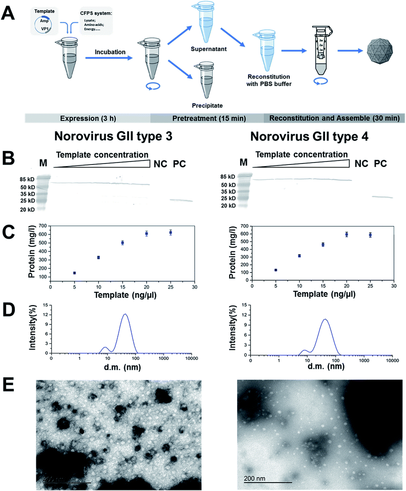
In this study, we reported for the first time that an E. coli-based CFPS system could be used to synthesize and assemble HuNoV-VLPs in 4 hours (Fig. 1A). To prepare the CFPS system, two types of S30 extract, the BL21 S30 extract and the CK-T7 extract, were used. The BL21 S30 extract was prepared from the E. coli strain BL21 as previously reported.20 The CK-T7 extract was used to bypass the exogenous addition of commercial creatine kinase (CK) and T7 RNA polymerase (T7 RNAP) into the cell-free reaction mixtures. To prepare the CK-T7 extract, a recombinant E. coli BL21 strain was constructed by harbouring a CK and T7 RNAP expression vector pETDuet-CK-T7. The CK and T7 RNAP were expressed during the cultivation of E. coli via IPTG induction (1.0 mM) at OD600 0.5. The recombinant E. coli BL21 strains were harvested at 2 h after induction and used for preparing the CK-T7 extract. In addition to the S30 extracts, we also prepared the CFPS reaction mix consisted of the following components in a total volume of 50 μL: 55 mM HEPES/KOH, pH 7.5, 1.2 mM ATP, 0.85 mM each of GTP, UTP, and CTP, 1.7 mM dithiothreitol, 200 mM K+-glutamate, 0.17 mg mL−1E. coli total tRNA, 34 mg mL−1 folinic acid, 0.65 mM cAMP, 2 mM each of amino acids, 80 mM creatine phosphate, 28 mM ammonium acetate, 11 mM magnesium acetate, 18 μL BL21 extract, 6 μL CK-T7 extract, and plasmid template harbouring the target gene. The extremely low endotoxin level of E. coli cell-free protein system could meet the demands for vaccine applications referencing guidelines for toxoid-based vaccines.21,22
We next used this CFPS system to synthesize two capsid proteins of HuNoV-VLPs: VP1 from HuNoV genotype GII.3 (VP1-GII.3) and genotype GII.4 (VP1-GII.4). The VP1 capsid gene of HuNoV GII.3 or GII.4 was cloned into cell-free protein expression vector pIVEX2.4c, respectively. A 6 × His-Tag was incorporated to the N-terminus of the P domain to facilitate their detection and purification. Subsequently, the two plasmids containing HuNoV GII.3 or GII.4 VP1 gene were expressed in the CFPS system at 37 °C for 3 hours. The product was then analysed by western-blotting using a protocol that was previously developed.23,24 To optimize the protein production in the CFPS system, we generated a dose–response curve by using various concentrations (range from 5–25 ng μL−1) of plasmid template harbouring the HuNoV GII.3 or GII.4 VP1 gene. We used the “empty” CFPS without any template as our negative control and used the defined amount of GFP (500 mg L−1) as our positive control. The expression level of target protein was quantified by QuantityOne (Bio-Rad, USA). As shown in Fig. 1B, the size of recombinant protein synthesized in the CFPS system matched our expectations: molecular mass of 63 kDa for VP1-GII.3 and 62 kDa for VP1-GII.4. GII.3 and GII.4 were produced at 0.62 g L−1 and 0.57 g L−1, respectively. Such protein yield is comparable to the cell-based system using P. pastoris. However, the production time is only 4 hours, which is much shorter than any cell-based system (>50 hours).
We then examined if VLPs were formed from the capsid proteins synthesized by the CFPS system. To start, the product of CFPS reaction was centrifuged for 10 min to remove the debris. The supernatant was diluted five times by PBS buffer (pH 7.4) and concentrated by centrifugal filter units with 30 kDa membrane (Millipore, USA). After buffer exchange for three times and high-speed centrifuge to remove the precipitate, the CFPS reaction was subjected to the morphological analysis. We used dynamic light scattering (DLS) to characterize the size distribution of the VLPs and electronic microscopy for VLP imaging. For DLS analysis, an aliquot was transferred to 400 μL disposable sizing cuvettes and the diffusion of the particles moving under Brownian motion using a Zetasizer Nano-ZS (Malvern Instruments Ltd, UK) was measured. The particle sizes of VLPs were calculated as the average of three consecutive measurements recorded at 25 °C. As shown in Fig. 1D, the average size of the VP1-GII.3 and VP1-GII.4 particles were 23.8 nm and 38.7 nm, respectively. This discovery was consistent with previous reports,25 which found that the sizes of HuNoV-VLPs vary from 23–40 nm. We also examined the morphology of HuNoV-VLPs via electronic microscopy as described previously.3,26 Basically, electron microscopy formvar carbon square grids (Electron Microscopy Sciences) were pre-treated with 1% aqueous alcian blue for 5 min. After three washes, 10 μL of assembled VLPs of VP1-GII.3 and VP1-GII.4 were absorbed to the grids for 1 min. The grids were stained with 3% phosphotungstic acid pH 7.0 for 1 min. The grid was then viewed with a JEOLJEM 1400 transmission electron microscopy. As shown in Fig. 1E, VLPs were formed from both the VP1-GII.3 and VP1-GII.4, which showed similar structures as reported previously.3,26 It is also worth mentioning that the different buffer systems might lead to different imaging of particle structure.27–30
It is worth mentioning that with the continuous decreasing cost of current CFPS system, the expense of HuNoV-VLPs when using CFPS system to produce could be reduced to <5 cents per μL with protein yields of 1.0 g L−1.21 Assuming a similar dosage of NoV-VLPs as that of Takeda candidate VLP vaccine, which contains 100 μg antigen per dose, the cost of 1 dose of HuNoV-VLPs could be as low as $2.5–5.0. Given the ease of the downstream purification process conferred by CFPS system,31 this platform could be a promising technology to produce both efficacious and affordable HuNoV vaccines.
Conclusions
In this study, we demonstrated that E. coli-based cell-free protein synthesis could be used to synthesize and assemble HuNoV VLPs. Compared to the cell-based system, our cell-free system led to similar protein yield but much shorter production time. This fast, high-yield system can be a promising manufacturing process for supplying HuNoV vaccines in the future.Notes and references
- S. G. Morillo and C. Timenetsky Mdo, Rev. Assoc. Med. Bras., 2011, 57, 453–458 CrossRef PubMed.
- D. Flynn, Takeda's norovirus vaccine first to reach human trials, http://www.foodsafetynews.com/2016/06/takedas-norovirus-vaccine-first-to-reach-human-trials/#.WNKSn_krJPY.
- J. Tome-Amat, L. Fleischer, S. A. Parker, C. L. Bardliving and C. A. Batt, Microb. Cell Fact., 2014, 13, 134 CrossRef PubMed.
- K. Ettayebi, S. E. Crawford, K. Murakami, J. R. Broughman, U. Karandikar, V. R. Tenge, F. H. Neill, S. E. Blutt, X. L. Zeng, L. Qu, B. Kou, A. R. Opekun, D. Burrin, D. Y. Graham, S. Ramani, R. L. Atmar and M. K. Estes, Science, 2016, 353, 1387–1393 CrossRef PubMed.
- M. Tan, W. Zhong, D. Song, S. Thornton and X. Jiang, J. Med. Virol., 2004, 74, 641–649 CrossRef CAS PubMed.
- T. A. Lamounier, L. M. de Oliveira, B. R. de Camargo, K. B. Rodrigues, E. F. Noronha, B. M. Ribeiro and T. Nagata, Braz. J. Microbiol., 2015, 46, 1265–1268 CrossRef CAS PubMed.
- L. Santi, L. Batchelor, Z. Huang, B. Hjelm, J. Kilbourne, C. J. Arntzen, Q. Chen and H. S. Mason, Vaccine, 2008, 26, 1846–1854 CrossRef CAS PubMed.
- A. G. Diamos, S. H. Rosenthal and H. S. Mason, Front. Plant Sci., 2016, 7, 200 Search PubMed.
- S. Hervas-Stubbs, P. Rueda, L. Lopez and C. Leclerc, J. Immunol., 2007, 178, 2361–2369 CrossRef CAS.
- M. G. Casteleijn, A. Urtti and S. Sarkhel, Int. J. Pharm., 2013, 440, 39–47 CrossRef CAS PubMed.
- W. Guo, J. Sheng and X. Feng, Comput. Struct. Biotechnol. J., 2017, 15, 161–167 CrossRef CAS PubMed.
- J. R. Mattingly Jr, J. Youssef, A. Iriarte and M. Martinez-Carrion, J. Biol. Chem., 1993, 268, 3925–3937 CAS.
- G. Yin and J. R. Swartz, Biotechnol. Bioeng., 2004, 86, 188–195 CrossRef CAS PubMed.
- D. N. Hebert, J. X. Zhang and A. Helenius, Biochem. Cell Biol., 1998, 76, 867–873 CrossRef CAS PubMed.
- F. Katzen, G. Chang and W. Kudlicki, Trends Biotechnol., 2005, 23, 150–156 CrossRef CAS PubMed.
- Y. Endo and T. Sawasaki, Curr. Opin. Biotechnol., 2006, 17, 373–380 CrossRef CAS PubMed.
- T. Sawasaki, T. Ogasawara, R. Morishita and Y. Endo, Proc. Natl. Acad. Sci. U. S. A., 2002, 99, 14652–14657 CrossRef CAS PubMed.
- K. Jackson, R. Khnouf and Z. H. Fan, Methods Mol. Biol., 2014, 1118, 157–168 CAS.
- P. Avenaud, M. Castroviejo, S. Claret, J. Rosenbaum, F. Megraud and A. Menard, Biochem. Biophys. Res. Commun., 2004, 318, 739–745 CrossRef CAS PubMed.
- M. C. Jewett and J. R. Swartz, Biotechnol. Bioeng., 2004, 86, 19–26 CrossRef CAS PubMed.
- K. Pardee, S. Slomovic, P. Q. Nguyen, J. W. Lee, N. Donghia, D. Burrill, T. Ferrante, F. R. McSorley, Y. Furuta, A. Vernet, M. Lewandowski, C. N. Boddy, N. S. Joshi and J. J. Collins, Cell, 2016, 167, 248–259 CrossRef CAS PubMed.
- L. A. Brito and M. Singh, J. Pharm. Sci., 2011, 100, 34–37 CrossRef CAS PubMed.
- T. Mahmood and P.-C. Yang, N. Am. J. Med. Sci., 2012, 4, 429–434 CrossRef PubMed.
- J. Sheng, H. Flick and X. Feng, Front. Microbiol., 2017, 8, 875 CrossRef PubMed.
- J. L. Cuellar, F. Meinhoevel, M. Hoehne and E. Donath, J. Gen. Virol., 2010, 91, 2449–2456 CrossRef CAS PubMed.
- S. Lei, A. Ramesh, E. Twitchell, K. Wen, T. Bui, M. Weiss, X. Yang, J. Kocher, G. Li, E. Giri-Rachman, N. V. Trang, X. Jiang, E. P. Ryan and L. Yuan, Front. Microbiol., 2016, 7, 1699 Search PubMed.
- M. Fang, W. Diao, B. Dong, H. Wei, J. Liu, L. Hua, M. Zhang, S. Guo, Y. Xiao, Y. Yu, L. Wang and M. Wan, Intervirology, 2015, 58, 318–323 CrossRef CAS PubMed.
- S. F. Ausar, T. R. Foubert, M. H. Hudson, T. S. Vedvick and C. R. Middaugh, J. Biol. Chem., 2006, 281, 19478–19488 CrossRef CAS PubMed.
- A. Roldão, M. C. M. Mellado, J. C. Lima, M. J. T. Carrondo, P. M. Alves and R. Oliveira, PLoS Comput. Biol., 2012, 8, e1002367 Search PubMed.
- J. Nilsson, N. Miyazaki, L. Xing, B. Wu, L. Hammar, T. C. Li, N. Takeda, T. Miyamura and R. H. Cheng, J. Virol., 2005, 79, 5337–5345 CrossRef CAS PubMed.
- E. D. Carlson, R. Gan, C. E. Hodgman and M. C. Jewett, Biotechnol. Adv., 2012, 30, 1185–1194 CrossRef CAS PubMed.
Footnote |
| † Electronic supplementary information (ESI) available. See DOI: 10.1039/c7ra03742b |
| This journal is © The Royal Society of Chemistry 2017 |

