Efficient two-photon luminescence for cellular imaging using biocompatible nitrogen-doped graphene quantum dots conjugated with polymers†
Ping-Ching
Wu
a,
Jiu-Yao
Wang
bcd,
Wen-Lung
Wang
e,
Chia-Yuan
Chang
f,
Chia-Hung
Huang
gh,
Kun-Lin
Yang
i,
Jui-Cheng
Chang
j,
Chih-Li Lilian
Hsu
d,
Shih-Yao
Chen
*e,
Ting-Mao
Chou
*k and
Wen-Shuo
Kuo
 *bcl
*bcl
aDepartment of Biomedical Engineering, National Cheng Kung University, Tainan 701, Taiwan, Republic of China
bAllergy & Clinical Immunology Research Center, National Cheng Kung University Hospital, College of Medicine, National Cheng Kung University, Tainan 701, Taiwan, Republic of China. E-mail: wenshuokuo@mail.ncku.edu.tw
cDepartment of Pediatrics, National Cheng Kung University Hospital, College of Medicine, National Cheng Kung University, Tainan 701, Taiwan, Republic of China. E-mail: wenshuokuo@mail.ncku.edu.tw
dDepartment of Microbiology & Immunology, National Cheng Kung University Hospital, College of Medicine, National Cheng Kung University, Tainan 701, Taiwan, Republic of China
eDepartment of Internal Medicine, National Cheng Kung University Hospital, College of Medicine, National Cheng Kung University, Tainan 701, Taiwan, Republic of China. E-mail: z9903038@email.ncku.edu.tw
fCenter for Micro/Nano Science and Technology, National Cheng Kung University, Tainan 701, Taiwan, Republic of China
gMetal Industries Research & Development Centre, Kaohsiung 811, Taiwan, Republic of China
hDepartment of Materials Science Engineering, National Cheng Kung University, Tainan 701, Taiwan, Republic of China
iDepartment of Biotechnology, National Kaohsiung Normal University, Kaohsiung 802, Taiwan, Republic of China
jDepartment of Chemical Engineering, National Cheng Kung University, Tainan 701, Taiwan, Republic of China
kDivision of Plastic Surgery, Department of Surgery, E-Da Hospital, Kaohsiung 824, Taiwan, Republic of China. E-mail: ed109842@edah.org.tw
lAdvanced Optoelectronic Technology Center, National Cheng Kung University, Tainan 701, Taiwan, Republic of China. E-mail: wenshuokuo@mail.ncku.edu.tw
First published on 17th November 2017
Abstract
Nitrogen-doped graphene quantum dot (N-GQD) nanomaterials conjugated with polyethylenimine (PEI)-polystyrene sulfonate (PSS)-anti-epidermal growth factor receptor (AbEGFR) antibody (N-GQD-PEI-PSS-AbEGFR) demonstrated impressive two-photon properties and stability, signifying that they can serve as an effective two-photon contrast agent in two-photon bioimaging. Furthermore, they provided high intensity, brightness, and signal-to-noise ratios at an ultra-low two-photon excitation (TPE) power level in an observation extending to a deep, three-dimensional depth.
The present study aimed to develop a promising contrast agent with a high quantum yield (QY) and the ability to exhibit favorable multiphoton properties for deeper biological specimen and tissue detection. Consequently, shifting the irradiation source from the range of visible wavelengths to the near-infrared (NIR) window, which offers low scattering, energy absorption, photodamage, background tissue autofluorescence and maximum penetration into deeper tissues, in addition to enhancing the detection limit through a coupled nonlinear multiphoton laser with noninvasive properties of subcellular features and a less than 1 mm deep penetration into tissue, is necessary. Currently, the use of carbon-based materials, especially graphene-based nanomaterials, as probes in biosensing and bioimaging has attracted considerable attention and substantial research interest. However, the zero-optical band gap of graphene limits its optical-related applications. Several strategies have been developed to enlarge the band gap in order to make it luminescent including reducing its geometric size to the nanometer regime,1 decorating graphene with various functional groups2 and introducing heteroatoms to form structural defects.3 QDs are a type of nanoparticle (NP) with size-dependent electronic and optical properties that offer promising advantages: they have been used in biological applications for sensing agents, diagnostics, and contrast probes.4 GQDs, a new type of zero-dimensional QD converted from two-dimensional graphene sheets, are emerging as potential optical nanomaterials in biomedical research. Furthermore, doping is crucial to semiconductors because the carrier density can be clearly engineered; therefore, the optical and electrical properties might be entirely distinct from their intrinsic counterparts.5 Nitrogen (N) doping or functionalization is highly advantageous for tailoring the intrinsic properties of GQDs. The incorporated five-valance-electron and electron-accepting N atoms, which are of a corresponding atomic size to carbon (C) atoms, impart a relatively high positive charge density to the adjacent C of graphene. Furthermore, N can substitute C in sp2-bonded molecular systems, leading to heterocyclic aromatic compounds. Atom doping is considered to be an efficient alternative for modifying the intrinsic properties of C-based nanomaterials including their local chemical features, surface features, and electronic characteristics.6 Consequently, for GQDs (<10 nm) exhibiting extraordinary edge effects and quantum confinement, N-GQDs could change the chemical composition and tune the band gap of the GQDs to that which would be expected to exhibit enhanced photochemical, electrochemical, and electrocatalytic actions, which are favorable for tunable luminescence in bioimaging and optoelectronic applications.7 Therefore, chemical doping is an efficacious method for modulating the electronic, chemical, and optical properties of graphene.
Because the band gap of N-GQDs can be manipulated by modifying their edge, shape, size, and surface functionalities,8 very little attention9,10 has been paid to the use of different polymers containing N and sulfur (S) atoms in conjugation with N-GQD (N-GQD-polymers). In the current study, N-GQD-polymers containing N (PEI) and S (PSS) were synthesized, and their two-photon properties after nonlinear multiphoton laser excitation in the NIR window were examined. The examination results demonstrated that the N-GQD-polymers possessed high photoluminescence (PL), QY, and discernible two-photon properties, such as two-photon absorption (TPA), absolute TPE cross sections, two-photon luminescence (TPL) emission, ratios of the radiative- to nonradiative-decay rate, and post-TPE stability. However, the shortened lifetime, which was likely due to the higher quantum confinement of emissive energy trapped on the surface of the N-GQD (which has a large surface-to-volume ratio in each particle), resulted in these two-photon properties of the N-GQD-polymers. In addition, the high absolute TPE cross-section value of the N-GQD-polymers (55![[thin space (1/6-em)]](https://www.rsc.org/images/entities/char_2009.gif) 899–57
899–57![[thin space (1/6-em)]](https://www.rsc.org/images/entities/char_2009.gif) 980 Goeppert–Mayer units (GM), with 1 GM = 10−50 cm4 per s per photon) renders them favorable for efficiently executing two-photon examinations because the ratio of the energy absorbed to the input-energy flux through a specimen is high, which minimizes the likely photodamage of the specimen.11,12 Furthermore, TPE-mediated high TPL intensity in acidic environments enables N-GQD-polymers to act as a promising contrast probe for tracking and localizing N-GQD-polymer-treated cancer cells at a deep, three-dimensional (3D) depth of 93 μm, and provides additional information on the status of cancer cells irradiated through an ultra-low TPE power of 0.598 mW (59.8 nJ per pixel; the calculations in detail are shown in the ESI;† for the laser system, the x–y axis focal spot is around 375.38 nm, and the z axis resolution is around 0.94193 μm, Fig. S1†); this reveals that the TPL intensity of the N-polymer-treated cells can use 35 times less power than that of two-photon autofluorescence (TPAF, 21.120 mW or 2112.0 nJ per pixel) to minimize the photodamage of the cells and achieve a similar collected intensity. Consequently, we expect the N-GQD-polymers, which generate nonreactive oxygen species-dependent oxidative stress effects, to act as two-photon contrast agents for biological applications and for obtaining images in deeper biological specimens and tissues in the human body in the future.
980 Goeppert–Mayer units (GM), with 1 GM = 10−50 cm4 per s per photon) renders them favorable for efficiently executing two-photon examinations because the ratio of the energy absorbed to the input-energy flux through a specimen is high, which minimizes the likely photodamage of the specimen.11,12 Furthermore, TPE-mediated high TPL intensity in acidic environments enables N-GQD-polymers to act as a promising contrast probe for tracking and localizing N-GQD-polymer-treated cancer cells at a deep, three-dimensional (3D) depth of 93 μm, and provides additional information on the status of cancer cells irradiated through an ultra-low TPE power of 0.598 mW (59.8 nJ per pixel; the calculations in detail are shown in the ESI;† for the laser system, the x–y axis focal spot is around 375.38 nm, and the z axis resolution is around 0.94193 μm, Fig. S1†); this reveals that the TPL intensity of the N-polymer-treated cells can use 35 times less power than that of two-photon autofluorescence (TPAF, 21.120 mW or 2112.0 nJ per pixel) to minimize the photodamage of the cells and achieve a similar collected intensity. Consequently, we expect the N-GQD-polymers, which generate nonreactive oxygen species-dependent oxidative stress effects, to act as two-photon contrast agents for biological applications and for obtaining images in deeper biological specimens and tissues in the human body in the future.
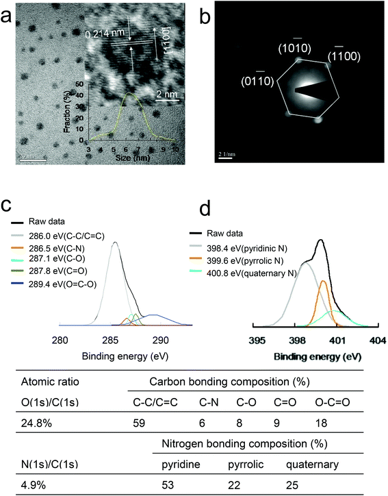
A modified Hummers’ method was used to synthesize graphene oxide from the same graphite sample (Fig. S2a†).13 N-GQDs were then prepared through an ultrasonic shearing reaction of the graphene oxide sheets. High-resolution transmission electron microscopy (HRTEM) images indicated the mean lateral size of the N-GQDs to be approximately 6.4 ± 1.1 nm (Fig. 1a). A good crystallinity with a lattice distance of 0.214 nm was obtained, and this corresponds to the d-spacing of graphene {1![[1 with combining macron]](https://www.rsc.org/images/entities/char_0031_0304.gif) 00} lattice fringes.14,15 The selected-area electron diffraction pattern of the N-GQDs was projected perpendicularly onto the basal plane of the N-GQDs, revealing definite diffraction spots in a hexagonal arrangement the same as that of graphite projected in the [0001] direction, which showed a single crystalline structure (Fig. 1b). A thickness of approximately 0.83 ± 0.04 nm was determined for single-layer N-GQD using atomic force microscopy (Fig. S2b†). Moreover, exposed epoxy, hydroxyl and carboxyl groups from the N-GQDs were observed via Fourier transform infrared spectroscopy (Fig. S2c†). Because of the presence of exposed carboxylic acid, a surface charge of approximately −20.3 mV was measured using zeta-potential spectroscopy. The absorptions for the π–π* transition of aromatic C
00} lattice fringes.14,15 The selected-area electron diffraction pattern of the N-GQDs was projected perpendicularly onto the basal plane of the N-GQDs, revealing definite diffraction spots in a hexagonal arrangement the same as that of graphite projected in the [0001] direction, which showed a single crystalline structure (Fig. 1b). A thickness of approximately 0.83 ± 0.04 nm was determined for single-layer N-GQD using atomic force microscopy (Fig. S2b†). Moreover, exposed epoxy, hydroxyl and carboxyl groups from the N-GQDs were observed via Fourier transform infrared spectroscopy (Fig. S2c†). Because of the presence of exposed carboxylic acid, a surface charge of approximately −20.3 mV was measured using zeta-potential spectroscopy. The absorptions for the π–π* transition of aromatic C![[double bond, length as m-dash]](https://www.rsc.org/images/entities/char_e001.gif) C bonds occurred at approximately 224 nm and were red-shifted by approximately 11 nm compared with those of GQDs of a similar size; the n–π* transitions of the C
C bonds occurred at approximately 224 nm and were red-shifted by approximately 11 nm compared with those of GQDs of a similar size; the n–π* transitions of the C![[double bond, length as m-dash]](https://www.rsc.org/images/entities/char_e001.gif) O shoulder and C–N occurred at approximately 326 nm, which corresponded to the π electron transition in oxygen-containing N-GQDs, and was indicative of N doping in the GQDs,16 according to ultraviolet-visible spectroscopy (Fig. S2d†). The D bands (approximately 1382 cm−1) and G bands (approximately 1603 cm−1) exhibited an integrated intensity ID/IG ratio of approximately 0.85 in the Raman spectrum, indicating their high quality. Furthermore, the insertion of N into the C backbone introduced abundant defects into the GQDs.17
O shoulder and C–N occurred at approximately 326 nm, which corresponded to the π electron transition in oxygen-containing N-GQDs, and was indicative of N doping in the GQDs,16 according to ultraviolet-visible spectroscopy (Fig. S2d†). The D bands (approximately 1382 cm−1) and G bands (approximately 1603 cm−1) exhibited an integrated intensity ID/IG ratio of approximately 0.85 in the Raman spectrum, indicating their high quality. Furthermore, the insertion of N into the C backbone introduced abundant defects into the GQDs.17
Nonoxygenated rings (C–C/C![[double bond, length as m-dash]](https://www.rsc.org/images/entities/char_e001.gif) C, 286.0 eV), C–N bonds (286.5 eV), and hydroxyl (C–O, 287.1 eV), carbonyl (C
C, 286.0 eV), C–N bonds (286.5 eV), and hydroxyl (C–O, 287.1 eV), carbonyl (C![[double bond, length as m-dash]](https://www.rsc.org/images/entities/char_e001.gif) O, 287.8 eV), and carboxylate (O
O, 287.8 eV), and carboxylate (O![[double bond, length as m-dash]](https://www.rsc.org/images/entities/char_e001.gif) C–O, 289.4 eV) groups from the deconvoluted C(1s) spectra of the N-GQDs were observed by X-ray photoelectron spectroscopy (XPS), whereas pyridinic N (398.4 eV), pyrrolic N (399.6 eV), and quaternary N (400.8 eV) were observed from the deconvoluted N(1s) spectra with a N(1s)/C(1s) atomic ratio of 4.9% (Fig. 1c and d); the PL emitted from the N-GQDs exhibited a Stokes shift from 497 to 548 nm at excitation wavelengths from 410 to 500 nm (Fig. S2e†). The aforementioned characterizations confirmed that the N-GQDs were successfully synthesized.
C–O, 289.4 eV) groups from the deconvoluted C(1s) spectra of the N-GQDs were observed by X-ray photoelectron spectroscopy (XPS), whereas pyridinic N (398.4 eV), pyrrolic N (399.6 eV), and quaternary N (400.8 eV) were observed from the deconvoluted N(1s) spectra with a N(1s)/C(1s) atomic ratio of 4.9% (Fig. 1c and d); the PL emitted from the N-GQDs exhibited a Stokes shift from 497 to 548 nm at excitation wavelengths from 410 to 500 nm (Fig. S2e†). The aforementioned characterizations confirmed that the N-GQDs were successfully synthesized.
C-Based materials engender a higher PL and QY, and this is attributed to doping and the quantum confinement of emissive energy trapped on the surface after surface passivation.18 In addition, the surface-passivated reaction of polymers containing N and S atoms leads to the same phenomenon.4,19–21 In the present study, PEI and PSS were coated on the surface of N-GQDs (N-GQD-polymers) via electrostatic interaction to determine whether the PL and QY were enhanced. Therefore, the characterizations of N-GQD-PEI and N-GQD-PEI-PSS were processed successfully and are presented in Table 1. A detailed discussion is presented in the ESI (Fig. S3–S11†).
| Mean lateral size (nm) | Thickness (nm) | Zeta potential (mV) | FTIR | XRD | UV-vis | Raman spectra (D band, G band (cm−1), and ID/IG ratio) | |
|---|---|---|---|---|---|---|---|
| N-GQD | 6.4 ± 1.1 | 0.83 ± 0.04 | −20.3 | Fig. S2c | Fig. S3c | Fig. S2d | 1382, 1603, 0.85 |
| N-GQD-PEI | 6.5 ± 1.2 | 0.96 ± 0.03 | 22.2 | Fig. S7a | Fig. S8a | 1364, 1596, 0.85 | |
| N-GQD-PEI-PSS | 6.6 ± 1.4 | 1.05 ± 0.05 | −18.9 | Fig. S7b | Fig. S8a | 1352, 1593, 0.86 |
| XPS | PL | Relative QY | Absolute cross section of TPE (GM) | Lifetime (ns) | Radiative decay rate (×108 s−1) | Non-radiative decay rate (×108 s−1) |
|---|---|---|---|---|---|---|
| a The calculations in detail are available in the experimental section of the ESI. One- or two-photon excitation yields the same QY.22–24 | ||||||
| Fig. 1c and d | Fig. S2e | 0.223 | 52![[thin space (1/6-em)]](https://www.rsc.org/images/entities/char_2009.gif) 594.5 594.5 |
1.3135 | 1.6979 | 5.9155 |
| Fig. S10b | 0.435 | 55![[thin space (1/6-em)]](https://www.rsc.org/images/entities/char_2009.gif) 899.5 899.5 |
1.0433 | 4.1695 | 5.4155 | |
| Fig. S10c | 0.565 | 57![[thin space (1/6-em)]](https://www.rsc.org/images/entities/char_2009.gif) 980.1 980.1 |
0.4769 | 11.8474 | 9.1214 | |
Both doping with heteroatoms and surface passivation can efficiently increase the QY of GQDs.5 Surface passivation can apparently improve the integrity of surface π electron networks and/or inhibit the nonradiative recombination of localized electron–hole pairs, resulting in a rise in the QY of GQDs.22–24 Furthermore, doping with heteroatoms can efficiently engineer the band gaps and local chemical features of graphene structures, resulting in changes in their optical properties and electronic characteristics.6,7 In the present study, the QYs of the N-GQD-polymers were determined using fluorescein dissolved in NaOH (0.1 M, pH 11) as a standard reference.25 The relative QY of the N-GQDs was calculated to be 0.223 (Table 1); its absolute QY26 had a similar value of 0.226. The newly formed energy level generated by the unique configuration of N played a crucial role in manipulating the conduction type, electrocatalytic activity, conductivity, and optical properties.5–7 The structure-related properties caused the N-GQD-polymers to exhibit a stronger PL and higher QY, which is crucial for fluorescent materials. After coating with PEI through electrostatic interaction, the relative and absolute QYs increased to 0.435 and 0.442, respectively. According to the crosslink enhanced emission effect,27 the C core from an N-GQD with a multilayer crystal lattice possessed nonradiative traps and structures, the PEI possessed potential fluorophores (secondary and tertiary amines), and the enhanced PL originated from the reduced rotation and vibration corresponding with the cross-linked PEI coated on the NPs. Consequently, an increased QY value was expected. In addition, the calculated relative and absolute QYs exhibited higher values of 0.565 and 0.573, respectively, after PSS was coated on the surface of the N-GQD-PEI. The introduced S and N atoms enhanced the effect of electrons trapped by the newly introduced surface states on the properties of the C-doped nanomaterials by a cooperative effect, which resulted in a high yield of radiative recombination, leading to higher QY.4,19 Following surface conjugation involving a higher quantum confinement of emissive energy trapped on the surface of the N-GQDs, the QY of N-GQD-PEI-PSS was increased dramatically.
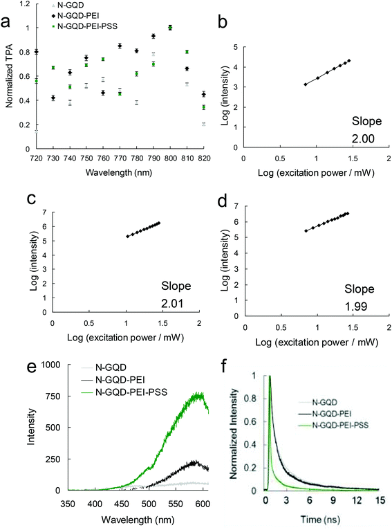
Using TPE could be advantageous to maintain a low average laser power and enable the excited wavelength to be lengthened to the NIR window, thereby improving the visibility for bioimaging processes. Additionally, the high values of the TPE cross section render fluorophores highly effective for the investigation of two-photon techniques because this is consistent with the high ratio of energy absorbed to the input-energy flux in biospecimens, thereby reducing probable photodamage.11 Moreover, TPL is a nonlinear phenomenon, and its intensity is proportional to the square of the excitation power, TPE cross section and QY. When the QY and TPE cross section are increased, modification of the intrinsic possession of the fluorophores and enhancement of the localized excitation power can engender an increased TPL signal.22,28 As demonstrated in Fig. 2a, the most efficient excitation wavelength for the N-GQD and N-GQD-polymers under TPE was approximately 800 nm in the NIR region. Due to the good absorption in the NIR region, the materials were able to detect deeper tissue, exhibiting potential application in the biomedical field via nonlinear lasers throughout this work. Through excitation of the N-GQD and N-GQD-polymers, this study confirmed a two-photon process in which the PL intensity demonstrated a quadratic dependence on the excitation power22,28 with exponents of 1.99–2.01 (Fig. 2b–d). Therefore, the TPL spectra of the N-GQDs and N-GQD-polymers displayed broad peaks at approximately 572 nm, 590 nm and 596 nm, respectively, after an identical investigation (Fig. 2e). By contrast, the N-GQD-polymers displayed higher intensities. For one-photon excitation, TPE yields the same QY22–24 and the absolute cross section of TPE for the N-GQDs was 52![[thin space (1/6-em)]](https://www.rsc.org/images/entities/char_2009.gif) 594.5 GM (Rhodamine B was chosen as the standard reference28 for determining the cross section) under TPE at a wavelength of 800 nm, whereas N-GQD-PEI-PSS demonstrated the highest value (57
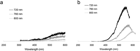
594.5 GM (Rhodamine B was chosen as the standard reference28 for determining the cross section) under TPE at a wavelength of 800 nm, whereas N-GQD-PEI-PSS demonstrated the highest value (57![[thin space (1/6-em)]](https://www.rsc.org/images/entities/char_2009.gif) 980.1 GM) (Tables 1, 2 and S1†). Additionally, the absolute cross section of TPE for the N-GQD-polymers surpassed that of conventional fluorophores by over 2 orders of magnitude and was larger than that of well-established semiconductor QDs (Table S2†).29 After further investigation, the lifetime was detected with a time-correlated single photon counting technique involving a triple-exponential fitting function; sequentially radiative and nonradiative decay rates were calculated according to the QY and lifetime (Table 1). The average lifetime of the N-GQDs was 1.3135 ns, which was derived from the observed lifetimes of 0.1805, 0.7828, and 3.1337 ns, whereas the average lifetimes of N-GQD-PEI and N-GQD-PEI-PSS were 1.0433 and 0.4769 ns, respectively (Fig. 2f and Table S3†). Hence, the ratio of the radiative and nonradiative decay rates of the N-GQDs was 0.287, which was derived from 1.6979 × 10⁸ and 5.9155 × 10⁸ s−1, respectively; the ratios of N-GQD-PEI and N-GQD-PEI-PSS were 0.770 (4.1695 × 10⁸ s−1/5.4155 × 108 s−1) and 1.299 (11.8474 × 10⁸ s−1/9.1214 × 108 s−1), respectively. These results indicate that when the QY increases and the lifetime decreases, the materials might primarily pass through the radiative pathway after TPE, instead of through the nonradiative pathway. Fig. 3 shows the TPL spectra measured for the materials with irradiation; peaks are observed from approximately 537 nm to 572 nm for the N-GQDs and from 548 nm to 596 nm for N-GQD-PEI-PSS under TPE wavelengths of 720–800 nm, exhibiting a similar trend to the emission wavelength by one-photon excitation in Fig. S10a and S10c.† Additionally, the TPL intensity of as-prepared N-GQD-PEI-PSS for 3 months exhibited nearly no drop after being subjected to two-photon irradiation for 5 min (Ex: 800 nm), indicating that its high photostability capability was maintained even after 3 months (Fig. S12†); good stability in physiological environments (Table S4†) in regard to further biological application was also displayed. Doping GQDs with bonded N atoms leads to more effective TPL radiative emission as a consequence of restoring sp2 hybridization and the donation of delocalized electrons entering the π* states, resulting in a higher absolute TPE cross section. Furthermore, the effect of strong electron donation and large π-conjugated systems in polymers containing N and S atoms coated on N-GQDs assisted the charge transfer efficiency; the interaction between the N-GQDs and the polymers generated oscillating dipoles;2,4,18,19 therefore, the two-photon properties of N-GQD-polymers can cause sharp increases or obvious changes,30 leading to efficacious two-photon studies.
980.1 GM) (Tables 1, 2 and S1†). Additionally, the absolute cross section of TPE for the N-GQD-polymers surpassed that of conventional fluorophores by over 2 orders of magnitude and was larger than that of well-established semiconductor QDs (Table S2†).29 After further investigation, the lifetime was detected with a time-correlated single photon counting technique involving a triple-exponential fitting function; sequentially radiative and nonradiative decay rates were calculated according to the QY and lifetime (Table 1). The average lifetime of the N-GQDs was 1.3135 ns, which was derived from the observed lifetimes of 0.1805, 0.7828, and 3.1337 ns, whereas the average lifetimes of N-GQD-PEI and N-GQD-PEI-PSS were 1.0433 and 0.4769 ns, respectively (Fig. 2f and Table S3†). Hence, the ratio of the radiative and nonradiative decay rates of the N-GQDs was 0.287, which was derived from 1.6979 × 10⁸ and 5.9155 × 10⁸ s−1, respectively; the ratios of N-GQD-PEI and N-GQD-PEI-PSS were 0.770 (4.1695 × 10⁸ s−1/5.4155 × 108 s−1) and 1.299 (11.8474 × 10⁸ s−1/9.1214 × 108 s−1), respectively. These results indicate that when the QY increases and the lifetime decreases, the materials might primarily pass through the radiative pathway after TPE, instead of through the nonradiative pathway. Fig. 3 shows the TPL spectra measured for the materials with irradiation; peaks are observed from approximately 537 nm to 572 nm for the N-GQDs and from 548 nm to 596 nm for N-GQD-PEI-PSS under TPE wavelengths of 720–800 nm, exhibiting a similar trend to the emission wavelength by one-photon excitation in Fig. S10a and S10c.† Additionally, the TPL intensity of as-prepared N-GQD-PEI-PSS for 3 months exhibited nearly no drop after being subjected to two-photon irradiation for 5 min (Ex: 800 nm), indicating that its high photostability capability was maintained even after 3 months (Fig. S12†); good stability in physiological environments (Table S4†) in regard to further biological application was also displayed. Doping GQDs with bonded N atoms leads to more effective TPL radiative emission as a consequence of restoring sp2 hybridization and the donation of delocalized electrons entering the π* states, resulting in a higher absolute TPE cross section. Furthermore, the effect of strong electron donation and large π-conjugated systems in polymers containing N and S atoms coated on N-GQDs assisted the charge transfer efficiency; the interaction between the N-GQDs and the polymers generated oscillating dipoles;2,4,18,19 therefore, the two-photon properties of N-GQD-polymers can cause sharp increases or obvious changes,30 leading to efficacious two-photon studies.
| Reference | Integrated emission intensity (counts) | Action cross-section (ησ) |
|---|---|---|
| Fluorescein | 167.5 | 34.4 |
| Sample | Integrated emission intensity (counts) | Relative quantum yield (η) | Absolute cross-section (σ) |
|---|---|---|---|
| a Fluorescein was selected as the standard reference for the cross section, and the relevant calculations are shown in the ESI.† | |||
| N-GQD | 57![[thin space (1/6-em)]](https://www.rsc.org/images/entities/char_2009.gif) 108.6 108.6 |
0.223 | 52![[thin space (1/6-em)]](https://www.rsc.org/images/entities/char_2009.gif) 594.5 594.5 |
| N-GQD-PEI | 118![[thin space (1/6-em)]](https://www.rsc.org/images/entities/char_2009.gif) 400.5 400.5 |
0.435 | 55![[thin space (1/6-em)]](https://www.rsc.org/images/entities/char_2009.gif) 899.5 899.5 |
| N-GQD-PEI-PSS | 159![[thin space (1/6-em)]](https://www.rsc.org/images/entities/char_2009.gif) 508.5 508.5 |
0.565 | 57![[thin space (1/6-em)]](https://www.rsc.org/images/entities/char_2009.gif) 980.1 980.1 |
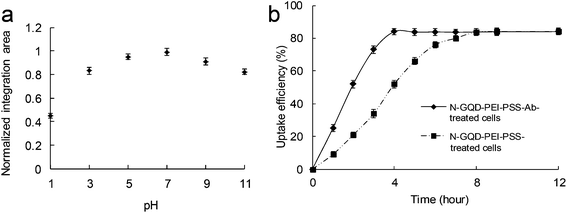
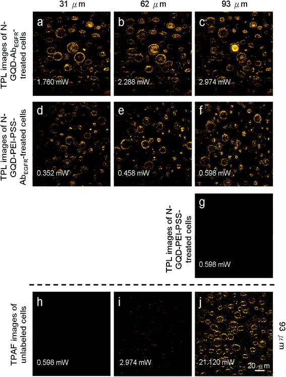
A431 skin cancer cells with an overexpression of EGFRs on their surfaces were selected as our experimental template. To increase the specificity and efficiency of targeting, the AbEGFR was coated onto the materials. The aim was to demonstrate the outstanding two-photon properties of N-GQD-PEI-PSS, the generation of nonreactive oxygen species-dependent oxidative stress31 on the cells (Fig. S13–S15†), the presence of high TPL intensity in the acidic environment of a cancerous specimen or tissue (Fig. 4a), and the effectiveness of the materials in serving as a two-photon contrast agent. According to the uptake assay (Fig. 4b), the results also showed that the initial profile of the material-Ab included a burst uptake rate of approximately 84% within 4 h, whereas it was 52% for the material on its own. The burst and rapid rate may be attributed to the substantial amount of material-Ab absorbed onto the cellular surface or taken up by the cells because of the highly specific binding ability of the antibody. From 4 h to 12 h, the uptake rate exhibited a saturated status due to the fact that the sites that the material-Ab absorbed onto were full, but it did not reach saturation until the 8th hour for the material on its own. Consequentially, the results not only showed that the antibody was successfully coated onto the material, but also that the material-Ab was more efficiently absorbed and taken up on the cellular surface than the material on its own, facilitating two-photon bioimaging. Fig. 5 displays TPL images of the material-AbEGFR-treated A431 cells at different depths at a wavelength of 800 nm under TPE. To imitate 3D epithelial tissue, embedded cells in a collagen matrix32,33 were also used. As a function of depth at every 31 μm to 93 μm, the TPL from the N-GQD-AbEGFR- (Fig. 5a–c) and N-GQD-PEI-PSS-AbEGFR-treated cells (Fig. 5d–f) was illuminated. For the material-AbEGFR-treated cells, the imaging process needed the same TPE power increase of 30% at each 31 μm imaging depth increment to maintain constant TPL intensity. The TPL imaging of the N-GQD-PEI-PSS-AbEGFR-treated cells needed 5 times less power than that of the N-GQD-AbEGFR emission, whereas the TPL imaging of the N-GQD-PEI-PSS-treated cells with no coating antibody demonstrated only little attachment on the cell surface and internalization into the cell (Fig. 5g). The TPL signal corresponded to bright rings with a distribution throughout the cellular membrane, which is associated with a characteristic pattern of successful AbEGFR labeling. In addition, TPAF images (Fig. 5h–j) emitted from intrinsic fluorophores of the cancer cells required 21.120 mW (2112.0 nJ per pixel) of TPE in unlabeled cells (Fig. 5j) to obtain the same signal level, compared with only 2.974 mW (297.4 nJ per pixel) being required for the N-GQD-AbEGFR-treated cells (Fig. 5c). Additionally, the TPE power was reduced to 0.598 mW (59.8 nJ per pixel) for the N-GQD-PEI-PSS-AbEGFR-treated cells (Fig. 5f), indicating a TPL intensity 35 times brighter than that of TPAF in the unlabeled cells. Besides, a photothermal effect of the N-GQD-PEI-PSS was not observed following identical laser treatments (Fig. S16†).
Conclusions
In the present study, the polymers containing N and S atoms coated on the N-GQDs exposed under TPE resulted in higher PL, QY, TPA, TPL emission, absolute cross sections of TPE, ratios of radiative decay rate/nonradiative decay rate, and resistance to photobleaching after TPE, and shortened lifetimes. Furthermore, the N-GQDs-polymers generated nonreactive oxygen species-dependent oxidative stress on cells after TPE. The N-GQDs-polymers also exhibited a high QY, impressive two-photon properties and highly emitted TPL intensity in an acidic environment, signifying that they can serve as promising two-photon contrast probes for the noninvasive detection of deep and 3D biological specimens by using TPE. These findings present possible future biological applications for obtaining images in deeper biological specimens and tissues in the interior of the human body.Conflicts of interest
There are no conflicts to declare.Acknowledgements
This study was supported by the Ministry of Science and Technology, Taiwan, R.O.C. (MOST-104-2314-B-006-076-MY3; MOST-106-2811-B-006-011), and the Aim for the Top University Project to the National Cheng Kung University (MOST-106-2221-E-006-002-, MOST-106-2119-M-006-008-, MOST-106-2119-M-038-001- and MOST-105-2812-8-006-002-). This research received funding from the Headquarters of University Advancement at the National Cheng Kung University, which is sponsored by the Ministry of Education, Taiwan, R.O.C.Notes and references
- D. Pan, J. Zhang, Z. Li and M. Wu, Adv. Mater., 2010, 22, 734–738 CrossRef CAS PubMed.
- H. Tetsuka, R. Asahi, A. Nagoya, K. Okamoto, I. Tajima, R. Ohta and A. Okamoto, Adv. Mater., 2012, 24, 5333–5338 CrossRef CAS PubMed.
- K. J. Jeon, Z. Lee, E. Pollak, L. Moreschini, A. Bostwick, C. M. Park, R. Mengelsberg, V. Radmilovic, R. Kostecki, T. J. Richardson and E. Rotenberg, ACS Nano, 2011, 5, 1042–1046 CrossRef CAS PubMed.
- Y. Dong, H. Pang, H. B. Yang, C. Guo, J. Shao, Y. Chi, C. M. Li and T. Yu, Angew. Chem., Int. Ed., 2013, 52, 7800–7804 CrossRef CAS PubMed.
- L. Hui, H. Haiping and Y. Zhizhen, Adv. Mater. Res., 2014, 950, 44–47 CrossRef.
- J. C. Carrero-Sanchez, A. L. Elias, R. Mancilla, G. Arrellin, H. Terrones, J. P. Laclette and M. Terrones, Nano Lett., 2006, 6, 1609–1616 CrossRef CAS PubMed.
- X. Wang, X. Li, L. Zhang, Y. Yoon, P. K. Weber, H. Wang, J. Guo and H. Dai, Science, 2009, 324, 768–771 CrossRef CAS PubMed.
- X. Yan, X. Cui and L. Li, J. Am. Chem. Soc., 2010, 132, 5944–5945 CrossRef CAS PubMed.
- L. Cao, X. Wang, M. J. Meziani, F. Lu, H. Wang, P. G. Luo, Y. Lin, B. A. Harruff, M. Veca, D. Murray, S. Y. Xie and Y. P. Sun, J. Am. Chem. Soc., 2007, 129, 11318–11319 CrossRef CAS PubMed.
- A. Ananthanarayanan, Y. Wang, P. Routh, M. A. Sk, A. Than, M. Lin, J. Zhang, J. Chen, H. Sun and P. Chen, Nanoscale, 2015, 7, 8159–8165 RSC.
- M. A. Albota, C. Xu and W. W. Webb, Appl. Opt., 1998, 37, 7352–7356 CrossRef CAS PubMed.
- W. S. Kuo, C. L. L. Hsu, H. H. Chen, C. Y. Chang, H. F. Kao, L. C. S. Chou, Y. C. Chen, S. J. Chen, W. T. Chang, S. W. Tseng, J. Y. Wang and Y. C. Pu, Nanoscale, 2016, 8, 16874–16880 RSC.
- W. S. Hummers and R. E. Offeman, J. Am. Chem. Soc., 1958, 80, 1339 CrossRef CAS.
- T. F. Yeh, C. Y. Teng, S. J. Chen and H. Teng, Adv. Mater., 2014, 26, 3297–3303 CrossRef CAS PubMed.
- W. S. Kuo, H. H. Chen, S. Y. Chen, C. Y. Chang, P. C. Chen, Y. I. Hou, Y. T. Shao, H. F. Kao, C. L. L. Hsu, Y. C. Chen, S. J. Chen, S. R. Wu and J. Y. Wang, Biomaterials, 2017, 120, 185–194 CrossRef CAS PubMed.
- L. Tang, R. Ji, X. Li, G. Bai, C. P. Liu, J. Hao, K. S. Teng, Z. Yang and S. P. Lau, ACS Nano, 2014, 8, 6312–6320 CrossRef CAS PubMed.
- D. Y. Pan, L. Guo, J. C. Zhang, C. Xi, Q. Xue, H. Huang, J. Li, Z. Zhang, W. Yu, Z. Chen, Z. Li and M. Wu, J. Mater. Chem., 2012, 22, 3314–3318 RSC.
- L. Li, G. Wu, G. Yang, J. Peng, J. Zhao and J. J. Zhu, Nanoscale, 2013, 5, 4015–4039 RSC.
- Q. Xu, Y. Liu, C. Gao, J. Wei, H. Zhou, Y. Chen, C. Dong, T. S. Sreeprasad, N. Li and Z. Xia, J. Mater. Chem. C, 2015, 3, 9885–9893 RSC.
- F. Jiang, D. Q. Chen, R. M. Li, C. Y. Wang, G. Q. Zhang, S. Li, J. Zhang, N. Huang, Y. Gu, C. Wang and C. Shu, Nanoscale, 2013, 5, 1137–1142 RSC.
- M. Zheng, S. Liu, J. Li, Z. Xe, D. Qu, X. Miao, X. Jing, Z. Sun and H. Fan, J. Mater. Res., 2015, 30, 3386–3393 CrossRef CAS.
- C. Y. Lin, C. H. Lien, K. C. Cho, C. Y. Chang, N. S. Chang, P. J. Campagnola, C. Y. Dang and S. J. Chen, Opt. Express, 2012, 20, 13669–13676 CrossRef CAS PubMed.
- J. Mertz, Eur. Phys. J. D, 1998, 3, 53–66 CrossRef CAS.
- C. Xu and W. W. Webb, Nonlinear and Two-Photon-Induced Fluorescence, in Topics in Fluorescence Spectroscopy, Plenum Press, New York, 1997, vol. 5 Search PubMed.
- D. M. Togashi, B. Szczupak, A. G. Ryder, A. Calvet and M. O'Loughlin, J. Phys. Chem. A, 2009, 113, 2757–2767 CrossRef CAS PubMed.
- C. Würth, M. Grabolle, J. Pauli, M. Spieles and U. Resch-Genger, Nat. Protoc., 2013, 8, 1535–1550 CrossRef PubMed.
- S. Zhu, L. Wang, N. Zhou, X. Zhao, Y. Song, S. Maharjan, J. Zhang, L. Lu, H. Wang and B. Yang, Chem. Commun., 2014, 50, 13845–13848 RSC.
- C. Xu and W. W. Webb, J. Opt. Soc. Am. B, 1996, 13, 481–491 CrossRef CAS.
- W. R. Zipfel, R. M. Williams and W. W. Webb, Nat. Biotechnol., 2003, 21, 1369–1377 CrossRef CAS PubMed.
- J. I. Gersten and A. Nitzan, Chem. Phys. Lett., 1984, 104, 31–37 CrossRef CAS.
- D. Y. Lyon, L. Brunet, G. W. Hinkal, M. R. Wiesner and P. J. J. Alvarez, Nano Lett., 2008, 8, 1539–1543 CrossRef CAS PubMed.
- N. J. Durr, T. Larson, D. K. Smith, B. A. Korgel, K. Sokolov and A. Ben-Yakar, Nano Lett., 2007, 7, 941–945 CrossRef CAS PubMed.
- W. S. Kuo, C. Y. Chang, H. H. Chen, C. L. L. Hsu, J. Y. Wang, H. F. Kao, L. C. S. Chou, Y. C. Chen, S. J. Chen, W. T. Chang, S. W. Tseng, P. C. Wu and Y. C. Pu, ACS Appl. Mater. Interfaces, 2016, 8, 30467–30474 CAS.
Footnote |
| † Electronic supplementary information (ESI) available: Experimental section, z-axis resolution of femtosecond laser, characterizations, TPE action/absolute cross sections, lifetime data, stability after photoexcitation or in physiological environments, cell viability and the amount of generated ROS, plots of dependence of TPL on excitation intensity and temperature dependence as a function of irradiation time. See DOI: 10.1039/c7nr06836k |
| This journal is © The Royal Society of Chemistry 2018 |

