 Open Access Article
Creative Commons Attribution 3.0 Unported Licence
3-(4-Hydroxyphenyl)propionic acid, a major microbial metabolite of procyanidin A2, shows similar suppression of macrophage foam cell formation as its parent molecule
Yu-Ying Zhang†
a,
Xiao-Le Li†b,
Tong-Yun Lia,
Mei-Ying Lia,
Ri-Ming Huanga,
Wu Li*b and
Rui-Li Yang *a
*a
aGuangdong Provincial Key Laboratory of Food Quality and Safety, College of Food Science, South China Agricultural University, Guangzhou 510642, China. E-mail: rlyang77@scau.edu.cn; Fax: +86-20-85280270; Tel: +86-20-85283448
bCollege of Food Science and Technology, Hainan University, Haikou 570228, China. E-mail: leewuu@163.com; Fax: +86-898-66193581; Tel: +86-898-66198861
First published on 7th February 2018
Abstract
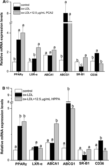
The effect of procyanidin A2 (PCA2) and its major colonic metabolite 3-(4-hydroxyphenyl)propionic acid (HPPA) on the suppression of macrophage foam cell formation, and underlying mechanism, were investigated for the first time. The results showed that 12.5 μg mL−1 PCA2 and HPPA significantly reduced cellular lipid accumulation and inhibited foam cell formation. HPPA promoted macrophage cholesterol efflux by up-regulating mRNA expressions of ABCA1 and SR-B1, while PCA2 significantly increased SR-B1 and LXR-α mRNA expression levels. Moreover, PCA2 and HPPA significantly lowered the elevated levels of CD36 mRNA expression in ox-LDL-treated macrophage cells. Besides these, the ox-LDL-induced cellular oxidative stress and inflammation was also restricted by PCA2 and HPPA treatment via nuclear factor kappa-B pathways. In conclusion, PCA2 and its major microbial metabolite, HPPA, inhibited the conversion of macrophage into foam cells via regulating cellular lipid metabolism and suppressing cellular oxidative stress and inflammation.
1. Introduction
Atherosclerosis (AS), a leading cause of death and illness in the world, has been considered to be an important pathological basis of cardiovascular disease. Excessive uptake of oxidized low-density lipoprotein (ox-LDL), excessive cholesterol esterification, and impaired cholesterol efflux occurring in the macrophages, results in the formation of foam cells.1 It has been shown that the conversion of macrophages into foam cells is a typical pathological feature of early stage atherosclerotic lesions.2 Therefore, suppressing macrophage derived foam cell formation is critical as an effective strategy for prevention and treatment of AS.In order to develop novel therapeutic interventions for AS, the mechanisms underlying the formation of foam cells have been studied in recent years.
Reverse cholesterol transport (RCT) is the only way to remove excess cholesterol, which is important for the removal of excess lipids from peripheral tissues to the liver for catabolism and excretion.3 The most important step in macrophage RCT is cholesterol efflux, by which intracellular cholesterol from macrophages is transferred to extracellular acceptors. ATP-binding cassette transporter A1 (ABCA1), ATP-binding cassette transporter G1 (ABCG1) and scavenger receptor BI (SR-BI) play key roles in RCT pathway.4 Studies have shown that the expressions of ABCA1 and ABCG1 are to some extent, regulated by peroxisome proliferator-activated receptor gamma (PPARγ) activators and liver X receptor alpha (LXR-α).5 CD36, a high affinity physiological receptor for ox-LDL, plays a vital role in the foam cell formation. ox-LDL can stimulate CD36 expression, then the up-regulation of CD36 make more ox-LDL endocytosed, and eventually lead to excess cholesterol buildup to form foam cells.6 Growing evidence establishes that the imbalanced expressions of CD36, ABCA1, ABCG1 and SR-B1 mediate the foam cell formation via impairing the balance of cholesterol influx and efflux.7 Furthermore, oxidative stress and inflammation play important roles in the pathogenesis of diverse cardiovascular diseases including AS. Macrophages can phagocytosed ox-LDL and other lipids and produce inflammatory cytokines and reactive oxygen species (ROS) while continuing to accumulate lipids and differentiate into foam cells to form the early atherosclerotic plaques.8
Procyanidins, which are among the most abundant dietary polyphenols, are recognized as biologically active in human health. Procyanidins have strong anti-oxidant,9 anti-inflammation,10,11 anti-aging capabilities12 and can help prevent cardiovascular and cerebrovascular diseases. The subunits of procyanidins are linked by C4–C8 or C4–C6 bond (B-type), or doubly linked by a C4–C8/C6 bond and an additional C2–O–C7 or C2–O–C5 ether bond (A-type). Moreover, the most ubiquitous procyanidins in foods are the B-type procyanidins, the A-types are less common and concerned in nature.13 B-type procyanidins mainly from apple,14 grape seed,15 cocoa16 exhibited great health benefits for preventing the development of AS. Recently, it is indicated that A-type procyanidins similarly have some different bioactivities, such as effective uropathogenic bacterial anti-adhesion activity.17,18 Wong et al., reported that the deleterious effect of p-cresol on HT29 cells were prevented by the extracts from cranberries and avocados (characterized by the presence of type-A procyanidins), whereas they became worse by extracts from apples and grape.19 However, the effect of A-type procyanidins on foam cells formation remains unclear.
Though potential health beneficial effects have been attributed to procyanidins, it is poorly absorbable in the gastrointestinal tract. Current evidences indicate that colonic microbiota can degrade procyanidins, producing a large number of metabolites that can be absorbed and exert biological effects in the body.20 In particular, colonic metabolites have demonstrated anti-oxidant,21 anti-inflammatory,22 and anti-glycative23 activities. Altogether, these findings support the hypothesis that not only procyanidins but also their microbial metabolites must be taken into account when assessing the impact of procyanidins on health.24 However, up to date, whether the microbial metabolite of procyanidin has an inhibitory effect on foam cells formation is unclear.
As mentioned above, most of studies assessing the anti-atherosclerosis activity of procyanidins have been performed using plant procyanidins extracts (mainly composed of B-type procyanidins). We sought to determine whether A-type procyanidins and its major microbial metabolite have a direct effect on the formation of foam cells. Procyanidin A2 (PCA2) is one structurally defined A-type procyanidin that is predominately found in cranberries,25 avocado,26 peanut red skins27 and litchi fruit pericarp,28 which has been found to anti-oxidant and anti-inflammatory activities.29 In this study, for the first time, we investigated the interference effects of PCA2 in comparison with 3-(4-hydroxyphenyl)propionic acid (HPPA), its major microbial metabolite30 on foam cells formation induced by ox-LDL. The aim of this study was to investigate the potential atheroprotective effects of A-type procyanidins and its major microbial metabolite, and to gain insights into the underlying molecular mechanisms.
2. Materials and methods
2.1. Chemicals and reagents
Procyanidin A2 and 3-(4-hydroxyphenyl)propionic acid were purchased from Sigma-Aldrich (Shanghai, China). RPMI-1640 medium, DMEM medium and fetal bovine serum were purchased from Gibco Life Technologies (California, USA). Human ox-LDL was purchased from Yiyuan Biotechnologies (Guangzhou, China). The kits for total cholesterol (TC) and free cholesterol (FC) measurement were purchased from Applygen Technologies Inc., (Beijing, China). A ROS kit was purchased from Beyotime Institute of Biotechnology (Shanghai, China). The kit for determination of MDA equivalent level was purchased from Nanjing Jiancheng Bioengineering Institute (Nanjing, China). The ELISA kits of IL-6 and IL-1β were obtained from R&D System (Minneapolis, MN, USA). SYBR-Green qPCR Master Mix and RevertAid First Strand cDNA Synthesis Kit were obtained by TransGen Biotech (Beijing, China). The primary antibodies against NF-κB-p65 and β-actin were obtained from Cell Signal Technology Inc. (Beverly, MA). All other reagents were of analytical grade and obtained locally.2.2. Cell line and cell culture
The mouse RAW264.7 cells were donated by Zhongshan School of Medicine, Sun Yat-sen University, Guangzhou, China. The cells were cultured in DMEM medium containing 10% fetal bovine serum (FBS) at 37 °C under 5% CO2.2.3. Cytotoxicity assay of PCA2 and HPPA
The effect of PCA2 and HPPA on the viability of RAW264.7 cells was measured using a cell counting kit-8 (CCK-8) assay (SAB, USA) according to the manufacturer's instructions. The cells were seeded at a density of 5 × 10⁴ cells per mL and incubated with PCA2 and HPPA at the concentrations of 6.25, 12.5, 25, 50, and 100 μg mL−1 for 24 h. After incubation, 10 μL of CCK-8 solution was added to each well. The cells were incubated for 1 h at 37 °C and the absorbance at 450 nm was measured.2.4. Oil red O staining and cellular cholesterol analysis
The raw264.7 macrophages were collected at the logarithmic growth phase and inoculated into the 6-well plates at 5 × 10⁶ cells per mL. After cells were treated with 80 μg mL−1 ox-LDL in the presence or absence of PCA2 and HPPA for 48 h, the cells were collected, washed three times with PBS buffer and subjected to oil-red O staining31 or harvested to determine the cellular cholesterol content according to the kit's instructions. The stained cells were observed and photographed under a microscope (Olympus, Tokyo, Japan). The TC and FC in cells were measured using the commercial kits according to the manufacturer's instructions. The difference between TC and FC represents the production of cellular cholesteryl ester (CE).2.5. ROS and thiobarbituric acid reactive substances (MDA) assay
After washing with PBS, cells were suspended in 10 μM 2′,7′-dichlorofluorescein diacetate (DCFH-DA) solution (Beyotime, Shanghai, China) and incubated at 37 °C for 30 min. Intracellular ROS generation was monitored by measuring the fluorescence intensity of cells with an EnSpire® Multimode Plate Reader, with excitation and emission wavelengths set at 488 and 525 nm, respectively. The level of thiobarbituric acid reactive substances (TBARS, reported as MDA equivalents) was measured using a kit according to the manufacturer's instructions.2.6. Determination of IL-6 and IL-1β
The cells were plated into a 96-well plate at 5 × 10⁴ cells per well and incubated with 80 μg mL−1 ox-LDL in the presence or absence of PCA2 and HPPA for 24 h. The cell liquid supernatant was collected by centrifugation at 5000g for 10 min. The levels of IL-6 and IL-1β were measured using ELISA kit (R&D System, Minneapolis, MN, USA).2.7. RT-QPCR analysis
The mRNA levels of the cholesterol-metabolism-related genes were determined by real-time quantitative PCR. Total RNA was extracted and transcribed into cDNA using a Reverse Transcription System kit (TransGen Biotech, Beijing, China). The PCR primers sequences used are listed in Table 1. Real-time PCR were performed with the incorporation of SYBR green using the real-time PCR System 7500 (Applied Biosystems, Waltham, MA). Melting curve analysis and agarose gel electrophoresis were used to monitor synthesis of the PCR products. β-Actin was used as an internal control.| Genes | Sequence (5′–3′) | Product size (bp) |
|---|---|---|
| β-Actin | F: GTCCCTCACCCTCCCAAAAG | 202 |
| R: GCTGCCTCAACACCTCAACCC | ||
| ABCA1 | F: CCCAGAGCAAAAAGCGACTC | 136 |
| R: GGTCATCATCACTTTGGTCCTTG | ||
| ABCG1 | F: AATGTCTGCTTTGCCTCGTT | 108 |
| R: GCAGCTACTGCATGTGATCAAGA | ||
| SR-B1 | F: TTTGGAGTGGTAGTAAAAAGGGC | 71 |
| R: TGACATCAGGGACTCAGAGTAG | ||
| LXR-α | F: CAGCGTCCATTCAGAGCAAGTGT | 140 |
| R: GTCAGTGAGCCTTCGCCATGTG | ||
| PPARγ | F: GCAGCTACTGCATGTGATCAAGA | 214 |
| R: GTCAGCGGGTGGGACTTTC | ||
| CD36 | F: GTGCTCTCCCTTGATTCTGC | 102 |
| R: CTCCAAACACAGCCAGGAC |
2.8. Western-blot analysis
RAW264.7 cells were plated in 6-well plates at a density of 1 × 10⁶ cells per well. After PCA2 and HPPA treatment, the cells were washed twice with ice-cold PBS, and then were lysed with RIPA lysis containing a protease inhibitor. Protein quantification was determined using the BCA assay. Then, protein samples were electrophoresed on 8% SDS-PAGE and transferred to a PVDF membrane. Each membrane was blocked for 2 h with 5% (w/v) skimmed milk, followed by incubation with primary antibodies overnight at 4 °C. After washing five times with TBST for 5 min, the membrane was incubated for 1 h with the secondary antibody, and then washed too. The immuno-reactive protein bands were quantified by UVP ChemiDoc-IT 510 imaging system (USA).2.9. Statistical analysis
Data are expressed as mean ± SD standard deviation. Significance of differences between groups was calculated by one-way ANOVA. P values <0.05 was considered statistically significant.3. Results
3.1. PCA2 and the microbial metabolite HPPA promoted the proliferation of macrophages
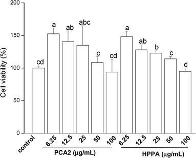
CCK-8 assay was conducted to assess the effects of PCA2 and the metabolites HPPA on the viability of raw264.7 macrophages. As shown in Fig. 1, compared to control cells, the cell growth was significantly stimulated by PCA2 and the metabolites HPPA within tested concentrations. These results showed that none of the assayed concentrations of PCA2 and HPPA were cytotoxic to macrophages.3.2. PCA2 and the metabolite HPPA inhibited ox-LDL-induced foam cell formation and lipid accumulation
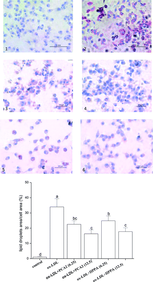
As shown in Fig. 2A, the oil-red O staining results were negative for the normal macrophages, while there was heavy reddish staining, and the lipid accumulation was significantly induced after the macrophages were exposed to 80 μg mL−1 ox-LDL for 24 h. However, treatment with PCA2 and the microbial metabolites HPPA dose-dependently diminished the reddish lipid droplets. The quantitative levels of the oil-red O staining exhibited that 12.5 μg mL−1 PCA2 and HPPA decreased the area of lipid droplets and cell area ratio by 51.96% and 47.72% compared with the model cells, respectively, which indicated that the formation of foam cells was blocked by PCA2 and HPPA.As shown in Table 2, TC, FC, and CE in the macrophages were significantly increased after exposure to 80 μg mL−1 ox-LDL for 24 h, and the ratio of CE to TC reached 54.70%, which means the conversion of macrophages to foam cells. When the macrophages were simultaneously treated with ox-LDL and different concentration of PCA2 and HPPA, the intracellular TC, FC, CE, and CE/TC levels decreased. 12.5 μg mL−1 PCA2 significantly decreased the levels of intracellular TC, FC, CE, and CE/TC of model cells by 31.36%, 11.77%, 47.55% and 23.64%, respectively. Likewise, treatment with 12.5 μg mL−1 HPPA partially precluded ox-LDL-induced increase in intracellular TC, FC, CE and CE/TC levels by 28.75%, 6.59%, 44.15% and 21.63%, respectively. These data revealed that PCA2 and its microbial metabolite HPPA decreased lipid deposition, and therefore restrained the formation of foam cells induced by ox-LDL.
| Groups | TC | FC | CE | CE/TC (%) |
|---|---|---|---|---|
| a Values are expressed as mean ± SD. Values in the same column with different superscripts are significantly different at P < 0.05 level | ||||
| Control | 59.52 ± 6.17d | 41.05 ± 1.62c | 18.46 ± 7.52d | 31.02 ± 10.14d |
| ox-LDL | 138.97 ± 13.70a | 62.95 ± 1.14a | 76.03 ± 13.21a | 54.70 ± 4.09a |
| ox-LDL + 6.25 μg mL−1 PCA2 | 112.33 ± 2.39b | 55.51 ± 2.50b | 56.79 ± 3.25b | 47.51 ± 2.10ab |
| ox-LDL + 12.5 μg mL−1 PCA2 | 95.39 ± 1.63c | 55.54 ± 1.80b | 39.88 ± 0.95c | 41.77 ± 1.67c |
| ox-LDL + 6.25 μg mL−1 HPPA | 114.11 ± 1.56b | 56.56 ± 1.46b | 55.31 ± 2.68b | 48.47 ± 1.83ab |
| ox-LDL + 12.5 μg mL−1 HPPA | 99.02 ± 2.84c | 58.80 ± 1.58b | 42.46 ± 4.11c | 42.87 ± 2.94bc |
3.3. PCA2 and the metabolite HPPA protected against ox-LDL-induced oxidative stress
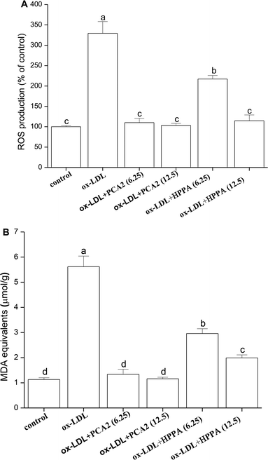
The incubation of macrophage cells with 80 μg mL−1 ox-LDL for 24 h resulted in significant increases in the levels of ROS and MDA equivalent. These effects were reversed by 6.25 and 12.5 μg mL−1 PCA2 treatment (Fig. 3A and B). Microbial metabolites HPPA were less effective preventing the ox-LDL-induced ROS and MDA equivalent level, with only partial recovery of MDA equivalent levels back to those in control cells.3.4. PCA2 and the metabolite HPPA inhibited the secretion of IL-6 and IL-1β
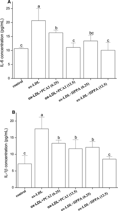
As shown in Fig. 4, ox-LDL promoted the production of IL-6 and IL-1β of macrophages. Compared with the model group, PCA2 significantly suppressed the ox-LDL-induced secretion of IL-6 and IL-1β, with a dose-dependent manner (P < 0.05). Macrophage IL-6 and IL-1β productions with 12.5 μg mL−1 PCA2 treatment were decreased by 46.39% and 33.75% than that of model group, respectively. 12.5 μg mL−1 HPPA totally inhibited ox-LDL-induced increase in intracellular IL-1β and IL-6 levels.3.5. Effects of PCA2 and the metabolite HPPA on the mRNA expression of cholesterol efflux/influx-related genes: ABCA1, ABCG1, SR-B1, LXR-α, PPARγ and CD36 in Raw264.7 foam cells
Cholesterol flux in macrophages is tightly regulated by several genes. ABCA1, ABCG1, SR-B1, LXR-α and PPARγ are key regulators of cholesterol efflux, while CD36 is critical scavenger receptors involved in cholesterol uptake. As shown in Fig. 5, 12.5 μg mL−1 PCA2 seperately increased the mRNA levels of LXR-α and SR-B1 by 1.88 and 2.89 times compared with the model group. 12.5 μg mL−1 HPPA significantly up-regulated the mRNA levels of ABCA1 and SR-B1 responsible for the efflux of cellular cholesterol. The expression levels of ABCA1 and SR-B1 treatment by 12.5 μg mL−1 HPPA were 2.13 and 2.10 times more than the model group, respectively. In addition, the ox-LDL significantly increased the mRNA level of CD36 in RAW264.7 cells, which is recognized as the macrophage mediating the ox-LDL uptake. However, PCA2 and HPPA remarkably down-regulated CD36 mRNA level by 58.99% and 57.42%, respectively.3.6. PCA2 and the metabolite HPPA inhibited the activation of NF-κB pathway induced by ox-LDL
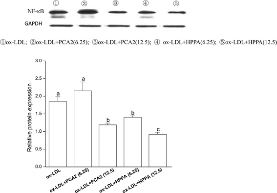
To further study the possible mechanisms that are responsible for PCA2 and the major metabolite HPPA action, the NF-κB protein expression was analyzed by western blot. As shown in Fig. 6, 12.5 μg mL−1 PCA2 or the metabolite HPPA-treated foam cells showed a marked decrease in NF-κB protein expression, showing PCA2 and the metabolite HPPA can inhibit the activation of NF-κB signaling pathway induced by ox-LDL.4. Discussion
Excessive lipids accumulation in macrophages can lead to the formation of foam cells, where the foam cells formation plays a crucial role in promoting AS. It is well accepted that generation of macrophage-derived foam cells is highly associated with the imbalance of cholesterol influx, esterification, and efflux.32 Therefore, cholesterol outflow from macrophages, the first step in RCT, has been suggested to play a vital role in the early stage of AS to lower its occurrence.1 ABCA1, ABCG1 and SR-B1 have been reported to play the key role in RCT pathway.33 Compared to the cells treated with ox-LDL alone, HPPA exhibited good ability to enhance the mRNA levels of ABCA1 and SR-B1, while PCA2 significantly up-regulated SR-B1 and LXR-α mRNA expression levels. Furthermore, CD36 is the principal receptor responsible for the uptake of lipoprotein-derived cholesterol.34 We initially observed that ox-LDL increased lipid accumulation associated with the elevated levels of CD36 mRNA level. The increase in CD36 caused by the ox-LDL was significantly decreased by PCA2 and its major microbial metabolite HPPA. Consistent with our results, procyanidins derived from grape seed (mainly composed of B-type procyanidins) attenuate the development of foam cell formation by reducing cholesterol accumulation and modulating the expression of CD36 and ABCA1 expression.15 Taken together, the results suggested that PCA2, an A-type procyanidin and its major microbial metabolite regulated the balance between cholesterol uptake and efflux through modulating gene expressions of CD36, ABCA1, SR-B1 and LXR-α, which play important roles in the transformation of macrophages into foam cells. The present work suggested that the inhibition of foam cell formation might be an important mechanism of PCA2 and its major microbial metabolite blocking AS.It has transpired that the pathogenesis of AS involves a network of vascular wall cells and mediators, in which macrophages play pivotal roles by producing proinflammatory cytokines and via transition to lipid-laden foam cells that initiate the formation of atherosclerotic lesion.35,36 NF-κB signaling pathways are well accepted to participate in inflammatory cytokine productions and regulate the transcription of downstream targets genes involved in AS.37 Ample evidence supports a central regulatory role of NF-κB pathway in atherogenesis.38 Our results showed that PCA2 and its major microbial metabolite significantly decreased the ox-LDL-increased levels of IL-6 and IL-1β. Procyanidin B2 inhibits inflammasome activation and IL-1β secretion through the inactivation of the NF-κB signaling pathway in lipopolysaccharide-stimulated macrophages.39 Terra et al., reported that grape-seed procyanidins significantly inhibited the overproduction of NO and PGE(2) and NF kappa B (p65) translocation to nucleus in RAW264.7 macrophages stimulated with lipopolysaccharide plus interferon-gamma.40 Consistent with these data, our results revealed that PCA2 and its major microbial metabolite significantly suppressed the activation of NF-κB pathways induced by ox-LDL, confirming the fact that inhibition of NF-κB can reduce foam cell formation.
Oxidative stress plays a critical role in the pathogenesis of diverse cardiovascular diseases including AS.31,41 Increasing evidence has revealed that ox-LDL can be phagocytosed by macrophages, resulting in macrophage activation and subsequent production of inflammatory cytokines and ROS.42,43 Therefore, suppression of ox-LDL-induced oxidative stress may be an efficient strategy to prevent and minimize the development of AS. Many antiatherosclerotic therapeutics exert their atheroprotective effects by inhibiting macrophage oxidative stress.44,45 Previous studies have shown a protective effect of PCA2 against oxidative stress, protecting against bisphenol A-induced apoptosis in islet cells and decreasing the MDA equivalents level in pancreas tissue.46 Similarly, our data also revealed that PCA2 and its major metabolite HPPA protected against ox-LDL-induced oxidative stress. HPPA was less effective than PCA2 and only partly moderating the oxidative stress induced by ox-LDL, not being able to recover the cellular MDA equivalents level completely.
Current evidences indicate that a large proportion of ingested procyanidins would reach the colon, where they undergo metabolism by the intestinal microbiota generating a wide variety of metabolites. As mentioned above, it has been reported that the bioavailability of PCA2 is low,47 generating metabolites including 5-phenylvaleric acid, 4-hydroxyphenylacetic acid, 3-(3-hydroxyphenyl)propionic acid and HPPA which is the major metabolites.30,48 Therefore, it is crucial to assess whether its metabolites retain the biological activity of the parent compounds. In the present work the microbial metabolite HPPA showed a protective effect against foam cells formation similar to that of PCA2, whilst other metabolites was less effective (data not shown). The results implied that the protective effect of PCA2 against foam cells formation is strengthened by the action of its major microbial metabolite. To the best of our knowledge, this is the first report on the atheroprotective activities of PCA2 and its major microbial metabolites in a foam cell model.
5. Conclusion
In summary, our study showed that PCA2, an A-type procyanidin and its major microbial metabolites, HPPA, effectively hinder the formation of foam cells. The cells treated with PCA2 or its major microbial metabolites were protected from the deleterious effect of ox-LDL as shown by the lower lipids accumulation, decreased ROS and MDA equivalents production, and diminished levels of inflammatory biomarkers (Fig. 7). The microbial metabolite HPPA showed a similar suppression effect on the macrophage foam cell formation with that of PCA2. The protective effect of PCA2 against foam cells formation might partly attribute to its microbial metabolites.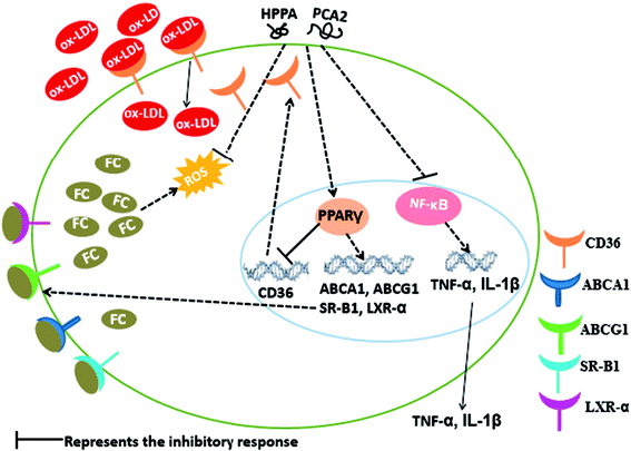 | ||
| Fig. 7 Possible molecular mechanism of PCA2 and its metabolite HPPA-mediated suppression of macrophage foam cell formation. | ||
Conflicts of interest
There are no conflicts to declare.Acknowledgements
The study was financially supported by the National Natural Science Foundation of China (Grant No. 31471705), the Earmarked Fund for China Agriculture Research System (Grant No. CARS-33) and Natural Science Foundation of Hainan Province, China (Project No. 317007).References
- X. H. Yu, Y. C. Fu, D. W. Zhang, K. Yin and C. K. Tang, Clin. Chim. Acta, 2013, 424, 245–252 CrossRef CAS PubMed.
- K. J. Moore and I. Tabas, Cell, 2011, 145, 341–355 CrossRef CAS PubMed.
- M. Hellerstein and S. Turner, Curr. Opin. Lipidol., 2014, 25, 40–47 CrossRef PubMed.
- G. H. Rothblat and M. C. Phillips, Curr. Opin. Lipidol., 2010, 21, 229–238 CrossRef CAS PubMed.
- R. Z. Zhao, J. Feng and G. X. He, Biochem. Biophys. Res. Commun., 2014, 448, 329–334 CrossRef CAS PubMed.
- I. Voloshyna, O. Hai, M. J. Littlefield, S. Carsons and A. B. Reiss, Eur. J. Pharmacol., 2013, 698, 299–309 CrossRef CAS PubMed.
- A. Chawla, W. A. Boisvert, C. H. Lee, B. A. Laffitte, Y. Barak, S. B. Joseph, D. Liao, L. Nagy, P. A. Edwards, L. K. Curtiss, R. M. Evans and P. Tontonoz, Mol. Cell, 2001, 7, 161–171 CrossRef CAS PubMed.
- P. Gao, X. M. Wang, D. H. Qian, Z. X. Qin, J. Jin, Q. Xu, Q. Y. Yuan, X. J. Li and L. Y. Si, Cardiovasc. Res., 2013, 97, 533–543 CrossRef CAS PubMed.
- K. E. Constanza, B. L. White, J. P. Davis, T. H. Sanders and L. L. Dean, J. Agric. Food Chem., 2012, 60, 10776–10783 CrossRef CAS PubMed.
- N. Martinez-Micaelo, N. Gonzalez-Abuin, A. Ardevol, M. Pinent and M. T. Blay, BioFactors, 2012, 38, 257–265 CrossRef CAS PubMed.
- G. M. Sawyer, D. E. Stevenson, T. K. McGhie and R. D. Hurst, J. Funct. Foods, 2017, 31, 141–151 CrossRef CAS.
- C. G. Fraga and P. I. Oteiza, Free Radical Biol. Med., 2011, 51, 813–823 CrossRef CAS PubMed.
- L. W. Gu, M. A. Kelm, J. F. Hammerstone, G. Beecher, J. Holden, D. Haytowitz and R. L. Prior, J. Agric. Food Chem., 2003, 51, 7513–7521 CrossRef CAS PubMed.
- L. Wang, T. Fumoto, S. Masumoto, T. Shoji, T. Miura, M. Naraoka, N. Matsuda, T. Imaizumi and H. Ohkuma, Atherosclerosis, 2017, 258, 56–64 CrossRef CAS PubMed.
- X. Terra, J. Fernandez-Larrea, G. Pujadas, A. Ardevol, C. Blade, J. Salvado, L. Arola and M. Blay, J. Agric. Food Chem., 2009, 57, 2588–2594 CrossRef CAS PubMed.
- D. M. Chen, X. Cai, C. L. Kwik-Uribe, R. Zeng and X. Z. Zhu, J. Cardiovasc. Pharmacol., 2006, 48, 54–70 CrossRef CAS PubMed.
- X. Q. Dong, B. Zou, Y. Zhang, Z. Z. Ge, J. Du and C. M. Li, Fitoterapia, 2013, 91, 128–139 CrossRef CAS PubMed.
- L. Y. Foo, Y. R. Lu, A. B. Howell and N. Vorsa, J. Nat. Prod., 2000, 63, 1225–1228 CrossRef CAS.
- X. N. Wong, C. Carrasco-Pozo, E. Escobar, P. Navarrete, F. Blachier, M. Andriamihaja, A. Lang, D. Tome, M. J. Cires, E. Pastene and M. Gotteland, J. Agric. Food Chem., 2016, 64, 3574–3583 CrossRef CAS PubMed.
- M. Monagas, M. Urpi-Sarda, F. Sanchez-Patan, R. Llorach, I. Garrido, C. Gomez-Cordoves, C. Andres-Lacueva and B. Bartolome, Food Funct., 2010, 1, 233–253 CAS.
- G. Baeza, B. Sarria, R. Mateos and L. Bravo, Food Res. Int., 2016, 87, 25–33 CrossRef CAS.
- M. Monagas, N. Khan, C. Andres-Lacueva, M. Urpi-Sarda, M. Vazquez-Agell, R. M. Lamuela-Raventos and R. Estruch, Br. J. Nutr., 2009, 102, 201–206 CrossRef CAS PubMed.
- E. Verzelloni, C. Pellacani, D. Tagliazucchi, S. Tagliaferri, L. Calani, L. G. Costa, F. Brighenti, G. Borges, A. Crozier, A. Conte and D. Del Rio, Mol. Nutr. Food Res., 2011, 55, S35–S43 CAS.
- F. Cardona, C. Andres-Lacueva, S. Tulipani, F. J. Tinahones and M. I. Queipo-Ortuno, J. Nutr. Biochem., 2013, 24, 1415–1422 CrossRef CAS PubMed.
- J. Lee, J. Funct. Foods, 2013, 5, 144–153 CrossRef CAS.
- A. Lopez-Cobo, A. M. Gomez-Caravaca, F. Pasini, M. F. Caboni, A. Segura-Carretero and A. Fernandez-Gutierrez, LWT--Food Sci. Technol., 2016, 73, 505–513 CrossRef CAS.
- M. M. Appeldoorn, M. Sanders, J. P. Vincken, V. Cheynier, C. Le Guerneve, P. C. H. Hollman and H. Gruppen, Food Chem., 2009, 117, 713–720 CrossRef CAS.
- S. Y. Li, J. Xiao, L. Chen, C. L. Hu, P. Chen, B. J. Xie and Z. D. Sun, Food Chem., 2012, 135, 31–38 CrossRef CAS.
- S. L. Coleman, M. C. Kruger, G. M. Sawyer and R. D. Hurst, Int. J. Mol. Sci., 2016, 17, 1–13 Search PubMed.
- A. Engemann, F. Hubner, S. Rzeppa and H. U. Humpf, J. Agric. Food Chem., 2012, 60, 749–757 CrossRef CAS PubMed.
- H. D. Zheng, D. J. Cui, X. J. Quan, W. L. Yang, Y. N. Li, L. Zhang and E. Q. Liu, Biochem. Biophys. Res. Commun., 2016, 477, 1017–1023 CrossRef CAS PubMed.
- L. Yvan-Charvet, M. Ranalletta, N. Wang, S. Han, N. Terasaka, R. Li, C. Welch and A. R. Tall, J. Clin. Invest., 2007, 117, 3900–3908 CAS.
- X. Ma, S. F. Li, Z. S. Qin, J. Ye, Z. L. Zhao, H. H. Fang, Z. W. Yao, M. N. Gu and Y. W. Hu, Cardiovasc. Pathol., 2015, 24, 230–235 CrossRef CAS PubMed.
- R. Chen, Z. B. Cheng, J. Huang, D. Liu, C. M. Wu, P. Guo and W. H. Lin, RSC Adv., 2017, 7, 49235–49243 RSC.
- X. Q. Zha, L. Xue, H. L. Zhang, M. N. Asghar, L. H. Pan, J. Liu and J. P. Luo, Mol. Nutr. Food Res., 2015, 59, 2008–2021 CAS.
- D. A. Chistiakov, Y. V. Bobryshev and A. N. Orekhov, J. Cell. Mol. Med., 2016, 20, 17–28 CrossRef CAS PubMed.
- Y. Wang, X. Wang, M. H. Sun, Z. Y. Zhang, H. Cao and X. Q. Chen, Biochem. Biophys. Res. Commun., 2011, 411, 543–548 CrossRef CAS PubMed.
- L. Sironi, C. Banfi, M. Brioschi, P. Gelosa, U. Guerrini, E. Nobili, A. Gianella, R. Paoletti, E. Tremoli and M. Cimino, Neurobiol. Dis., 2006, 22, 445–451 CrossRef CAS PubMed.
- N. Martinez-Micaelo, N. Gonzalez-Abuin, M. Pinent, A. Ardevol and M. Blay, Mol. Nutr. Food Res., 2015, 59, 262–269 CAS.
- X. Terra, J. Valls, X. Vitrac, J. M. Merrillon, L. Arola, A. Ardevol, C. Blade, J. Fernandez-Larrea, G. Pujadas, J. Salvado and M. Blay, J. Agric. Food Chem., 2007, 55, 4357–4365 CrossRef CAS PubMed.
- B. Fischer, A. von Knethen and B. Brune, J. Immunol., 2002, 168, 2828–2834 CrossRef CAS.
- J. Khallou-Laschet, A. Varthaman, G. Fornasa, C. Compain, A. T. Gaston, M. Clement, M. Dussiot, O. Levillain, S. Graff-Dubois, A. Nicoletti and G. Caligiuri, PLoS One, 2010, 5, 1–10 Search PubMed.
- F. Li, S. H. Cui, X. Q. Zha, V. Bansal, Y. L. Jiang, M. N. Asghar, J. H. Wang, L. H. Pan, B. F. Xu and J. P. Luo, Int. J. Biol. Macromol., 2015, 72, 664–672 CrossRef CAS PubMed.
- X. Cheng, D. X. Gao, J. J. Song, F. Z. Ren and X. Y. Mao, RSC Adv., 2015, 5, 4511–4523 RSC.
- M. Z. Xiao, X. B. Jin, F. T. Tang, L. J. Wang, J. W. Mao, X. Q. Li, F. J. Chu, Y. T. Huang, J. Ding, W. Q. Chen, J. G. Geng and J. Y. Zhu, RSC Adv., 2016, 6, 105363–105370 RSC.
- A. Ahangarpour, G. Afshari, S. A. Mard, A. Khodadadi and M. Hashemitabar, J. Physiol. Pharmacol., 2016, 67, 243–252 CAS.
- K. Q. Ou, S. S. Percival, T. Zou, C. Khoo and L. W. Gu, J. Agric. Food Chem., 2012, 60, 1390–1396 CrossRef CAS PubMed.
- Z. Z. Ge, X. Q. Dong, W. Zhu, Y. Zhang and C. M. Li, J. Agric. Food Chem., 2015, 63, 8991–8998 CrossRef CAS PubMed.
Footnote |
| † These authors contributed equally to this work. |
| This journal is © The Royal Society of Chemistry 2018 |

