 Open Access Article
Creative Commons Attribution 3.0 Unported Licence
Disarming the virulence arsenal of Pseudomonas aeruginosa by blocking two-component system signaling†
Manibarsha
Goswami
a,
Adeline
Espinasse
a and
Erin E.
Carlson
 *abc
*abc
aDepartment of Chemistry, University of Minnesota, 225 Pleasant St. SE, Minneapolis, MN 55454, USA. E-mail: carlsone@umn.edu
bDepartment of Medicinal Chemistry, University of Minnesota, USA
cDepartment of Biochemistry, Molecular Biology and Biophysics, University of Minnesota, USA
First published on 10th July 2018
Abstract
Pseudomonas aeruginosa infections have reached a “critical” threat status making novel therapeutic approaches required. Inhibiting key signaling enzymes known as the histidine kinases (HKs), which are heavily involved with its pathogenicity, has been postulated to be an effective new strategy for treatment. Herein, we demonstrate the potential of this approach with benzothiazole-based HK inhibitors that perturb multiple virulence pathways in the burn wound P. aeruginosa isolate, PA14. Specifically, our compounds significantly reduce the level of toxic metabolites generated by this organism that are involved in quorum-sensing and redox-balancing mechanisms. They also decrease the ability of this organism to swarm and attach to surfaces, likely by influencing their motility appendages. Quantitative transcription analysis of inhibitor-treated cultures showed substantial perturbations to multiple pathways including expression of response regulator GacA, the cognate partner of the “super regulator” of virulence, HK GacS, as well as flagella and pili formation. These promising results establish that blocking of bacterial signaling in P. aeruginosa has dramatic consequences on virulence behaviours, especially in the context of surface-associated infections.
Introduction
Bacterial virulence mechanisms have become promising new targets for the development of antibacterial agents, including ubiquitous signaling pathways that play key roles in many of these processes, the two-component systems (TCSs).1–4 Most bacteria rely heavily on TCSs; (>20 distinct TCSs per organism) to respond to their external environment and transmit various cell signals.5 TCSs are typically composed of a membrane-bound sensory protein, a histidine kinase (HK), and a cognate response regulator (RR) protein (Fig. 1). HKs respond to external stimuli by autophosphorylation, using ATP, of a catalytic histidine residue in the DHp (dimerization and histidine phosphotransfer) region and subsequent phosphoryl group transfer to the RR, which is often a transcription factor and induces altered gene expression (Fig. 1).6 TCSs and their corresponding HKs are postulated to be critical in many bacterial survival, metabolic, and virulence mechanisms.1–4,7 For example, HKs have been linked to severe infections caused by Gram-positive and Gram-negative bacteria, including the ESKAPE pathogens (Enterococcus faecium, Staphylococcus aureus, Klebsiella pneumoniae, Acinetobacter baumannii, Pseudomonas aeruginosa, and Enterobacter species).8 Antibiotic resistance has also been attributed to TCS signaling – for example, resistance to cationic antimicrobial peptides is thought to be associated with PhoQ/PhoP in Salmonella and GraS/GraR in S. aureus, whereas CzcR/CzcS is implicated in carbapenem-resistance in P. aeruginosa.7 While some TCSs are essential, most are non-essential and are quintessential in virulence processes in Gram-negative bacteria such as quorum sensing (QS), toxin production, and formation of bacterial communities.2,7 In light of these advantages, HKs have attained considerable interest as antivirulence targets.2,7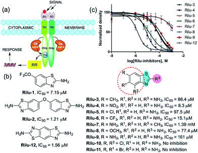 | ||
| Fig. 1 (a) TCS signaling pathway; transmembrane protein, HK and phosphoryl transfer to its cognate RR. Domains present in transmembrane HK portion; SD = sensor, TM = transmembrane, CA = catalytic and ATP-binding, DHp = dimerization and histidine phosphotransfer. His = histidine, ADP-P = adenosine triphosphate, (b) structures of compounds and their IC50 values for HK853 inhibitory activity, (c) dose–response curves of the compounds for HK inhibition (Fig. S1, Table S1†). | ||
Results and discussion
We postulated that targeting the highly-conserved catalytic ATP-binding (CA) domain within these proteins would be broadly applicable to a range of bacteria, and that simultaneous inhibition of multiple HKs within an organism would decrease its ability to quickly evolve resistance.7,9 In addition, HKs utilize a Bergerat fold to bind this substrate, which is not found in eukaryotic kinases, potentially providing a means for the generation of selective inhibitors.7,9 Recently, we reported nine lead compounds, identified from a high-throughput screen (HTS), which bind to the CA domain of multiple HKs.9 Two leads from the HTS share a common scaffold, a benzothiazole core, making this structure attractive for further study (Fig. 1b; Rilu-1 and 2).9 One of these compounds, riluzole (Rilu-1; IC50 = 7.2 μM) is an FDA approved amyotrophic lateral sclerosis drug, indicating that this chemotype has drug-like properties.10 In this work, we sought to assess the ability of these potent inhibitors to affect bacterial signaling in live cells and lay the foundation for the development of potent antivirulence agents.Rilu-1 has a trifluoromethoxy (OCF3) group at the 6-position on the ring and based on this core scaffold, we generated a small library of compounds to evaluate their biochemical potency and to provide a diversity of molecules for in cell assessment (Rilu-3–11, Fig. 1b and c). Compounds containing an electron-withdrawing group, such as –OCF3, –NO2, –CF3 (Rilu-1, 4, 6; 7–15 μM), were found to be potent inhibitors of HK autophosphorylation (HK853, Fig. 1b), while a mildly deactivating –Cl group, decreased potency (Rilu-5; 98 μM). Electron-donating groups such as –OCH3, –NH2 or even a weakly donating moiety such as –CH3 yielded ∼10-fold increase in the IC50 values (Rilu-3, 8, 9; 77–161 μM). Interestingly, functionalization with multiple deactivating groups yielded inactive compounds (Rilu-10, 11). Recently, we demonstrated that a –N–NH–N– or similar triad, such as the –S–NH2–N–, is critical for CA domain binding (highlighted in cyan and pink, Fig. 1b).11 Consistent with this, we found that the positional isomer in which the –NH2 and –CH3 groups are switched shows minimal activity (comparison of Rilu-3 and 7, IC50 = 1.4 mM). The more potent lead from the HTS, Rilu-2, contains two benzothiazole rings, likely promoting additional polar interactions within the active site. Assessment of Rilu-12, which still features two amino moieties but does so in a rigid tricyclic structure, revealed it to be equivalently potent (IC50 1.6 μM; Fig. 1b and c). From our HTS pilot screen library, ∼10 compounds containing a similar scaffold were “hits”, but several were ultimately found to be either low-binders or protein-aggregators (Fig. S2†).9 Thus, we selected four compounds (Rilu-1, 2, 4, 12) containing one or two thiazole rings that were the most potent in our biochemical assay, as well as non-aggregators (Fig. S3†),9 and investigated their effects in live cells.
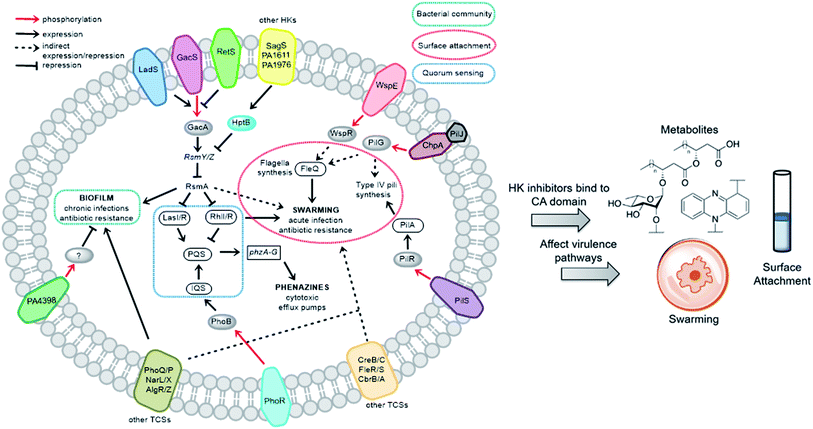
We sought to study the opportunistic Gram-negative organism P. aeruginosa, one of the most prevalent nosocomial pathogens and named on the most “critical” threats list by the World Health Organization.12 It is the leading cause of death associated with immune-compromised and CF patients. The virulent P. aeruginosa strain PA14, obtained from a burn wound, encodes a large number of TCS regulatory proteins in its genome, with >64 sensor HKs and >72 RRs.13 Among these HKs, many are intricately linked and influence virulence and antibiotic-resistance mechanisms such as biofilm formation, swarming, and toxin production (Fig. 2). These traits strongly suggest that inhibitors of these TCSs or HKs could provide viable leads for treatment of P. aeruginosa infection. Given our stated goal of identifying antivirulence agents, we first assessed Rilu-1, 2, 4, 12 for bacteriostatic and bactericidal properties in PA14 cultures. None of these compounds substantially slowed growth or killed the organism as determined both by OD620 measurements (Fig. S4†) and CFU studies (data not shown), even at high concentrations (500 μM). Given that some thiazoles have been reported to have off-target effects,14 we tested our inhibitors against a mammalian heat-shock protein (HSP90α), which possesses a similar ATP-binding Bergerat fold. None of the compounds were found to substantially inhibit this protein, even at concentrations of 1.25 mM (Fig. S5†). We have also demonstrated that Rilu-1, 2 are not cytotoxic to Vero cells at <180 μM in earlier studies.9 Recently, molecules containing a similar scaffold, 2-aminoimidazole, were reported as inhibitors of RR QseB that led to reduced biofilms in Francisella,15 further exemplifying the potential of this general class of compounds as promising antivirulence candidates.16
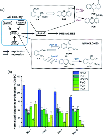
To determine if the HK inhibitors affect the pathogenicity of P. aeruginosa, we examined its production of two major classes of molecules involved in signaling and infection establishment, the Pseudomonas quinolone signals (PQSs) and the phenazines. At the TCS level, pathways such as GacS/GacA-RetS and PhoR/PhoB are associated with the formation of PQSs and phenazine metabolites via the QS system and small RNAs machinery (Fig. 2).17 However, a complete picture of the roles that the TCSs play in QS regulation is not yet available.13,17,18 Looking deeper into the QS network, production of the QS signals is tightly regulated by several circuits (Fig. 3a).18 We were interested in the PQS system as it autoregulates the production of quinolone-type compounds, which have roles in controlling other toxins and virulence behavior in Pseudomonas. When a quorum is reached, biosynthesis of 4-hydroxy-2-heptylquinoline (HHQ) and 3,4-dihydroxy-2-heptylquinoline (PQS) is initiated by the production of their precursor, anthranilic acid (AA).18,19 In another pathway, AA can also be converted to 4-hydroxy-2-heptylquinoline-N-oxide (HQNO). The release of PQS activates the phenazine-producing genes phzA-G that convert chorismic acid (CA) to the primary phenazine metabolite, phenazine-1-carboxylic acid (PCA; Fig. 3a).17 From different cues, PCA can be modified to phenazine-1-carboxamide (PCN) and to the other crucial toxin of P. aeruginosa, pyocyanin (PYO; Fig. 3a).17 Although not direct, it is evident that the expulsion of these toxic metabolites (quinolones, phenazines) is regulated by TCSs and as such, inhibiting the production of these toxins is a viable antibacterial therapy.20
To measure these metabolites, we carried out LC-MS/MS analyses on the supernatant of PA14 cultures grown in the presence or absence of inhibitors at various growth phases (OD620 = 0.9 in Fig. 3b, OD620 = 1.8 in Fig. S6, mass spectrometry data in Fig. S7†).19,21 Although the levels of HHQ were slightly increased upon treatment (Rilu-1, ∼24%), PQS production was markedly decreased (∼50%) by all inhibitors compared to the DMSO control (Fig. 3b). This result is quite exciting as null mutation of the pqs system affects other virulence factors such as PYO, elastase, and rhamnolipids (RLs).18 All of the inhibitors also significantly reduced the levels of HQNO by up to ∼50%. This is an important finding as HQNO, unlike QS-activity related quinolones,22 is an extremely crucial toxin for P. aeruginosa as it enables the bacteria to survive in hosts by aiding in extracellular DNA production and killing of other microbiota.18 In addition, up to a 70% reduction of PYO was achieved (Rilu-2), while PCN was reduced by ∼85% (Rilu-2) and finally, only negligible amounts of PCA (∼8%) were produced with Rilu-12 (Fig. 3b). These results were corroborated by dramatic reduction in the characteristic green hue of P. aeruginosa cultures, which is due to multiple metabolites, including the phenazines and the siderophore pyoverdine (24 h inhibitor treatment; Fig. S6†). The turbidity of these cultures further validates that the compounds do not impede bacterial growth and that interfering with the aforementioned TCS machinery component(s) likely causes the striking reductions in toxin production.
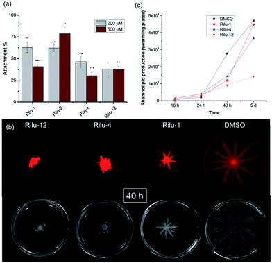
We next sought to assess the ability of our molecules to interfere with P. aeruginosa virulence mechanisms. Biofilm formation (Fig. S8†) is indisputably the best way for a microorganism to establish its cellular community and in P. aeruginosa, a complex web of many TCSs controls biofilm formation. For example, TCSs GacS/GacA, PhoQ/PhoP, and NarL/NarX positively regulate biofilm formation, but when sensor kinase PA4398 was mutated in PA14, a 1.8 fold increase in biofilm production was seen (Fig. 2).13 Using a microtitre assay, we evaluated biofilm formation of PA14 cultures and observed 25% and 40% reduction of biofilm mass with high concentrations of Rilu-2 and 4, respectively (Fig. S8†). Although the leads caused only a modest reduction in biofilm formation, deeper examination of these microbial films can provide insights into the organism's ability to attach to solid surfaces, as alterations in appendages like flagella and type IV pili can result in dramatic architectural changes. Accordingly, an important corollary analysis is assessment of the initial surface adhesion of the pathogen (Fig. 4a) that is dependent on flagella/pili formation, with a “rapid attachment assay”.8,23 A notable difference in surface attachment was observed with all of the Rilu-compounds. In particular, a 70% reduction occurred in cultures containing Rilu-4 or 12 (Fig. 4a).
Activation of HKs, such as chemotaxis-related WspE, ChpA and the nitrogen acquisition-related, PilS, has been linked to flagella and pili-synthesis and the pronounced effects of our molecules in the attachment assay implied that the Rilu-compounds may be affecting the motility of P. aeruginosa. Pseudomonas is unique in its application of versatile motility modes and among these, swarming enables the deadly pathogen to move through mucosal layers in CF patients based on nutritional and viscosity variations.24,25 The coordinated movement of bacteria using both flagella and type IV pili exhibit a greater resistance to multiple antibiotics and express higher levels of virulence-related factors compared to planktonic cultures.24 Swarming is TCS-dependent, as HKs gacS, and fleS, as well as alginate RR algR mutants are all impaired in swarming motility.13,24 We investigated if our inhibitors modify swarming behavior, as this is essential for pathogenicity. The Rilu-compounds (200 μM) dramatically affected swarming growth.26 After 40 h, a marked reduction in tendril formation is clearly observed with all compounds, most prominently with Rilu-12 (Fig. 4b, bottom images). Even after 5 days, a moderate difference in swarming can be noted with Rilu-1, 4, and a major impairment in motility is still visible with Rilu-12 (Fig. S9†). A similar reduction in swarming was also seen with 125 μM of Rilu-4, 12 (Fig. S10†), indicating the efficacy of the compounds at lower concentrations.
To reduce surface tension and swarm adequately, P. aeruginosa cells secrete RLs, amphiphilic glycolipids.25 We utilized a fluorescent lipophilic stain, Nile red that can bind to these glycolipids, and visualized the swarming growth of PA14.25,27 Similar to the phase-contrast images, the stain is concentrated at the center of the swarm colony for all of the samples at 40 h (Fig. 4b, top images). Whereas in the 5 days images, the RLs are evenly distributed throughout the swarm area (Fig. S9†). We quantified the RL levels at 16 h, 24 h, 40 h and 5 days (Fig. 4c, S9 and S11†) and found that RL production is lowest in the presence of Rilu-12 (70% reduction). The loss of swarming behavior provides ample evidence that the benzothiazole compounds heavily affect motility machinery and surfactant production.
To dig deeper into our TCS regulation postulation and to quantify genetic level changes, we performed RT-qPCR experiments. We examined the transcript levels of 14 genes that are linked to the pathogenicity, as well as the TCS machinery of P. aeruginosa. Two inhibitor concentrations (200 and 500 μM) were chosen to examine if transcription levels change in a dose-dependent manner and PA14 cultures from the OD620 experiments were used for RNA isolation for consistent results. We selected 8 genes that are QS signal-related and indirectly govern swarming and biofilm formation (colored in grey bars; Fig. 5 and S12†).28,29lasI, lasR and rhlI, rhlR encode the synthases and receptors of the QS systems, Las and Rhl, respectively. pqsA, pqsR govern PQS synthesis and response and phzA, phzB are involved in phenazine biosynthesis.30 For many of these genes namely, lasI, lasR, rhlI, rhlR, pqsA and phzA, only small transcriptional changes were observed with the Rilu-compounds (Fig. 5 and S12†). This is at odds with the substantial decrease in production of RLs in the swarm plates that we noted, which is dictated by rhl. This discrepancy is likely due to the fact that the microorganism requires more glycolipids to move/colonize on solid surfaces, while it does not need much in liquid media. To investigate this, we quantified RLs in treated PA14 liquid cultures using a phenol-orcinol assay and found no difference in the levels of glycolipids, corroborating the qPCR results (Fig. S13†). This observation indicated that transcription levels could vary greatly in Pseudomonas from liquid cultures to swarm semi-solid cultures. In fact, Deziel et al. reported vast differences in gene expression between the center and tendril tips of a P. aeruginosa swarm colony.31 On the other hand, the PQS response gene (pqsR), also known as multi-virulence factor (mvfR), showed a ∼3-fold decrease in expression with all of the Rilu-inhibitors.18 This regulation is quite significant as pqsR, the cognate receptor and co-inducer of PQS, is essential for executing PQS signal transduction, and controls synthesis of the phenazines. Thus, the lower expression of pqsR is consistent with the reduced metabolite production, such as PQS and HQNO, seen in Fig. 3b. This may affect other phenazine biosynthetic enzymes (PhzC-G or PhzMS/H) as is suggested by the dramatically altered PCA/PCN/PYO levels (Fig. 3b).17,18
Next, we examined gacA, the RR of the super-regulator TCS GacA/GacS whose expression directly upregulates expression of the small regulatory RNA RsmY, which captures the small RNA-binding regulatory protein RsmA (Fig. 2; encoded by rsmA, also quantified).32 All of these genes are colored as blue bars in Fig. 5 and S12.† The expression of gacA was dramatically reduced (up to ∼15 fold), suggesting that the sensor kinase, GacS is inhibited, which controls its cognate RR GacA levels. In turn, we expected to see reduction of RsmY, but observed the opposite trend. This outcome possibly stems from the fact that RsmY regulation is complex and while GacS positively regulates RsmY, four other HKs, namely BfiS (not shown in Fig. 2), PA1611, PA1976, and SagS repress it through the histidine phosphotransfer protein, HptB (Fig. 2).33 Similarly, rsmA transcription is not only controlled by RsmY but another small regulatory RNA, RsmZ influences its expression.33 We noted a slight up-regulation of rsmA that potentially has effects on the Las, Rhl systems. While gacA reduction is promising and supports our HK-inhibition hypothesis, full transcriptome analysis along with affinity probe-pull down experiments will be needed to provide conclusive evidence for the TCS inhibition events that are most critical to the observed phenotypic results. Finally, since we hypothesized that the reduction in swarming and surface attachment is related to motility mechanisms, we investigated the transcript levels of fleQ (flagellar regulator) and pilA (type IV pili fimbrial precursor).24,25,34 Both are colored as green bars in Fig. 5 and S12.† These genes are heavily regulated by chemotaxis-related TCSs and HKs involved with cyclic-di-GMP messenger levels. In fact, the Sintim lab recently demonstrated that a cyclic-di-GMP esterase inhibitor can interfere with swarming and RL production in PAO1.35 Other reports have connected mutants of fleQ and pilA with deficiencies in swarming and twitching motility.24,25,34 Gratifyingly, expression of both of these genes was decreased by up to ∼3.5 fold, which corroborates the significant defects observed in the swarming and surface attachment experiments.
Finally, to provide further evidence that the in vitro activity of the HK inhibitors is correlated with what is observed in the whole cell assays, we performed transcription analysis with a Rilu-analog that was inactive for HK inhibition, Rilu-7. We observed that this compound does not significantly affect the expression of virulence-associated genes (pqsR, fleQ and pilA), which were substantially downregulated by our lead molecules, Rilu-4 and 12 (Fig. S12†). We also found that although Rilu-4 and 12 caused a dramatic downregulation (12–15 fold) of gacA, the “super regulator” of virulence, the expression of this gene was essentially unchanged in the presence of Rilu-7 (1.2 fold change). These promising results further strengthen our hypothesis that the antivirulence activity that we observe with the Rilu-leads is due to inhibition of crucial HK targets and subsequent signal transduction in bacteria.
Conclusions
P. aeruginosa is a versatile organism and has an extremely intricate virulence network that is tightly controlled by TCSs.36 We have shown that by blocking TCS signaling, a remarkable attenuation in virulence behavior can be achieved. Inhibition of HKs, the kinase partner in TCSs, was carried out with benzothiazole-containing compounds. Among these, Rilu-4 and 12 are the most promising as they significantly reduce the production of virulence factors such as PQS, toxins like PYO, PCN, and PCA, and severely impact the motility behavior of PA14. Transcription analyses showcased the regulation of a crucial TCS GacS/GacA that is essential to P. aeruginosa's pathogenicity and provides evidence of other TCS signaling inhibition. Current efforts in our lab are aimed at improving the efficacy of the Rilu-inhibitors, understanding the key inhibition mechanisms, and evaluating the identified molecules in additional drug-resistant strains of P. aeruginosa.Conflicts of interest
There are no conflicts to declare.Acknowledgements
We thank H. Blackwell for providing the PA14 strain and W. Pomerantz for the use of a plate reader. This work was supported by an Alfred P. Sloan Fellowship (E. E. C.), the University of Minnesota NIH Biotechnology Training Grant (T32GM008347; A. E.), and the University of Minnesota. We thank the University of Minnesota's Supercomputing Institute for the use of various modeling and data analysis software.Notes and references
- A. Beceiro, M. Tomás and G. Bou, Clin. Microbiol. Rev., 2013, 26, 185–230 CrossRef PubMed.
- D. Beier and R. Gross, Curr. Opin. Microbiol., 2006, 9, 143–152 CrossRef PubMed.
- B. Heras, M. J. Scanlon and J. L. Martin, Br. J. Clin. Pharmacol., 2015, 79, 208–215 CrossRef PubMed.
- S. Ruer, N. Pinotsis, D. Steadman, G. Waksman and H. Remaut, Chem. Biol. Drug Des., 2015, 86, 379–399 CrossRef PubMed.
- G. E. Schaller, S.-H. Shiu and J. P. Armitage, Curr. Biol., 2011, 21, R320–R330 CrossRef PubMed.
- A. M. Stock, V. L. Robinson and P. N. Goudreau, Annu. Rev. Biochem., 2000, 69, 183–215 CrossRef PubMed.
- A. E. Bem, N. Velikova, M. T. Pellicer, P. v. Baarlen, A. Marina and J. M. Wells, ACS Chem. Biol., 2015, 10, 213–224 CrossRef PubMed.
- A. Neidig, A. T. Yeung, T. Rosay, B. Tettmann, N. Strempel, M. Rueger, O. Lesouhaitier and J. Overhage, BMC Microbiol., 2013, 13, 77 CrossRef PubMed.
- K. E. Wilke, S. Francis and E. E. Carlson, ACS Chem. Biol., 2015, 10, 328–335 CrossRef PubMed.
- A. Doble, Neurology, 1996, 47, 233S–241S CrossRef.
- M. Goswami, K. E. Wilke and E. E. Carlson, J. Med. Chem., 2017, 60, 8170–8182 CrossRef PubMed.
- Global priority list of antibiotic-resistant bacteria to guide research, discovery, and development of new antibiotics, http://www.who.int/medicines/publications/WHO-PPL-Short_Summary_25Feb-ET_NM_WHO.pdf?ua=1.
- W. J. Gooderham and R. E. W. Hancock, FEMS Microbiol. Rev., 2009, 33, 279–294 CrossRef PubMed.
- S. M. Devine, M. D. Mulcair, C. O. Debono, E. W. W. Leung, J. W. M. Nissink, S. S. Lim, I. R. Chandrashekaran, M. Vazirani, B. Mohanty, J. S. Simpson, J. B. Baell, P. J. Scammells, R. S. Norton and M. J. Scanlon, J. Med. Chem., 2015, 58, 1205–1214 CrossRef PubMed.
- M. E. Milton, C. L. Allen, E. A. Feldmann, B. G. Bobay, D. K. Jung, M. D. Stephens, R. J. Melander, K. E. Theisen, D. Zeng, R. J. Thompson, C. Melander and J. Cavanagh, Mol. Microbiol., 2017, 106, 223–235 CrossRef PubMed.
- R. J. Melander and C. Melander, Antibiotic Adjuvants, Springer Berlin Heidelberg, Berlin, Heidelberg, 2017, pp. 1–30, DOI:10.1007/7355_2017_10.
- H. Sakhtah, A. Price-Whelan and L. E. P. Dietrich, in Microbial Phenazines: Biosynthesis, Agriculture and Health, ed. S. Chincholkar and L. Thomashow, Springer Berlin Heidelberg, Berlin, Heidelberg, 2013, pp. 19–42, DOI:10.1007/978-3-642-40573-0_2.
- J. Lee and L. Zhang, Protein Cell, 2015, 6, 26–41 CrossRef PubMed.
- G. Allegretta, C. K. Maurer, J. Eberhard, D. Maura, R. W. Hartmann, L. Rahme and M. Empting, Front Microbiol., 2017, 8, 924 CrossRef PubMed.
- G. W. Lau, D. J. Hassett, H. Ran and F. Kong, Trends Mol. Med., 2004, 10, 599–606 CrossRef PubMed.
- B. R. Lundgren, W. Thornton, M. H. Dornan, L. R. Villegas-Peñaranda, C. N. Boddy and C. T. Nomura, J. Bacteriol., 2013, 195, 2087–2100 CrossRef PubMed.
- R. Hazan, Y. A. Que, D. Maura, B. Strobel, P. A. Majcherczyk, L. R. Hopper, D. J. Wilbur, T. N. Hreha, B. Barquera and L. G. Rahme, Curr. Biol., 2016, 26, 195–206 CrossRef PubMed.
- A. Siryaporn, S. L. Kuchma, G. A. O'Toole and Z. Gitai, Proc. Natl. Acad. Sci. U. S. A., 2014, 111, 16860–16865 CrossRef PubMed.
- N. Verstraeten, K. Braeken, B. Debkumari, M. Fauvart, J. Fransaer, J. Vermant and J. Michiels, Trends Microbiol., 2008, 16, 496–506 CrossRef PubMed.
- J. D. Morris, J. L. Hewitt, L. G. Wolfe, N. G. Kamatkar, S. M. Chapman, J. M. Diener, A. J. Courtney, W. M. Leevy and J. D. Shrout, Appl. Environ. Microbiol., 2011, 77, 8310–8317 CrossRef PubMed.
- J. Tremblay and E. Déziel, J. Basic Microbiol., 2008, 48, 509–515 CrossRef PubMed.
- H. Du, Z. Xu, M. Anyan, O. Kim, W. M. Leevy, J. D. Shrout and M. Alber, Biophys. J., 2012, 103, 601–609 CrossRef PubMed.
- M. A. Welsh and H. E. Blackwell, Cell Chem. Biol., 2016, 23, 361–369 CrossRef PubMed.
- S. L. McKnight, B. H. Iglewski and E. C. Pesci, J. Bacteriol., 2000, 182, 2702–2708 CrossRef PubMed.
- M. A. Welsh, N. R. Eibergen, J. D. Moore and H. E. Blackwell, J. Am. Chem. Soc., 2015, 137, 1510–1519 CrossRef PubMed.
- J. Tremblay and E. Déziel, BMC Genomics, 2010, 11, 587 CrossRef PubMed.
- T. Rasamiravaka, Q. Labtani, P. Duez and M. El Jaziri, BioMed Res. Int., 2015, 2015, 17 CrossRef PubMed.
- L. E. P. Dietrich, A. Price-Whelan, A. Petersen, M. Whiteley and D. K. Newman, Mol. Microbiol., 2006, 61, 1308–1321 CrossRef PubMed.
- T. S. Murray and B. I. Kazmierczak, J. Bacteriol., 2008, 190, 2700–2708 CrossRef PubMed.
- Y. Zheng, G. Tsuji, C. Opoku-Temeng and H. O. Sintim, Chem. Sci., 2016, 7, 6238–6244 RSC.
- D. Balasubramanian, L. Schneper, H. Kumari and K. Mathee, Nucleic Acids Res., 2013, 41, 1–20 CrossRef PubMed.
Footnote |
| † Electronic supplementary information (ESI) available. See DOI: 10.1039/c8sc02496k |
| This journal is © The Royal Society of Chemistry 2018 |

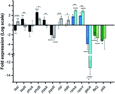
![[thin space (1/6-em)]](https://www.rsc.org/images/entities/char_2009.gif) 2 scale. Genes are colored based on three classes: QS-related (grey), TCS GacSA-related (blue) and motility-related (green). Darker and lighter bars (grey, blue and green) are measurements for Rilu-12 at 200 μM and 500 μM, respectively. Error bars represent the standard error of three independent experiments (
2 scale. Genes are colored based on three classes: QS-related (grey), TCS GacSA-related (blue) and motility-related (green). Darker and lighter bars (grey, blue and green) are measurements for Rilu-12 at 200 μM and 500 μM, respectively. Error bars represent the standard error of three independent experiments (