
High resolution multielement XRF spectroscopy of extended and diffused sources with a graphite mosaic crystal based Von Hamos spectrometer
A.
Scordo
 *a,
L.
Breschi
bca,
C.
Curceanu
a,
M.
Miliucci
a,
F.
Sirghi
ad and
J.
Zmeskal
e
*a,
L.
Breschi
bca,
C.
Curceanu
a,
M.
Miliucci
a,
F.
Sirghi
ad and
J.
Zmeskal
e
aLaboratori Nazionali di Frascati INFN, Via E. Fermi 40, 00044 Frascati, Rome, Italy. E-mail: alessandro.scordo@lnf.infn.it
bFondazione Policlinico Universitario A. Gemelli IRCCS, Rome, Italy
cUniversità Cattolica del Sacro Cuore, Rome, Italy
dHoria Hulubei National Institute of Physics and Nuclear Engineering (IFIN-HH), Magurele, Romania
eStefan-Meyer-Institut für Subatomare Physik, Vienna, Austria
First published on 5th November 2019
Abstract
Bragg spectroscopy is a consolidated experimental method for high energy resolution X-ray measurements. However, this technique is limited to the detection of photons produced from point-like or well collimated sources and becomes quite inefficient for photons generated in extended and diffused ones; also, the possibility to perform simultaneous measurements of several X-ray lines is of great benefit when low rate signals are expected and individual angular scans require long exposure times. We present a prototype of a high resolution Highly Annealed Pyrolytic Graphite (HAPG) mosaic crystals based X-ray spectrometer, developed by the VOXES (high resolution VOn hamos X-ray spectrometer using HAPG for extended sources) collaboration at INFN Laboratories of Frascati, able to work with extended (millimetric) and isotropic sources. The spectrometer has been tested in the energy range 6–19 keV, with the aim to deliver a cost effective system having an energy resolution (FWHM) at the level of few eV, able to perform sub-eV precision measurements of single and multielement targets in a broad energy range. In this paper, the working principle of the VOXES spectrometer is presented, including a detailed description of the geometry, of the calibration methods and of the spectral fitting functions. Results of X-ray tracing simulations are compared to the experiment. The main results achieved by our spectrometer regard the peak resolutions. In particular, we report values as low as 4.02 ± 0.08 eV and 3.60 ± 0.05 eV for the Fe(Kα1,2) and Cu(Kα1,2) lines; for larger dynamic ranges, we report single shot spectra with Cu(Kα1,2) Ni(Kβ) and Zn(Kα1,2) lines with best resolution values of 5.15 ± 0.13 eV, 6.02 ± 0.24 eV and 6.20 ± 0.34 eV, respectively. Finally, we explored the capabilities of our spectrometer for energies above 15 keV, obtaining for Mo(Kα1,2) and Nb(Kβ) lines resolution values of 19.44 ± 0.75 eV and 39.90 ± 1.48 eV, respectively. The proposed spectrometer has possible applications in several fields, going from fundamental physics, synchrotron radiation and X-FEL applications, medicine and industry to hadronic physics experiments, where its performances make it a fundamental tool for a series of measurements like the energies of kaonic atoms transitions, allowing to extract fundamental parameters in the low energy QCD in the strangeness sector.
1 Introduction
The possibility to perform high precision measurements of soft X-rays, strongly required in many fields of fundamental science, from particle and nuclear physics to quantum mechanics tests, as well as in astronomy and in several applications using synchrotron light sources or X-FEL beams, in biology, medicine and industry, is still a big challenge. Typical large area spectroscopic detectors used for wide and isotropic targets are solid state devices, like the Silicon Drift Detectors (SDDs), employed for the first time as spectroscopic devices by the SIDDHARTA experiment1,2 for exotic atoms transition lines measurements at the DAΦNE e+e− collider of the INFN National Laboratories of Frascati.3 The intrinsic resolution of such kind of detectors is nevertheless limited to ≃120 eV FWHM by the Fano Factor, making them unsuitable for those cases in which the photon energy has to be measured with a precision below 1 eV.4Few eV resolutions are achievable using superconducting microcalorimeters, like the Transition Edge Sensors developed at NIST, able to obtain few eV FHWM at 6 keV;5 in spite of this excellent resolution, these kind of detectors still have some limitations: a very small active area, prohibitively high costs of the complex cryogenic system needed to reach the operational temperature of ≃50 mK, and a response function which is not fully understood.5
As a third possibility, Bragg spectroscopy is one of the best established high resolution X-ray measurement technique; however, when the photons emitted from extended sources (like a gaseous or liquid target) need to be measured, this method has been ruled out by the constraint to reduce the dimension of the target to a few tens of microns.6,7 Recently, the development of the Pyrolytic Graphite mosaic crystals,8 renewed the interest on Bragg spectrometers not only for X-ray Absorption Fine Structure (XAFS) spectroscopy (see ref. 9 for example), but also as possible candidates for millimetric isotropic sources X-ray Fluoresce (XRF) and X-ray Emission (XES) spectroscopy.
Mosaic crystals consist of a large number of nearly perfect small pyrolytic graphite crystallites, randomly misoriented around the lattice main direction. The FWHM of this random angular distribution is called mosaicity (ω) and it allows that even a photon not reaching the crystal with the exact Bragg energy-angle relation, can find a properly oriented crystallite and be reflected.10 This, together with a lattice spacing constant d002 = 3.514 Å, enables them to be highly efficient in diffraction in the 2–20 keV energy range, for the n = 1 reflection order, while even higher energies can be reached at higher reflection orders.
Thanks to their production mechanism, Highly Annealed Pyrolytic Graphite crystals (HAPG) can be realised with different ad-hoc geometries, making them suitable to be used in the Von Hamos configuration,11 combining the standard dispersion of a flat crystal with the focusing properties of cilindrically bent crystals.
The main issue to overcome is represented by the source size. Von Hamos spectrometers have been extensively used in the past years providing very promising results in terms of spectral resolution6,12–14 but all the measurements were done in conditions where the effective source dimensions were of some tens of microns. This configuration was achieved either with microfocused X-ray tubes, or with a set of slits and collimators in front of the target to minimize the activated area.
The possibility to use a mosaic crystal based Von Hamos spectrometer for isotropic sources of millimetric dimensions has been investigated by the VOXES collaboration at the INFN Laboratories of Frascati (LNF), where in particular the influence of the mosaic spread and of the thickness of the crystal on the obtainable resolutions have been studied.15–17
In this work we report the possibility to combine HAPG crystals properties with the vertical focus of the Von Hamos configuration, to realize a spectrometer able to maintain a resolution in the order of 0.1% (FWHM/E), for energies below 10 keV, and of 0.3% up to 20 keV, using a source size ranging from 500 μm to 2 mm in the Bragg dispersion plane. We also explore the possibility to exploit the “semi” – Von Hamos configuration (see Section 3.3) to enlarge the dynamic range of the spectrometer, allowing for a simultaneous detection of different lines in a ≃1 keV energy range. In Section 2 we present the experimental setup, the beam geometry in both the dispersive and sagittal planes and the used instrumentation, while in Section 3 the calibration procedure, the fitting function, the spectra and the obtained results are reported. In Section 4 we present the ray tracing simulations of our setup and compare them with the measurements.
2 Experimental setup
2.1 Von Hamos (VH) geometry
The spectrometer configuration used in the measurements presented in this work is the Von Hamos (VH) one, in which the X-ray source and the position detector are placed on the axis of a cylindrically bent crystal (see Fig. 1). This geometrical scheme allows an improvement in the reflection efficiency due to the sagittal focusing.11 For each X-ray energy the source-crystal (L1) and the source-detector (2L2) distances are then determined by the Bragg angle (θB) and the curvature radius of the crystal (ρc): | (1) |
L2 = L1![[thin space (1/6-em)]](https://www.rsc.org/images/entities/char_2009.gif) sinϕ sinϕ | (2) |
 | ||
| Fig. 1 VH schematic geometry of the dispersive plane (not in scale): the X-ray source is pictured in orange, the HAPG crystal in red and the position detector in blue and green for the standard and the SVH configuration (see Section 3.3), respectively. ρc is the crystal curvature radius, θB is the Bragg angle, ϕ = π − θB, θM is the position detector rotation angle with respect to standard VH configuration, L1 is the source-crystal distance and L2 is half the resulting source-detector one. | ||
In Fig. 1, a schematic of the dispersive plane is shown where the X-ray source (target) is sketched in orange, the HAPG crystal in red and the position detector in blue and green for the standard and the “semi” VH configuration (SVH, see Section 3.3), respectively. In the figure, ρc is the crystal curvature radius, θB is the Bragg angle, ϕ = π − θB, L1 is the source-crystal distance and L2 is half of the resulting source-detector one.
The HAPG crystals used for the measurements, all having a thickness of 100 μm, have curvature radius and mosaicity (ω) values of 206.7 mm (ω = 0.1°), 103.4 mm (ω = 0.11°) and 77.5 mm (ω = 0.8°). They are deposited, by the OPTIGRAPH GmbH producer,18 on Thorlabs N-BK7 30 × 32 mm2 uncoated plano-concave cylindrical lenses, held by a motorized mirror mount (STANDA 8MUP21-2), able to rotate around the cylinder axis and its orthogonal (Y axis in Fig. 5) with <1 arcsec resolution, and coupled to a 0.08 μm full-step resolution motorized vertical translation stage (STANDA 8MVT40-13), a 0.01° full-step resolution rotation stage (STANDA 8MR191-28) and two motorized 1.25 μm full-step resolution linear translation stages (STANDA 8MT167-25LS).
The position detector is a commercial MYTHEN2-R-1D 640 channels strip detector produced by DECTRIS (Zurich, Switzerland), having an active area of 32 × 8 mm2 and whose strip pitch and thickness are, respectively, 50 μm and 420 μm. The MYTHEN2-1D detector is also coupled to a positioning motorized system identical to that of the HAPG holder. Finally, a cooling Peltier Cell is kept on top of the strip detector in order to stabilize its temperature in the working range of 18–28 °C. The resulting 10-axis motorized positioning system is mounted on a set of Drylin rails and carriers to ensure a better stability and alignment and, in addition, to easily adjust source-crystal-detector positions for each energy to be measured.
The size of the target portion illuminated by the X-ray tube can be calculated starting from the initial anode position h = 95.3 mm and the half angular conical aperture α = 11° of the tube (see Fig. 2).
 | ||
| Fig. 2 Drawing of the YZ projection of the 45° target holder and its corresponding illuminated portion (not in scale). See text for more details. | ||
The intersection of the conical beam and the prism, rotated at 45°, is resulting to be ellipsoidal with axes:
| ρx = h × tgα = 95.3 mm × tg(11°) = 18.5 mm | (3) |
 | (4) |
 | (5) |
Referring to Fig. 3, this configuration is obtained setting the position (z1 and z2) and the aperture (S1 and S2) of each slit such as to create a virtual source between the two slits (zf, green solid lines on Fig. 3), an angular acceptance Δθ′, and an effective source  (green) which can be set to millimetric dimensions. The Δθ′ angular acceptance can also be set to any value, provided it is large enough to ensure that all the θB corresponding to the lines to be measured are included.
 | ||
Fig. 3 Beam geometry on the dispersive plane (not in scale): the position (z1 and z2) and the aperture (S1 and S2) of two slits are used to create a virtual source between the two slits (zf, green solid lines), and effective source  (green) and an angular acceptance Δθ′. The HAPG crystal is pictured in red, the two slits are shown in black while zh and Sh are the position and the diameter of the circular exit window of the aluminum box front panel (light blue), respectively. (green) and an angular acceptance Δθ′. The HAPG crystal is pictured in red, the two slits are shown in black while zh and Sh are the position and the diameter of the circular exit window of the aluminum box front panel (light blue), respectively. | ||
There is also a second component of the X-ray beam, depicted as a dotted green line in Fig. 3, due to the X-rays emitted in the inner part of the target (S0) with an angular divergence Δθ ≤ Δθ′. In Fig. 3, zh and Sh are the position and the diameter of the circular exit window of the aluminum box front panel, respectively.
The increase of the effective source size introduces a background, as shown in Fig. 4. For simplicity we describe this situation only for the dotted green lines scheme of Fig. 3 but it applies also in the solid line case.
 | ||
| Fig. 4 Signal and background scheme (not in scale): two slits S1 and S2 are used to shape the X-ray beam divergence (Δθ) and effective source size (S0). Depending on the HAPG mosaic spread, photons emitted parallel (solid lines) and not (dashed lines) to the nominal ones matching the Bragg condition (yellow) are shown. Some of them are reflected under the signal peak (solid and dashed green), some are source of background (solid red) and some are not reflected (dashed red). See the text for more details. | ||
The yellow lines on the figure represent the photons which, matching the Bragg condition (θB, λB), form the signal peak on the reflected Bragg spectrum (we call them nominal from now on). Since the photons are isotropically emitted from the whole target foil, some of them may have the correct energy and angle to be reflected but originate from a point of the target near the nominal one (on the yellow line). As far as this mislocation is below the limit given by the mosaic spread of the crystal, such photons are also reflected under the signal peak worsening the spectral resolution (solid green lines in the figure). On the contrary, when this mislocation exceeds this limit (solid red line) these photons are reflected outside the signal peak. In the same way, photons not emitted in parallel to the nominal ones may still impinge on the HAPG crystals with an angle below its mosaic spread (dashed green lines) and be then reflected under the signal peak also affecting the spectral resolution. On the contrary, if the impinging angle is out of this limit, those photons are not reflected on the position detector. The goal of the VOXES spectrometer is to verify, for each energy, if there is the possibility to find the right slits configuration leading at the maximum source size keeping the resolution below the desired limit.
For each chosen Δθ′,  pair, the corresponding values of the slits apertures S1 and S2 can be found. First, we define the position of the intersection point zf:
 | (6) |
Then, the two slits apertures and the vertical illuminated region of the HAPG are defined by:
 | (7) |
 | (8) |
Concerning the second component, the Δθ and S0 parameters can be obtained from z1, S1 and z2, S2 as:
 | (9) |
 | (10) |
Since both positions of the various components of the setup (slits, crystal and position detector) with respect to the target are not known with infinite precision, it is useful to derive from the above formula also the errors on the most important parameters:
 | (11) |
 | (12) |
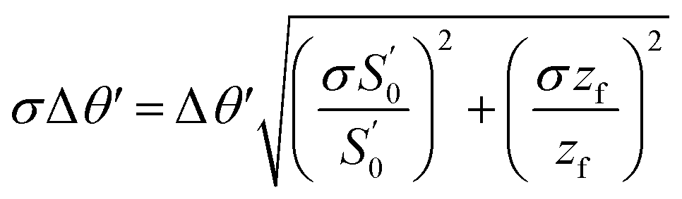 | (13) |
 | ||
| Fig. 5 Beam geometry on the focusing plane. The vertical spread of the X-ray beam is fixed by the slits positions (z1, z2) and their frame size (As), together with the exit circular hole in the front panel of the setup box (zh, Ah). See text for more details. | ||
The ϕ angle is then defined as:
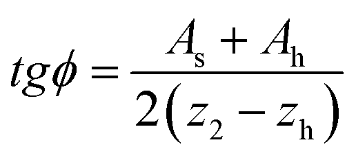 | (14) |
We first define, as in the horizontal case, the position of the intersection point zf:
 | (15) |
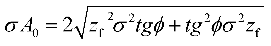
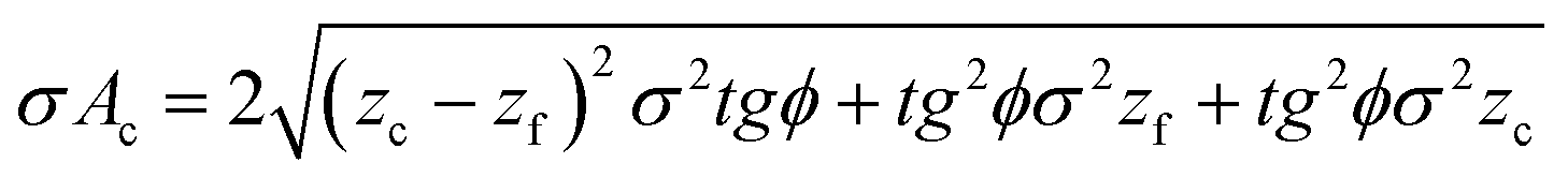
The vertical spread of the beam on the target (A0) and on the HAPG crystal (Ac) are then:
| A0 = 2zftgϕ | (16) |
| Ac = 2(zc − zf)tgϕ | (17) |
The final effective source area is then determined by 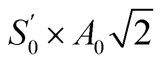 .
.
Following the same criteria of Section 2.1.2, we define the errors of the sagittal plane fundamental parameters:
 | (18) |
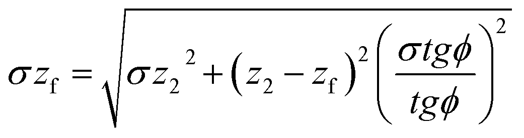 | (19) |
 | (20) |
 | (21) |
An eventual small roughness of the target surface would create fluctuations of the photon starting position in the Z direction; however, in the VH configuration the Z starting position may only influence the vertical focusing properties of the spectrometer. Since a strip detector is used, this would only cause a deviation on the arrival position along the same strip which is not detectable.
3 Experimental results
In this section we present the spectra obtained for various elements (see Table 1). For each measurement, the radius of curvature of the used crystal and the slits positions are listed in Table 2. The crystal curvature radius is chosen in order to make a compromise between the energy resolution and the signal rate, since higher ρc values lead to longer paths meaning better resolution but higher X-ray absorption from the air. Slits positions z1 and z2 are chosen such as to have a vertical dispersion at the HAPG position (Ac) smaller than the crystal size (30 mm).| Line | E (eV) | θ B (°) |
|---|---|---|
| Fe(Kα1) | 6403.84 | 16.77 |
| Fe(Kα2) | 6390.84 | 16.81 |
| Cu(Kα1) | 8047.78 | 13.28 |
| Cu(Kα2) | 8027.83 | 13.31 |
| Ni(Kβ) | 8264.66 | 12.92 |
| Zn(Kα1) | 8638.86 | 12.35 |
| Zn(Kα2) | 8615.78 | 12.39 |
| Mo(Kα1) | 17479.34 |
6.07 |
| Mo(Kα2) | 17374.30 |
6.11 |
| Nb(Kβ) | 18622.50 |
5.70 |
| Target | ρ c (mm) | z 1 (mm) | z 2 (mm) | L 1 (mm) |
|---|---|---|---|---|
| Fe | 77.5 | 60 | 107 | 268.68 |
| Fe | 103.4 | 76 | 257 | 358.81 |
| Fe | 206.7 | 60 | 650 | 716.58 |
| Cu | 77.5 | 60 | 289 | 337.65 |
| Cu | 103.4 | 75 | 352 | 450.49 |
| Cu | 206.7 | 60 | 820 | 900.54 |
| CuNiZn | 103.4 | 65 | 416 | 463.07 |
| MoNb | 77.5 | 99 | 650 | 733.35 |
3.1 Data analysis
In this section, we present more in details the most important aspects of the data analysis procedure. First, the method used for the fitting model selection is presented, then the choice of a simple Gaussian as fitting function for our spectra is motivated, and finally the calibration procedure, used to obtain energy spectra from position ones, is illustrated. The whole analysis is performed using the 6.12 version of the ROOT framework developed by R. Brun and F. Rademakers at CERN.19 In particular, the fits are performed using the MINUIT function minimizations20 implemented in the ROOT TMinuit class, which makes use of χ2 minimization. | (22) |
 is the sum of the residuals between the measured xi value and the expected yi value resulting from the fit. When sample size N is small compared to the number of parameters, (
 , that is our case), the use of a second-order corrected AIC (AICc) is recommended:22
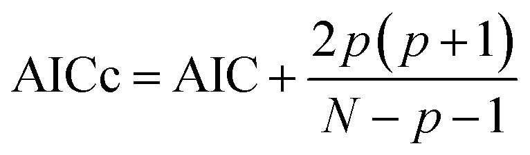 | (23) |
Given a set of candidate models for the data, the preferred model is the one with the minimum AICc value. Thus, AICc rewards goodness of fit (residuals are directly connected to the χ2 function), but it also includes a penalty that is an increasing function of the number of estimated parameters. The penalty discourages overfitting, because increasing the number of parameters in the model almost always improves the goodness of the fit. To apply AIC in practice to a dataset, for each model i the quantity e−0.5(AICcmin−AICi) is proportional to the probability of the i-th model to minimize the (estimated) information loss as good as the minimum AICc one.
The usage of sources with dimension ranging between few hundreds of microns and 2 millimeters require, instead, a more careful investigation on the effect of the background on the final measured lineshape; also, some distorsion from the pure Lorentzian shape may be induced by the mosaic spread of the crystals. We performed such a detailed investigation by fitting our spectra with either a Gaussian, a Lorentzian or a voigt function, this latter being a convolution of the former two of the form
 | (24) |
In this section we present, as an example, the results obtained on one of the spectra shown later in this work (see Section 3.2).
In Fig. 6, the MYTHEN2-1D recorded spectra, uncalibrated, are fitted with gauss, Lorentz or voigt functions. In each pad, the overall fitting function (red), the polynomial background (blue) and the Kα1 (green) and Kα2 (violet) components are overimposed to the spectrum. In the text boxes, the mean value of the two peaks, the individual or common σ and Γ are reported, together with the obtained reduced χ2 and the AICc values. Except for the plot in the lower pad, the spectra are fitted leaving the two individual linewidths as separate parameters. From these fits, even if the resulting parameters are compatible within the errors, one can immediately see how the pure Lorentzian functions are not reproducing the data as well as, on the contrary, both the gauss and voigt functions do. So, the wider source dimension and the mosaic spread are inducing a Gaussian broadening of the peaks with respect to their natural Lorentzian shape. This is also confirmed by our ray tracing simulations presented in Section 4.
 | ||
| Fig. 6 CuK(α1,2) MYTHEN2-1D recorded spectra fitted with Lorentz, voigt, Gaussian or Gaussian with common σ functions. In each pad, the overall fitting function (red), the polynomial background (blue) and the Kα1 (green) and Kα2 (violet) components are overimposed to the spectrum. In the text boxes, the mean value of the two peaks, the individual or common σ and Γ are reported, together with the obtained reduced χ2 and the AICc values. | ||
Comparing the voigtian and the Gaussian fit, the small improvement in the χ2 value is compensated by a small difference in the AICc values, suggesting that the Gaussian model is minimizing the information loss. The same criteria has been applied for the investigation of the possibility to use a common σ parameter for the two lines, because even if their natural linewidths could be a bit different, this effect is overwhelmed by this background induced broadening of the lines, as also confirmed by ray tracing simulations (see Section 4). The larger difference in AICc values between the common and individual σ case, implies an information loss when using the first one; however, as clearly visible from the parameter values, this information loss is not influencing the mean positions and their errors, which represents the most important parameters to be studied in this work. On the contrary, the possibility to use a common σ for the two peaks turns out to be very useful for spectra in which the background induced broadening prevent the clear separation between the Kα1 and Kα2 lines and a fit with individual σs as free parameters becomes unreliable.
These differences between the single and double σ gauss and voigt fits in terms of mean positions, triggered our decision to fit all the reported spectra with common σ Gaussian functions, which provide a more direct comparison in terms of peak resolutions between different measurements taken in various beam conditions.
g(θB) (see Fig. 1), and the energy-position relation can be then inferred.
The situation for our spectrometer is instead depicted in Fig. 7 where, for a nominal and a generic photon (see Section 2.1.2, Fig. 4), the different paths and the hit positions on MYTHEN2-1D (SrN and Sr, respectively) are shown.
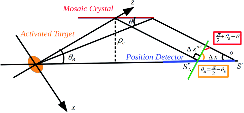 | ||
| Fig. 7 Schematic of the spectrometer parameters to be taken into account for the energy-position calibration (not in scale). The activated target, the mosaic crystal and the position detector, together with the main geometrical parameters are shown. | ||
With respect to Fig. 7, we want to calculate the difference in the arrival points of the rotated MYTHEN2-1D detector between a general photon and a nominal one, assuming both of them are reflected from a microcrystallite following the Bragg rule λ = 2dsinθ.
First, we calculate this relative distance in the standard VH configuration, assuming the center of the activated target on the cylinder axis as origin. On this axis, the arrival point of the nominal photon is SrN = 2ρc × cotgθB; on the contrary, the arrival point of a generic photon is Sr = 2ρc × cotgθ. The difference is then given by
| Δx = 2ρc(cotgθB − cotgθ) | (25) |
Now we want to calculate this distance on the position detector when this latter one is rotated by  with respect to the crystal axis. From the law of sines, applied to the Δx and Δxrot of Fig. 7, it follows that
 | (26) |
This formula is exact in the limit of source, crystal and strip detector X, Z positions known with infinite precisions and corresponding to the nominal values obtainable from the equations of Section 3. Also, the MYTHEN2-1D recorded spectra are in strip numbers while the curvature radius is in mm. To take into account this conversion, some possible small misalignments (uncertainties in the motorized positioners and rotators) and an offset due to the edge of the MYTHEN2-1D active area, the spectra can be calibrated with a parametrical function of the form:
 | (27) |
To test this calibration procedure, we applied it to our CuNiZn spectra (see Table 1) for wich, according to the Bragg rule, a broad angular acceptance of Δθ′ ≥ 0.96° is needed to include all the transition lines.
Based on the low  ratio for all the measurements, we also investigated the possibility to calibrate the spectra with a second order polynomial, directly using the known transition energy values28 in Fig. 8 we report, as an example, the results for a CuNiZn spectrum obtained in the beam conditions with
  and Δθ′ = 0.27°. The fitted MYTHEN2-1D recorded spectra and the calibration plots corresponding to eqn (27) and to the 2nd order polynomial fits are shown in the top, mid and low pad respectively. The spectrum is fitted using Gaussian peaks plus a polynomial background, according to the outcomes of Section 3.1.2.
 | ||
Fig. 8 Calibration details for a CuNiZn spectrum obtained in the beam conditions with  and Δθ′ = 0.27°. The fitted MYTHEN2-1D recorded spectra and the calibration plots corresponding to eqn (27) and to the 2nd order polynomial fits are shown in the top, mid and low pad respectively. and Δθ′ = 0.27°. The fitted MYTHEN2-1D recorded spectra and the calibration plots corresponding to eqn (27) and to the 2nd order polynomial fits are shown in the top, mid and low pad respectively. | ||
Based on considerations similar to the one used for the fitting function selection (see Section 3.1.2), one can see how the differences in the χ2 and AICc values imply some information loss when moving from using eqn (27) to a second order polynomial; however, this information loss is not influencing the mean values of the peaks, which are the only relevant parameters for the calibration procedure. In both cases, the resulting parameters are then used to obtain the final energy spectrum. We performed this analysis for all the measurements subject of this work, and in Table 3 we summarize the obtained peak positions values obtained using eqn (27) or a 2-nd order polynomial calibration function. For each target we only report, as an example, the results corresponding to the  , Δθ′ pair minimizing the errors on the peak positions.
| Line | ρ c (mm) | Eqn (27) (eV) | 2-nd order polynomial (eV) |
|---|---|---|---|
| Fe(Kα1) | 103.4 | 6403.83 ± 0.09 | 6403.80 ± 0.09 |
| Fe(Kα2) | 103.4 | 6390.78 ± 0.13 | 6390.75 ± 0.13 |
| Cu(Kα1) | 206.7 | 8047.79 ± 0.04 | 8046.79 ± 0.04 |
| Cu(Kα2) | 206.7 | 8027.83 ± 0.07 | 8027.76 ± 0.07 |
| Cu(Kα1) | 103.4 | 8047.83 ± 0.10 | 8047.83 ± 0.10 |
| Cu(Kα2) | 103.4 | 8027.53 ± 0.21 | 8027.49 ± 0.21 |
| Ni(Kβ) | 103.4 | 8264.67 ± 0.13 | 8264.67 ± 0.13 |
| Zn(Kα1) | 103.4 | 8639.13 ± 0.26 | 8639.15 ± 0.26 |
| Zn(Kα2) | 103.4 | 8615.48 ± 0.42 | 8615.42 ± 0.42 |
| Mo(Kα1) | 77.5 | 17480.02 ± 0.78 |
17479.49 ± 0.78 |
| Mo(Kα2) | 77.5 | 17372.32 ± 1.45 |
17374.04 ± 1.45 |
| Nb(Kβ) | 77.5 | 18622.34 ± 1.27 |
18622.53 ± 1.27 |
From Table 3, one can immediately see how the small differences in θB of the measured lines allow to calibrate MYTHEN2 raw spectra (in strip numbers) either using eqn (27) or a 2nd order polynomial function which, based on the very small C coefficient, can be also approximated with a linear function, especially for single element measurements. In the following sections, we present only energy calibrated spectra, according to what described above.
3.2 Single element Cu and Fe spectra
In this section we present the results of the single element measurements performed on the copper and iron Kα lines, listed in Table 2. For each measurement, the spectra have been acquired with different , ρc and Δθ′ settings but with a fixed integration time (1 hour). In Fig. 9 and 10 we show one fitted spectrum per measurement corresponding to the best achieved resolution.
 | ||
| Fig. 9 Fitted spectrum of Fe(Kα1,2) lines: Kα1, Kα2, polynomial background and total fitting function correspond to the green, violet, blue and red curves, respectively. | ||
 | ||
| Fig. 10 Fitted spectrum of Cu(Kα1,2) lines: Kα1, Kα2, polynomial background and total fitting function correspond to the green, violet, blue and red curves, respectively. | ||
As introduced in Section 3.1.2, the individual peak fitting functions are Gaussians with a common σ parameter for Kα1,2. Kα1 and Kα2 correspond to the green and violet curves, respectively and are, together with a polynomial background (blue) and the overall fit (red), overimposed to the calibrated spectrum. The values of the  , Δθ′ pair, the corresponding S0, Δθ pair and the curvature radius ρc of the used crystal are also reported in the figures.
The effective source sizes  these spectra are acquired with, are almost one order of magnitude bigger than those presented in Section 1.
In Fig. 11 we show the results from the copper target measurements done with a ρc = 206.7 mm curvature radius crystal. In the upper corner, the peak resolutions (σ) resulting from the Gaussian fits are presented, while in the middle and lower ones the precision obtained on the Kα1,2 peak positions is reported. The different colors refer to the scan of Δθ′ values for a fixes  (upper panel insight). For each point, the integration time is 1 hour and the errors are calculated according to the equations given in Sections 2.1.2 and 2.1.3.
 | ||
Fig. 11 Results from the copper target measurements done with a ρc = 206.7 mm curvature radius crystal. In the upper corner, the peak resolutions (σ) resulting from the Gaussian fits are presented, while in the middle and lower ones the precision obtained on the Kα1,2 peak positions can be seen. The different colors refer to the scan of Δθ′ values for a fixes  (upper panel insight). For each point, the integration time is 1 hour and the errors are calculated according to the equations given in Sections 2.1.2 and 2.1.3. (upper panel insight). For each point, the integration time is 1 hour and the errors are calculated according to the equations given in Sections 2.1.2 and 2.1.3. | ||
These plots show how, for a given crystal and for a given integration time, an optimal  , Δθ′ pair can be found which maximizes the peak precision. This is a very important result because it allows to set the beam geometry in the best configuration in any possible application. The presence of a minimum in the plots, despite of a constantly decreasing σ, is due to an increase in the number of events (N) for bigger Δθ′. In a pure Gaussian limits, the statistical error on the mean parameter is indeed
 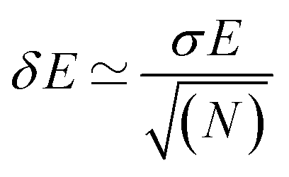 . The decreasing of σ for larger Δθ′, which is not intuitive, is explained by the parallel decrease of the S0 (not
  , see Fig. 4) values: photons coming from the central part of the target have a wider angular acceptance and their contribute to the background, as shown in Fig. 4, is higher than that of the periferical one. This explains why lower values of S0 lead to better resolutions.
The same scan has been performed for other targets and ρc values, and the resulting  , Δθ′ pairs giving the best resolutions and precisions are summarized in Table 4.
| Element | ρ c (mm) | Parameter | Value (eV) |
|
|---|---|---|---|---|
| Fe | 77.5 | σ(Kα1,2) | 4.17 ± 0.16 | 0.3/0.24 |
| δ(Kα1) | 0.11 | 0.6/0.44 | ||
| δ(Kα2) | 0.18 | 0.6/0.44 | ||
| 103.4 | σ(Kα1,2) | 4.05 ± 0.13 | 0.3/0.18 | |
| δ(Kα1) | 0.09 | 0.7/0.34 | ||
| δ(Kα2) | 0.13 | 0.7/0.34 | ||
| 206.7 | σ(Kα1,2) | 4.02 ± 0.08 | 1.1/0.60 | |
| δ(Kα1) | 0.1 | 1.2/0.70 | ||
| δ(Kα2) | 0.15 | 1.2/0.70 | ||
| Cu | 77.5 | σ(Kα1,2) | 6.8 ± 0.07 | 0.3/0.16 |
| δ(Kα1) | 0.07 | 0.6/0.32 | ||
| δ(Kα2) | 0.1 | 0.6/0.32 | ||
| 103.4 | σ(Kα1,2) | 4.77 ± 0.05 | 0.3/0.16 | |
| δ(Kα1) | 0.04 | 0.7/0.32 | ||
| δ(Kα2) | 0.07 | 0.7/0.32 | ||
| 206.7 | σ(Kα1,2) | 3.60 ± 0.05 | 0.8/0.60 | |
| δ(Kα1) | 0.04 | 1.1/0.70 | ||
| δ(Kα2) | 0.07 | 1.1/0.70 | ||
| Cu | 103.4 | σ(Kα1,2) | 5.15 ± 0.13 | 0.5/0.27 |
| δ(Kα1) | 0.10 | 0.6/0.22 | ||
| δ(Kα2) | 0.21 | 0.6/0.22 | ||
| Ni | 103.4 | σ(Kβ) | 6.02 ± 0.24 | 0.5/0.27 |
| δ(Kβ) | 0.13 | 0.6/0.22 | ||
| Zn | 103.4 | σ(Kα1,2) | 6.20 ± 0.34 | 0.5/0.27 |
| δ(Kα1) | 0.26 | 0.6/0.22 | ||
| δ(Kα2) | 0.42 | 0.6/0.22 | ||
| Mo | 77.5 | σ(Kα1,2) | 19.44 ± 0.75 | 1.6/0.80 |
| δ(Kα1) | 0.78 | 1.6/0.70 | ||
| δ(Kα2) | 1.45 | 1.6/0.70 | ||
| Nb | 77.5 | σ(Kβ) | 39.90 ± 1.48 | 1.6/0.80 |
| δ(Kβ) | 1.27 | 1.6/0.70 |
3.3 Semi VH (SVH) configuration
One of the most critical parameter of an X-ray spectrometer is its dynamic range. In particular, the possibility to record multiple lines on a single spectrum is crucial if low rates physical processes have to be measured simultaneously. A possible method to increase the dynamic range is to rotate the position detector of an angle θM, as shown in Fig. 12, where the comparisons between two measurements in the standard (red) and SVH (blue) configuration are reported, as an example, for a copper target, ρc = 206.7 mm crystal, and Δθ′ = 0.5° measurement.
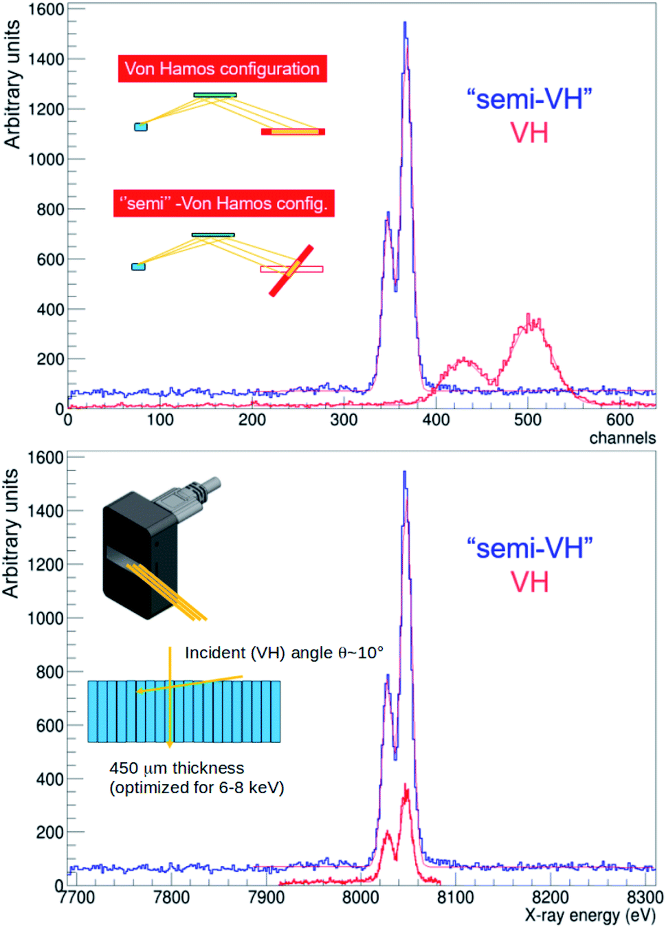 | ||
Fig. 12 Comparison between the standard (red) and the SVH (blue) configuration uncalibrated spectra of Cu(Kα1,2) lines, for a ρc = 206.7 mm crystal,  and Δθ′ = 0.5° measurement. In the upper panel, the MYTHEN2-1D uncalibrated spectra are shown for a standard (red) and a SVH (blue) configuration. In the insight, the geometrical difference between the two cases is given, in wich the source is pictured in blue, the crystal in green, the position detector in red and its illuminated portion in yellow as well as the impinging photons. In the lower panel, the calibrated spectra are shown with the same color code, while in the insight a schematic of the impinging angles and strip thickness is shown (see text). and Δθ′ = 0.5° measurement. In the upper panel, the MYTHEN2-1D uncalibrated spectra are shown for a standard (red) and a SVH (blue) configuration. In the insight, the geometrical difference between the two cases is given, in wich the source is pictured in blue, the crystal in green, the position detector in red and its illuminated portion in yellow as well as the impinging photons. In the lower panel, the calibrated spectra are shown with the same color code, while in the insight a schematic of the impinging angles and strip thickness is shown (see text). | ||
In the upper panel, the MYTHEN2-1D uncalibrated spectra are shown for a standard (red) and a SVH (blue) configuration. In the insight, the geometrical difference between the two cases is shown, in wich the source is pictured in blue, the crystal in green, the position detector in red and its illuminated portion in yellow as well as the impinging photons. In the lower panel, the calibrated spectra are shown with the same color code. From these latter, one can immediately see how the dynamic range (in eV) of the SVH spectrum is wider than the standard one. This configuration has, also, another big advantage: the Bragg rule implies that for increasing energies the reflection angle lowers, reaching very small values (θB < 10°) above 10 keV. For a strip detector, having a strip thickness tuned to maximize the detection efficiency, this may drop because of the too short path of the photons in each strip. This situation, which only stands for the standard VH configuration, is avoided in the SVH one as sketched in the insight of the lower panel in Fig. 12.
In order to check how the peak resolution is influenced by this rotation, we report as an example in Fig. 13 the results of a set of measurements of the Cu(Kα1,2), for different θM values ranging from 0° to θB − 90°, using a 206.7 mm radius HAPG with  and Δθ′ = 0.5°. The θM = 0 position corresponds to the SVH configuration in which the position detector is rotated in order to have the photons reflected with the nominal Bragg angle θB impinging orthogonally to the detector surface. The fitting function is a double Gaussian with common σ for the Cu lines and a polynomial for the background.
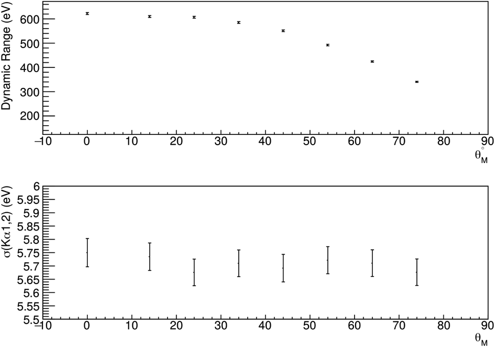 | ||
Fig. 13 Results of the Cu(Kα1,2) lines measured with a 206.7 mm radius HAPG for different rotation angles of the position detector θM, where θM = 0 refers to the SVH configuration. In the top panel the dynamic range (eV), defined as Emax − Emin of the calibrated spectrum is plotted, while in the bottom one the resolutions (σ) of the Kα1,2 peaks are shown. The fitting function is a double Gaussian with common σ for the Cu lines and a polynomial for the background. In this measurement,  and Δθ′ = 0.5°. and Δθ′ = 0.5°. | ||
3.4 Multi-element CuNiZn and MoNb spectra
Triggered by the perspectives opened by the SVH configuration described in Section 3.3, we performed another two sets of measurements having the aim of detecting photons coming from multielement targets and of testing the capabilities of the spectrometer in the ≃20 keV region. To do this, we used a copper–zinc–nitrogen and a molybdenum–niobium target, looking in particular at the characteristic X-ray transition lines listed in Table 2. For each measurement, the spectra have been acquired with different and Δθ′ settings but with a fixed integration time. In Fig. 14 and 15 we show one fitted spectrum per measurement corresponding to the best achieved resolution. The individual peak fitting functions are Gaussians with a common σ parameter for Kα1,2. Kα1, Kα2 and Kβ fitting functions correspond to the green, violet and light blue curves, respectively and are, together with a polynomial background (blue) and the overall fit (red), overimposed to the calibrated spectrum. The values of the
  , Δθ′ pair, the corresponding S0, Δθ pair and the curvature radius ρc of the used crystal are also reported in the figures.
 | ||
| Fig. 14 Fitted spectrum of Mo(Kα1,2) + Nb(Kβ) lines: Kα1, Kα2 and Kβ fitting function correspond to the green, violet and light blue curves, respectively and are, together with a polynomial background (blue) and the overall fit (red), overimposed to the calibrated spectrum. | ||
 | ||
| Fig. 15 Fitted spectrum of Cu(Kα1,2) + Zn(Kβ) + Ni(Kα1,2) lines: Kα1, Kα2 and Kβ fitting function correspond to the green, violet and light blue curves, respectively and are, together with the overall fit (red), overimposed to the calibrated spectrum. | ||
These measurements show how, at higher energies, the effective source size can become even wider, and resolutions of σ ≃ 20 eV have been obtained using a ρc = 77.5 mm radius HAPG crystal to avoid the almost 2 m path lengths resulting from bigger ρc values, for which indeed a better resolution could be achieved.
It has to be mentioned that the worsening in resolution could be avoided exploiting the n = 2 reflection order. This would correspond to a measurement of a line around 8 keV for n = 1, leading to a resolution similar to the one obtained for the Cu target. As a drawback, this would require a longer exposure time since the n = 2 peak reflectivity is an order of magnitude smaller than the n = 1 one.10
The possibility to exploit a wide dynamic range is highlighted, together with the MoNb spectrum, by the CuNiZn one, in which the combined effect of the Δθ′ angular acceptance and the mosaicity allow to have, in a single measurement, lines which are almost 1.2 keV and 600 eV distant, respectively.
3.5 Summary of the results
In the previous sections we presented some of the spectra and of the , Δθ′ scans we performed on our targets with the VOXES spectrometer. In this section we summarize, in Table 4, the best results, in terms of peak resolution (σE) and mean precision (δE), we obtained for each target using different ρc crystals. For each measurement, the parameter (σE or δE), the obtained value and the corresponding geometrical conditions
  are reported.
4 Ray tracing simulations
In this section, we present the results of the X-ray tracing simulations we performed to check the consistency of our measurements. We show, as an example, the simulations corresponding to the measurement reported in Fig. 10.4.1 Description
The simulations are performed using the Shadow29 and XOP 2.4 (ref. 30) software integrated in the OASYS environment.31,32Following the snapshot of the OASYS workflow in Fig. 16, the simulation main parameters are:
 | ||
| Fig. 16 Snapshot of the OASYS workflow used for the ray tracing simulations. | ||
•The target is generated using a 12 × 5 mm rectangular 0.125 mm thick target with the Shadow Geometrical Source widget. The energy spectrum of the copper X-rays is sampled with a double Lorentzian function, the widths of which are obtained from an average of several experimental papers found in literature.23–25,27,33,34 By means of two python scripts widgets, the target is then rotated of 45° and its divergence set to a random angular distribution to match the experimental conditions.
•The box hole and the two slits are included using the Shadow Screen-Slits widget, setting the corresponding positions and apertures.
•The HAPG crystal is simulated using the Shadow Spherical Crystal, where the cylindrical shape can be set with the desired ρc value. The mosaicity, set to 0.1° according to the sample test performed by the producer, is introduced with a python script where the mosaic crystal model developed by M. Sanchez del Rio35 is used.
•The MYTHEN2-1D Empty Element of Fig. 16 is used to rotate the reference frame to simulate the SVH conditions.
•Along the whole beamlines, 1D and 2D histograms and plots widgets are used to obtain the simulation results.
4.2 Horizontal and vertical beam dimensions
In Fig. 17, the ray tracing simulations on both the dipersive and sagittal planes are shown. The vertical dimensions of the beam at the source (up), crystal (mid) and MYTHEN2-1D (low) positions are displayed on the left, while the corresponding horizontal ones are shown on the right. Plots related to the source and the crystal dimensions include all the photons which hits this latter, while the ones rejected by the slits and not reaching the HAPG are discarded. On the other side, among all these photons, only the ones actually reflected by the mosaic crystal and reaching the MYTHEN2-1D detector are included in the lower pads plots of Fig. 17. | ||
| Fig. 17 Vertical (left) and horizontal (right) dimensions of the beam at the source (up), crystal (mid) and MYTHEN2-1D (low) positions. | ||
From Fig. 17, some important confirmations can be deduced. First, all the plots show horizontal and vertical dimensions in agreement with the formulae introduced in Sections 2.1.2 and 2.1.3. Second, looking at the middle plots, one can see that the vertical size of the beam on the HAPG crystal is still below the crystal dimensions (32 × 30 mm), while the confirmation of the sagittal focusing properties of its cylindrical shape is given by the almost identical vertical dispersions at the source and MYTHEN2-1D position. Since the MYTHEN2-1D strips vertical size is 8 mm, only few photons in the tails of the distribution are lost. Finally, the horizontal hit position plot in the lower right pad gives the Bragg spectrum. In the next section, this will be analysed and compared to the measured one.
4.3 Bragg spectrum comparison
In this section, we report the comparison between the simulated and measured copper Bragg spectra for the case of ρc = 206.7 mm HAPG crystal, and Δθ′ = 0.7°. In Fig. 18, Gaussian fits with individual and common σs for the Kα1 and Kα2 lines are presented in the upper and middle panel, respectively. The corresponding measured energy spectrum, fitted with common σ for the two peaks, is shown in the lower one, where Kα1 and Kα2 fitting functions correspond to the green and violet curves, respectively and are, together with a polynomial background (blue) and the overall fit (red), overimposed to the calibrated spectrum.
 | ||
| Fig. 18 Gaussian fits of the reconstructed raytrace simulation Bragg spectrum with individual (up) and common (mid) σ. In the lower panel, the corresponding measured energy spectrum, fitted with common σ for the two peaks, is shown, where Kα1 and Kα2 fitting functions correspond to the green and violet curves, respectively and are, together with a polynomial background (blue) and the overall fit (red), overimposed to the calibrated spectrum. | ||
The results of these fits bring along two important evidences: first, the perfectly compatible values for the mean positions in the individual and common σ case are confirmed, allowing to use a common σ for all the measured spectra. Second, the experimental resolution of σE = 4.18 ± 0.03 eV is well reproduced (the small difference is due to the MYTHEN2-1D cross talk between adjacent strips). In particular, as already stated in Section 3.1.2, the first implication is crucial, because for large values of  and Δθ′ the two peaks become hard to disentangle, and a fit with two separate σ is not anymore reliable. Since we are more interested in the final peak positions and their errors, it is crucial to have the possibility to compare measurements up to those large
  , Δθ′ pair values.
5 Conclusions
In this paper we presented the results obtained with the VOXES compact Von Hamos spectrometer based on mosaic crystals of pyrolytic graphite (HAPG). We demonstrated how the proposed spectrometer allows to obtain energy resolutions of few eV when used with extended and diffused isotropic sources. In particular, resolution in the order of 0.1% (FWHM/E), for energies below 10 keV, and up to 0.3% for 20 keV, using a source size ranging from 500 μm to 2 mm in the Bragg dispersion plane have been achieved.We achieved best values of peak resolutions as low as 4.02 ± 0.08 eV and 3.60 ± 0.05 eV for the individual Fe(Kα1,2) and Cu(Kα1,2) lines measurements. For larger dynamic ranges, we report the possibility to have a single shot spectrum with Cu(Kα1,2) Ni(Kβ) and Zn(Kα1,2) lines, obtaining best resolutions values of 5.15 ± 0.13 eV, 6.02 ± 0.24 eV and 6.20 ± 0.34 eV, respectively. Finally, we explored the capabilities of our spectrometer for energies above 15 keV, obtaining for a Mo(Kα1,2) + Nb(Kβ) target best resolutions of 19.44 ± 0.75 eV and 39.90 ± 1.48 eV, respectively.
Depending on the spectrometer geometries, for a fixed integration time of 1 hour we obtained precisions on the peak positions ranging from 0.04 eV at 8 keV (Cu(Kα1)) to 1.45 eV at 17.4 keV (Mo(Kα2)). In general, for each energy line to be measured it is always possible to find a configuration, in terms of  , Δθ′ pair, providing the best resolution or the best peak position precision.
We described the possibility to use the spectrometer in a “semi” – Von Hamos configuration, improving its energy range without losses in resolution. We proved, for this geometry, that the calibration and peak fitting procedures are under control, as well as the geometrical parameters.
The obtained resolutions are of the same order of magnitude as those obtainable with Transition Edge Sensors,5 with respect to which the VOXES spectrometer has the big advantages of much lower costs, no need of powerfull and costly cooling systems and a calibration function that, as shown in Section 3.1.3, is well under control.
All these results make this spectroscopy technique a serious candidate for high precision X-ray measurements from diffused and extended sources, with possible applications in several fields, going from fundamental physics, synchrotron radiation and X-FEL applications, astronomy, medicine and industry to hadronic physics experiments, where its performances make it a fundamental tool for a series of measurements like the energies of kaonic atoms radiative transitions which allow to extract fundamental parameters in the low energy QCD in the strangeness sector.
Conflicts of interest
There are no conflicts to declare.Acknowledgements
This work is supported by the 5th National Scientific Committee of INFN in the framework of the Young Researcher Grant 2015, no. 17367/2015. This project has received funding from the European Union's Horizon 2020 research and innovation programme under grant agreement No 824093. We thank the LNF and SMI staff, in particular the LNF SPCM service and Doris Pristauz-Telsnigg, for the support in the preparation of the setup.Notes and references
- M. Bazzi, et al. , Phys. Lett. B, 2011, 704, 113–117 CrossRef CAS.
- C. Curceanu, et al. , Rev. Mod. Phys., 2019, 91, 025006 CrossRef.
- A. Gallo, et al., Conf. Proc. C060626, 2006, pp. 604–606 Search PubMed.
- M. Miliucci, et al. , Condens. Matter, 2019, 4, 31 CrossRef CAS.
- W. B. Doriese, et al. , Rev. Sci. Instrum., 2017, 88, 053108 CrossRef CAS.
- H. Legall, et al., Proceedings of FEL 2006, 2006, p. 798 Search PubMed.
- R. Barnsley, et al. , Rev. Sci. Instrum., 2003, 74, 2388 CrossRef CAS.
- M. Sanchez del Rio, et al. , Proc. SPIE, 1998, 3448, 246–255 CrossRef.
- C. Schlesiger, et al. , J. Anal. At. Spectrom., 2015, 30, 1080–1085 RSC.
- M. Gerlach, et al. , J. Appl. Crystallogr., 2015, 48, 1381–1390 CrossRef CAS.
- L. V. Von Hamos, Ann. Phys., 1933, 409, 716 CrossRef.
- A. P. Shevelko, et al. , Rev. Sci. Instrum., 2002, 73, 3458 CrossRef CAS.
- U. Zastrau, et al. , J. Instrum., 2013, 8, P10006 CrossRef.
- L. Anklamm, et al. , Rev. Sci. Instrum., 2014, 85, 053110 CrossRef.
- A. Scordo, et al. , Condens. Matter, 2019, 4, 59 CrossRef.
- A. Scordo, et al. , J. Instrum., 2018, 13, C04002 CrossRef.
- A. Scordo, et al. , Acta Phys. Pol. B Proc. Suppl., 2017, 48, 1715 Search PubMed.
- I. Grigorieva, et al. , Condens. Matter, 2019, 4, 18 CrossRef CAS.
- R. Brun and F. Rademakers, AIHENP’96 Workshop, Lausanne, 1996, pp. 81–86 Search PubMed.
- F. James, MINUIT Function Minimization and Error Analysis: Reference Manual Version 94.1, 1994 Search PubMed.
- H. Akaike, in Information Theory and an Extension of the Maximum Likelihood Principle, Springer New York, New York, NY, 1973, pp. 199–213 Search PubMed.
- K. Burnham and D. Anderson, Model Selection and Inference, Springer, New York, NY, 1998 Search PubMed.
- H. Sorum, J. Phys. F: Met. Phys., 1987, 17, 417–425 CrossRef.
- M. H Mendenhall, et al. , J. Phys. B: At., Mol. Opt. Phys., 2017, 50, 115004 CrossRef.
- A. C. Illig, et al. , J. Phys. B: At., Mol. Opt. Phys., 2013, 46, 235001 CrossRef.
- M. Deutsch, et al. , Phys. Rev. A: At., Mol., Opt. Phys., 1995, 51, 283–296 CrossRef CAS.
- G. Hölzer, et al. , Phys. Rev. A, 1997, 56, 4554–4568 CrossRef.
- X-ray Data Booklet, ed. A. C. Thompson and D. Vaughan, L. B. N. Laboratory, 2000 Search PubMed.
- M. Sanchez del Rio, et al. , J. Synchrotron Radiat., 2011, 18, 708–716 CrossRef CAS.
- M. Sanchez del Rio and R. J. Dejus, Proc. SPIE, 2011, 8141 Search PubMed.
- L. Rebuffi and M. Sanchez del Rio, J. Synchrotron Radiat., 2016, 23, 1357–1367 CrossRef.
- L. Rebuffi and M. Sanchez del Rio, OASYS (OrAnge SYnchrotron Suite): an open-source graphical environment for x-ray virtual experiments, 2017, p. 28 Search PubMed.
- M. O. Krause and J. H. Oliver, J. Synchrotron Radiat., 2016, 23, 1357–1367 CrossRef.
- M. Deutsch, et al. , Phys. Rev. A, 1995, 51, 283–296 CrossRef CAS.
- M. Sanchez del Rio, et al. , Rev. Sci. Instrum., 1992, 63, 932–935 CrossRef CAS.
| This journal is © The Royal Society of Chemistry 2020 |