 Open Access Article
Creative Commons Attribution 3.0 Unported Licence
Correction: A highly X-ray sensitive iridium prodrug for visualized tumor radiochemotherapy
Zhennan
Zhao
,
Pan
Gao
,
Li
Ma
* and
Tianfeng
Chen
*
Department of Chemistry, Jinan University, Guangzhou 510632, China. E-mail: tchentf@jnu.edu.cn; chem-mali@foxmail.com
First published on 15th February 2021
Abstract
Correction for ’A highly X-ray sensitive iridium prodrug for visualized tumor radiochemotherapy’ by Zhennan Zhao et al., Chem. Sci., 2020, 11, 3780–3789, DOI: 10.1039/D0SC00862A
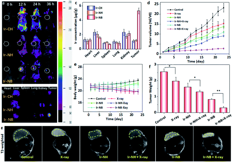
The authors regret errors in Fig. 5a in the original version of this Chemical Science article, specifically the 0 h and 24 h images in the top row (the Ir–CH treated group) and the 12 h and 36 h images in the bottom row (the Ir–NB treated group) which were used in Fig. 5a in error. The authors therefore wish to replace Fig. 5a and b with data from parallel experiments to ensure uniform intensity units in the colour bar. The correct version of Fig. 5 is shown below. This does not affect any of the discussions or conclusions reported in the article.
The Royal Society of Chemistry apologises for these errors and any consequent inconvenience to authors and readers.
| This journal is © The Royal Society of Chemistry 2021 |

