 Open Access Article
Creative Commons Attribution 3.0 Unported Licence
Synthesis of textured discontinuous-nanoisland Ca3Co4O9 thin films†
Binbin
Xin
 *,
Arnaud
Le Febvrier
 ,
Jun
Lu
,
Biplab
Paul‡
,
Jun
Lu
,
Biplab
Paul‡
 and
Per
Eklund
 *
*
Thin Film Physics Division, Department of Physics, Chemistry and Biology (IFM), Linköping University, SE-58183 Linköping, Sweden. E-mail: binbin.xin@liu.se; per.eklund@liu.se
First published on 4th July 2022
Abstract
Controllable engineering of the nanoporosity in layered Ca3Co4O9 remains a challenge. Here, we show the synthesis of discontinuous films with islands of highly textured Ca3Co4O9, effectively constituting distributed nanoparticles with controlled porosity and morphology. These discontinuously dispersed textured Ca3Co4O9 nanoparticles may be a candidate for hybrid thermoelectrics.
The misfit-layered calcium cobaltate Ca3Co4O9 has a complex crystal structure composed of CoO2 conductive layers and oxygen deficient rock-salt type Ca2CoO3 insulating layers.1 Ca3Co4O9 can be used in various energy-harvesting systems because of its high thermal stability and oxidation resistance. This material is an attractive p-type thermoelectric material with a high Seebeck coefficient S, moderate electrical conductivity σ and low thermal conductivity. It also can be used as an active material in Li-ion-battery anodes,2,3 hydrogen evolution and oxygen reduction reactions4,5 and supercapacitors with high cycling stability.6,7
Nanostructures such as nanoparticles8 and nanoporous films9,10 are common means to alter electrical, catalytic, and thermal properties of inorganic materials. In previous work, we have shown that nanoporous Ca3Co4O9 films on sapphire exhibit a thermal conductivity of 0.82 W m−1 K−1, which is nearly twofold lower than that obtained from comparable nonporous Ca3Co4O9 films.11 Furthermore, nanoporous Ca3Co4O9 films grown on mica can be obtained by reactions in hydrated CaO/CoO multilayers.12 The volume shrinkage in Ca(OH)2/Co3O4 multilayers and the out-of-plane orientation relationship between Ca(OH)2 and Co3O4 induce the formation of faceted and oriented nanopores in textured Ca3Co4O9 films.
Here, we show control of morphology and porosity in textured Ca3Co4O9 films, to form discontinuous films with islands of highly textured Ca3Co4O9, effectively constituting distributed nanoparticles. The discontinuous films with islands of highly textured Ca3Co4O9 were synthesized by radio-frequency (rf) sputtering followed by post-deposition annealing without any templates. Such films of discontinuously dispersed Ca3Co4O9 nanoparticles may be a promising filler in polymer matrixes for hybrid and composite materials in, e.g., thermoelectrics.13–16
The Ca3Co4O9 nanoparticles were obtained by a similar method to that published in our earlier work.12 First, the CaO/Co3O4 multilayer films were deposited on muscovite mica (00l) and sapphire substrates (001) at 600 °C by reactive radio-frequency magnetron sputtering. The multilayers consisted of eight alternative bilayers of CaO (top layer) and Co3O4. The overall Ca![[thin space (1/6-em)]](https://www.rsc.org/images/entities/char_2009.gif) :
:![[thin space (1/6-em)]](https://www.rsc.org/images/entities/char_2009.gif) Co elemental ratio in the multilayer films was varied and set to 1
Co elemental ratio in the multilayer films was varied and set to 1![[thin space (1/6-em)]](https://www.rsc.org/images/entities/char_2009.gif) :
:![[thin space (1/6-em)]](https://www.rsc.org/images/entities/char_2009.gif) 1.38 (close to stoichiometric Ca3Co4O9), 1
1.38 (close to stoichiometric Ca3Co4O9), 1![[thin space (1/6-em)]](https://www.rsc.org/images/entities/char_2009.gif) :
:![[thin space (1/6-em)]](https://www.rsc.org/images/entities/char_2009.gif) 0.82, 1
0.82, 1![[thin space (1/6-em)]](https://www.rsc.org/images/entities/char_2009.gif) :
:![[thin space (1/6-em)]](https://www.rsc.org/images/entities/char_2009.gif) 0.67, and 1
0.67, and 1![[thin space (1/6-em)]](https://www.rsc.org/images/entities/char_2009.gif) :
:![[thin space (1/6-em)]](https://www.rsc.org/images/entities/char_2009.gif) 0.52 by varying CaO and Co3O4 deposition times of their respective layer. Then, all the as-deposited multilayer films were exposed to a humid environment (0.88 relative humidity at constant temperature) to form Ca(OH)2/Co3O4 multilayer films at room temperature for two days, as described earlier.12,17 At the final stage, the different Ca(OH)2/Co3O4 multilayer films were annealed at 700 °C in air for 2 h.
0.52 by varying CaO and Co3O4 deposition times of their respective layer. Then, all the as-deposited multilayer films were exposed to a humid environment (0.88 relative humidity at constant temperature) to form Ca(OH)2/Co3O4 multilayer films at room temperature for two days, as described earlier.12,17 At the final stage, the different Ca(OH)2/Co3O4 multilayer films were annealed at 700 °C in air for 2 h.
X-ray diffraction (XRD) measurements were performed using an X'Pert PRO MRD diffractometer from PANalytical using Cu Kα1,2 radiation with a nickel filter in the Bragg–Brentano configuration (θ–2θ scans). The surface morphology of the films was studied by scanning electron microscopy (SEM) using a LEO Gemini 1550 Zeiss with a 10 kV operating voltage. Transmission electron microscopy (TEM) was carried out on an FEI Tecnai G2 TF20 UT instrument operated at 200 kV. The Ca/Co elemental ratio was determined using energy-dispersive X-ray spectroscopy (EDS) by measuring at several positions on each sample growing on sapphire. The surface porosity fraction or coverage was determined from the SEM micrographs analysed using the software ImageJ (Java version).18 The electrical conductivity σ was calculated from the sheet resistance measured by using a four-point probe Jandel RM3000 station, and the film thickness was determined from the cross-sectional SEM images. The Seebeck coefficient was determined from the slope of the temperature gradient–voltage characteristics measured using a homemade Seebeck measurement setup system described elsewhere.12,17
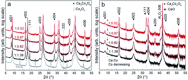
Fig. 1 shows the X-ray diffraction patterns of the Ca3Co4O9 films on mica and sapphire as a function of the Ca/Co ratio: 1![[thin space (1/6-em)]](https://www.rsc.org/images/entities/char_2009.gif) :
:![[thin space (1/6-em)]](https://www.rsc.org/images/entities/char_2009.gif) 1.38, 1
1.38, 1![[thin space (1/6-em)]](https://www.rsc.org/images/entities/char_2009.gif) :
:![[thin space (1/6-em)]](https://www.rsc.org/images/entities/char_2009.gif) 0.82, 1
0.82, 1![[thin space (1/6-em)]](https://www.rsc.org/images/entities/char_2009.gif) :
:![[thin space (1/6-em)]](https://www.rsc.org/images/entities/char_2009.gif) 0.67, and 1
0.67, and 1![[thin space (1/6-em)]](https://www.rsc.org/images/entities/char_2009.gif) :
:![[thin space (1/6-em)]](https://www.rsc.org/images/entities/char_2009.gif) 0.52, respectively. The Ca/Co elemental ratios were measured from the different annealed films grown on sapphire by EDS and were the same as those in the as-deposited multilayer films grown on sapphire and mica and the annealed films on mica, respectively. Diffraction peaks for 001, 002, 003, 004, 005, and 006 reflections from Ca3Co4O9 and 111 and 222 reflections from Co3O4 can be observed in the film on mica with the initial composition (1
0.52, respectively. The Ca/Co elemental ratios were measured from the different annealed films grown on sapphire by EDS and were the same as those in the as-deposited multilayer films grown on sapphire and mica and the annealed films on mica, respectively. Diffraction peaks for 001, 002, 003, 004, 005, and 006 reflections from Ca3Co4O9 and 111 and 222 reflections from Co3O4 can be observed in the film on mica with the initial composition (1![[thin space (1/6-em)]](https://www.rsc.org/images/entities/char_2009.gif) :
:![[thin space (1/6-em)]](https://www.rsc.org/images/entities/char_2009.gif) 1.38, the closest to the stoichiometric Ca3Co4O9) in Fig. 1a. With decreasing Co content (Ca
1.38, the closest to the stoichiometric Ca3Co4O9) in Fig. 1a. With decreasing Co content (Ca![[thin space (1/6-em)]](https://www.rsc.org/images/entities/char_2009.gif) :
:![[thin space (1/6-em)]](https://www.rsc.org/images/entities/char_2009.gif) Co 1
Co 1![[thin space (1/6-em)]](https://www.rsc.org/images/entities/char_2009.gif) :
:![[thin space (1/6-em)]](https://www.rsc.org/images/entities/char_2009.gif) 0.82 → 1
0.82 → 1![[thin space (1/6-em)]](https://www.rsc.org/images/entities/char_2009.gif) :
:![[thin space (1/6-em)]](https://www.rsc.org/images/entities/char_2009.gif) 0.52), pure-phase Ca3Co4O9 can be identified in the films on mica from the XRD patterns (Fig. 1a). The intensity of peaks of Ca3Co4O9 growing on mica remains approximately the same in Fig. 1a. As known and observed from our earlier work,19–21 the excess Ca migrates and is incorporated in an amorphous layer between the nanoporous Ca3Co4O9 films and the mica substrate and will be discussed below. However, the pure-phase Ca3Co4O9 can be seen from the film growing on sapphire with Ca
0.52), pure-phase Ca3Co4O9 can be identified in the films on mica from the XRD patterns (Fig. 1a). The intensity of peaks of Ca3Co4O9 growing on mica remains approximately the same in Fig. 1a. As known and observed from our earlier work,19–21 the excess Ca migrates and is incorporated in an amorphous layer between the nanoporous Ca3Co4O9 films and the mica substrate and will be discussed below. However, the pure-phase Ca3Co4O9 can be seen from the film growing on sapphire with Ca![[thin space (1/6-em)]](https://www.rsc.org/images/entities/char_2009.gif) :
:![[thin space (1/6-em)]](https://www.rsc.org/images/entities/char_2009.gif) Co = 1
Co = 1![[thin space (1/6-em)]](https://www.rsc.org/images/entities/char_2009.gif) :
:![[thin space (1/6-em)]](https://www.rsc.org/images/entities/char_2009.gif) 1.38 (Fig. 1b). With increasing Ca content, additional CaO can be observed for the films on sapphire (Fig. 1b). This result indicates that some CaO remained in the annealed films on sapphire.
1.38 (Fig. 1b). With increasing Ca content, additional CaO can be observed for the films on sapphire (Fig. 1b). This result indicates that some CaO remained in the annealed films on sapphire.
 | ||
Fig. 1 X-ray diffractograms of the annealed films grown on mica (a) and sapphire (b) with decreasing Co content in the films with Ca/Co ratios: 1![[thin space (1/6-em)]](https://www.rsc.org/images/entities/char_2009.gif) : :![[thin space (1/6-em)]](https://www.rsc.org/images/entities/char_2009.gif) 1.38, 1 1.38, 1![[thin space (1/6-em)]](https://www.rsc.org/images/entities/char_2009.gif) : :![[thin space (1/6-em)]](https://www.rsc.org/images/entities/char_2009.gif) 0.82, 1 0.82, 1![[thin space (1/6-em)]](https://www.rsc.org/images/entities/char_2009.gif) : :![[thin space (1/6-em)]](https://www.rsc.org/images/entities/char_2009.gif) 0.67, and 1 0.67, and 1![[thin space (1/6-em)]](https://www.rsc.org/images/entities/char_2009.gif) : :![[thin space (1/6-em)]](https://www.rsc.org/images/entities/char_2009.gif) 0.52, respectively. 0.52, respectively. | ||
The SEM images of the morphology of Ca3Co4O9 films on mica are shown in Fig. 2. The film on mica with the initial composition (1![[thin space (1/6-em)]](https://www.rsc.org/images/entities/char_2009.gif) :
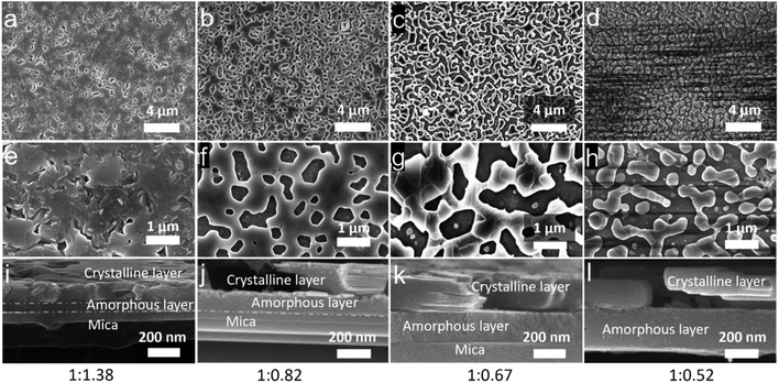
:![[thin space (1/6-em)]](https://www.rsc.org/images/entities/char_2009.gif) 1.38) shows morphology with few nanopores (Fig. 2a and e). With decreasing Co content, the morphology of Ca3Co4O9 in the annealed films change from a nanoporous continuous film morphology (Fig. 2b and f), via larger pores (Fig. 2c and g), to a discontinuous film of textured islands (Fig. 2d and h). The surface porosity fraction increases from 1.2% and 22% to 37% for the first three films (Table 1). For the discontinuous films, the corresponding value obtained from image analysis is an apparent “porosity” of 46% (Table 1), i.e. a surface coverage of 54%. This morphology is fundamentally different from the nanoporous films, though, in that the film is discontinuous and cannot be described as a porous film. The size of the nanoislands is mainly distributed from 50 nm to 1000 nm, as shown in Fig. S1.†
1.38) shows morphology with few nanopores (Fig. 2a and e). With decreasing Co content, the morphology of Ca3Co4O9 in the annealed films change from a nanoporous continuous film morphology (Fig. 2b and f), via larger pores (Fig. 2c and g), to a discontinuous film of textured islands (Fig. 2d and h). The surface porosity fraction increases from 1.2% and 22% to 37% for the first three films (Table 1). For the discontinuous films, the corresponding value obtained from image analysis is an apparent “porosity” of 46% (Table 1), i.e. a surface coverage of 54%. This morphology is fundamentally different from the nanoporous films, though, in that the film is discontinuous and cannot be described as a porous film. The size of the nanoislands is mainly distributed from 50 nm to 1000 nm, as shown in Fig. S1.†
![[thin space (1/6-em)]](https://www.rsc.org/images/entities/char_2009.gif) :
:![[thin space (1/6-em)]](https://www.rsc.org/images/entities/char_2009.gif) Co ratio in the films
Co ratio in the films
Ca![[thin space (1/6-em)]](https://www.rsc.org/images/entities/char_2009.gif) : :![[thin space (1/6-em)]](https://www.rsc.org/images/entities/char_2009.gif) Co elemental ratio Co elemental ratio |
1![[thin space (1/6-em)]](https://www.rsc.org/images/entities/char_2009.gif) : :![[thin space (1/6-em)]](https://www.rsc.org/images/entities/char_2009.gif) 1.38 1.38 |
1![[thin space (1/6-em)]](https://www.rsc.org/images/entities/char_2009.gif) : :![[thin space (1/6-em)]](https://www.rsc.org/images/entities/char_2009.gif) 0.82 0.82 |
1![[thin space (1/6-em)]](https://www.rsc.org/images/entities/char_2009.gif) : :![[thin space (1/6-em)]](https://www.rsc.org/images/entities/char_2009.gif) 0.67 0.67 |
1![[thin space (1/6-em)]](https://www.rsc.org/images/entities/char_2009.gif) : :![[thin space (1/6-em)]](https://www.rsc.org/images/entities/char_2009.gif) 0.52 0.52 |
|---|---|---|---|---|
| Thickness of the amorphous layer (nm) | 81 ± 5 | 128 ± 6 | 179 ± 9 | 250 ± 12 |
| Thickness of Ca3Co4O9 (nm) | 121 ± 7 | 178 ± 9 | 194 ± 10 | 170 ± 9 |
| Porosity (%) | 1.2 ± 0.1 | 22 ± 1 | 37 ± 1.9 | 46 ± 2.3 |
The electrical conductivity and the Seebeck coefficient of the Ca3Co4O9/Co3O4 film are 27 S cm−1 and 139 μV K−1, respectively. The electrical conductivity of the nanoporous pure Ca3Co4O9 film decreases from 112 to 38 S cm−1 with increasing the porosity from 22% up to 37%. The Seebeck coefficient of the nanoporous Ca3Co4O9 films with different porosities is approximately 127 μV K−1, essentially the same for both. The Seebeck coefficient and electrical conductivity of the discontinuous film of textured Ca3Co4O9 islands cannot be measured by using these setups since there is no continuous conduction path.
The cross-sectional SEM micrographs (Fig. 2i–l) reveal that the films are composed of a crystalline layer on top of an amorphous layer. As is known from our earlier work,20,21 this amorphous layer forms due to a reaction between the mica substrate and the initial films during annealing. The amorphous layer contains O, Al, and Si elements from mica and Ca element from the initial films. The film growing on mica with the initial composition (1![[thin space (1/6-em)]](https://www.rsc.org/images/entities/char_2009.gif) :
:![[thin space (1/6-em)]](https://www.rsc.org/images/entities/char_2009.gif) 1.38) shows a crystalline layer with a thickness of 121 nm and an amorphous layer with a thickness of 81 nm (Fig. 2i). This indicates that the formation of Ca3Co4O9 and amorphous layers occurs at same time during annealing. With increasing Ca content in the initial films, the pure crystalline Ca3Co4O9 layer of the last three films shows a similar apparent thickness of around 170 nm for all the films, but the thickness of the amorphous layer increases from 130 nm to 240 nm (Fig. 2j–l and Table 1).
1.38) shows a crystalline layer with a thickness of 121 nm and an amorphous layer with a thickness of 81 nm (Fig. 2i). This indicates that the formation of Ca3Co4O9 and amorphous layers occurs at same time during annealing. With increasing Ca content in the initial films, the pure crystalline Ca3Co4O9 layer of the last three films shows a similar apparent thickness of around 170 nm for all the films, but the thickness of the amorphous layer increases from 130 nm to 240 nm (Fig. 2j–l and Table 1).
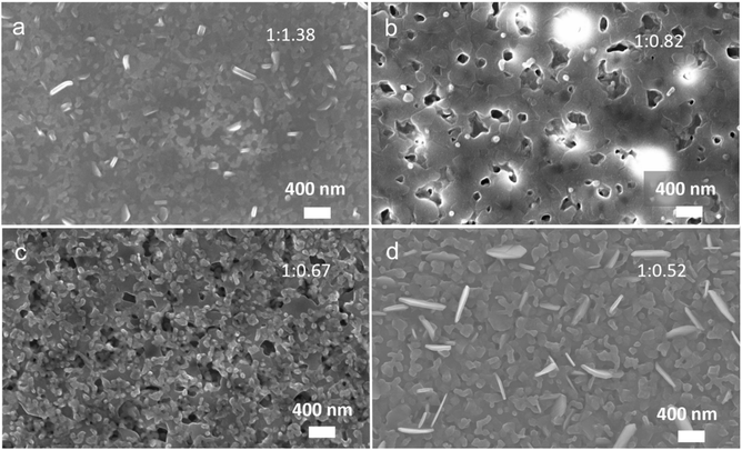
The SEM images of the morphology of Ca3Co4O9 films on sapphire are shown in Fig. 3a–d. A dense film can be observed in the annealed film with Ca![[thin space (1/6-em)]](https://www.rsc.org/images/entities/char_2009.gif) :
:![[thin space (1/6-em)]](https://www.rsc.org/images/entities/char_2009.gif) Co = 1
Co = 1![[thin space (1/6-em)]](https://www.rsc.org/images/entities/char_2009.gif) :
:![[thin space (1/6-em)]](https://www.rsc.org/images/entities/char_2009.gif) 1.38 (Fig. 3a). The nanoporous morphology with a nanopore size of ∼200 nm can be observed in the annealed film with low Ca
1.38 (Fig. 3a). The nanoporous morphology with a nanopore size of ∼200 nm can be observed in the annealed film with low Ca![[thin space (1/6-em)]](https://www.rsc.org/images/entities/char_2009.gif) :
:![[thin space (1/6-em)]](https://www.rsc.org/images/entities/char_2009.gif) Co = 1
Co = 1![[thin space (1/6-em)]](https://www.rsc.org/images/entities/char_2009.gif) :
:![[thin space (1/6-em)]](https://www.rsc.org/images/entities/char_2009.gif) 0.82 (Fig. 3b). Upon further decreasing the Co content, the surface morphology seems to be composed of a mixture of two families of grains (Fig. 3c). The similar round grains with a “size” (∼100 nm) can be observed on the top of the flat grains and nanopores at films 1
0.82 (Fig. 3b). Upon further decreasing the Co content, the surface morphology seems to be composed of a mixture of two families of grains (Fig. 3c). The similar round grains with a “size” (∼100 nm) can be observed on the top of the flat grains and nanopores at films 1![[thin space (1/6-em)]](https://www.rsc.org/images/entities/char_2009.gif) :
:![[thin space (1/6-em)]](https://www.rsc.org/images/entities/char_2009.gif) 0.82 and 1
0.82 and 1![[thin space (1/6-em)]](https://www.rsc.org/images/entities/char_2009.gif) :
:![[thin space (1/6-em)]](https://www.rsc.org/images/entities/char_2009.gif) 0.6 in Fig. 3b and c. For the lowest Co containing film, different family of grains can be observed forming a dense film (without apparent nanopores) (Fig. 3d).
0.6 in Fig. 3b and c. For the lowest Co containing film, different family of grains can be observed forming a dense film (without apparent nanopores) (Fig. 3d).
 | ||
Fig. 3 Surface SEM images for the annealed films grown on sapphire with the different ratios of Ca/Co: (a) 1![[thin space (1/6-em)]](https://www.rsc.org/images/entities/char_2009.gif) : :![[thin space (1/6-em)]](https://www.rsc.org/images/entities/char_2009.gif) 1.38; (b) 1 1.38; (b) 1![[thin space (1/6-em)]](https://www.rsc.org/images/entities/char_2009.gif) : :![[thin space (1/6-em)]](https://www.rsc.org/images/entities/char_2009.gif) 0.82; (c) 1 0.82; (c) 1![[thin space (1/6-em)]](https://www.rsc.org/images/entities/char_2009.gif) : :![[thin space (1/6-em)]](https://www.rsc.org/images/entities/char_2009.gif) 0.67; (d) 1 0.67; (d) 1![[thin space (1/6-em)]](https://www.rsc.org/images/entities/char_2009.gif) : :![[thin space (1/6-em)]](https://www.rsc.org/images/entities/char_2009.gif) 0.52. 0.52. | ||
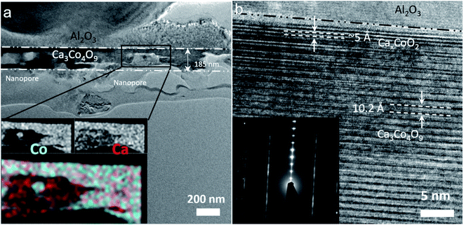
The cross-sectional TEM images of the annealed film with the ratio of Ca![[thin space (1/6-em)]](https://www.rsc.org/images/entities/char_2009.gif) :
:![[thin space (1/6-em)]](https://www.rsc.org/images/entities/char_2009.gif) Co = 1
Co = 1![[thin space (1/6-em)]](https://www.rsc.org/images/entities/char_2009.gif) :
:![[thin space (1/6-em)]](https://www.rsc.org/images/entities/char_2009.gif) 0.82 deposited on sapphire are shown in Fig. 4a and b. The nanopore structure in the Ca3Co4O9 layer with an apparent thickness of 185 nm can be observed in Fig. 4a, with the EDS maps of Co and Ca elements showing a uniform distribution in the Ca3Co4O9 layer but a higher Ca concentration in the nanopores. At the interface film substrate, a thin CaxCoO2 layer can be seen near the sapphire substrate in Fig. 4b. The formation of CaxCoO2 has been observed in earlier work.22 The lattice images for the Ca3Co4O9 layer and the SAED patterns (Fig. 4b) confirm that the (001) basal planes are oriented parallel to the film surface, corroborating the XRD results.
0.82 deposited on sapphire are shown in Fig. 4a and b. The nanopore structure in the Ca3Co4O9 layer with an apparent thickness of 185 nm can be observed in Fig. 4a, with the EDS maps of Co and Ca elements showing a uniform distribution in the Ca3Co4O9 layer but a higher Ca concentration in the nanopores. At the interface film substrate, a thin CaxCoO2 layer can be seen near the sapphire substrate in Fig. 4b. The formation of CaxCoO2 has been observed in earlier work.22 The lattice images for the Ca3Co4O9 layer and the SAED patterns (Fig. 4b) confirm that the (001) basal planes are oriented parallel to the film surface, corroborating the XRD results.
The dense Ca3Co4O9 film can be synthesized with the right Ca![[thin space (1/6-em)]](https://www.rsc.org/images/entities/char_2009.gif) :
:![[thin space (1/6-em)]](https://www.rsc.org/images/entities/char_2009.gif) Co elemental ratio (close to stoichiometric Ca3Co4O9) when the film grows on sapphire. The nanoporous film but a non-phase pure film mixing CaO and Ca3Co4O9 can form on sapphire with increasing Ca content. Comparing the results for the films grown on sapphire with those on mica allows determining the mechanism of the increase in the porosity fraction and formation of a discontinuous film of islands, effectively constituting distributed nanoparticles.
Co elemental ratio (close to stoichiometric Ca3Co4O9) when the film grows on sapphire. The nanoporous film but a non-phase pure film mixing CaO and Ca3Co4O9 can form on sapphire with increasing Ca content. Comparing the results for the films grown on sapphire with those on mica allows determining the mechanism of the increase in the porosity fraction and formation of a discontinuous film of islands, effectively constituting distributed nanoparticles.
This discontinuous structure is correlated with the reaction between Ca in the Ca(OH)2/Co3O4 multilayer films with the mica layer. In our previous work, pore formation could be attributed to the basal plane removal driven by local densification of textured Ca3Co4O9 nuclei during growth.12 In the present case, this mechanism yields formation of a discontinuous film of islands, i.e., distributed nanoparticles, for the high initial Ca content in the starting multilayers. A schematic illustration is shown in Fig. 5. When Ca![[thin space (1/6-em)]](https://www.rsc.org/images/entities/char_2009.gif) :
:![[thin space (1/6-em)]](https://www.rsc.org/images/entities/char_2009.gif) Co = 1
Co = 1![[thin space (1/6-em)]](https://www.rsc.org/images/entities/char_2009.gif) :
:![[thin space (1/6-em)]](https://www.rsc.org/images/entities/char_2009.gif) 1.38 (close to 3
1.38 (close to 3![[thin space (1/6-em)]](https://www.rsc.org/images/entities/char_2009.gif) :
:![[thin space (1/6-em)]](https://www.rsc.org/images/entities/char_2009.gif) 4), the film with few nanopores is composed of a crystalline Ca3Co4O9/Co3O4 layer on top of a thin amorphous layer, which proves that Ca diffuses and reacts with the mica substrate to form an amorphous layer during formation of Ca3Co4O9. During annealing and with increased Ca content, the excess Ca from Ca(OH)2 will be attracted to the interface substrate/film where the reaction occurs to form a thicker amorphous layer underneath phase pure crystalline Ca3Co4O9 layers with nanopores. With further increase of Ca content, the nanopore size and porosity significantly increase, while the apparent thickness of the crystalline Ca3Co4O9 layer remains constant. This result indicates that the volume shrinkage of Ca3Co4O9 preferentially occurs in the in-plane direction and not in the out-of-plane direction. As expected, the more excess Ca results in a thicker amorphous layer with even lower Co content. Instead of forming nanoporous Ca3Co4O9, the Ca3Co4O9 instead forms a discontinuous film of islands, constituting distributed nanoparticles with a larger apparent “porosity”.
4), the film with few nanopores is composed of a crystalline Ca3Co4O9/Co3O4 layer on top of a thin amorphous layer, which proves that Ca diffuses and reacts with the mica substrate to form an amorphous layer during formation of Ca3Co4O9. During annealing and with increased Ca content, the excess Ca from Ca(OH)2 will be attracted to the interface substrate/film where the reaction occurs to form a thicker amorphous layer underneath phase pure crystalline Ca3Co4O9 layers with nanopores. With further increase of Ca content, the nanopore size and porosity significantly increase, while the apparent thickness of the crystalline Ca3Co4O9 layer remains constant. This result indicates that the volume shrinkage of Ca3Co4O9 preferentially occurs in the in-plane direction and not in the out-of-plane direction. As expected, the more excess Ca results in a thicker amorphous layer with even lower Co content. Instead of forming nanoporous Ca3Co4O9, the Ca3Co4O9 instead forms a discontinuous film of islands, constituting distributed nanoparticles with a larger apparent “porosity”.
The growth of discontinuous films with islands of highly textured Ca3Co4O9 effectively constituting distributed nanoparticles has been demonstrated by sequential sputtering-annealing without any templates. The volume shrinkage in the initial Ca(OH)2/Co3O4 multilayers with different Ca/Co overall ratios can be used to tailor morphology and surface coverage porosity in textured Ca3Co4O9 films. Such films of discontinuously dispersed Ca3Co4O9 nanoparticles may be a promising filler in polymer matrixes for hybrid and composite materials in, e.g., hybrid thermoelectrics.
Conflicts of interest
There are no conflicts to declare.Notes and references
- Y. Miyazaki, M. Onoda, T. Oku, M. Kikuchi, Y. Ishii, Y. Ono, Y. Morii and T. Kajitani, J. Phys. Soc. Jpn., 2002, 71, 491–497 CrossRef CAS.
- D. W. Kim, Y. D. Ko, J. G. Park and B. K. Kim, Angew. Chem., Int. Ed., 2007, 46, 6654–6657 CrossRef CAS PubMed.
- S. Guan, Q. Fan, L. Liu, J. Luo, Y. Zhong, W. Zhao, Z. Huang and Z. Shi, Sci. China: Technol. Sci., 2020, 64, 673–679 CrossRef.
- C. S. Lim, C. K. Chua, Z. Sofer, O. Jankovský and M. Pumera, Chem. Mater., 2014, 26, 4130–4136 CrossRef CAS.
- V. D. Silva, T. A. Simões, F. J. A. Loureiro, D. P. Fagg, E. S. Medeiros and D. A. Macedo, Mater. Lett., 2018, 221, 81–84 CrossRef CAS.
- Z. Wang, Y. Wang, X. Yue, G. Shi, M. Shang, Y. Zhang, Z. Lv and G. Ao, J. Alloys Compd., 2019, 792, 357–364 CrossRef CAS.
- R. Mendoza, J. Oliva, K. P. Padmasree, A. I. Oliva, A. I. Mtz-Enriquez and A. Zakhidov, J. Energy Storage, 2022, 46, 103818 CrossRef.
- J. Snyder, I. McCue, K. Livi and J. Erlebacher, J. Am. Chem. Soc., 2012, 134, 8633–8645 CrossRef CAS PubMed.
- D.-J. Guo and Y. Ding, Electroanalysis, 2012, 24, 2035–2043 CrossRef CAS.
- J. Tang, H. T. Wang, D. H. Lee, M. Fardy, Z. Huo, T. P. Russell and P. Yang, Nano Lett., 2010, 10, 4279–4283 CrossRef CAS PubMed.
- B. Paul, Y. Zhang, W. Zhu, B. Xin, G. Ramanath, T. Borca-Tasciuc and P. Eklund, Appl. Phys. Lett., 2022, 120, 061904 CrossRef CAS.
- B. Xin, A. L. Febvrier, R. Shu, A. Elsukova, V. Venkataramani, Y. Shi, G. Ramanath, B. Paul and P. Eklund, ACS Appl. Nano Mater., 2021, 4, 9904–9911 CrossRef CAS.
- Y. Du, J. Xu, B. Paul and P. Eklund, Appl. Mater. Today, 2018, 12, 366–388 CrossRef.
- Y. Wang, L. Yang, X. L. Shi, X. Shi, L. Chen, M. S. Dargusch, J. Zou and Z. G. Chen, Adv. Mater., 2019, 31, e1807916 CrossRef PubMed.
- L. Zhang, X.-L. Shi, Y.-L. Yang and Z.-G. Chen, Mater. Today, 2021, 46, 62–108 CrossRef CAS.
- L. Wang, Z. Zhang, Y. Liu, B. Wang, L. Fang, J. Qiu, K. Zhang and S. Wang, Nat. Commun., 2018, 9, 3817 CrossRef PubMed.
- B. Xin, A. L. Febvrier, L. Wang, N. Solin, B. Paul and P. Eklund, Mater. Des., 2021, 210, 110033 CrossRef CAS.
- M. D. Abràmoff, P. J. Magalhães and S. J. Ram, Biophot. Int., 2004, 11, 36–42 Search PubMed.
- B. Paul, J. Lu and P. Eklund, ACS Appl. Mater. Interfaces, 2017, 9, 25308–25316 CrossRef CAS PubMed.
- B. Paul, E. M. Björk, A. Kumar, J. Lu and P. Eklund, ACS Appl. Energy Mater., 2018, 1, 2261–2268 CrossRef CAS PubMed.
- B. Xin, E. Ekström, Y.-T. Shih, L. Huang, J. Lu, A. Elsukova, Y. Zhang, W. Zhu, T. Borca-Tasciuc, G. Ramanath, A. Le Febvrier, B. Paul and P. Eklund, Nanoscale Adv., 2022 10.1039/D2NA00278G.
- B. Paul, J. L. Schroeder, S. Kerdsongpanya, N. V. Nong, N. Schell, D. Ostach, J. Lu, J. Birch and P. Eklund, Adv. Electron. Mater., 2015, 1, 1400022 CrossRef.
Footnotes |
| † Electronic supplementary information (ESI) available. See https://doi.org/10.1039/d2na00373b |
| ‡ Present address: Platit AG, Selzach, Switzerland. |
| This journal is © The Royal Society of Chemistry 2022 |


