 Open Access Article
Biomimicry in nanotechnology: a comprehensive review
Mehedi Hasan
Himel
ab,
Bejoy
Sikder
a,
Tanvir
Ahmed
 ab and
Sajid Muhaimin
Choudhury
 *a
*a
aDepartment of Electrical and Electronic Engineering, Bangladesh University of Engineering and Technology, Dhaka 1205, Bangladesh. E-mail: sajid@eee.buet.ac.bd
bDepartment of Computer Science and Engineering, Brac University, 66 Mohakhali, Dhaka 1212, Bangladesh
First published on 22nd December 2022
Abstract
Biomimicry has been utilized in many branches of science and engineering to develop devices for enhanced and better performance. The application of nanotechnology has made life easier in modern times. It has offered a way to manipulate matter and systems at the atomic level. As a result, the miniaturization of numerous devices has been possible. Of late, the integration of biomimicry with nanotechnology has shown promising results in the fields of medicine, robotics, sensors, photonics, etc. Biomimicry in nanotechnology has provided eco-friendly and green solutions to the energy problem and in textiles. This is a new research area that needs to be explored more thoroughly. This review illustrates the progress and innovations made in the field of nanotechnology with the integration of biomimicry.
1 Introduction
Nature, through billions of years of evolution, has often provided unique but elegant designs and structures that have aided researchers and engineers to invent new technologies to enhance the capabilities of existing ones. The term ‘biomimetics’ or ‘biomimicry’, coined by American biophysicist O. H. Schmitt in the 1950s1,2 is composed of two words that are derived from Greek words: ‘bios’ (meaning life) and ‘mimesis’ (meaning imitation). Biomimicry is the emulation of natural structures, designs, and elements with a view to develop novel devices with the desired functionalities.The evolving field of biomimicry is highly multidisciplinary and it is found on almost every engineering scale, from microscopic applications to macroscopic applications. The idea of mimicking nature to solve complex problems is implemented in every branch of science and engineering. Photonic structures that are prevalent in nature, such as the moth-eye, the structural colors of insects, etc., have provided inspiration for the construction of many novel photonic materials. Photosynthesis of plants and trees has provided the idea for newer energy harvesting methods. Mimicking the human brain's activity to solve problems such as object identification, pattern recognition, etc. has led to a new computation algorithm called ‘neural networks,’ which is now successfully implemented in many branches of science to solve complicated problems. Japan's ‘Shinkansen’ or high-speed bullet train, was redesigned, taking inspiration from nature when faced with the alarming problem of loud tunnel boom noise. The front of the train was remodeled like a kingfisher's face to achieve better aerodynamics. We are now able to make wind turbines with improved energy efficiency and performance, thanks to the ‘tubercles’ (meaning large bumps) design found in humpback whales. Airplanes nowadays are designed like birds, and advanced robots are looking more and more like humans and animals. To push the limits of new innovations or to solve a complex engineering problem, humans have always looked at nature to seek answers.
Engineered biomimicry includes three methodologies: bioinspiration, biomimetics, and bioreplication.3 Bioinspiration is the implementation of an idea taken from nature without reproducing the actual structure or mechanism. For example, both helicopters and bumblebees hover, but the mechanisms are different. Biomimetics require the reproduction of the actual mechanism to obtain a certain functionality. Robots that walk on four or more legs are instances of biomimetics. However, the distinction between these two is very slight, and differentiating them is not always an easy task. The third approach, bioreplication is the direct replication of a biological structure to obtain a certain functionality. All these methodologies exploit natural instances to develop structures or devices with desirable functionalities.4
Biomimicry enables researchers to exploit natural solutions to solve a problem, and nanotechnology provides the methods for observing matters at a nanoscale level. Nanotechnology is the manipulation of matter at the nanometer (10−9 m) scale. It is one of the most promising technologies of this century, which has paved the way for many extraordinary innovations such as nano-sensors and nano-robots. There exists a close relationship between nanotechnology and biomimicry. All biological systems are comprised of units that are observed at the nanoscale, and biology inspires the creation of new devices and systems to enhance the capabilities of existing technology. Hence the integration of nanotechnology and biomimicry proves to be very essential in the amelioration of science and technology.
Although biomimicry in nanotechnology is a relatively new research direction, there are many excellent articles that go into great depth to review the progress in the individual application domains of nanotechnology. Most recently, M. Simovic-Pavlovic et al. discussed the advancements of MEMS (microelectromechanical systems) and NEMS (nanoelectromechanical systems), along with the trends in designing new nano-devices for advanced materials and sensing applications.5 The clavate-hair-based sensory system of crickets has inspired a one-axis biomimetic MEMS accelerometer6 and the lateral-line sensors of fish have inspired hydrodynamic flow sensor MEMS arrays, which are two of the most recent examples.7 Biomimetic NEMS plays an important role in medicine. An inspiring advancement in this field was made in 2018 by Karthick et al., who provided an improved insight into the blood plasma separation from whole blood within blood capillaries by using acoustophoresis, a natural phenomenon.8 The lab-on-chip (LOC), an emerging technology with enormous potential in health and medicine, is also an instance of bioinspired nano-systems. As biomimetic MEMS do not fall into the scope of this paper, interested readers are encouraged to go through the excellent articles by F. Khoshnoud et al.,9 M. Kruusmaa et al.,10 J. Song et al.,11 and A. Rahaman et al.12 for applications in robotics, sensors, and audio technologies at the microscopic scale, respectively. P. P. Vijayan et al. discussed the scope of biomimetic functionalities such as self-healing, self-cleaning, etc., for everyday materials.13 L. Zhang et al. and E. S. Kim et al. discussed the advancements of biomimetic tissue engineering and regenerative nanomedicine in their review studies.14,15 T. Li et al. and C. Guido et al. reviewed the recent progress of biomimetic cell membrane-coated nanoparticles and nanocarriers for cancer therapy.16,17 Z. Jakšić et al. and A. Giwa et al. reviewed the development of biomimetic nanomembranes18,19 while R. Bogue reviewed biomimetic nano-adhesives.20 N. Soudi et al. reviewed biomimetic photovoltaic solar cells,21 Y. Saylan et al. reviewed biomimetic molecular recognition and biosensing,22 and S. Rauf et al. reviewed biomimetic optical nanosensors.23 While many accomplished researchers and scientists of different fields have reviewed biomimicry in nanotechnology in specific application areas, to our knowledge, there exists no comprehensive review of biomimicry in the general field of nanotechnology that encompasses different areas of the field in a single study. With that inspiration, in this work, we will review various applications, advancements, and advantages of biomimicry in the field of nanotechnology.
A comprehensive overview of this review paper is given in Table 1, which summarizes the application of biomimicry in various fields of nanotechnology.
| Fields | Applications | Biological inspiration | Significant instances | Features |
|---|---|---|---|---|
| Nano-sensor | Optical nanosensor | • Built-in IR sensing capabilities of snakes and vampire bats24 | • Heat sensors27 | • Temperature sensing |
| • Chemical sensing | ||||
| • Light weight | ||||
| • Wide field-of-view of the common housefly25 | • Aircraft wing deflection measurement sensor25 | • Low cost | ||
| • Better form factor | ||||
| • Camouflage capabilities of cephalopods26 | • Vapor sensors28 | |||
| Peptide nanosensor | • Peptide bonds | • Nanoelectronic noses for distinguishing chemical vapors29 | • Linking peptides to nanosensors | |
| Tactile nanosensor | • Human skin neuroreceptors | • Skin attachable adhesives or E-skins30 | • Reversible adhesion30 | |
| • Ease of deformability30 | ||||
| Stimuli responsive nanosensor | • Venus flytrap | • Thermo-responsive self-folding microgrippers31 | • Self-actuating action without any external power sources31 | |
| • Contains stimuli responsive hydrogel/polymers32 | ||||
| Nano-medicine | Tissue engineering | • Human bone extracellular matrix (ECM) | • Generation of bony tissues33 | • Self-assembling nanostructured gel33 |
| • Extracellular matrix proteins33–37 | • Cell attachment, proliferation, differentiation and ultimately tissue generation | • Creates scaffolds that replicate natural tissues33–37 | ||
| • Native tissue ECM38 | • Hydrogels maintain the cell structure, repair bonds38,39 | • Hydrogels provide strong mechanical support40 and adjust themselves to match the shape of the injury38 | ||
| Surgery | • Reconstruction of tissues | • Vascularized composite tissue allotransplantation (VCA)41 | • Recovering tissues lost due to injury41 | |
| • Tissue engineering | ||||
| Nanoparticles | • Encapsulation of dendritic cells42 | • Encapsulation of internal influenza proteins on the VLPs42 | • Mimicking virus properties increased vaccine effectiveness43 | |
| • Nanocage | • Protein cage nanoparticle, encapsulin44 | • Protein based nanocages to deliver drugs to specific sites of the human body44 | ||
| Nano-robots | Bacteria inspired nanorobots | • Bacteria45 | • Self-assembled flagellar nanorobotic swimmers45 | • Imitates the rotary motion of helical bacteria flagella for propulsion45 |
| Platelet-camouflaged nanorobots | • Hybrid red blood cells46 and platelets45 | • Flagellar nano-swimmers47 | • Platelet imitating properties like binding to toxins or special kinds of pathogens46 | |
| Nano energy harvest | Solar energy | • Photosynthesis | • Gratzel cell49,50 | • Anthocyanin to recreate photosynthesis48,49 |
| • Plant leaves48 | • Nanoleaves and nano trees51 | • Thin photovoltaic film converts light into energy50 | ||
| • Butterfly wings52 | • Solar energy harvesting52,53 | • Increase in the optical path length via light trapping52 | ||
| • Compound eye of insects53,54 | • Wide angular field and decreases the reflectance53,54 | |||
| Ocean energy | • Kelp | • Bio-inspired triboelectric nanogenerator (BITENG)55–59 | • Replicates the gentle sway of kelps60 | |
| Nano-photonics | Anti-reflecting coating | • Moth-eye | • Integration of moth-eye nanostructures into solar cells61–74 | • Increase of external quantum and power conversion efficiencies of solar cells |
| • Superhydrophobic surface in foldable displays74 | • Incident angle and polarization independent reflectance | |||
| • Foldable displays exhibit excellent mechanical resilience with good thermal and chemical resistance | ||||
| Anti-reflective transparent nanostrcutures | • Transparent wings of Greta Oto | • Omnidirectional anti-reflective glass78 | • Less than 1% reflectivity irrespective of the angle of incidence for S–P linearly polarized light | |
| • Cicada Cretensis75–77 | ||||
| Textile engineering | Water repellent textiles | • Lotus leaf79–83 | • Superhydrophobic surfaces on cotton textiles84 | • Superhydrophobic, antibacterial and UV-blocking fabric |
| • Carnauba Palm (Copernicia Prunifera) | • Water repellent nano-coating on cotton, nylon and cotton-nylon fabrics85 | • Significant hydrophobicity for 30 s and even after washing | ||
| • Environment friendly water repellent textiles exhibiting good air permeability and antibacterial effect | ||||
| Textile dyeing | • Prodigiosin86,87 | • Production of a pyrrole structure pigment by cell metabolization88 | • Better mass transfer rate for positively charged substances |
2 Biomimicry and nanosensors
Nanosensors are nano-devices used for sensing physical, real-life quantities that can later be converted into detectable signals. Although nanosensors are used in a variety of domains, they have a similar workflow: (i) binding, (ii) signal generation, and (iii) processing. Major advantages of using nanosensors (compared to their macro counterpart) are increased sensitivity and specificity, lower cost, and better response times. Taking inspiration from mother nature, researchers and scientists are now trying to design nanosensors that very closely emulate nature's sophisticated way of effortlessly binding to an analyte or generating signals. While there are many studies that review nanosensors in light of specific applications,89–92 there still ceases to exist any comprehensive review on “biomimicry and nanosensors”. In this section, we will review some of the nanosensors which were designed by taking inspirations from the biosphere, specifically in the following domains: optical nanosensing, peptide nanosensing, tactile nanosensing, and stimuli responsive nanosensing (Fig. 1).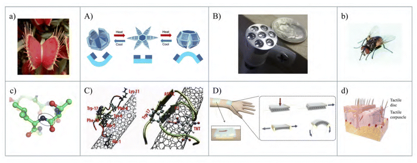 | ||
| Fig. 1 Biomimicry in nanosensors.* (a) Venus flytrap [self-published photograph93]. (A) Thermo-responsive self-folding microgrippers [inspired by (a). Figure reproduced from ref. 31 with permission from the American Chemical Society, copyright 2015]. (b) Common housefly [self-published photograph94]. (B) Aircraft wing deflection sensor [inspired by (b). Figure reproduced from ref. 25 with permission from the American Institute of Aeronautics and Astronautics, Inc., copyright 2015]. (c) Peptide bonds [self-published photograph95]. (C) Nano-electronic noses [inspired by (c). Figure reproduced from ref. 29 with permission from the American Chemical Society, copyright 2012]. (d) Human skin neuroreceptors [self-published work96]. (D) Skin-attachable adhesives (E-skins) [inspired by (d). Figure reproduced from ref. 30 with permission from the American Chemical Society, copyright 2014]. *In this article, when we present natural inspiration and the technology that it led to in the same figure, we use a lowercase English letter for the inspiration and the corresponding uppercase English letter for the technology. For example, (a) inspires (A). | ||
2.1 Biomimetic optical nanosensors
Research in the field of nanosensors has paved the way for the successful implementation of chemical, infrared and mechanical sensors, all of which have vast applications in modern-day technology, from tensile strength of wires to the design of an aerospace vehicle.97Many biological creatures have “built-in” infrared (IR) sensing capabilities. Emulating this particular characteristic has led to the fabrication of a biomimetic IR receptor micro-sensor. Some families of pythons, boas, and even beetles have infrared sensing organs, which sometimes convert IR radiation into heat, which in turn results in a thermal-sensitive system. In the inspired sensor, the incident IR radiation warms a liquid, thus inducing a mechanical deformation of the membrane, which is spotted in a capacitor.98 This is possible because temperature changes can be detected by closely monitoring the changes in the capacitance. The main advantage of such a sensor is that it can be used at room temperature without any additional active cooling, unlike other photo detectors.
Recently, another optical sensor that is aided by the common housefly has been developed which can be used for aircraft wing deflection. The functionality of a compound eye is the wider field-of-view detection and other image processing techniques. The advantage of such a sensor is its low weight, less power consumption, faster computation, and better form factor.
Many animals use their ability to change their coloration for communication, predator capturing, camouflage, etc. This ability to change color has pushed engineers to make sensors that change color in the presence of certain chemicals, hence acting as a chemical sensing sensor. The iridescent scales of some butterflies also show variation in their optical properties when exposed to different kinds of vapors.99 These phenomena of the natural system aided researchers to build photonic vapor sensors. Another device which consisted of periodic ridges and lamella deposited on a silicon wafer showed strong reflectance in well-defined spectral regions. These are some of the prime examples of how researchers, scientists and engineers have mimicked Mother Nature's light exploitation techniques to fabricate optical nanosensors.
2.2 Biomimetic peptide nanosensors
By coupling peptides to a common nanomaterial surface (graphene and carbon nanotubes) a peptide nanosensor is created, which is capable of distinguishing chemically camouflaged mixtures of vapors and detecting chemical warfare agents at a parts-per-billion level.292.3 Biomimetic tactile nanosensors
Tactile wearable sensors that can receive information upon physical contact have gained a lot of attention from researchers and have potential usage in healthcare, robotic, and wearable device applications. Researchers are now working on tactile nanosensors that are based on biological tactile systems.100 The main challenge of such a device is mimicking the human skin mechanoreceptors-receiving and transducing physical stimuli to electrical signals i.e. action potentials, and the biomimetic signal processing mechanism.1012.4 Biomimetic stimuli responsive grippers
Inspired by the Venus flytrap, many stimuli-responsive, biomimetic shape-changing robots/actuators have been proposed by combining stimuli-responsive hydrogels, polymers, or their hybrid combination.32 These grippers actually have far-reaching applications in the fields of biosensors, smart medicine, robotics, and surgery. Hydrogels and polymers are best suited for this purpose because they have the same moduli ranges (∼kPa) of biological tissues and they can extend up to several orders of magnitude in volume when perturbed with external stimuli. This extending and shrinking mechanism replicates the self-actuating action without any external electrical power sources. The main material for this type of gripper is N-isopropylacrylamide (NIPAM) based stimuli-responsive hydrogels, and liquid crystalline material-based stimuli-responsive hydrogels.3 Biomimicry in nanomedicine
Biomimicry means technological achievements that have been developed using inspiration found in nature. The application of nanotechnology in healthcare is referred to as nanomedicine, and biomimicry is the inspiration for it because the human body is a gift of nature and riddled with questions and answers. In the sense of understanding natural structures and using this knowledge to perform and develop many tasks, nanomedicine may be regarded as a specialized division of biomimicry. Biomimicry assists in bone regeneration by use of self-assembled nanostructure tissues,33 hydrogels38–40,102 or electrospinning.103 Skin104,105 and nerve restoration are another important aspect of biomimicry application in nanomedicine.106 Biomimetic nanorobots are another application of biomimicry in nanomedicine. Inspired by platelets107,108 and platelet-membrane-cloaked nanomotors, they perform the task of adsorption and isolation of platelet-targeting biological agents.1093.1 Tissue engineering and surgery
Plastic surgery is used to reconstruct tissues lost due to injury, but it has evolved so much from the surgical practice of 30 years ago. Previously, doctors used synthetic implants using silicone,110 cellulose,111etc. but they might interact with the host in a harmful manner, resulting in infection and rare tumors. And those implants did not replicate biological tissues. Vascularized composite tissue allotransplantation (VCA) was the first reconstructive milestone, but it had inevitable probabilities like transplant rejection. Tissue engineering then became the best alternative solution since this process does not have those limitations.41 Research on self-assembling biomaterials that mimic complex biological structures has also been going on for a long time. Molecules that self-assemble into a nanostructured gel, replicate the key characteristics of the human bone extracellular matrix (ECM). This is extremely important for the generation of bony tissues.33Biomimetic materials also support a microenvironment for cell attachment, proliferation, differentiation, and ultimately tissue regeneration. For example, the most abundant ECM protein in our body is collagen.112 A number of natural micromolecules (collagen,34 silk fibrin,35 fibrinogen, synthetic polymers (poly[glycolic acid],36 poly[L-lactic acid],37 and poly[lactic-co-glycolic acid]) have been processed using electrospinning. This technique builds scaffolds that mimic natural tissues. To produce good scaffolds the biomaterial has to be both biocompatible and support cell growth. This will allow the scaffold to continue the metabolic functions. Moreover, it must promote integration with host tissue and allow vascularization.103
Since the early 90s, multiple biomaterials have been exploited for cartilage and bone applications. And recently, hydrogels have become popular due to their network of interacting hydrophilic polymer chains that have striking similarities with a native tissue ECM.38 Hydrogels maintain the cell structure39 and provide strong mechanical support.40 In the case of bone repair, hydrogels bond with the natural ECM strongly.102 And hydrosols can be injected at the injury site, and they adjust themselves to match the shape of the injury.38 Artificial bone substitutes have been produced from mineral compositions, ceramic or material, bio-glass, or other fiber materials to replicate the organic tissue fraction as boney tissues.113 However, due to the inability to fully master the level of growing fully functional bone with cortical bone components, bone replacement surgical techniques such as non-vascularized or vascularized bone transfer or the Masquelet technique to support bone construction by maintaining flap coverage are used.113–115
Skin is also an important part of the human body that can receive damage from various sources, such as burns and infections. When a skin defect reaches the deep dermal layer, the self-regeneration potential of the skin is obstructed, and artificial skin substitutes using biomimicry can assist in skin repair.105 Artificial skin substitutes have been constructed with different compositions and material properties, mimicking the organization of normal skin.116 Examples are the collagen matrix bonded to a flexible nylon fabric/silicone rubber epidermis116 or collagen-elastin matrix.104 Biomimetic scaffolds can also be used for nerve replacement by mimicking the inner structure and surface structure of original nerves.106
3.2 Biomimetic nanorobots in medical science
One exciting current application of biomimicry is the development of biomimetic nanorobots. These are micro-nanoscale robots for biomedical operations. Industrial robots are used mainly for the automation of dangerous tasks, but biomedical nanorobots are designed to tackle biological events. Biomimetic nanorobot design has recently started to take inspiration from nature, especially from circulating cells such as platelets and leukocytes. Human platelets become the main source of inspiration owing to their work in tackling immune evasion,107,108 pathogen interactions, and hemostasis. The platelet membrane cloaking method is the wrapping of natural platelet cell membranes onto the surface of nanostructures and nanodevices.117 These platelet-membrane-cloaked nanomotors are enclosed by human platelets and perform the adsorption and isolation of platelet-targeted biological agents.109 These PL-motors possess a membrane coating containing multiple functional proteins related to platelets. These PL-motors evade the body's immune system and display rapid locomotion in whole blood. PL-motors can also effectively absorb Shiga toxin using a Vero cell assay and also show binding to pathogens that can be used for rapid bacterial isolation.109Minimally invasive surgery (MIS) and natural orifice transluminal endoscopic surgery (NOTES) is gradually taking the place of open surgery as this will decrease patient trauma and recovery length. Viewing the inside of a patient's lumen is a must for MIS. And now soft endoscopic nanorobots, aided by nature, are being used for reaching distant surgical targets. Robot bodies are composed of soft materials that will reduce the potential of damage to tissue or organs. They are also bendable and flexible enough to move inside the patient's lumen. The meshworm design developed by Bernth et al. was inspired by the earthworm.118 The robot is made up of three segments of a soft plastic-silicone mesh composite. The total length of all segments is 50 cm and each segment is actuated by tendon wound mounted on DC motors, which are small enough to be embedded inside biomimetic meshworms. A PID controller controls the length of each tendon, and a USB camera is mounted at the tip (Fig. 2).
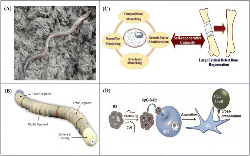 | ||
| Fig. 2 Biomimicry in nanomedicine. (A) Earthworm [self-published photograph121]. (B) A meshworm equipped with an endoscopic camera [inspired by (A). Figure reproduced from ref. 118], (C) schematic diagram of how a combination of nanoelements and growth factor administration may allow regeneration of large critical defect bone. [Figure reproduced from ref. 122 and 123 with permission from the Royal Society of Chemistry, copyright 2017]. (D) Encapsulation of internal influenza proteins on the virus-like-particles (VLPs). [Figure reproduced from ref. 42 with permission from the American Chemical Society, copyright 2013]. | ||
Then there can be nanorobots that have hybrid cell membranes – a combination of red blood cell (RBC) and platelet (PL) membranes. This shows better binding than PL-motors and neutralization of PL-adhering pathogens (Staphylococcus aureus bacteria) and neutralization of pore-forming toxins (α-toxin).119 Some drugs can be carried by platelets because of the site-specific adhesion of platelets. PL – motors can also be used for this purpose.120
3.3 Nanoparticles
Nanoparticles (NPs) are usually defined as particles having a diameter of between 11 and 100![[thin space (1/6-em)]](https://www.rsc.org/images/entities/char_2009.gif) 100 nm. Nanoparticles have unique material characteristics owing to their submicroscopic size. For example, these nanoparticles can be used to improve the activity of different medicines. Many cancer vaccines suffer from the inability to mount a T-cell response. But viruses possess this size, and the immune system can recognize these physical properties. So, replicating these properties with nanoparticles provides a better platform for vaccine design. A viral mimicking vaccine platform capable of encapsulating dendritic cells showed a threefold better reaction than normal cases.42 A controlled immune response towards a specific virus-cell is also possible, which results in a vaccine that is active against the specific virus at a greater rate. Encapsulation of internal influenza proteins on the interior of virus-like particles (VLPs) results in a vaccine that provides protection against 100
100 nm. Nanoparticles have unique material characteristics owing to their submicroscopic size. For example, these nanoparticles can be used to improve the activity of different medicines. Many cancer vaccines suffer from the inability to mount a T-cell response. But viruses possess this size, and the immune system can recognize these physical properties. So, replicating these properties with nanoparticles provides a better platform for vaccine design. A viral mimicking vaccine platform capable of encapsulating dendritic cells showed a threefold better reaction than normal cases.42 A controlled immune response towards a specific virus-cell is also possible, which results in a vaccine that is active against the specific virus at a greater rate. Encapsulation of internal influenza proteins on the interior of virus-like particles (VLPs) results in a vaccine that provides protection against 100![[thin space (1/6-em)]](https://www.rsc.org/images/entities/char_2009.gif) 100 times the lethal dose of an influenza.43 Protein-based nanocages are also of particular interest to researchers because of their excellent ability to deliver drugs to specific sites in the human body. A protein cage nanoparticle, encapsulin, isolated from Thermotoga Maritima, has been developed for multifunctional delivery nanoplatforms for both chemical and genetic engineering44
100 times the lethal dose of an influenza.43 Protein-based nanocages are also of particular interest to researchers because of their excellent ability to deliver drugs to specific sites in the human body. A protein cage nanoparticle, encapsulin, isolated from Thermotoga Maritima, has been developed for multifunctional delivery nanoplatforms for both chemical and genetic engineering44
Nanoparticles increase the bioavailability of compounds via their large surface-to-volume ratio. Many different nanoparticle systems have been created over the years to influence wound care in the medicine industry. Nanoparticles' responses depend on external stimuli such as temperature, pH, ultrasound, light, etc. Dual emission responses of PL polymeric hydrogel Eu-doped PS-co-PNIPAM/d-TPE-doped PNIPAM-co-PAA nanoparticles are controlled strongly and independently by temperature and pH. These different emission colors can be utilized to distinguish cancer cells from normal cells.124 pH and temperature variation of the wound help a hydrogel, which is prepared by cross-linking N-isopropylacrylamide with acrylic acid and silver nanoparticles (AgNPs) to eliminate 95% of pathogens between pH 7.4 and 10.125 Ultrasound, which is another stimulus, can disrupt ionically cross-linked hydrogels which can be used to enhance the toxicity of mitoxantrone toward breast cancer cells.126 Similarly, ultrasound can be used as a trigger to release drugs at specific locations,127,128 and the length and intensity of the ultrasound pulse can be used to control the amount of drugs used.129 Upon exposure to either one- or two-photon excitation, a mesoporous silica nanoparticle (MSN) based drug delivery system (DDS) for controlled anticancer drug release was disclosed.130 In this method, the MSN served as a “photo trigger” for drug release. The precise timing of the anticancer drug's release can be controlled by external light manipulations like varying the irradiation wavelength, intensity, and time. Degradable nanoparticles such as polymeric NPs accelerate wound healing by controlling inflammation, and fibroblast, and osteoclast activity,131 and they can also be used as antibacterial agents and stem cells.132
4 Biomimicry and nanorobots
The recent research explosion in nanotech, with the new discoveries in the bio-molecular field, has brought about a new era in nanorobotics. Nanorobots of any nanostructure can currently handle the task of actuation, intelligence, information processing, sensing, etc. at the nano-scale. Some of the important functionalities of a nanorobot are (i) decentralized intelligence or “swarm intelligence” (ii) self-assembly and replication at the nano-scale (iii) signal processing at the nano-scale and (iv) nano to macro interface architecture.133 In this section, we review some of the recent approaches in this field.4.1 Biomimetic bacteria inspired nanorobots
Wireless nanorobots have had far-reaching biomedical applications. But so far most of the micro/nano swimmers have been imitating the rotary motion of helical bacteria flagella for propulsion, while none of them have the ability to reform themselves in the presence of different external stimuli. Recently, magnetic actuation of self-assembled flagellar nanorobotic swimmers has been discovered.1364.2 Biomimetic platelet-camouflaged nanorobots
While the main purpose of the industrial and life-sized robots is to work in a controlled manner and perform some pre-determined tasks that are set beforehand, the task of biomedical nanorobots is to handle different biological environments, and adapt and improvise to the situation. In 2017, a study was published on a biologically interfaced nanorobot, which is made of magnetic helical nanomotors and is cloaked with the plasma membrane of human platelets.109 This kind of nanorobot shows platelet imitating properties, like binding to toxins or special kinds of pathogens.A different but kind of similar study in 2018, reported the design, construction, and evaluation of a dual-cell membrane functionalized nanorobot for the removal of biological threat agents.46 In this study, the nanorobots consisted of gold nanowires and are camouflaged in hybrid RBC and platelet membranes. This kind of nanorobots can fight against biological threats with no apparent biofouling, while imitating natural cells. In Fig. 3, the preparation and characterization of such a nanorobot are shown.
 | ||
| Fig. 3 Biomimicry in nanorobots. (a) Bacillus subtilis, a Gram-positive bacterium [self-published work134]. (A) Bacteria inspired flagellar curly swimmer nanorobot [inspired by (a). Figure reproduced from ref. 45 with permission from The Author(s), copyright 2017]. (b) SEM image from normal circulating human blood showing red blood cells, several white blood cells, and many small disc-shaped platelets [figure reproduced from ref. 135]. (B) Fluorescent images of PL-motors covered with rhodamine-labeled platelet membranes. Scale bars, 20 μm (left) and 1 μm (right) [inspired by (b). Figure reproduced from ref. 47 with permission from WILEY-VCH Verlag GmbH & Co., copyright 2017]. | ||
5 Biomimicry and nano energy harvesting technology
The population of our beautiful world is expected to increase to 9 billion by 2050.137 This will result in an increase in the global energy consumption rate of 40.8 TW in 2050.138 And the primary means of satisfying this enormous demand (around 80%) is through fossil fuels.139,140 But fossil fuels are detrimental to both human health and the environment. Burning fossil fuels releases mercury, sulfur dioxide, carbon monoxide, nitrogen oxide and other dangerous pollutants that can cause cancer, blood disorders and respiratory conditions.141,142 Groundwater and streams are also at risk from the increased use of fossil fuels.143 Oil spills can endanger aquatic ecosystems and contaminate supplies of drinking water.144 Fossil fuel extraction and processing significantly degrade the environment and ecosystem as well.145–147 Furthermore, the amount of remaining fossil fuels are depleting at an alarming rate with the increasing usage148 leading to the dire need for new alternative energy sources. Renewable energy sources such as solar, wind and wave energy can play a significant role as a replacement for fossil fuels. Nature provides numerous instances of harnessing energy efficiently from these renewable sources. Plants can convert solar energy into simple sugars through the process of photosynthesis. Biological cells can also be an inspiration for energy generation. Different types of biological skin textures can aid in enhancing the absorption ability of solar cells and the movement of kelp can be an inspiration for triboelectric nanogenerators for harnessing ocean energy.52,53,55–60,149–152 All of these alternative energy-generating and harnessing sources aided by nature will greatly assist in meeting the huge energy demand of the future.5.1 Solar energy
Energy is a must for humankind. The current focus of the world is on non-polluting sources of energy, and solar energy is the most desirable of them. 98% of photovoltaic cells are silicon based153 but solar cells require 99.999% pure silicon, which is very energy intensive and whose production steps also create hazardous byproducts.Biomimicry is now being used to harvest solar energy from sunlight. The photosynthesis process used by plants leaves no waste. So the inspiration is there to use biomimicry in a solar cell. One such cell is the Gratzel cell49 in which Gratzel used anthocyanin to recreate photosynthesis. This cell does not use silicon or other heavy metals, and this is much greener for the environment. The downside is that they are not as efficient as silicon cells.
A Gratzel solar cell (also known as a dye-sensitized solar cell or DSSC) mimics the photosynthetic system by having titanium dioxide (TiO2) as NADP+ and carbon dioxide, iodide as a water molecule, and triiodide as oxygen. They harvest the solar energy together in nano-scale systems.49 A basic DSC cell has an energy conversion efficiency of 1% under direct radiation. But using thymol, which has 10% of the mass of the dye from cyanin 3-glucoside and cyanin 3-rutinoside, improved the efficiency.154 The efficiency of DSSCs has exceeded 12% after the last twenty years of development. The amount of light absorbed within the dye is an important part of determining efficiency. For this reason, suitable structures can improve the power conversion efficiency further.155,156
Another approach to improving the efficiency of DSC cells has been the modification and optimization of titania photoelectrodes. One approach was to use sphere voids and large-sized solid particles as a scattering center149,150 or use a multilayer structure151 on the surface of the photoanode. These steps resulted in an increase in the optical path length of the photoanode and a subsequent increase in the light absorption efficiency. Another option is to use new structural materials that can increase the optical path length via light trapping. Butterfly wings have microstructures that are effective solar collectors or blocks. Solar heat is absorbed at a faster rate at the ribs in the wing, which increases the body temperature faster. Then scientists discovered that honeycomb structures are less reactive than cross-ribbing structures, and that this structure also has a greater advantage in terms of light trapping. Scales have a high refractive index, and so total internal reflection occurs more, and nearly all incident light is absorbed. So, inspired by this, a butterfly wing-scale titania film photoanode improved the light absorptivity of the DSC photoanode.52 This structure showed perfect light absorptivity and a higher surface area. This increased the total light harvesting efficiency and dye absorption.
Bioreplication has also become recently popular due to its application in solar energy harvesting. Its application in solar energy is based on two observations, the first observation is the wide angular field that many insects, such as flies have. Each eye of a fly is a compound eye, consisting of multiple elementary eyes. These are arranged radially on a carved surface. The second observation is the almost halving of the reflectance, which is calculated by using a simulation of a prismatic compound lens adhering to a silicon solar cell.53
This experimental technique called the Nano4Bio technique54 has been designed to replicate the layer of a compound eye from an actual specimen. The idea is that by covering the surface of a solar cell with numerous replicas, the angular field of view of the solar cell will increase. The Nano4bio technique can produce multiple replicas simultaneously of multiple biotemplates.
Solar cell efficiency is mainly bound by light-harvesting efficiency, and some research is being done to use the photosynthetic leaf structures to increase the solar cell efficiency. Using a nano-structured poly-carbonate thin-film inspired by the leaves of Salvinia cucullata and Pistia stratiotes helped design a crystalline Si semiconductor with 18.1% power conversion efficiency (PCE).157 The light-trapping coating inspired by the epidermal cells in leaves helped in the fabrication of a graphene/Si Schottky junction.158 The leaf anatomy combined the upper and lower epidermis, palisade, and spongy mesophylls. This upper epidermal layer increases the path length and allows light to traverse deep into the leaf. The middle layer has spongy mesophylls that scatter the light, and this scattered light has a higher chance of absorption by the lower part of the chlorophylls.
5.2 Ocean energy
Ocean waves are a great source of renewable energy as they cover almost 70% of the earth. But unlike solar and wind energy, ocean wave energy harnessing techniques have not developed so much yet.159–161 The integration of offshore wind power plants with ocean wave energy technology could enable power generation in a low-cost and more efficient way, which is obstructed by the deficit of mature wave energy harvesting technology.162,163To collect energy from wave energy, inspiration from kelp, a marine seaweed, has enabled the design of a triboelectric nanogenerator (TENG). Using a conjunction of triboelectric and electrostatic induction effects, a TNG can convert mechanical energy into electrical energy.55–59,152 A kelp-inspired TENG is made of vertically free-standing polymer strips which could sway independently to cause a contact separation with the neighboring strips, with the vibration of a TENG in water mimicking the gentle sway of kelp. A single unit of a BITENG (bio-inspired TENG) provides an output short circuit current of about 10 μA and an open circuit voltage of 260 volts with a maximum power density of 25 μW cm−2 which is high enough to drive 60 LEDs.60 A network of BITENGs can collect more energy from ocean waves and be integrated with an offshore windmill for generating electricity.
5.3 Nanoleaves and nano trees
Another emerging application of biomimicry in the field of energy harvesting is nanoleaves and stems of artificially created trees or plants.51 The nanoleaves are distributed throughout artificial trees and plants, and they can supply a whole household's electricity demand under optimum conditions.164,165 A nanoleaf has two sides, one side has a very thin photovoltaic film that converts the light from the sun into energy. On the other hand, thin thermo-voltaic films convert heat from solar energy into electricity.50 So, the sunlight is then converted into electricity when it falls onto the nanoleaves.Inspired by the leaf structure in nature, a biomimetic nanoleaf was developed for the first time.48 The fabricated nanoleaves contain a thin lamina and parallel veins, forming a monocot leaf structure. CuO nanowires with a high density on a conductive Cu mesh were used as the veins to support the layered double hydroxide (LDH) nanosheet lamina and promote charge transfer. Using CuO/GNS composites with microwave assistance is another quick, time-saving, and environmentally responsible way to make nanoleaves. Due to the strong coordination between graphene and CuO, this performs better than pristine CuO nanoleaves166 (Fig. 4).
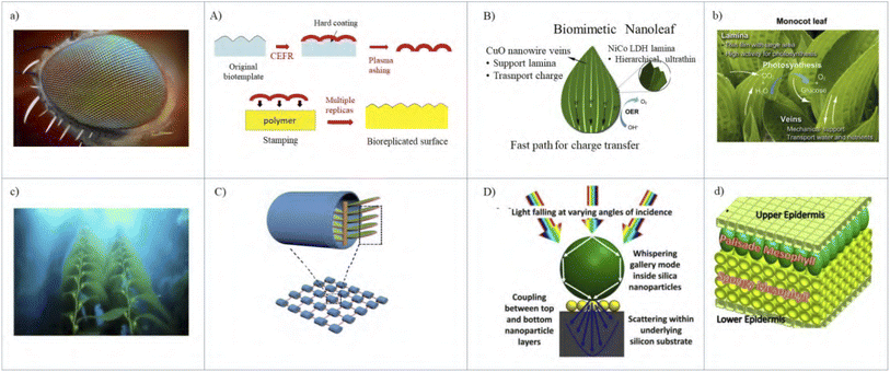 | ||
| Fig. 4 Biomimicry in nanoenergy harvest. (a) Compound eye of fly [self-published photograph167]. (A) Nano4Bio technique developed to replicate the corneal layer of a compound eye from an actual specimen recreated from ref. 54 [inspired by (a)]. (b) Photograph of a monocot leaf [figure reproduced from ref. 48 with permission from Elsevier, copyright 2019]. (B) Biomimetic nanoleaf48 [inspired by (b). Figure reproduced from ref. 48 with permission from Elsevier, copyright 2019]. (c) Images of a kelp plant [figure reproduced from ref. 60 with permission from Elsevier, copyright 2019]. (C) Schematic diagrams of a TENG-unit, the kelp-inspired TENG design and the networks [inspired by (c). Figure reproduced from ref. 60 with permission from Elsevier, copyright 2019]. (d) Schematic of leaf anatomy which inspired the light-trapping coating [figure reproduced from ref. 158 with permission from Elsevier, copyright 2019]. (D) Light management mechanism in a bilayer light-trapping scheme with an all-dielectric sphere. [inspired by (d). Figure reproduced from ref. 158 with permission from Elsevier, copyright 2019]. | ||
6 Biomimetic nanostructures and photonics
Over the years, nature, through evolution, has aided photonics researchers by providing a great variety of biological nanostructures. This has enabled them to design new devices and modify the existing ones to enhance performance. Prevalent biological photonic structures are developed and optimized to perform necessary tasks for that creature, such as camouflage, courtship, warning or attraction to conspecifics, pollination, and so on.173–175 Structural coloration and iridescence of insects, birds' feathers, plants,174,176–179 densely arrayed triangular hairs on Saharan ants for thermoregulation180, anti-reflecting coating of moth and butterfly eyes181,182 and on the transparent wings of hawkmoth,183 the apparent ‘metallic’ view of a fish scale173etc. are the instances of photonic structures existing in nature. In this section, we will review the applications of such natural structures in the area of nanophotonics. Mainly we will review how biomimetic structures can improve the absorption, reflectance, and transmittance spectra. Fig. 5 summarizes the applications of a few such biomimetic structures in nanophotonics.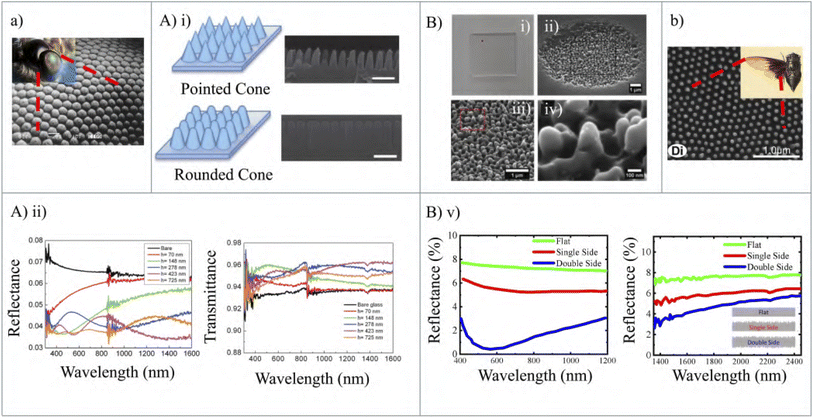 | ||
| Fig. 5 Biomimetic structures in nanophotonics. (a) Actual and SEM images of the eyes of a moth [self-published work168,169]. (A) (i) Moth eye inspired anti-reflective nanocones. The upper one is a pointed cone and its SEM image. The lower one is a rounded cone and its SEM image. The scale bars in the SEM images are all 500 nm (ii) measured reflectance and transmittance of the moth eye inspired nanocones and the bare structures [both inspired by (a). Figure reproduced from ref. 170 with permission from The RSC Pub, copyright 2012]. (b) SEM image of Cryptotympana atrata (cicada species) wing surfaces [(inset) self-published work,171 figure reproduced from ref. 172 with permission from PLoS One]. (B) (i–iv) Photographs and SEM image of an omnidirectional anti-reflective glass inspired from a Cicada Cretensis scale [inspired by (b)]. (v) Reflectance spectra of a pristine (green lines) and laser-treated at one (red lines) or both side (blue lines) fused silica plate. Reflectance in both cases decreases with the introduction of a biomimetic anti-reflection coating [all inspired by (b). Figure reproduced from ref. 78 with permission from The WILEY-VCH Verlag GmbH & Co., copyright 2019]. | ||
In 1967, Bernhard discovered that the reflection over the visible range (400 to 800 nm) from the corneas of night flying moths is reduced because of their having tiny conical burls.184,185 This has led to the discovery of an artificial moth eye, which is a very fine array of protuberances. The working of an artificial moth-eye surface may be understood with the help of the surface layer, which has a gradually changing refractive index having a value from unity to that of the bulk. Due to the presence of gradually varying refractive index layers, the net reflectance is the resultant of an infinite series of reflections at each incremental change in the index. Each of these reflections has different phase factors because of the change in the depth of different layers. Now if this occurs over a distance of  , all the phases will be present, which will result in destructive interference and so the net reflectance will be zero.
6.1 Moth-eye nanostructures
The spacing of the protuberances of the moth-eye must be small enough that the array cannot be resolved by the incident light; otherwise, the array will work as a diffraction grating and the light will simply be redistributed into the diffracted orders.181 The optical properties of an artificial moth-eye surface depend on the height of the protuberances and their spacing. The pattern of the moth-eye array is a photo-resist interference fringe at the intersection of two coherent laser beams.185 The advantage of moth eye anti-reflection coating applied to a high-quality optical component comes at larger angles of incidence. In the case of normal incidence, the moth-eye has the same performance as conventional multilayer coating techniques. But at a larger angle of incidence, the performance of the former is better than that of the latter. In a work presented in 2013, a group showed a comparative study of three different anti-reflecting structures (ARSs) for broadband and angle-independent anti-reflection,61 both theoretically and experimentally. These structures were single diameter nanorods (ARS I), dual diameter nanorods (ARS II), and biomimetic nanotips (ARS III) which take after the submicron structure of a moth's eyes. The simulations were performed using finite difference time domain calculations. All the structures were made of Si. The structures along with the refractive index profile are shown in Fig. 6(a) and (b).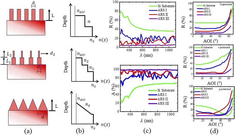 | ||
| Fig. 6 (a) Schematic diagram of three types of Si ARSs with the corresponding geometrical parameters. (b) The refractive index profile for each ARS. The refractive index of air, Si and the mixture of air/Si is represented by nair, ns, n1/2 respectively. nz represents the gradient index of refraction as a function of depth, which is in the z direction. (c) Reflectance and absorption as a function of wavelength for three types of ARSs with a reference planar Si substrate. R stands for reflectance and A stands for absorption. (d) Reflectance data for a Si wafer, ARS I, ARS II, ARS III as a function of angle of incidence (AOI) for unpolarized light, s-polarized light, and p-polarized light with a wavelength of 633 nm . The spectrum is almost independent of the incident angle for a biomimetic ARS [figure reproduced from ref. 61 with permission from the Society of Photo-Optical Instrumentation Engineers (SPIE), copyright 2013]. | ||
The three structures, along with a planar Si wafer (used as a reference) were simulated using FDTD to obtain the results of reflectance and absorption over 250–1100 nm. Fig. 6(c) shows the resulting reflectance and absorption over the mentioned wavelength region. From Fig. 6(c) it is seen that ARS-III has the lowest reflectance value among the others and in the case of absorption shown in Fig. 6(b), it has almost 98% absorption over the wavelength range.
The dependence of reflectance on the angle of incidence (AOI) was studied from 5° to 85° using unpolarized, s-polarized, and p-polarized light with a wavelength of 633 nm. It is seen from Fig. 6(d) that irrespective of polarization, the reflectance of the ARS III structure shows almost no AOI dependence below the Brewster angle (∼75°). From these results, it is concluded that ARS III inspired by the structure of moth eyes, has shown better performance as an anti-reflector than both ARS I and ARS II.
In the case of a solar cell, the greater the absorption of the incident light, the greater the efficiency. So a moth-eye is integrated into solar cells to increase the efficiency of solar cells. As we have seen earlier, a moth-eye anti-reflecting coating has virtually zero dependence of reflectance on the angle of incidence. This property can be exploited in an organic solar cell to enhance performances.62 Forberich et al.62 fabricated a type of organic solar cell with a moth-eye anti-reflective coating as an effective medium at the air–substrate interface. The organic solar cell consisted of a thin (∼20 nm) layer of poly(3,4-ethylenedioxythiophene) doped with poly(styrene sulfonate) (PEDOT:PSS Baytron, Bayer AG) on indium tin oxide (ITO) coated on a glass substrate. The moth-eye was introduced at the air–glass interface.
The measurement results are shown in Fig. 7(a) and (b). From the reflectance vs. wavelength curve for glass/ITO/PEDOT stacks, both curves show oscillatory behavior due to interference in the dielectric slab. However, in the case of moth eye/glass/ITO/PEDOT, the overall reflection is reduced due to the introduction of a moth-eye layer. Fig. 7(b) shows the result of state-of-the-art P3HT/PCBM solar cells with and without moth eyes.
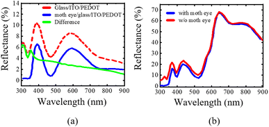 | ||
| Fig. 7 Measured reflection with and without a moth eye from a layer stack of (a) glass/ITO/PEDOT and (b) a P3HT:PCBM solar cell [figure reproduced from ref. 62 with permission from Elsevier B.V., copyright 2007]. | ||
Here it is observed that the highest reduction of reflection occurs in the 350 to 600 nm wavelength range. And beyond that, the reflection becomes almost identical. This happens because, in the case of longer wavelengths, most of the incident light is reflected from the metal electrode, and so the reduction of reflection at the air–glass interface has no significant effect on the overall reflection. From the measured incident angle dependence of external quantum efficiency (EQE) of the P3HT:PCBM solar cell with a moth-eye anti-reflective coating, it is found that at 0° incidence i.e., normal incident, the EQE increases by around 3.5% with the introduction of moth eyes in a complete solar cell. In fact, EQE is greater with moth eyes than with a normal solar cell for all angles of incidence.
Zhu et al.63 reported a nanocone solar cell. It had efficient optical properties with a great processing advantage over nanowire and thin-film solar cells. The work found that the nanocone arrays had shown absorption above 93% which was much greater than the nanowire arrays (75%) or thin films (64%) in the wavelength range of 400–600 nm.64,65
In another work, biomimetic nanostructured antireflection coatings with polymethyl methacrylate (PMMA) layers were integrated with silicon crystalline solar cells. The structure could reduce the average reflectance of solar cells from 13.2% to 7.8%. As a result, power conversion efficiency increased from 12.85% to 14.2%.66
The anti-reflector moth eye used in perovskite solar cells can improve performance significantly. All-inorganic carbon-based perovskite solar cells (PSCs) have drawn increasing interest among researchers due to their low cost and the balance between bandgap and stability.67 However, due to the reflection at the air/glass interface, such PSCs suffer an optical loss of about 10% of the incident photons.186,187 This optical loss results in a reduction in the short circuit current density (Jsc) for PSCs. The inclusion of moth-eye antireflection (AR) nanostructures with wide-range-wavelength AR properties have paved the way to solve the optical loss issue of the above-mentioned PSCs and enhance the power conversion efficiency (PCE).67–73 Compared to the conventional AR, the additional benefit of moth-eye ARs is the enhancement of broadband antireflection through the tuning of structural and geometrical parameters i.e. height, periodic distance, shape, and arrangement.73 Such modulation of optical properties of moth-eye ARs gives rise to new directions to increase the PCE of PSCs. 300 nm and 1000 nm inverted moth-eye structured polydimethylsiloxane (PDMS) films were fabricated using soft lithography and are reported in ref. 72. The former one abated the optical loss at the air/glass interface and enhanced the solar cell efficiency by about 21% from 19.66% and Jsc from 23.83 mA cm−2 to 25.11 mA cm−2. The 1000 nm moth-eye structured PDMS films exhibit elegant coloration due to the interference originating from the diffraction grating effect. Ormostamp-based moth-eye AR has been fabricated using the nano-imprinting method and included on the glass side of the all-inorganic carbon-based CsPbIBr2 PSCs in ref. 67. This resulted in an increase of Jsc from 10.89 mA cm−2 to 11.91 mA cm−2 and PCE increased from 9.17% to 10.08%. The solar cell also showed excellent adaptability to higher temperatures up to 200°.
A superhydrophobic surface is another application of moth-eye structures apart from the anti-reflection coatings. Moth-eye textured surfaces, due to their roughness, possess enhanced water-repellent properties, resulting in a superhydrophobic surface. Such structures have been utilized to fabricate foldable displays for various electronic devices. The transmission property of the structures remains almost invariant under different bending, thermal, and chemical conditions. Hence, the resulting displays show very good anti-reflective function, excellent mechanical resilience, and foldability, as well as superhydrophobicity with good thermal and chemical resistance.74
6.2 Anti-reflective transparent nanostructures
Greta Oto, commonly known as the glasswing butterfly, and the cicada Cretensis species possesses unique transparent wings with broadband and omnidirectional anti-reflection property. This property originates from the presence of nanopillars having periodicity in the range of 150–250 nm on the wing's surface resulting in a gradient of refractive index between air and the wing's membrane.75–77 This property aided the fabrication of omnidirectional anti-reflective glass which can show a reflectivity smaller than 1% irrespective of incident angles in the visible spectrum for S–P linearly polarized configurations.78 Such biomimetic anti-reflective transparent materials have pervasive applications including in displays, solar windows, optical components, and devices.188–195 A single-step laser texturing approach, and nanosecond, and femtosecond laser processing systems are used to fabricate such anti-reflective transparent structures.78,196,1977 Biomimetic nanotechnology in textile engineering
Textiles have been an integral part of human civilization for a long time. Humans started using textiles to protect themselves from the cold and other adverse conditions. With time, the trend and manufacturing process in textiles has changed tremendously. Natural elements showing superhydrophobicity, self-cleaning properties, structural color, and fine quality of fabric have aided textile engineers and researchers to develop more sophisticated textiles with enhanced performances and functionalities. In this section, we will review some of the research work related to nano-biomimicry in the field of textile engineering.7.1 Water repellent textiles
Superhydrophobicity and self-cleaning properties are prevalent in nature in various plants. The most common and notable example is the lotus. Due to the higher contact angle (≥150°), virtually no wetting of the surface takes place on a superhydrophobic surface, leading to the property of self-cleaning. Whenever rain falls on a lotus leaf, the water droplets roll off as a result of a higher contact angle, and on its way, it collects dirt or other materials, cleaning the surface and keeping the leaves dry and free of pathogens.79–81,83Nanotex was founded in 1998. It was based on the idea of superhydrophobicity i.e., repelling water from a surface. They implemented this natural process using nanotechnology, where nanomolecules bonded to the textiles provided an efficient stain-repellent mechanism. This eventually led to the removal of stains and dirt. This technology is now used in over 100 brands around the world. In another research work published in 2013, the superhydrophobicity of lotus leaves inspired the development of a simple coating method using silver nanoparticles (AgNPs). This could facilitate the bionic creation of superhydrophobic surfaces on cotton textiles with the functionality of water repellency, and antibacterial, and UV blocking.84 In this work, NaOH-soaked cotton fiber was immersed into the aqueous solution which resulted in the formation of silver cellulosate which finally formed AgNPs through reduction of Ag cellulosate by C6H6O8 solution. This Ag-coated cotton fabric was then immersed into prehydrolyzed OTES (octyltriethoxysilane) and the final fabric was called “Cot-Ag-OTES”. This fabric was then subjected to ultraviolet-visible reflectance spectrophotometry, scanning electron microscopy, energy dispersive X-ray spectroscopy, and X-ray diffractometry for characterization. The superhydrophobicity was determined by measuring a 5 μL water droplet contact angle (CA) and shedding angle (SHA). Cot-OTES (the sample without AgNPs) had a CA of 135° ± 5.2° and water SHA of 31°. But with the introduction of a AgNP coating, the CA was found to be 156° ± 3.8° and SHA, 8°. This proves the performance enhancement of the water repellence properties of the Cot-Ag-OTES sample. Fig. 8(A)(i–iii) show the optical micrographs of 5 μL water droplets on the surfaces of the untreated (i) Cot-OTES (ii) Cot-Ag-OTES and (iii) fabric samples. With the introduction of AgNPs, the increase of contact angle and hence the hydrophobic properties is evident in this figure. SEM micrographs of untreated and AgNP treated cotton samples are given in Fig. 8(A)(iv and v) and (A)(vi and vii), respectively. In 8(A)(vii), the water droplets are visible on the sample, ensuring its hydrophobic properties. Gram-negative E. coli and Gram-positive S. aureus bacteria were used to evaluate the anti-bacterial properties of the samples. It turned out that the inhibition zone against E. coli and S. aureus was about 2 and 3 nm, respectively, which indicates high antibacterial activity. Transmittance spectra (shown in Fig. 8(A)(viii)) of the samples in the UV range (280–400 nm) were also observed to assess the UV-blocking properties. From this figure, it is pretty clear that the transmittance is very low for AgNP-coated fabrics with and without the OTES layer, which manifests itself as excellent at blocking UV. The sample Cot-Ag-OTES happened to have a UPF (ultraviolet protection factor) value of 266.01 where UPF > 40 is considered to be excellent against UV radiation. So the fabric produced in this work with a silver nanoparticle coating on cotton mimicking the lotus leaf showed excellent performance as superhydrophobic, anti-bacterial, and UV-blocking fabric.
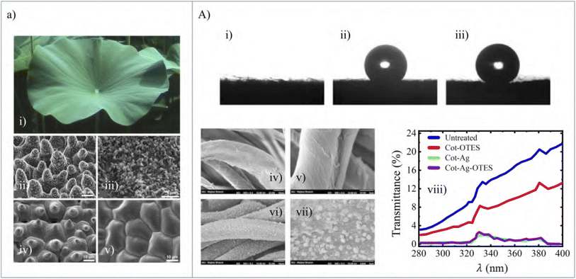 | ||
| Fig. 8 (a) (i) A natural super-hydrophobic surface lotus leaf (ii) SEM image of the upper leaf side prepared by ‘glycerol substitution’ with the hierarchical surface structure consisting of papillae, wax clusters, and wax tubules. (iii) Wax tubules on the upper leaf side. (iv) Upper leaf side after critical-point (CP) drying and dissolution of wax tubules, which make the stomata visible. (v) CP dried underside leaf shows convex cells without stomata [figure reproduced from ref. 82 with permission from Ensikat et al., copyright 2011]. (A) A 5 μL water droplet on the surface of (i) untreated (ii) the Cot-OTES (iii) Cot-Ag-OTES samples. It is observed from these figures that with Ag coating the contact angle is greater than that of the other two and it shows superhydrophobicity. This is inspired by the superhydrophobicity of the lotus leaf. SEM figure of (iv) and (v) untreated and (vi) and (vii) Cot-Ag samples. The water droplets are seen in the cases of (vi) and (vii). (viii) UV-transmission spectra for different types of cotton samples. The UV blocking performance is better in the case of Ag-coated samples [figure reproduced from ref. 84 with permission from Taylor & Francis Group, copyright 2013]. | ||
Bashari et al. proposed a natural element-based water repellent nano-coating, instead of using C8 fluorochemical compounds, onto different fabrics using layer by layer self assembly.85 To replicate the superhydrophobicity observed in nature, various water-repellent finishing agents are used. Among them, fluorocarbons (FCs) possess a unique property of repelling not only water but also oil, providing a surface tension of 10–20 mN m−1 while coated on a fabric. However, it has been reported to exert a detrimental effect on the environment and humans leading to the need for alternative green solutions. Carnauba wax obtained from a Brazilian tree carnauba palm, formally known as Copernicia Prunifera, is a natural water repellent agent which has been used in this proposed method to develop hydrophobic textiles using layer-by-layer self-assembly. This natural water-repellent nano-coating was deposited on three types of samples, cotton, nylon, and cotton-nylon fabrics. Characterization of the solid carnauba nanoparticles was performed through various measurements and experiments. Table 2 shows the contact angles and the antibacterial effects of the untreated and treated samples. It is seen that all of the treated samples have significant hydrophobicity for 30 s and even after washing. The coated fabrics show enhanced antibacterial effects against Staphylococcus aureus (S. aureus) and Escherichia coli (E. coli). The coated fabrics also show good air permeability and thus have paved the way for green ingredient-based environmentally friendly water repellent textiles.
| Sample | Treatment | Averaged contact angle (degree) | Antibacterial effect (%) | ||
|---|---|---|---|---|---|
| 0 s | 30 s | S. aureus | E. coli | ||
| Cotton | Untreated | 45.36 | 0 | — | — |
| Treated | 127.6 | 130.9 | 95.75 | 95.1 | |
| Treated after washing | 109.9 | 101.7 | — | — | |
| Nyco | Untreated | 141.6 | 48.7 | — | — |
| Treated | 132.1 | 135 | 95.75 | 94.6 | |
| Treated after washing | 122.8 | 124.9 | — | — | |
| Nylon 6 | Untreated | 128.6 | 77.3 | — | — |
| Treated | 127 | 131.4 | 97.78 | 94.1 | |
| Treated after washing | 105.2 | 102.4 | — | — | |
7.2 Textile dyeing
Natural instances show a great variety of structural colors that inspired engineers and designers to design various dyeing techniques and processes. Biomass pigments, with their sustainability, naturality, and biocompatibility, have become an interesting research object in cleaner production-focused textile dyeing. Microbial pigments have the advantage of not being constrained by season, climate, or geography.198–204 Prodigiosin, an intracellular metabolite with a pyrrole structure, has bright colors and an antibacterial function, making it attractive to the researchers.86,87 In the past, the pyrrole structure red pigment was insoluble in water and organic solvents had to be used to extract it from the interior of thalli which increased the cost.205,206 Now, if the pyrrole structure pigment is produced by cell metabolization, it will result in cost reduction and extraction of microbial pigments can be avoided.88 To design this trans-membrane culture, it is required to mimic the mechanism and substance transport through the cell membrane. Through the simulation of an artificial membrane, the chemistry of the preparation and kinetic study of the actual cell membrane can be conducted. Phospholipid bilayers are dynamic and closely similar to cell membranes, and this can be used to research trans-membrane behavior. These phospholipid membrane microbial pigments can cross the membrane easily for smaller molecular structure media.88 It was found in the work presented in ref. 88 for positively charged substances that the pigments had a better mass transfer rate than negatively charged segments. The application of electric fields also boosted the extracellular substance concentration. Using a permeability agent also improved the permeability of pigments.8 Conclusion
This article provides a comprehensive review of how different biological instances have contributed to a number of subfields within the area of nanotechnology, including nanosensors, nanomedicine, nanorobots, nanoenergy harvesting, nanophotonics, and nanotextiles. We briefly discussed how biomimicry led to the invention of several cost-efficient nano-sensors with different notable features.Further applications of biomimicry in the field of nano-medicine and nano-robots to provide assistance in surgery, tissue engineering and drug delivery to different sites of human body have been discussed in adequate detail. Biomimetic innovations have also contributed to the development of novel methods and enhancement of existing ones to harness energy from natural sources to meet the ever increasing demand for eco-friendly green energy. Later we discussed the ways bio-aided innovations have contributed to the development of different nano-photonic structures which significantly improves the performance of solar cells and other such optical devices. Finally, the role of biomimetic nanotechnology in the field of textile engineering has been described to develop superior performing fabric and dyeing pigments compared to the conventional ones.
It should be noted that this review touches on almost all the recent biomimetic innovations and ideas developed in the field of nanosensors, nano-medicine, nano-robots, nano-energy harvesting, nano-photonics and textile engineering. Nevertheless, the studies presented here provide a glimpse into the vast possibilities of biomimicry. According to a study in 2016, the estimated number of living species on Earth ranges from 2 million to 1 trillion, of which approximately 1.74 million have been catalogued.207,208 So, more than 80 percent of the living creatures have not yet been described. Each of these organisms possesses unique physiological and exterior characteristics to perform distinct functions. Furthermore, these species are constantly evolving over time to adapt to the ever changing nature. Hence, the scope of taking inspirations for novel innovations and improving current ones is infinite. Consequently, the potential applications of biomimicry are limitless. We deem that biologists will discover more astounding information from this vast number of existing species in the future and nanotech researchers will utilize them to build a better future for human civilization.
Author contributions
The authors confirm their contribution to the paper as follows: conceptualization M. H. H., B. S., T. A. and S. M. C., writing-original draft, literature review, data collection, figure preparation and referencing M. H. H., B. S. and T. A., review and editing, supervision S. M. C. All authors have read and agreed to the published version of the manuscript.Conflicts of interest
The authors declare no conflicts of interest.Acknowledgements
The authors acknowledge the support of both the Department of Electrical and Electronic Engineering, Bangladesh University of Engineering and Technology, Dhaka, Bangladesh, and the Department of Computer Science and Engineering, Brac University, Dhaka, Bangladesh.Notes and references
- J. F. Vincent, O. A. Bogatyreva, N. R. Bogatyrev, A. Bowyer and A.-K. Pahl, J. R. Soc., Interface, 2006, 3, 471–482 CrossRef PubMed.
- O. H. Schmitt, Some Interesting and Useful Biomimetic Transforms, Proceeding, Third International Biophysics Congress, Boston, Mass., 1969, p. 297 Search PubMed.
- D. P. Pulsifer, A. Lakhtakia, R. J. Martín-Palma and C. G. Pantano, Bioinspiration Biomimetics, 2010, 5, 036001 CrossRef PubMed.
- R. J. Martín-Palma and A. Lakhtakia, Int. J. Smart Nano Mater., 2013, 4, 83–90 CrossRef.
- M. Simovic-Pavlovic, B. Bokic, D. Vasiljevic and B. Kolaric, Appl. Sci., 2022, 12, 905 CrossRef CAS.
- H. Droogendijk, M. J. de Boer, R. G. Sanders and G. J. Krijnen, J. R. Soc., Interface, 2014, 11, 20140438 CrossRef CAS PubMed.
- M. Asadnia, A. G. P. Kottapalli, J. Miao, M. E. Warkiani and M. S. Triantafyllou, J. R. Soc., Interface, 2015, 12, 20150322 CrossRef PubMed.
- S. Karthick and A. Sen, Phys. Rev. Appl., 2018, 10, 034037 CrossRef CAS.
- F. Khoshnoud and C. W. de Silva, IEEE Instrum. Meas. Mag., 2012, 15, 14–24 Search PubMed.
- M. Kruusmaa, P. Fiorini, W. Megill, M. de Vittorio, O. Akanyeti, F. Visentin, L. Chambers, H. El Daou, M.-C. Fiazza and J. Ježov, et al. , IEEE Robot. Autom. Mag., 2014, 21, 51–62 Search PubMed.
- J. Song, R. Wang, G. Zhang, Z. Shang, L. Zhang and W. Zhang, IEEE Access, 2019, 7, 102366–102376 Search PubMed.
- A. Rahaman, H. Jung and B. Kim, Appl. Sci., 2021, 11, 1305 CrossRef CAS.
- P. P. Vijayan and D. Puglia, Emergent Mater., 2019, 2, 391–415 CrossRef.
- L. Zhang and T. J. Webster, Nano Today, 2009, 4, 66–80 CrossRef CAS.
- E.-S. Kim, E. H. Ahn, T. Dvir and D.-H. Kim, Int. J. Nanomed., 2014, 9, 1 CAS.
- T. Li, X. Qin, Y. Li, X. Shen, S. Li, H. Yang, C. Wu, C. Zheng, J. Zhu and F. You, et al. , Front. Bioeng. Biotechnol., 2020, 8, 371 CrossRef PubMed.
- C. Guido, G. Maiorano, B. Cortese, S. D'Amone and I. E. Palamà, Bioengineering, 2020, 7, 111 CrossRef CAS PubMed.
- Z. Jakšić and O. Jakšić, Biomimetics, 2020, 5, 24 CrossRef PubMed.
- A. Giwa, S. Hasan, A. Yousuf, S. Chakraborty, D. Johnson and N. Hilal, Desalination, 2017, 420, 403–424 CrossRef CAS.
- R. Bogue, Assemb. Autom., 2008, 28, 282–288 CrossRef.
- N. Soudi, S. Nanayakkara, N. M. Jahed and S. Naahidi, Sol. Energy, 2020, 208, 31–45 CrossRef CAS.
- Y. Saylan, Ö. Erdem, F. Inci and A. Denizli, Biomimetics, 2020, 5, 20 CrossRef CAS PubMed.
- S. Rauf, M. A. Hayat Nawaz, M. Badea, J. L. Marty and A. Hayat, Sensors, 2016, 16, 1931 CrossRef PubMed.
- Q. Shen, Z. Luo, S. Ma, P. Tao, C. Song, J. Wu, W. Shang and T. Deng, Adv. Mater., 2018, 30, 1707632 CrossRef PubMed.
- S. A. Frost, C. Wright and M. A. Khan, AIAA Guidance, Navigation, and Control Conference, 2015, p. 0083 Search PubMed.
- R. Hanlon, Curr. Biol., 2007, 17, R400–R404 CrossRef CAS PubMed.
- H. Schmitz and H. Bousack, PLoS One, 2012, 7, e37627 CrossRef CAS PubMed.
- K. Kertész, G. Piszter, Z. Bálint and L. P. Biró, Sensors, 2018, 18, 4282 CrossRef PubMed.
- Y. Cui, S. N. Kim, R. R. Naik and M. C. McAlpine, Acc. Chem. Res., 2012, 45, 696–704 CrossRef CAS PubMed.
- J. Park, Y. Lee, J. Hong, Y. Lee, M. Ha, Y. Jung, H. Lim, S. Y. Kim and H. Ko, ACS Nano, 2014, 8, 12020–12029 CrossRef CAS PubMed.
- J. C. Breger, C. Yoon, R. Xiao, H. R. Kwag, M. O. Wang, J. P. Fisher, T. D. Nguyen and D. H. Gracias, ACS Appl. Mater. Interfaces, 2015, 7, 3398–3405 CrossRef CAS PubMed.
- C. Yoon, Nano Convergence, 2019, 6, 1–14 CrossRef CAS PubMed.
- J. D. Hartgerink, E. Beniash and S. I. Stupp, Science, 2001, 294, 1684–1688 CrossRef CAS PubMed.
- J. A. Matthews, G. E. Wnek, D. G. Simpson and G. L. Bowlin, Biomacromolecules, 2002, 3, 232–238 CrossRef CAS PubMed.
- C. Li, C. Vepari, H. J. Jin, H. J. Kim and D. L. Kaplan, Biomaterials, 2006, 27, 3115–3124 CrossRef CAS PubMed.
- G. E. Wnek, M. E. Carr, D. G. Simpson and G. L. Bowlin, Nano Lett., 2003, 3, 213–216 CrossRef CAS.
- E. D. Boland, G. E. Wnek, D. G. Simpson, K. J. Pawlowski and G. L. Bowlin, J. Macromol. Sci., Pure Appl. Chem., 2001, 38A, 1231–1243 CrossRef.
- M. Liu, X. Zeng, C. Ma, H. Yi, Z. Ali, X. Mou, S. Li, Y. Deng and N. He, Bone Res., 2017, 5, 1–20 Search PubMed.
- H. Yamaoka, H. Asato, T. Ogasawara, S. Nishizawa, T. Takahashi, T. Nakatsuka, I. Koshima, K. Nakamura, H. Kawaguchi, U. I. Chung, T. Takato and K. Hoshi, J. Biomed. Mater. Res., Part A, 2006, 78, 1–11 CrossRef PubMed.
- R. L. Mauck, M. A. Soltz, C. C. Wang, D. D. Wong, P. H. G. Chao, W. B. Valhmu, C. T. Hung and G. A. Ateshian, J. Biomech. Eng., 2000, 122, 252–260 CrossRef CAS PubMed.
- K. Amin, R. Moscalu, A. Imere, R. Murphy, S. Barr, Y. Tan, R. Wong, P. Sorooshian, F. Zhang, J. Stone, J. Fildes, A. Reid and J. Wong, Nanomedicine, 2019, 14, 2679–2696 CrossRef CAS PubMed.
- N. M. Molino, A. K. Anderson, E. L. Nelson and S. W. Wang, ACS Nano, 2013, 7, 9743–9752 CrossRef CAS PubMed.
- D. P. Patterson, A. Rynda-Apple, A. L. Harmsen, A. G. Harmsen and T. Douglas, ACS Nano, 2013, 7, 3036–3044 CrossRef CAS.
- E. J. Lee, N. K. Lee and I.-S. Kim, Adv. Drug Delivery Rev., 2016, 106, 157–171 CrossRef CAS PubMed.
- J. Ali, U. K. Cheang, J. D. Martindale, M. Jabbarzadeh, H. C. Fu and M. Jun Kim, Sci. Rep., 7, 1–10 Search PubMed.
- B. Esteban-Fernández de Ávila, P. Angsantikul, D. E. Ramírez-Herrera, F. Soto, H. Teymourian, D. Dehaini, Y. Chen, L. Zhang and J. Wang, Sci. Robot., 2018, 3(18), eaat0485 CrossRef PubMed.
- J. Li, P. Angsantikul, W. Liu, B. Esteban-Fernández de Ávila, X. Chang, E. Sandraz, Y. Liang, S. Zhu, Y. Zhang and C. Chen, et al. , Adv. Mater., 2018, 30, 1704800 CrossRef PubMed.
- B. Chen, Z. Zhang, S. Kim, M. Baek, D. Kim and K. Yong, Appl. Catal., B, 2019, 259, 118017 CrossRef CAS.
- G. P. Smestad and M. Gratzel, J. Chem. Educ., 1998, 75, 752 CrossRef CAS.
- C. Bhuvaneswari, R. Rajeswari, C. Kalaiarasan and K. Muthukumararajaguru, Int. J. Sci. Res. Publ., 2013, 3, 1 Search PubMed.
- K. V. Kumar, G. A. Kumar and G. A. K. Reddy, Caribb. J. Sci. Technol., 2014, 2, 424–430 Search PubMed.
- W. Zhang, D. Zhang, T. Fan, J. Gu, J. Ding, H. Wang, Q. Guo and H. Ogawa, Chem. Mater., 2009, 21, 33–40 CrossRef.
- F. Chiadini, V. Fiumara, A. Scaglione and A. Lakhtakia, Bioinspiration Biomimetics, 2010, 6, 014002 CrossRef PubMed.
- R. J. Martín-Palma and A. Lakhtakia, Int. J. Smart Nano Mater., 2013, 4, 83–90 CrossRef.
- S. Cui, Y. Zheng, J. Liang and D. Wang, Nano Res., 2018, 11, 1873–1882 CrossRef CAS.
- C. He, W. Zhu, G. Q. Gu, T. Jiang, L. Xu, B. D. Chen, C. B. Han, D. Li and Z. L. Wang, Nano Res., 2018, 11, 1157–1164 CrossRef.
- J. He, T. Wen, S. Qian, Z. Zhang, Z. Tian, J. Zhu, J. Mu, X. Hou, W. Geng and J. Cho, et al. , Nano Energy, 2018, 43, 326–339 CrossRef CAS.
- D. Y. Kim, H. S. Kim, D. S. Kong, M. Choi, H. B. Kim, J.-H. Lee, G. Murillo, M. Lee, S. S. Kim and J. H. Jung, Nano Energy, 2018, 45, 247–254 CrossRef CAS.
- C. Xu, Y. Zi, A. C. Wang, H. Zou, Y. Dai, X. He, P. Wang, Y.-C. Wang, P. Feng and D. Li, et al. , Adv. Mater., 2018, 30, 1706790 CrossRef PubMed.
- N. Wang, J. Zou, Y. Yang, X. Li, Y. Guo, C. Jiang, X. Jia and X. Cao, Nano Energy, 2019, 55, 541–547 CrossRef CAS.
- Y. F. Huang and S. Chattopadhyay, J. Nanophotonics, 2013, 7, 073594 CrossRef.
- K. Forberich, G. Dennler, M. C. Scharber, K. Hingerl, T. Fromherz and C. J. Brabec, Thin Solid Films, 2008, 516, 7167–7170 CrossRef CAS.
- J. Zhu, Z. Yu, S. Fan and Y. Cui, Mater. Sci. Eng., R, 2010, 70, 330–340 CrossRef.
- W. Yi, D.-B. Xiong and D. Zhang, Nano Adv., 2016, 1, 62–70 Search PubMed.
- M. Yu, Y.-Z. Long, B. Sun and Z. Fan, Nanoscale, 2012, 4, 2783–2796 RSC.
- J. Chen, W.-L. Chang, C. Huang and K. Sun, Opt. Express, 2011, 19, 14411 CrossRef CAS PubMed.
- W. Lan, D. Chen, Q. Guo, B. Tian, X. Xie, Y. He, W. Chai, G. Liu, P. Dong and H. Xi, et al. , Nanomaterials, 2021, 11, 2726 CrossRef CAS PubMed.
- S. Ju, M. Byun, M. Kim, J. Jun, D. Huh, D. S. Kim, Y. Jo and H. Lee, Nano Res., 2020, 13, 1156–1161 CrossRef CAS.
- W. Qarony, M. I. Hossain, R. Dewan, S. Fischer, V. B. Meyer-Rochow, A. Salleo, D. Knipp and Y. H. Tsang, Adv. Theory Simul., 2018, 1, 1800030 CrossRef.
- F. Tao, P. Hiralal, L. Ren, Y. Wang, Q. Dai, G. A. Amaratunga and H. Zhou, Nanoscale Res. Lett., 2014, 9, 1–7 CrossRef CAS PubMed.
- J. Sun, X. Wang, J. Wu, C. Jiang, J. Shen, M. A. Cooper, X. Zheng, Y. Liu, Z. Yang and D. Wu, Sci. Rep., 2018, 8, 1–10 Search PubMed.
- M.-c. Kim, S. Jang, J. Choi, S. M. Kang and M. Choi, Nano-Micro Lett., 2019, 11, 1–10 CrossRef PubMed.
- S. Ji, K. Song, T. B. Nguyen, N. Kim and H. Lim, ACS Appl. Mater. Interfaces, 2013, 5, 10731–10737 CrossRef CAS PubMed.
- H.-W. Yun, G.-M. Choi, H. K. Woo, S. J. Oh and S.-H. Hong, Curr. Appl. Phys., 2020, 20, 1163–1170 CrossRef.
- V. R. Binetti, J. D. Schiffman, O. D. Leaffer, J. E. Spanier and C. L. Schauer, Integr. Biol., 2009, 1, 324–329 CrossRef PubMed.
- R. H. Siddique, G. Gomard and H. Hölscher, Nat. Commun., 2015, 6, 1–8 Search PubMed.
- J. Morikawa, M. Ryu, G. Seniutinas, A. Balcytis, K. Maximova, X. Wang, M. Zamengo, E. P. Ivanova and S. Juodkazis, Langmuir, 2016, 32, 4698–4703 CrossRef CAS PubMed.
- A. Papadopoulos, E. Skoulas, A. Mimidis, G. Perrakis, G. Kenanakis, G. D. Tsibidis and E. Stratakis, Adv. Mater., 2019, 31, 1901123 CrossRef PubMed.
- A. Marmur, Langmuir, 2004, 20, 3517–3519 CrossRef CAS PubMed.
- L. Zhang, Z. Zhou, B. Cheng, J. M. DeSimone and E. T. Samulski, Langmuir, 2006, 22, 8576–8580 CrossRef CAS PubMed.
- K. Balani, R. G. Batista, D. Lahiri and A. Agarwal, Nanotechnology, 2009, 20, 305707 CrossRef PubMed.
- H. J. Ensikat, P. Ditsche-Kuru, C. Neinhuis and W. Barthlott, Beilstein J. Nanotechnol., 2011, 2, 152–161 CrossRef CAS PubMed.
- S. Das, N. Shanmugam, A. Kumar and S. Jose, Bioinspired, Biomimetic Nanobiomater., 2017, 6, 224–235 CrossRef.
- M. Shateri-Khalilabad and M. E. Yazdanshenas, J. Text. Inst., 2013, 104, 861–869 CrossRef CAS.
- A. Bashari, A. H. Salehi K and N. Salamatipour, J. Text. Inst., 2020, 111, 1148–1158 CrossRef CAS.
- X. Liu, Y. Wang, S. Sun, C. Zhu, W. Xu, Y. Park and H. Zhou, Prep. Biochem. Biotechnol., 2013, 43, 271–284 CrossRef CAS PubMed.
- O. Falklöf and B. Durbeej, ChemPhysChem, 2016, 17, 954–957 CrossRef.
- J. Gong, J. Liu, X. Tan, Z. Li, Q. Li and J. Zhang, Nanomaterials, 2019, 9, 114 CrossRef PubMed.
- A. Munawar, Y. Ong, R. Schirhagl, M. A. Tahir, W. S. Khan and S. Z. Bajwa, RSC Adv., 2019, 9, 6793–6803 RSC.
- S. A. Perdomo, J. M. Marmolejo-Tejada and A. Jaramillo-Botero, J. Electrochem. Soc., 2021, 168, 107506 CrossRef CAS.
- A. K. Srivastava, A. Dev and S. Karmakar, Environ. Chem. Lett., 2018, 16, 161–182 CrossRef CAS.
- P. J. Vikesland, Nat. Nanotechnol., 2018, 13, 651–660 CrossRef CAS PubMed.
- N. Elhardt, Self-published photo. From Wikimedia Commons, distributed under a CC BY-SA 2.5 license.
- USDAgov, From Wikimedia Commons, distributed under a CC BY 2.0 license.
- Webridge, From Wikimedia Commons, distributed under a CC BY-SA 3.0 license.
- B. Blaus, Medical Gallery of Blausen Medical 2014, Wiki. J. Med., 1 (2), DOI:10.15347/wjm/2014.010. ISSN 2002-4436. Own work, distributed under a CC BY 3.0.
- R. J. Martín-Palma and M. Kolle, Opt. Laser Technol., 2019, 109, 270–277 CrossRef.
- H. Herzog, A. Klein, H. Bleckmann, P. Holik, S. Schmitz, G. Siebke, S. Tätzner, M. Lacher and S. Steltenkamp, Bioinspiration Biomimetics, 2015, 10, 036001 CrossRef PubMed.
- R. A. Potyrailo, H. Ghiradella, A. Vertiatchikh, K. Dovidenko, J. R. Cournoyer and E. Olson, Nat. Photonics, 2007, 1, 123–128 CrossRef CAS.
- Y. Lee, J. Park, A. Choe, S. Cho, J. Kim and H. Ko, Adv. Funct. Mater., 2020, 30, 1904523 CrossRef CAS.
- Y. Lee and J.-H. Ahn, ACS Nano, 2020, 14, 1220–1226 CrossRef CAS PubMed.
- G. Wu, C. Feng, J. Quan, Z. Wang, W. Wei, S. Zang, S. Kang, G. Hui, X. Chen and Q. Wang, Carbohydr. Polym., 2018, 182, 215–224 CrossRef CAS PubMed.
- M. M. Martino, S. Brkic, E. Bovo, M. Burger, D. J. Schaefer, T. Wolff, L. Gürke, P. S. Briquez, H. M. Larsson and R. Gianni-Barrera, et al. , Front. Bioeng. Biotechnol., 2015, 3, 45 Search PubMed.
- D. Girard, B. Laverdet, V. Buhé, M. Trouillas, K. Ghazi, M. M. Alexaline, C. Egles, L. Misery, B. Coulomb and J.-J. Lataillade, et al. , Tissue Eng., Part B, 2017, 23, 59–82 CrossRef PubMed.
- B. Weyand and P. Vogt, Biomimetics and Bionic Applications with Clinical Applications, Springer, 2021, pp. 29–43 Search PubMed.
- J. Du, H. Chen, L. Qing, X. Yang and X. Jia, Biomater. Sci., 2018, 6, 1299–1311 RSC.
- M. Olsson, P. Bruhns, W. A. Frazier, J. V. Ravetch and P. A. Oldenborg, Blood, 2005, 105, 3577–3582 CrossRef CAS PubMed.
- P. Sims, S. Rollins and T. Wiedmer, J. Biol. Chem., 1989, 264, 19228–19235 CrossRef CAS PubMed.
- J. Li, P. Angsantikul, W. Liu, B. Esteban-Fernández de Ávila, X. Chang, E. Sandraz, Y. Liang, S. Zhu, Y. Zhang, C. Chen, W. Gao, L. Zhang and J. Wang, Adv. Mater., 2018, 30, 1–8 CAS.
- S. Barr and A. Bayat, Aesthetic Surg. J., 2011, 31, 56–67 CrossRef PubMed.
- S. Barr, E. W. Hill and A. Bayat, Acta Biomater., 2017, 49, 260–271 CrossRef CAS PubMed.
- P. X. Ma, Adv. Drug Delivery Rev., 2008, 60, 184–198 CrossRef CAS PubMed.
- E. Brett, J. Flacco, C. Blackshear, M. T. Longaker and D. C. Wan, BioRes. Open Access, 2017, 6, 1–6 CrossRef CAS PubMed.
- D. W. Buck and G. A. Dumanian, et al. , Plast. Reconstr. Surg., 2012, 129, 950e–956e CrossRef CAS PubMed.
- R. Verboket, M. Leiblein, C. Seebach, C. Nau, M. Janko, M. Bellen, H. Bönig, D. Henrich and I. Marzi, Eur. J. Trauma Emerg. Surg., 2018, 44, 649–665 CrossRef CAS PubMed.
- F. M. Wood, Int. J. Biochem. Cell Biol., 2014, 56, 133–140 CrossRef CAS PubMed.
- R. H. Fang, Y. Jiang, J. C. Fang and L. Zhang, Biomaterials, 2017, 128, 69–83 CrossRef CAS PubMed.
- J. E. Bernth, A. Arezzo and H. Liu, IEEE Rob. Autom. Lett., 2017, 2, 1718–1724 Search PubMed.
- B. E. F. De Ávila, P. Angsantikul, D. E. Ramírez-Herrera, F. Soto, H. Teymourian, D. Dehaini, Y. Chen, L. Zhang and J. Wang, Sci. Robot., 2018, 3, 485 Search PubMed.
- Y. Lu, Q. Hu, C. Jiang and Z. Gu, Curr. Opin. Biotechnol., 2019, 58, 81–91 CrossRef CAS PubMed.
- R. Hille, Distributed under a CC BY-SA 3.0 license, https://candide.com/GB/insects/607a3d43-0eb0-4100-8f0f-43117cb332a0.
- Y. Li and C. Liu, Nanoscale, 2017, 9, 4862–4874 RSC.
- A. Khademhosseini and R. Langer, Nat. Protoc., 2016, 11, 1775–1781 CrossRef CAS PubMed.
- Y. Zhao, C. Shi, X. Yang, B. Shen, Y. Sun, Y. Chen, X. Xu, H. Sun, K. Yu and B. Yang, et al. , ACS Nano, 2016, 10, 5856–5863 CrossRef CAS PubMed.
- H. Haidari, K. Vasilev, A. J. Cowin and Z. Kopecki, ACS Appl. Mater. Interfaces, 2022, 14(46), 51744–51762 CrossRef CAS PubMed.
- N. Huebsch, C. J. Kearney, X. Zhao, J. Kim, C. A. Cezar, Z. Suo and D. J. Mooney, Proc. Natl. Acad. Sci. U. S. A., 2014, 111, 9762–9767 CrossRef CAS PubMed.
- A. Schroeder, Y. Avnir, S. Weisman, Y. Najajreh, A. Gabizon, Y. Talmon, J. Kost and Y. Barenholz, Langmuir, 2007, 23, 4019–4025 CrossRef CAS PubMed.
- A. Schroeder, R. Honen, K. Turjeman, A. Gabizon, J. Kost and Y. Barenholz, J. Controlled Release, 2009, 137, 63–68 CrossRef CAS PubMed.
- H. Epstein-Barash, G. Orbey, B. E. Polat, R. H. Ewoldt, J. Feshitan, R. Langer, M. A. Borden and D. S. Kohane, Biomaterials, 2010, 31, 5208–5217 CrossRef CAS PubMed.
- Q. Lin, Q. Huang, C. Li, C. Bao, Z. Liu, F. Li and L. Zhu, J. Am. Chem. Soc., 2010, 132, 10645–10647 CrossRef CAS PubMed.
- R. M. Baxter, T. Dai, J. Kimball, E. Wang, M. R. Hamblin, W. P. Wiesmann, S. J. McCarthy and S. M. Baker, J. Biomed. Mater. Res., Part A, 2013, 101, 340–348 CrossRef PubMed.
- E. M. Ahmed, J. Adv. Res., 2015, 6, 105–121 CrossRef CAS PubMed.
- A. Ummat, A. Dubey and C. Mavroidis, Biomimetics: Biologically Inspired Technologies, CRC Press, Boca Raton, FL, 2005, pp. 201–227 Search PubMed.
- D. G. Beards, From Wikimedia Commons, distributed under a CC BY-SA 4.0 license.
- B. Wetzel and H. Schaefer, National Cancer Institute., 1982, retreived from, https://visualsonline.cancer.gov/details.cfm?imageid=2129.
- J. Ali, U. K. Cheang, J. D. Martindale, M. Jabbarzadeh, H. C. Fu and M. Jun Kim, Sci. Rep., 2017, 7, 1–10 CrossRef CAS PubMed.
- L. Roberts, 9 Billion?, 2011 Search PubMed.
- N. S. Lewis and D. G. Nocera, Proc. Natl. Acad. Sci. U. S. A., 2006, 103, 15729–15735 CrossRef CAS PubMed.
- A. Hassan, S. Z. Ilyas, A. Jalil and Z. Ullah, Environ. Sci. Pollut. Res., 2021, 28, 21204–21211 CrossRef PubMed.
- W. Bank, Pakistan Strategic Country Environmental Assessment, 2006 Search PubMed.
- K. B. Karnauskas, S. L. Miller and A. C. Schapiro, GeoHealth, 2020, 4(5), e2019GH000237 CrossRef PubMed.
- F. Perera, Int. J. Environ. Res. Public Health, 2018, 15, 16 CrossRef PubMed.
- L. Allen, M. J. Cohen, D. Abelson and B. Miller, The World's Water, Springer, 2012, pp. 73–96 Search PubMed.
- J. M. Navas, M. Babín, S. Casado, C. Fernández and J. V. Tarazona, Mar. Environ. Res., 2006, 62, S352–S355 CrossRef CAS PubMed.
- N. Butt, H. Beyer, J. Bennett, D. Biggs, R. Maggini, M. Mills, A. Renwick, L. Seabrook and H. Possingham, Science, 2013, 342, 425–426 CrossRef CAS PubMed.
- S. Wylie, E. Wilder, L. Vera, D. Thomas and M. McLaughlin, Sci. Technol. Soc., 2017, 3, 426 Search PubMed.
- M. B. Harfoot, D. P. Tittensor, S. Knight, A. P. Arnell, S. Blyth, S. Brooks, S. H. Butchart, J. Hutton, M. I. Jones and V. Kapos, et al. , Conserv. Lett., 2018, 11, e12448 CrossRef.
- S. Shafiee and E. Topal, Energy Policy, 2009, 37, 181–189 CrossRef.
- C. J. Barbe, F. Arendse, P. Comte, M. Jirousek, F. Lenzmann, V. Shklover and M. Grätzel, J. Am. Ceram. Soc., 1997, 80, 3157–3171 CrossRef CAS.
- S. Hore, P. Nitz, C. Vetter, C. Prahl, M. Niggemann and R. Kern, Chem. Commun., 2005, 2011–2013 RSC.
- Z.-S. Wang, H. Kawauchi, T. Kashima and H. Arakawa, Coord. Chem. Rev., 2004, 248, 1381–1389 CrossRef CAS.
- J. Ren, Q. Liu, Y. Pei, Y. Wang, S. Yang, S. Lin, W. Chen, S. Ling and D. L. Kaplan, Adv. Mater. Technol., 2021, 6, 2001301 CrossRef CAS.
- T. Wang and T. F. Ciszek, Purified silicon production system, US Pat., US6712908B2, 2004, https://patents.google.com/patent/US6712908 Search PubMed.
- S. Ali and J. Matthew, Biomimicry in Solar Energy Conversion with Natural Dye Sensitized Nanocrystalline Photovoltaic Cells, Post graduate thesis, Department of Chemistry and Biochemistry Obelin College, Ohio, 2007, pp. 1–22.
- T. W. Hamann, R. A. Jensen, A. B. Martinson, H. Van Ryswyk and J. T. Hupp, Energy Environ. Sci., 2008, 1, 66–78 RSC.
- A. Yella, H.-W. Lee, H. N. Tsao, C. Yi, A. K. Chandiran, M. K. Nazeeruddin, E. W.-G. Diau, C.-Y. Yeh, S. M. Zakeeruddin and M. Grätzel, Science, 2011, 334, 629–634 CrossRef CAS PubMed.
- W. Yan, Y. Huang, L. Wang, F. Vuellers, M. N. Kavalenka, H. Hoelscher, S. Dottermusch, B. S. Richards and E. Klampaftis, Sol. Energy Mater. Sol. Cells, 2018, 186, 105–110 CrossRef CAS.
- S. Das, M. J. Hossain, S.-F. Leung, A. Lenox, Y. Jung, K. Davis, J.-H. He and T. Roy, Nano Energy, 2019, 58, 47–56 CrossRef CAS.
- Z. L. Wang, Faraday Discuss., 2015, 176, 447–458 RSC.
- Q. Schiermeier, Nature, 2016, 535, 212–214 CrossRef CAS PubMed.
- J. Wang, S. Li, F. Yi, Y. Zi, J. Lin, X. Wang, Y. Xu and Z. L. Wang, Nat. Commun., 2016, 7, 1–8 Search PubMed.
- B. Sørensen, Nature, 2017, 543, 491 CrossRef PubMed.
- Z. L. Wang, Nature, 2017, 542, 159–160 CrossRef CAS PubMed.
- S. Save and K. Mishra, Int. J. Comput. Appl., 2015, 975, 8887 Search PubMed.
- D. Bogdanov and C. Breyer, Proceedings of the 19th Sede Boqer Symposium on Solar Electricity Production February 23–25, 2015, 2015 Search PubMed.
- X. Zhou, J. Zhang, Q. Su, J. Shi, Y. Liu and G. Du, Electrochim. Acta, 2014, 125, 615–621 CrossRef CAS.
- Tomatito26, https://www.dreamstime.com/stock-images-macro-fly-compound-eye-surface-magnification-image29981764.
- R. Cowen, https://physicsworld.com/a/moth-eyes-inspire-more-efficient-solar-cell/.
- S. J. Wright, https://academic.udayton.edu/shirleywright/sem/images/Samples/SEM_MothEye.jpg.
- S. Ji, J. Park and H. Lim, Nanoscale, 2012, 4, 4603–4610 RSC.
- W. Distant, https://www.cicadamania.com/cicadas/category/researchers/william-lucas-distant/page/3/.
- M. Sun, A. Liang, G. S. Watson, J. A. Watson, Y. Zheng, J. Ju and L. Jiang, PLoS One, 2012, 7, e35056 CrossRef CAS PubMed.
- A. R. Parker and H. E. Townley, Nat. Nanotechnol., 2007, 2, 347–353 CrossRef CAS PubMed.
- P. Vukusic and J. R. Sambles, Nature, 2003, 424, 850–855 CrossRef PubMed.
- J. Xu and Z. Guo, J. Colloid Interface Sci., 2013, 406, 1–17 CrossRef CAS PubMed.
- H. Ghiradella, Appl. Opt., 1991, 30, 3492–3500 CrossRef CAS PubMed.
- R. O. Prum and R. Torres, J. Exp. Biol., 2003, 206, 2409–2429 CrossRef PubMed.
- D. G. Stavenga, H. L. Leertouwer, N. J. Marshall and D. Osorio, Proc. R. Soc. B, 2011, 278, 2098–2104 CrossRef PubMed.
- S. Vignolini, E. Moyroud, B. J. Glover and U. Steiner, J. R. Soc., Interface, 2013, 10, 20130394 CrossRef PubMed.
- N. N. Shi, C.-C. Tsai, F. Camino, G. D. Bernard, N. Yu and R. Wehner, Science, 2015, 349, 298–301 CrossRef CAS PubMed.
- S. Wilson and M. Hutley, Opt. Acta, 1982, 29, 993–1009 CrossRef.
- C. Bernhard, G. Gemne and G. Seitz, Physiology of Photoreceptor Organs, 1972, pp. 357–379 Search PubMed.
- A. Yoshida, M. Motoyama, A. Kosaku and K. Miyamoto, Zool. Sci., 1997, 14, 737–741 CrossRef.
- C. Bernhard, Endeavour, 1967, 26, 79–84 Search PubMed.
- P. Clapham and M. Hutley, Nature, 1973, 244, 281–282 CrossRef.
- A. S. Rad, A. Afshar and M. Azadeh, Opt. Mater., 2020, 107, 110027 CrossRef CAS.
- T. Sertel, Y. Ozen, V. Baran and S. Ozcelik, J. Alloys Compd., 2019, 806, 439–450 CrossRef CAS.
- J. Choi, J. Mech. Sci. Technol., 2016, 30, 2707–2711 CrossRef.
- A. Gombert, W. Glaubitt, K. Rose, J. Dreibholz, B. Bläsi, A. Heinzel, D. Sporn, W. Döll and V. Wittwer, Sol. Energy, 2000, 68, 357–360 CrossRef.
- P. Yu, C.-H. Chang, C.-H. Chiu, C.-S. Yang, J.-C. Yu, H.-C. Kuo, S.-H. Hsu and Y.-C. Chang, Adv. Mater., 2009, 21, 1618–1621 CrossRef CAS.
- A. Jannasch, A. F. Demirörs, P. D. Van Oostrum, A. Van Blaaderen and E. Schäffer, Nat. Photonics, 2012, 6, 469–473 CrossRef CAS.
- D. Gao, W. Ding, M. Nieto-Vesperinas, X. Ding, M. Rahman, T. Zhang, C. Lim and C.-W. Qiu, Light: Sci. Appl., 2017, 6, e17039 CrossRef CAS PubMed.
- J.-Q. Xi, M. F. Schubert, J. K. Kim, E. F. Schubert, M. Chen, S.-Y. Lin, W. Liu and J. A. Smart, Nat. Photonics, 2007, 1, 176–179 CrossRef CAS.
- Z. Diao, M. Kraus, R. Brunner, J.-H. Dirks and J. P. Spatz, Nano Lett., 2016, 16, 6610–6616 CrossRef CAS PubMed.
- K. Pfeiffer, U. Schulz, A. Tünnermann and A. Szeghalmi, Coatings, 2017, 7, 118 CrossRef.
- R. Lou, G. Zhang, G. Li, X. Li, Q. Liu and G. Cheng, Micromachines, 2019, 11, 20 CrossRef PubMed.
- N. Livakas, E. Skoulas and E. Stratakis, Opto-Electron. Adv., 2020, 3, 190035 Search PubMed.
- R. Panesar, S. Kaur and P. S. Panesar, Curr. Opin. Food Sci., 2015, 1, 70–76 CrossRef.
- A. Kumar, H. S. Vishwakarma, J. Singh, S. Dwivedi and M. Kumar, Int. J. Pharm., Chem. Biol. Sci., 2015, 5(1), 203–212 CAS.
- H. S. Tuli, P. Chaudhary, V. Beniwal and A. K. Sharma, J. Food Sci. Technol., 2015, 52, 4669–4678 CrossRef CAS PubMed.
- P. S. Nigam and J. S. Luke, Curr. Opin. Food Sci., 2016, 7, 93–100 CrossRef.
- L. Dufosse, M. Fouillaud, Y. Caro, S. A. Mapari and N. Sutthiwong, Curr. Opin. Biotechnol., 2014, 26, 56–61 CrossRef CAS PubMed.
- C. K. Venil, C. A. Aruldass, L. Dufossé, Z. A. Zakaria and W. A. Ahmad, RSC Adv., 2014, 4, 39523–39529 RSC.
- C. K. Venil, Z. A. Zakaria and W. A. Ahmad, Process Biochem., 2013, 48, 1065–1079 CrossRef CAS.
- F. Alihosseini, K.-S. Ju, J. Lango, B. D. Hammock and G. Sun, Biotechnol. Prog., 2008, 24, 742–747 CrossRef CAS PubMed.
- Y. Kim and J. Choi, Fibers Polym., 2015, 16, 1981–1987 CrossRef CAS.
- K. J. Locey and J. T. Lennon, Proc. Natl. Acad. Sci. U. S. A., 2016, 113, 5970–5975 CrossRef CAS PubMed.
- C. of Life, 2018, https://www.catalogueoflife.org/.
| This journal is © The Royal Society of Chemistry 2023 |


