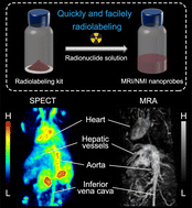
Magnetic resonance imaging (MRI)/nuclear medicine imaging (NMI) dual-modality imaging based on radiolabeled nanoparticles has been increasingly exploited for accurate diagnosis of tumor and cardiovascular diseases by virtue of high spatial resolution and high sensitivity. However, significant challenges exist in pursuing truly clinical applications, including massive preparation and rapid radiolabeling of nanoparticles. Herein, we report a clinically translatable kit for the convenient construction of MRI/NMI nanoprobes relying on the flow-synthesis and anchoring group-mediated radiolabeling (LAGMERAL) of iron oxide nanoparticles. First, homogeneous iron oxide nanoparticles with excellent performance were successfully obtained on a large scale by flow synthesis, followed by the surface anchoring of diphosphonate-polyethylene glycol (DP-PEG) to simultaneously render the underlying nanoparticles biocompatible and competent in robust labeling of radioactive metal ions. Moreover, to enable convenient and safe usage in clinics, the DP-PEG modified nanoparticle solution was freeze-dried and sterilized to make a radiolabeling kit followed by careful evaluations of its in vitro and in vivo performance and applicability. The results showed that 99mTc labeled nanoprobes are effectively obtained with a labeling yield of over 95% in 30 minutes after simply injecting Na[99mTcO4] solution into the kit. In addition, the Fe3O4 nanoparticles sealed in the kit can well stand long-term storage even for 300 days without deteriorating the colloidal stability and radiolabeling yield. Upon intravenous injection of the as-prepared radiolabeled nanoprobes, high-resolution vascular images of mice were obtained by vascular SPECT imaging and magnetic resonance angiography, demonstrating the promising clinical translational value of our radiolabeling kit.
