
Dimensional regulation of the aggregation-induced emission properties for complexes†
Shuyu
Wang
,
Lei
Wang
,
Fang
Fang
,
Xun
Ma
,
Yuanyuan
Guo
,
Ruidong
Wang
,
Suoshu
Zhang
,
Zhen
Zhang
,
Lin
Du
* and
Qi-Hua
Zhao
 *
*
Key Laboratory of Medicinal Chemistry for Natural Resource, Ministry of Education and Yunnan Province, Yunnan Characteristic Plant Extraction Laboratory, School of Chemical Science and Technology, Yunnan University, Kunming, 650500, PR China. E-mail: qhzhao@ynu.edu.cn
First published on 15th May 2023
Abstract
Aggregation-induced emission (AIE) is a fascinating luminescence phenomenon that provides new ideas for developing new fluorescent devices. However, research on photofunctional complexes with AIE properties is still in its infancy, mainly because the motion of the AIE molecules that can move freely will be greatly restricted after immobilization in the complex. Therefore, extending the AIE properties of organic molecules to their constructed complexes has been difficult. To explore a simple and efficient way to introduce the AIE properties of organic molecules into complexes, two similar tetraphenyl ethylene (TPE) carboxylic acid derivatives: tpemc and tpedc with different coordination sites were used to design and construct a series of Eu-complexes with three different dimensions in this work, namely, complex 1 ({[Eu2(tpedc)3(DMA)(H2O)2]·3DMA·2H2O}n, two-dimensional), complex 2 ([Eu(tpemc)2(DMA)(AcO)]n, one-dimensional), and complex 3 ([Eu(tpemc)3(phen)(DMA)(H2O)]·H2O, zero-dimensional). Complexes 1–3 showed an incremental AIE phenomenon, which indicated that the restriction of the AIE groups in low-dimensional complexes was limited. They still exhibited a certain movement space; viscosity response experiments and the analysis of a single crystal model with variable temperature also confirmed this result. Therefore, the construction of similar low-dimensional complexes could be regarded as a simple and effective method to introduce AIE properties. In addition, we found that both complexes 2 and 3 exhibited characteristic emissions of Eu3+ at low temperatures, and we explored this particular phenomenon in detail through time-dependent density functional theory (TD-DFT) calculations and energy level analysis. Moreover, they also have potential applications in temperature sensing and cell bioimaging.
Introduction
Aggregation-induced emission (AIE) is a photophysical phenomenon related to the molecular aggregation state,1 which is the opposite of the traditional aggregation-caused quenching (ACQ) phenomenon, where the chromophore is in a molecularly dissolved state. No, or a very weak, fluorescence effect will occur, but it can emit strong fluorescence in an aggregated state.2–4 Over the past two decades, AIE smart materials have been used for toxic chemical detection,5 environmental monitoring,6 chemical sensing,7 biosensing8 and biomedical imaging,9 clinical diagnosis and cancer treatment,10–12 and other fields13 and have received extensive attention. AIE-based sensing materials have been intensively studied due to their excellent optical properties, fast response, tunable structures, and high sensitivity,14 some of which have been used to detect metal ions,15 anions, and excellent small organic sensors for molecular pollutants16–18 in practical applications. At present, research on the properties and applications of AIE has mainly focused on organic molecules,19,20 and research on functional complexes with AIE properties, especially rare earth complexes, is still in its infancy mainly owing to coordination-induced emission (CIE), which is also with the same limitation of intramolecular motion (RIM) mechanism as AIE.21Tetraphenylethylene (TPE) is widely used in the synthesis of AIE molecules due to its simple structure, good chemical stability, and easily modifiable structure with a core structure to maintain AIE properties.22–24 Coordination polymers (CPs) constructed with the ligands of TPE as the core have attracted extensive attention mainly due to their unique rigid structure with a diversity of modifying groups, which can form a variety of coordination structures during the formation of complexes with metal ions. TPE and its derivatives often have excellent fluorescence properties.25 The use of TPE-based derivatives to synthesize CPs with specific functions started in 2011, and with increased research, scientists have introduced TPE-based derivatives into CPs and have explored their fluorescence sensing, mechanochromic, and adsorption characteristics and other properties such as separation and catalysis.26–29 Ligands tpedc (4,4′-(2,2-diphenylethenylidene)bis(benzoic acid)) and tpemc (4-(1,2,2-triphenylethenyl)benzoic acid) belong to the TPE class and consist of a typical tetrastyrene derivative with aggregation-induced luminescence properties with two carboxyl coordination sites and one carboxyl coordination site. Due to fewer coordination sites and more relatively free benzene rings, it is possible to reduce the CIE effect and construct CPs with AIE properties.30–32
We used tpedc and tpemc as the ligands and rare earth metal ion (Eu3+) with an oxidation number of +3 as the central metal ion to construct Eu-CPs with AIE properties. We first synthesized complex 1 using tpedc and Eu3+ with a two-dimensional planar structure, and then, we measured its AIE properties. Unfortunately, no obvious AIE properties, except for the ACQ phenomenon, were observed. Considering the many limitations of 2D structures on benzene ring rotors, we decided to further reduce the dimensionality of the complex to try and introduce the AIE properties of the ligand. The key was to further reduce the carboxyl coordination sites to increase the number of unanchored benzene ring rotors in the complex. Then, complex 2 with a one-dimensional chain structure was synthesized using tpemc and Eu3+, and complex 2 showed obvious AIE properties in subsequent tests. The success of this approach made us want to further consider if the AIE properties could be improved by further reducing the dimensionality. Considering the multi-coordination number characteristics of rare earth compounds, if we chose suitable chelating agents to occupy the space, we could obtain a lower-dimensional complex. Finally, complex 3 was obtained using tpemc, 1,10-phenanthrolinehydrate (a ligand that did not exhibit fluorescence or phosphorescence emission of itself during the formation of the complex with Eu3+![[thin space (1/6-em)]](https://www.rsc.org/images/entities/char_2009.gif) 33,34), and Eu3+ with a zero-dimensional structure. Consistent with our expected results, complex 3 had more obvious AIE properties than complex 2. Subsequently, we studied its optical properties and potential applications. By comparing the relationship between the structures and fluorescence properties, we explained the reasons for the corresponding properties of the complexes at a molecular level. In the past, the design and construction of AIE-CPs were always conducted by designing AIE linkers through post-synthesis modification methods, selecting reasonable metal nodes, or integrating CPs with the construction of CP-AIEgen nanocomposites,20 in such a way that adjusting the properties of AIEs by designing a series of complexes with different structures for controlling the dimensionality of the complex formed by the same class of ligands, which has never been done before (Fig. 1).
33,34), and Eu3+ with a zero-dimensional structure. Consistent with our expected results, complex 3 had more obvious AIE properties than complex 2. Subsequently, we studied its optical properties and potential applications. By comparing the relationship between the structures and fluorescence properties, we explained the reasons for the corresponding properties of the complexes at a molecular level. In the past, the design and construction of AIE-CPs were always conducted by designing AIE linkers through post-synthesis modification methods, selecting reasonable metal nodes, or integrating CPs with the construction of CP-AIEgen nanocomposites,20 in such a way that adjusting the properties of AIEs by designing a series of complexes with different structures for controlling the dimensionality of the complex formed by the same class of ligands, which has never been done before (Fig. 1).
 | ||
| Fig. 1 Synthetic route and structure of complexes 1–3, where C: gray; O: red; N: blue; Eu: pink; H atoms were omitted for clarity. | ||
Experimental
Materials
In this study, we utilized 4-(1,2,2-triphenylethylene) benzoic acid (tpemc) (Jilin Chinese Academy of Sciences-Yanshen Technology Co., Ltd), 4,4′-(2,2-diphenylethenylidene) bis benzoic acid (tpedc) (Jilin Chinese Academy of Sciences-Yanshen Technology Co., Ltd), 1,10-phenanthroline monohydrate (phen) (Jilin Chinese Academy of Sciences-Yanshen Technology Co., Ltd), N,N-dimethylacetamide (DMA) (Adamas Reagent Co., Ltd), and nitrate Eu(NO3)3·6H2O (Adamas Reagent Co., Ltd). All reagents were of AR grade and were used without further purification.Preparation of {[Eu2(tpedc)3(DMA)(H2O)2]·3DMA·2H2O}n(1)
First, 3 mg (0.008 mmol) of tpedc and 30 μL of Eu(NO3)3 aqueous solution (1 M, 0.03 mmol) in a mixed solvent of DMA (1.05 mL) and water (0.95 mL) were placed in a 5 mL glass tube. After the system was uniformly dispersed and clear under ultrasound, the glass tube was sealed with an oxyacetylene flame. The system was placed in a programmed temperature-controlled oven, reacted at 110 °C for 24 h, and then the temperature was lowered to room temperature at a rate of 5 °C h−1. Finally, colorless crystals of 1 were obtained, which were washed with DMA and EtOH, and after three cycles, they were placed in a ventilated place overnight to dry. The yield was 58% (based on Eu).Preparation of [Eu(tpemc)2(DMA)(AcO)]n(2)
First, 3 mg (0.008 mmol) of tpemc and 30 μL of Eu(NO3)3 aqueous solution (1 M, 0.03 mmol) in a mixed solvent of DMA (1.05 mL) and water (0.95 mL) were placed in a 5 mL glass tube. After the system was uniformly dispersed and became clear under ultrasound, the glass tube was sealed with an oxyacetylene flame. Then, the system was placed in a programmed temperature-controlled oven, reacted at 110 °C for 24 h, and the temperature was lowered to room temperature at a rate of 5 °C h−1. Finally, light yellow crystals of 2 were obtained, which were washed with DMA and EtOH, and after three cycles, they were placed in a ventilated place overnight to dry. The yield was 38% (based on Eu).Preparation of [Eu(tpemc)3(phen)(DMA)(H2O)]·H2O(3)
First, 2 mg (0.005 mmol) of tpemc, 2 mg (0.01 mmol) of 1,10-phenanthroline monohydrate, and 30 μL of Eu(NO3)3 aqueous solution (1 M, 0.03 mmol) in a mixed solvent of DMA (1.05 mL) and water (1.35 mL) were placed in a 5 mL glass tub. After the system was uniformly dispersed and became clear under ultrasound, the glass tube was sealed with an oxyacetylene flame. Then, the system was placed in a programmed temperature-controlled oven, reacted at 110 °C for 24 h, and the temperature was lowered to room temperature at a rate of 5 °C h−1. Finally, white crystals of 3 were obtained, which were washed with DMA and EtOH, and after three cycles, they were placed in a ventilated place overnight to dry. The yield was 42% (based on Eu).Characterization
A Bruker Smart AXS CCD diffractometer (Mo Kα, λ = 0.71073 Å) was used for the single-crystal X-ray diffraction (SCXRD) testing of complexes 1–3. In addition, complexes 1–3 were analyzed by thermogravimetric analysis (TGA) using a Mettler-Toledo synchronous differential thermal analyzer in a temperature range of 25–800 °C and at a heating rate of 10 °C min−1 under N2 conditions. Powder X-ray diffraction (PXRD) was performed on a Rigaku TTRIII-18KW diffractometer (Rigaku, Japan) with Cu Kα radiation (λ = 1.5418 Å) and an angle range (2θ) of 3°–55°. The IR spectra were recorded on an FT-IR Thermo Nicolet Avatar 360 using KBr pellets in the 4000–400 cm−1 region. Luminescence spectroscopy of all complexes was performed on a Lengguang (Shanghai, China) F98 fluorescence spectrophotometer.Results and discussion
Crystal structures and physical characterization
The photographs of the crystals of complexes 1–3 under daylight and UV light are shown in Fig. S1.† SCXRD showed that complex 1 crystallized in the P![[1 with combining macron]](https://www.rsc.org/images/entities/char_0031_0304.gif) space group in the triclinic system. Each asymmetric unit contained an [Eu2(tpedc)3(DMA)(H2O)2]·3DMA·2H2O unit (Fig. S2a†). There were two crystallographically independent metal central atoms in complex 1, in which the Eu1 atom adopted an eight-coordinate coordination mode, forming dodecahedrons that were coordinated with six oxygen atoms on five tpedc molecules (Fig. S5a†) and two oxygen atoms on the two coordinated water molecules. Eu2 adopted a seven-coordinate coordination mode, forming a single-cap triangular prism (Fig. S5b†). However, unlike Eu1, Eu2 was associated with six oxygen atoms on six ligands and one oxygen atom on the amide of the coordinated DMA molecule. Eu1 was bridged by the carboxylic acid of the two tpedc ligands with μ4-η1:η1:η1:η1 mode, while Eu2 was bridged by two ligands with μ4-η1:η1:η1:η1 and μ3-η1:η1:η1:η1 modes. Finally a two-dimensional structure/planar on the ab plane was formed (Fig. S2b†).
space group in the triclinic system. Each asymmetric unit contained an [Eu2(tpedc)3(DMA)(H2O)2]·3DMA·2H2O unit (Fig. S2a†). There were two crystallographically independent metal central atoms in complex 1, in which the Eu1 atom adopted an eight-coordinate coordination mode, forming dodecahedrons that were coordinated with six oxygen atoms on five tpedc molecules (Fig. S5a†) and two oxygen atoms on the two coordinated water molecules. Eu2 adopted a seven-coordinate coordination mode, forming a single-cap triangular prism (Fig. S5b†). However, unlike Eu1, Eu2 was associated with six oxygen atoms on six ligands and one oxygen atom on the amide of the coordinated DMA molecule. Eu1 was bridged by the carboxylic acid of the two tpedc ligands with μ4-η1:η1:η1:η1 mode, while Eu2 was bridged by two ligands with μ4-η1:η1:η1:η1 and μ3-η1:η1:η1:η1 modes. Finally a two-dimensional structure/planar on the ab plane was formed (Fig. S2b†).
Complex 2 crystallized in the P space group in the triclinic system. Each asymmetric unit contained an [Eu(tpemc)2(AcO)(DMA)] unit, in which the AcO-ligand (acetate) was obtained by the decomposition of some DMA solvent molecules at high temperatures (Fig. S3a†). Eu3+ in this complex adopted the coordination mode of distorted pentagonal bipyramids (Fig. S5c†), which coordinated with the oxygen atoms on the two tpemc ligands carboxylic acid, two oxygen atoms on one acetate group, and an oxygen atom on the amide of the DMA molecule. Tpemc ligands bridged Eu3+ with the μ2-η1:η1 mode to construct an entire complex, forming a one-dimensional chain extending in the space along the b-direction axis (Fig. S3b†).
Complex 3 crystallized in the P21/c space group in the monoclinic system. This was a zero-dimensional structure (Fig. S4†), and Eu3+ adopted the nine-coordinate coordination mode to form a distorted single-cap quadrilateral inverse prism (Fig. S5d†). Each Eu3+ was coordinated with three tpemc molecules, in which two tpemc ligands adopted the μ1-η1:η0 mode, and the other adopted the μ1-η1:η1 mode, with one phenanthroline molecule and one DMA molecule.
To determine the purity of the complexes 1–3 synthesized by following the above-mentioned steps, X-ray powder diffraction was performed on these complexes at room temperature. Comparing the X-ray powder diffraction patterns of these complexes with the powder diffraction patterns obtained from the crystal data through Mercury software simulation (Fig. S6†), we found that their peak positions were basically the same, and we determined that complexes 1–3 had high purity. The baseline drift of the powder diffraction pattern by the test was possibly caused by the test instrument itself, and the change in peak intensity was possibly due to the uneven preparation or change in the crystal planes of the complexes. In addition, the FT-IR spectra, thermogravimetric curves, and UV-Vis absorption spectra of the three complexes are shown in Fig. S7, S8, and S9,† respectively. In addition, Fig. 2 illustrates the HOMO and LUMO energy levels of the three ligands for further discussion of the fluorescence emission behavior of the complexes through calculation.35
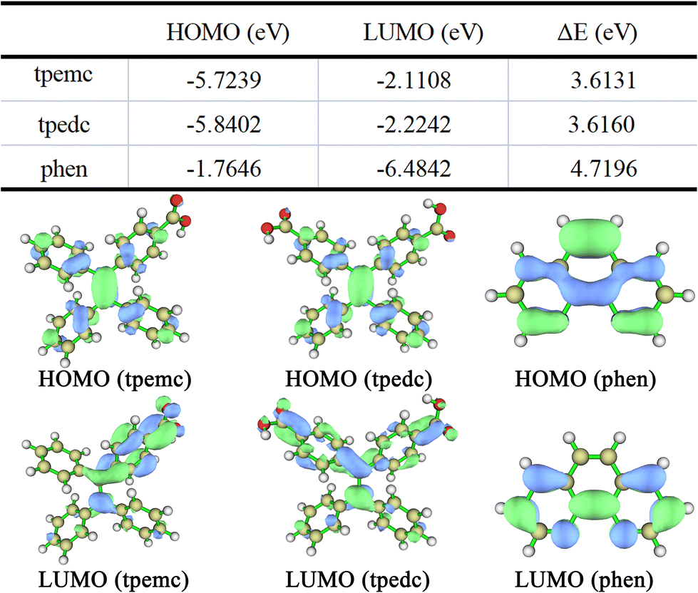 | ||
| Fig. 2 HOMO and LUMO energy levels with their differences (ΔE) among the different ligands, and molecular orbital diagrams of LUMO and HOMO of the three ligands. | ||
Fluorescence properties
The luminescence parameters of complexes 1–3 are shown in Table 1, and the additional details regarding solid-state fluorescence are shown in Fig. S10.†Fig. 3 shows the solid-state fluorescence excitation and emission spectra of complexes 1–3. The maximum excitation peak of the solid-state fluorescence of complex 1 was located at 397 nm, and its maximum emission peak was observed at 459 nm. The solid-state fluorescence excitation peak of complex 2 was located at 361 nm, and its maximum emission peak was observed at 450 nm. The solid-state fluorescence excitation peak of complex 3 was located at 389 nm, while the maximum emission peak was observed at 439 nm. Comparing the solid-state fluorescence spectra of ligands tpedc (λex = 399 nm, λem = 469 nm) and tpemc (λex = 400 nm, λem = 464 nm), the emission peak of complex 1 showed a blue shift of 10 nm relative to that of tpedc, and similar changes for complexes 2 and 3 showed a blue shift of about 14 nm and 25 nm, respectively, and the CIE properties of the three complexes are shown in Fig. S11.† The quantum yield and fluorescence lifetime test results are shown in Fig. S12–S17,† the fluorescence quantum yield of complexes 1–3 increases successively. Complex 3 has the longest fluorescence lifetime while complex 2 has the shortest fluorescence lifetime, the fluorescence lifetime of complex 1 is between them. | ||
| Fig. 3 Solid-state fluorescence emission spectra of complexes 1–3: (a) intensity normalized fluorescence contrast and (b) comparison diagram of the optimal emission spectrum of complexes 1–3. | ||
| Complex | λ ex (nm) | λ em (nm) | QY (%) | τ (ns) | Dimension |
|---|---|---|---|---|---|
| 1 | 397 | 459 | 5.07 | 0.668 | 2D |
| 2 | 361 | 450 | 6.41 | 0.591 | 1D |
| 3 | 389 | 439 | 12.72 | 1.012 | 0D |
AIE fluorescence properties of complexes 1–3
The stability of complexes 1–3 in different solvents for 24 h was tested. The PXRD powder patterns of complexes 1–3 after the treatment with different solvents are shown in Fig. S18,† for complex 1, except for the obvious shift of the position of the main peak after being treated with CH3CN, the powder patterns of the samples treated with other solvents (H2O, DMA, DMF, CH3OH, EtOH, glycerol and THF) could correspond to the simulated spectrum, but the intensity of some peaks was changed, which may be related to the crystallinity of the samples after immersion. For complex 2, the position of the most main peaks remained but many small peaks became weak or even disappeared after being treated with CH3CN, CH3OH, and EtOH, indicating that these crystal faces may have been damaged or dissolved partially. For complex 3, the pattern of the sample treated with THF changed a lot, indicating that the structure was destroyed in THF. In addition, we also found that many small peaks disappeared after treatment with CH3CN and EtOH, indicating that these crystal faces were also dissolved or destroyed but the main structure was maintained.Then, we observed the fluorescence spectra of complexes 1–3 dispersed in different solvents (Fig. S19†). We found that the emission position of complex 1 in CH3CN shifted greatly, and complex 3 had almost no emission peak in THF, which was consistent with the results of the solvent stability experiment due to the destruction of their structure.
We first studied the AIE property of complex 1 in THF–H2O solutions, but no AIE phenomenon was observed (Fig. 4a and d), the weakening of fluorescence in aqueous solution may be related to solvent effects.36 Then, the AIE properties of complexes 2 and 3 were explored. Considering that complex 3 has poor stability in THF solvent, we used DMA–H2O solutions instead of the THF–H2O solvent system, and the experimental process was completely consistent with that of complex 1. Complexes 2 and 3 showed obvious AIE fluorescence properties (Fig. 4b and c), where the volume fraction of water in the DMA–H2O mixed solution continued to increase, the degree of dispersion of complexes 2 and 3 in the mixed solution decreased and started to aggregate, a significantly enhanced fluorescence emission peak appeared, with a fluorescence emission peak that increased with increasing volume fraction of water. In addition, the AIE properties of complexes 2 and 3 in various solvent systems under the same experimental conditions were explored, and the results are shown in Fig. S20.† Fig. S20a–20e† are the fluorescence spectra of complex 2 when the ratio of H2O in different solvent systems is 90% and 10% and Fig. S20f–20j† are the fluorescence spectra of complex 3 at the same condition. During the experiment, the concentration of complexes 2 and 3 dispersed in the solvent was the same. As shown in Fig. S20,† the AIE property of complex 3 was much more obvious than that of complex 2. The fluorescence photos under UV light and Transmission Electron Microscope (TEM) images of complexes 1–3 in the corresponding solvents are shown in Fig. S21,† a significant aggregation phenomenon of complexes 1–3 from THF/DMA to aqueous solutions was observed.
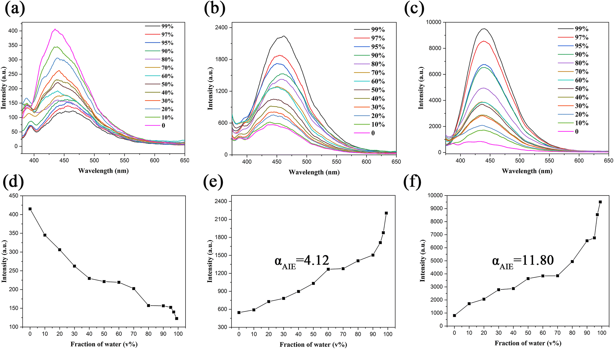 | ||
| Fig. 4 Emission spectra of complex 1 (a) in the THF–H2O solutions with different water volume fractions; complex 2 (b), and complex 3 (c) in the DMA–H2O solutions with different water volume fractions; relative luminescence intensities of complex 1 (d), complex 2 (e), and complex 3 (f) as a function of ƒw. | ||
To further verify that the AIE effect is not caused by the ligand decomposed from the complexes, we determined the fluorescence spectra of the ligands tpemc and phen in the DMA–H2O solutions with different water volume fractions (Fig. S22†), the results showed that tpemc had obvious AIE properties while phen had almost no fluorescence emission. So, if complexes were decomposed in the solvent, the fluorescence peaks position of AIE experiments should correspond to the peaks position of tpemc. By comparing Fig. 4 with Fig. S22a,† it also confirmed that complexes 2 and 3 were not damaged by solvent during the AIE test. Meanwhile, there was an obvious emission peak at about 388 nm, which increased with the increase in the THF component in THF–H2O mixtures (Fig. 4a). Therefore, we tested the fluorescence spectrum of pure THF solvent (Fig. S23a†) and found that the peak is the solvent peak of THF (λem = 387 nm). At the same time, we determined the fluorescence spectrum of pure DMA solution, which can be ignored because the intensity is far lower than that of complexes 2, 3, and tpemc during testing (Fig. S23b†). However, due to the weak emission of complex 1 in AIE testing, the emission peak of DMA near 450 nm may disturb the results; this is also the reason why we chose the THF–H2O system to test the AIE property of complex 1.
By analyzing the structures of the three complexes, we found that the two-dimensional structure formed by complex 1 was more compact than that of complexes 2 and 3, and the interactions between the surfaces, the filling of the solvent molecules, the high steric hindrance, and the fewer number of rotated benzene rings were enough to form sufficient rotational constraints on the benzene ring in tpedc, which would greatly weaken or even eliminate its AIE properties. For complexes 2 and 3, the benzene rings were not subjected to many restrictions, and in a dispersed state, the benzene rotors of the complexes could rotate freely, which means that the CIE effect was reduced in low dimensional structure and provided a nonradiative transition channel for the excited-state electrons. In an aggregated state, the motion of the benzene rotors was considerably restricted, causing a radiative transition in the primary pathway for the excited-state electrons back to the ground state and leading to intense fluorescence. In addition, we found from Fig. 4e and f that the αAIE value, which was calculated by I/I0 (I is the emission intensity in 99% H2O/DMA mixtures, and I0 is the emission intensity in DMA)37,38 of complex 2 was less than that of complex 3, which was possibly due to the existence of certain interactions between the adjacent phenyl groups on the same side of the chains in the one-dimensional chain structure of complex 2, such as π–π interactions,39 compared to the structure of complex 3, where the presence of these weak interaction forces also limited their motion to a certain extent; thus, the AIE effect of complex 2 was weaker than that of complex 3. To further verify the above mechanism, the fluorescence spectra of the complexes in solvents with high viscosity (obtained by the addition of glycerol) were recorded.
Luminescence response to viscosity stimuli of complexes 2 and 3
Changes in the ambient viscosity may affect the fluorescence emissions of complexes by restricting the motion of the benzene rotors.9,40 Therefore, we explored the effect of solvent viscosity on the fluorescence emissions of complexes 2 and 3. The increase in the glycerol fraction (ƒg) of the solvent significantly enhanced the fluorescence emissions of complexes 2 and 3. Fig. 5(a) and (b) show the luminescence emission spectra of complexes 2 and 3 in the glycerol/DMA mixtures, respectively, and Fig. 5(c) and (d) show the luminescence intensities increased as a function of glycerol fraction for complexes 2 and 3 in the glycerol/DMA mixtures, respectively. The viscosity values for glycerol/DMA mixtures were calculated according to the formula:| Lgη = Σχilgηi |
 | ||
| Fig. 5 Luminescence spectra of complexes 2 (a) and complex 3 (b) in the glycerol/DMA mixtures; the linear relationship between the fluorescence intensity and the viscosity values (complex 2 (c) and complex 3 (d)). | ||
Variable temperature fluorescence properties complexes 1–3
Generally, materials with AIE properties will often have a corresponding response to the temperature, because low temperatures will limit molecular motion.42–44 Thus, the temperature-dependent luminescence spectra of complexes 1–3 in the solid state in the different ranges were investigated, as shown in Fig. 6. As the temperature increased, the vibrational, rotational, and intermolecular collisions of the complex molecules intensified. Most of the received energy was dissipated in the form of non-radiation, which caused the fluorescence emission intensities of the complexes to become increasingly weaker. Of note, complexes 2 and 3 not only exhibited the above phenomenon but also showed obvious characteristic peaks belonging to Eu3+ at high wavelengths at low temperatures, and these characteristic peaks gradually weakened with increasing temperature. | ||
| Fig. 6 (a) Temperature-responsive emissions of complex 1 in the solid state between 250 K and 320 K; (b) temperature-responsive emissions of complex 2 in the solid state between 80 K and 170 K; (c) temperature-responsive emissions of complex 3 in the solid state between 77 K and 337 K. | ||
To further explore this special phenomenon, we analyzed the relationship between ligands and Eu3+ at the energy level. Based on the UV-Vis absorption spectra of the three ligands (Fig. S24†), the single-state level was 26315 cm−1 (380 nm) for tpedc, 25839 cm−1 (387 nm) for tpemc, and 30121 cm−1 (332 nm) for phen. While the lowest excited state energy of Gd3+ (32500 cm−1) was higher than the singlet state energy of the three ligands, the energy could not be transferred from the ligand to Gd3+, we can determine the triplet energy of the ligands through the phosphorescence spectrum of Gd-complexes, we synthesized the heteromorphic Gd-complexes 1′–3′ with the same way based Gd(NO3)3. The PXRD patterns and solid fluorescence spectra of Gd-complexes 1′–3′ are shown in Fig. S25 and S26,† the characterization data are basically consistent with that of Eu-complexes, revealing that they have the same structure. Then, we determined the triplet-state energy level of the ligand by testing the phosphorescence of three Gd-complexes at 77 K (Fig. S27†), the phosphorescence emission peaks of Gd-complex 1′–3′ are 491 nm, 471 nm, and 451 nm, respectively. In addition, the fluorescence lifetime and phosphorescence lifetime of Gd-complexes 1′–3′ were tested and data are shown in Fig. S28 and S29,† the fluorescence lifetimes of Gd-complexes 1′–3′ are 3.14 ns, 0.72 ns, and 3.08 ns, respectively. The triplet-state level of tpedc (T′1) was 20367 cm−1 (491 nm), and because we observed that the emission peak of tpedc blue-shifted by 9 nm after forming the Gd-complex, the energy of its triplet-state (T1) was 20000 cm−1.21 Similarly, the T′1 of tpemc was 21231 cm−1, while T1 was 20833 cm−1. The schematic energy transfer process in complexes 1 and 2 is shown in Fig. 7, where according to Reinhold's empirical rule, the energy gap between S1 and T1 (ΔE1) should be larger than about 5000 cm−1, with effective intersystem crossing.45 We found that the ΔE1 values were 6395 cm−1 and 5006 cm−1 in complexes 1 and 2, respectively; therefore, the intersystem crossing was efficient for the antenna effect procedure. In addition, only the sensitization of Eu3+ ions was effective when the energy gap between T1 and 5D0 (ΔE2) of Eu3+ (17500 cm−1) was larger than about 3500 cm−1.21,46 The ΔE2 values were 2500 cm−1 and 3333 cm−1 in complexes 1 and 2, respectively; therefore, the energy gap of complex 1 was too low to sensitize Eu3+ effectively, and it did not show the characteristic peak of Eu3+ ions. Meanwhile, the energy gap of complex 2 was at a critical value, and when the temperature increased, non-radiative energy dissipation in the excited ligand molecules increased, and the sensitivity of Eu3+ gradually decreased until it could not be sensitized. This explained why complex 2 exhibited the characteristic emission of Eu3+ only at low temperatures. For complex 3, although there were two different ligands, 1,10-phenanthrolinehydrate would not show fluorescence or phosphorescent emissions after forming a complex with Eu3+, because its singlet–triplet ISC and energy transfer from the triplet state of phen (in the complex) to the Eu3+ state were efficient (over 80%),33,34 while there was a far greater number of tpemc in complex 3 than phen, and we mainly considered the influence of tpemc. Similarly, we knew from the UV spectrum and phosphorescence spectrum that ΔE1 = 4918 cm−1 and ΔE2 = 3421 cm−1 in complex 3, which was basically consistent with complex 2, and they showed similar emission behaviour. In addition, the peak at 615 nm was assigned to the hypersensitive electric dipole transition (5D0 → 7F2) of Eu3+, which was greatly affected by the symmetry of the Eu3+ microenvironment, and symmetry reduction would result in greater transition intensity of 5D0 to 7F2,33 complex 3 belonged to the monoclinic system, while complex 2 belonged to the triclinic system, thus, the symmetry of Eu3+ in complex 3 was higher, which led to the obvious weakening of Eu3+ emissions.
 | ||
| Fig. 7 Schematic energy transfer process in complexes 1 and 2. | ||
From the perspective of molecular orbitals, the complex can be regarded as a discrete molecular system.47–49 Therefore, their fluorescence emission mechanism was explored on the optimized structure of the ground state (S0) through TD-DFT approaches. However, because there were too many atoms in the fragments of complexes 1 and 2, resulting in the low matching of the calculated results to actual results, we only discussed the calculation results of complex 3. The suitable model grown from its X-ray structure (Fig. S30†) was used for calculation, and we found that the calculated UV absorption curve (calculated by CAM-B3LYP functional) and experimental UV absorption curve fit well50–54 (Fig. S31†). The calculation results showed that there were two main peaks at 270 nm and 327 nm for complex 3, while the experimental results were 282 nm and 342 nm, with an error of less than 5%. Table S6† shows the maximum absorption wavelength (nm), oscillator strengths (f), and assignments for the electronic transitions with the largest f. The involvement of the frontier orbitals in the lowest-energy transition process is shown in Fig. 8 and S32.† In combination with the differences in electron density upon excitation from the ground S0 state to the singlet excited state for complex 3 (Fig. S33†) through hole–electron analysis,35 we found that the energy transfer in the luminescence process was mainly concentrated in ligand-to-ligand charge transfer (LLCT) and intra ligand charge transfer (ILCT) but did not involve the metal centre, and this may also be the reason for the higher quantum yield of complex 3 compared to 1 and 2.
 | ||
| Fig. 8 Molecular orbital energy diagrams for complex 3 showing the vertical electronic transitions for the maximum absorption (327 nm) band at the theoretical level when B3LYP/def2-TZVPP for C H O N and BLYP/MWB52 for Eu. | ||
When the temperature-dependent luminescence spectra data were further analyzed within a certain temperature range, the fluorescent intensity changed linearly as the temperature changed, and this feature could be used to design sensors for different ambient temperatures. Fig. 9 shows the linear relationship between the emission peak intensities and temperatures of the different complexes in different temperature ranges. Fig. 9a shows the emission intensity of complex 1 at 468 nm behaving linearly with a temperature range of 250–320 K (R2 = 0.9700), with the drop degree of emission intensity reaching 32.9%, and an average slope of 0.47% per K. Fig. 9b shows the intensity of the characteristic peaks belonging to Eu3+ at 616 nm behaving linearly with a temperature range of 110–170 K (R2 = 0.9927), where the temperature increased from 110 K to 170 K; the intensity of the emission peak of complex 2 at 616 nm decreased by 77.01%, with an average slope of 1.28% per K. Compared to the fluorescent temperature sensor reported earlier, the above results response to temperature was quite sensitive.6,44,55 Furthermore, the same phenomenon was observed in complex 3, indicating that both the characteristic peaks belonging to Eu3+ and the fluorescence emissions belonging to tpemc showed a linear relationship within a certain temperature range. As shown in Fig. 9c, the fluorescence emission intensity at 317 K was 23.88% lower than that at 197 K, with an average slope of 0.20%/K (R2 = 0.9961). Meanwhile, the peak intensity of Eu3+ at 615 nm showed an 84% reduction when the temperature increased from 97 K to 197 K, with an average slope of 0.84%/K (Fig. 9d). In addition, the linear relationship between temperature and other characteristic emission intensities is shown in Fig. S34.† From the above data, we found that complexes 1–3 had a sensitive response to temperature and that this response had a good linear relationship within a certain temperature range. The AIE-responsive CPs could be potentially used in luminescence thermometry. The trends of the CIE coordinates of complexes 2 and 3 with temperature are shown in Fig. S35.†
 | ||
| Fig. 9 (a) Relative fluorescence intensity of complex 1 under various temperatures (250–320 K) at a wavelength of 468 nm; (b) relative fluorescence intensity of complex 2 under various temperatures (110–170 K) at a wavelength of 616 nm; (c) relative fluorescence intensity of complex 3 under various temperatures (197–317 K) at a wavelength of 451 nm; (d) relative fluorescence intensity of complex 3 under various temperatures (97–197 K) at a wavelength of 615 nm. | ||
The properties of the materials could be revealed from their microscopic structures at the atomic level. Therefore, the single-crystal X-ray diffraction of complexes 2 and 3 was tested under 100 K and 298 K, as an example analysis. Fig. 10a and b show the eight and twelve benzene planes in the asymmetric units of complex 2 and complex 3, respectively. In complex 2, P1 and P5 were the two benzene planes that were directly linked by a coordinating metal ion through the carboxyl group and were relatively fixed. We analyzed the motion of the relatively free benzene rotors of complex 2 at different temperatures, and the angles between the different benzene planes are shown in Table S7.† In complex 3, P1, P5, and P9 were the three relatively fixed benzene planes directly linked by a coordinating metal ion through the carboxyl group, and the angles between the different benzene planes are shown in Table S8.† From Table S7 and Table S8,† we found that almost all of the dihedral angles (except P5–P7 in complex 2 and P1–P2, P9–P10, and P9–P11 in complex 3) between the corresponding benzene planes were larger under high temperatures. In addition, we also found from a comparison of the two tables that the angles between the greatest number of benzene ring planes in complex 3 changed more significantly when the ambient temperature value changed equivalently. From the data in Tables S5 and S6,† we calculated that the average change angle of the benzene ring rotor in complex 2 was 1.89°, while in complex 3, this value was 3.23°, which also revealed that the motion of these benzene ring rotors was more freely in complex 3 than in complex 2. The angle data suggested that the benzene rotors were possibly restricted at low temperatures, and this result indicated that the motion of the benzene rotors at room temperature was occurring more freely than at low temperatures. Thus, the increase in fluorescence intensity with decreasing temperature could be attributed to the restriction of the molecular motion at low temperatures. Therefore, the presence of more free benzene ring rotors and more free movement of the benzene ring rotors in complex 3 was the main reason for its stronger AIE properties compared to complex 2.
 | ||
| Fig. 10 Different benzene planes in the asymmetric units of complex 2 (a) and complex 3 (b); some solvent molecules were removed for brevity. | ||
Cell imaging
In addition, through cytotoxicity screening, we further found that complex 2 had low toxicity (the inhibition rate was only 2.28%) on HeLa cells (Fig. S36†); therefore, complex 2 was selected for the study of in vivo HeLa cell bioimaging. Fig. S37† shows the bioimaging photos of the HeLa cells for complex 2 at a concentration of 2 μM. We found that at lower concentrations, complex 2 could fluorescently label the cytoplasm of living HeLa cells within a short period of time, indicating that complex 2 had potential applications in live cell bioimaging.Conclusions
In summary, we synthesized a series of complexes with different dimensions using Eu3+ and ligands with the same chromophore, and among these, the two complexes with lower dimensions successfully inherited the AIE properties of the ligands, and the AIE properties were incremental as the dimension decreased. Comparing the structures of the three complexes and combined with the αAIE values, viscosity sensitivity, and analysis of a single crystal model with variable temperature, we concluded that the lower dimensional complexes contained the AIE molecules more freely. Moreover, we found that the characteristic peaks belonging to Eu3+ appeared in complexes 2 and 3 at low temperatures, and we further studied their fluorescence emission behavior quantitatively through energy level analysis and TD-DFT calculations. We found a good linear relationship between emission intensity and temperature with high sensitivity, and the phenomenon has potential application value for temperature detection at low temperatures. In addition, we found that complex 2 possessed excellent luminescence properties with low cytotoxicity, which could fluorescently label living cells in a short time, indicating its application value in cell imaging.Author contributions
Shuyu Wang: conceptualisation, data curation, formal analysis, investigation, methodology, software, validation, visualization, writing – original draft, writing – review, and editing. Lei Wang: methodology, software. Fang Fang: project administration. Xun Ma: supplement experimental data. Yuanyuan Guo: project administration. Ruidong Wang: validation and formal analysis. Suoshu Zhang: validation and formal analysis. Zhen Zhang: review and editing the manuscript. Lin Du: conceptualization, resources, review and editing, supervision, project administration, and funding acquisition. Qi-Hua Zhao: conceptualisation, funding acquisition, project administration, supervision, reviewing, and editing.Conflicts of interest
There are no conflicts to declare.Acknowledgements
This work was supported by the National Natural Science Foundation of China (Project No. 22061047), The Ten Thousand Talent Plans for Young Top-notch Talents of Yunnan Province (No. YNWR-QNBJ-2019-088), the Yunnan University's Research Innovation Fund for Graduate Students (No. 2021Z107). The authors thank the Advanced Analysis and Measurement Center of Yunnan University for the sample testing service. We thank LetPub for its linguistic assistance during the preparation of this manuscript.References
- J. Luo, Z. Xie, J. W. Lam, L. Cheng, H. Chen, C. Qiu, H. S. Kwok, X. Zhan, Y. Liu, D. Zhu and B. Z. Tang, Aggregation-induced emission of 1-methyl-1,2,3,4,5-pentaphenylsilole, Chem. Commun., 2001, 1740–1741, 10.1039/b105159h.
- H. J. Tracy, J. L. Mullin, W. T. Klooster, J. A. Martin, J. Haug, S. Wallace, I. Rudloe and K. Watts, Enhanced photoluminescence from group 14 metalloles in aggregated and solid solutions, Inorg. Chem., 2005, 44, 2003–2011 CrossRef CAS PubMed.
- W. Shang, X. Zhu, T. Liang, C. Du, L. Hu, T. Li and M. Liu, Chiral Reticular Self-Assembly of Achiral AIEgen into Optically Pure Metal-Organic Frameworks (MOFs) with Dual Mechano-Switchable Circularly Polarized Luminescence, Angew. Chem., Int. Ed., 2020, 59, 12811–12816 CrossRef CAS PubMed.
- Y. Li, Y. Dong, L. Cheng, C. Qin, H. Nian, H. Zhang, Y. Yu and L. Cao, Aggregation-Induced Emission and Light-Harvesting Function of Tetraphenylethene-Based Tetracationic Dicyclophane, J. Am. Chem. Soc., 2019, 141, 8412–8415 CrossRef CAS PubMed.
- B. Zhu, L. Zhu, Y. Wan, S. Deng, C. Zhang and J. Luo, Multicomponent metal-organic frameworks with aggregation-induced emission characteristics as fluorescence sensor array for the identification of energetic compounds, Sens. Actuators, B, 2021, 341, 130011 CrossRef CAS.
- J. Dong, P. Shen, S. Ying, Z.-J. Li, Y. D. Yuan, Y. Wang, X. Zheng, S. B. Peh, H. Yuan, G. Liu, Y. Cheng, Y. Pan, L. Shi, J. Zhang, D. Yuan, B. Liu, Z. Zhao, B. Z. Tang and D. Zhao, Aggregation-Induced Emission-Responsive Metal–Organic Frameworks, Chem. Mater., 2020, 32, 6706–6720 CrossRef CAS.
- C. Mu, Z. Zhang, Y. Hou, H. Liu, L. Ma, X. Li, S. Ling, G. He and M. Zhang, Tetraphenylethylene-Based Multicomponent Emissive Metallacages as Solid-State Fluorescent Materials, Angew. Chem., Int. Ed., 2021, 60, 12293–12297 CrossRef CAS PubMed.
- G. Niu, R. Zhang, X. Shi, H. Park, S. Xie, R. T. K. Kwok, J. W. Y. Lam and B. Z. Tang, AIE luminogens as fluorescent bioprobes, TrAC, Trends Anal. Chem., 2020, 123, 115769 CrossRef CAS.
- J. Dong, Y. Pan, H. Wang, K. Yang, L. Liu, Z. Qiao, Y. D. Yuan, S. B. Peh, J. Zhang, L. Shi, H. Liang, Y. Han, X. Li, J. Jiang, B. Liu and D. Zhao, Self-Assembly of Highly Stable Zirconium(IV) Coordination Cages with Aggregation Induced Emission Molecular Rotors for Live-Cell Imaging, Angew. Chem., Int. Ed., 2020, 59, 10151–10159 CrossRef CAS PubMed.
- Y. Wang, S. Xu, L. Shi, C. Teh, G. Qi and B. Liu, Cancer-Cell-Activated in situ Synthesis of Mitochondria-Targeting AIE Photosensitizer for Precise Photodynamic Therapy, Angew. Chem., Int. Ed., 2021, 60, 14945–14953 CrossRef CAS PubMed.
- J. G. Yu, L. Y. Sun, C. Wang, Y. Li and Y. F. Han, Coordination-Induced Emission from Tetraphenylethylene Units and Their Applications, Chemistry, 2021, 27, 1556–1575 CrossRef CAS PubMed.
- L. Ma, X. Feng, S. Wang and B. Wang, Recent advances in AIEgen-based luminescent metal–organic frameworks and covalent organic frameworks, Mater. Chem. Front., 2017, 1, 2474–2486 RSC.
- L. He, R. D. Wang, S. Wang, R. R. Zhu, Z. Li, Y. Y. Wu, J. Ma, L. Du and Q. H. Zhao, An AIE material with time-dependent luminescence conversion obtained by 2D coordination polymer modification via covalent post-synthetic modification, Dalton Trans., 2021, 50, 16685–16693 RSC.
- Z. H. Zhu, Z. Ni, H. H. Zou, G. Feng and B. Z. Tang, Smart Metal–Organic Frameworks with Reversible Luminescence/Magnetic Switch Behavior for HCl Vapor Detection, Adv. Funct. Mater., 2021, 31, 2106925 CrossRef CAS.
- R.-D. Wang, W.-Q. Zhang, S. Zhou, J. Tang, M. He, S. Zhang, L. Du and Q.-H. Zhao, A novel dual-functional coordination polymer for detection and ultra-effectively removal of Fe(III) in water, J. Mol. Liq., 2022, 355, 118942 CrossRef CAS.
- M. H. Xie, W. Cai, X. Chen, R. F. Guan, L. M. Wang, G. H. Hou, X. G. Xi, Q. F. Zhang, X. L. Yang and R. Shao, Novel CO2 Fluorescence Turn-On Quantification Based on a Dynamic AIE-Active Metal-Organic Framework, ACS Appl. Mater. Interfaces, 2018, 10, 2868–2873 CrossRef CAS PubMed.
- C. Y. Liu, X. R. Chen, H. X. Chen, Z. Niu, H. Hirao, P. Braunstein and J. P. Lang, Ultrafast Luminescent Light-Up Guest Detection Based on the Lock of the Host Molecular Vibration, J. Am. Chem. Soc., 2020, 142, 6690–6697 CrossRef CAS PubMed.
- K. Li, Z. Li, D. Liu, M. Chen, S. C. Wang, Y. T. Chan and P. Wang, Tetraphenylethylene(TPE)-Containing Metal-Organic Nanobelt and Its Turn-on Fluorescence for Sulfide (S(2-)), Inorg. Chem., 2020, 59, 6640–6645 CrossRef CAS PubMed.
- Y. Cai, H. Zhu, W. Zhou, Z. Qiu, C. Chen, A. Qileng, K. Li and Y. Liu, Capsulation of AuNCs with AIE Effect into Metal-Organic Framework for the Marriage of a Fluorescence and Colorimetric Biosensor to Detect Organophosphorus Pesticides, Anal. Chem., 2021, 93, 7275–7282 CrossRef CAS PubMed.
- L. Zhu, B. Zhu, J. Luo and B. Liu, Design and Property Modulation of Metal–Organic Frameworks with Aggregation-Induced Emission, ACS Mater. Lett., 2020, 3, 77–89 CrossRef.
- H. Q. Yin, X. Y. Wang and X. B. Yin, Rotation Restricted Emission and Antenna Effect in Single Metal-Organic Frameworks, J. Am. Chem. Soc., 2019, 141, 15166–15173 CrossRef CAS PubMed.
- Z. Zhou, C. He, J. Xiu, L. Yang and C. Duan, Metal-Organic Polymers Containing Discrete Single-Walled Nanotube as a Heterogeneous Catalyst for the Cycloaddition of Carbon Dioxide to Epoxides, J. Am. Chem. Soc., 2015, 137, 15066–15069 CrossRef CAS PubMed.
- Q. Zhang, J. Su, D. Feng, Z. Wei, X. Zou and H. C. Zhou, Piezofluorochromic Metal-Organic Framework: A Microscissor Lift, J. Am. Chem. Soc., 2015, 137, 10064–10067 CrossRef CAS PubMed.
- N. B. Shustova, B. D. McCarthy and M. Dinca, Turn-on fluorescence in tetraphenylethylene-based metal-organic frameworks: an alternative to aggregation-induced emission, J. Am. Chem. Soc., 2011, 133, 20126–20129 CrossRef CAS PubMed.
- D. C. Mayer, J. K. ZarÈ©ba, G. Raudaschl-Sieber, A. Pöthig, M. Chołuj, R. Zaleśny, M. Samoć and R. A. Fischer, Postsynthetic Framework Contraction Enhances the Two-Photon Absorption Properties of Pillar-Layered Metal–Organic Frameworks, Chem. Mater., 2020, 32, 5682–5690 CrossRef CAS.
- Z. Zhou, C. He, L. Yang, Y. Wang, T. Liu and C. Duan, Alkyne Activation by a Porous Silver Coordination Polymer for Heterogeneous Catalysis of Carbon Dioxide Cycloaddition, ACS Catal., 2017, 7, 2248–2256 CrossRef CAS.
- Y. Dou, L. Yang, L. Qin, Y. Dong, Z. Zhou and D. Zhang, Efficient hydrolytic cleavage of phosphodiester with a lanthanide-based metal-organic framework, J. Solid State Chem., 2021, 293, 121820 CrossRef CAS.
- X. L. Wu, Z. J. Li, H. Zhou, G. Yang, X. Y. Liu, N. Qian, W. Wang, Y. S. Zeng, Z. H. Qian, X. X. Chu and W. Liu, Enhanced Adsorption and Separation of Xenon over Krypton via an Unsaturated Calcium Center in a Metal-Organic Framework, Inorg. Chem., 2021, 60, 1506–1512 CrossRef CAS PubMed.
- X. Guo, N. Zhu, S. P. Wang, G. Li, F. Q. Bai, Y. Li, Y. Han, B. Zou, X. B. Chen, Z. Shi and S. Feng, Stimuli-Responsive Luminescent Properties of Tetraphenylethene-Based Strontium and Cobalt Metal-Organic Frameworks, Angew. Chem., Int. Ed., 2020, 59, 19716–19721 CrossRef CAS PubMed.
- G. Huang, Y. Jiang, J. Wang, Z. Li, B. S. Li and B. Z. Tang, The influence of intermolecular interactions and molecular packings on mechanochromism and mechanoluminescence – a tetraphenylethylene derivative case, J. Mater. Chem. C, 2019, 7, 12709–12716 RSC.
- A. Miyagawa, M. Harada, G. Fukuhara and T. Okada, Space Size-Dependent Transformation of Tetraphenylethylene Carboxylate Aggregates by Ice Confinement, J. Phys. Chem. B, 2020, 124, 2209–2217 CrossRef CAS PubMed.
- Y. Zhou, H. Gao, F. Zhu, M. Ge and G. Liang, Sensitive and rapid detection of aliphatic amines in water using self-stabilized micelles of fluorescent block copolymers, J. Hazard. Mater., 2019, 368, 630–637 CrossRef CAS PubMed.
- Z. Zhou, J. Zhang, Z. Zhang, Z. Yao and Z. Wang, Enhanced fluorescence and ion adsorption/sensing properties of europium(III) complex with porous structure, J. Solid State Chem., 2022, 309, 122985 CrossRef CAS.
- I. Georgieva, N. Trendafilova, T. Zahariev, N. Danchova and S. Gutzov, Theoretical insight in highly luminescent properties of Eu(III) complex with phenanthroline, J. Lumin., 2018, 202, 192–205 CrossRef CAS.
- T. Lu and F. Chen, Multiwfn: a multifunctional wavefunction analyzer, J. Comput. Chem., 2012, 33, 580–592 CrossRef CAS PubMed.
- N. D. Rudd, H. Wang, E. M. Fuentes-Fernandez, S. J. Teat, F. Chen, G. Hall, Y. J. Chabal and J. Li, Highly Efficient Luminescent Metal-Organic Framework for the Simultaneous Detection and Removal of Heavy Metals from Water, ACS Appl. Mater. Interfaces, 2016, 8, 30294–30303 CrossRef CAS PubMed.
- J. Zhang, Q. Liu, W. Wu, J. Peng, H. Zhang, F. Song, B. He, X. Wang, H. H. Sung, M. Chen, B. S. Li, S. H. Liu, J. W. Y. Lam and B. Z. Tang, Real-Time Monitoring of Hierarchical Self-Assembly and Induction of Circularly Polarized Luminescence from Achiral Luminogens, ACS Nano, 2019, 13, 3618–3628 CrossRef CAS PubMed.
- Y. J. Kong, Z. P. Yan, S. Li, H. F. Su, K. Li, Y. X. Zheng and S. Q. Zang, Photoresponsive Propeller-like Chiral AIE Copper(I) Clusters, Angew. Chem., Int. Ed., 2020, 59, 5336–5340 CrossRef CAS PubMed.
- J. H. Deng, J. Luo, Y. L. Mao, S. Lai, Y. N. Gong, D. C. Zhong and T. B. Lu, pi-pi stacking interactions: Non-negligible forces for stabilizing porous supramolecular frameworks, Sci. Adv., 2020, 6, eaax9976 CrossRef CAS PubMed.
- M. K. Kuimova, G. Yahioglu, J. A. Levitt and K. Suhling, Molecular rotor measures viscosity of live cells via fluorescence lifetime imaging, J. Am. Chem. Soc., 2008, 130, 6672–6673 CrossRef CAS PubMed.
- Y. Y. Liu, X. Zhang, K. Li, Q. C. Peng, Y. J. Qin, H. W. Hou, S. Q. Zang and B. Z. Tang, Restriction of Intramolecular Vibration in Aggregation-Induced Emission Luminogens: Applications in Multifunctional Luminescent Metal-Organic Frameworks, Angew. Chem., Int. Ed., 2021, 60, 22417–22423 CrossRef CAS PubMed.
- M. Jin, T. S. Chung, T. Seki, H. Ito and M. A. Garcia-Garibay, Phosphorescence Control Mediated by Molecular Rotation and Aurophilic Interactions in Amphidynamic Crystals of 1,4-Bis[tri-(p-fluorophenyl)phosphane-gold(I)-ethynyl]benzene, J. Am. Chem. Soc., 2017, 139, 18115–18121 CrossRef CAS PubMed.
- J. H. Tang, Y. Sun, Z. L. Gong, Z. Y. Li, Z. Zhou, H. Wang, X. Li, M. L. Saha, Y. W. Zhong and P. J. Stang, Temperature-Responsive Fluorescent Organoplatinum(II) Metallacycles, J. Am. Chem. Soc., 2018, 140, 7723–7729 CrossRef CAS PubMed.
- Y. Cui, H. Xu, Y. Yue, Z. Guo, J. Yu, Z. Chen, J. Gao, Y. Yang, G. Qian and B. Chen, A luminescent mixed-lanthanide metal-organic framework thermometer, J. Am. Chem. Soc., 2012, 134, 3979–3982 CrossRef CAS PubMed.
- W. V. F. J. Steemers, D. N. Reinhoudt, E. B. vander Tol and J. W. Verhoeven, New Sensitizer-Modified Calix[4]arenes Enabling Near-UV Excitation of Complexed Luminescent Lanthanide Ions, J. Am. Chem. Soc., 1995, 117, 9408–9414 CrossRef.
- S. Wu, Y. Lin, J. Liu, W. Shi, G. Yang and P. Cheng, Rapid Detection of the Biomarkers for Carcinoid Tumors by a Water Stable Luminescent Lanthanide Metal-Organic Framework Sensor, Adv. Funct. Mater., 2018, 28, 1707169 CrossRef.
- J. Poater, M. Gimferrer and A. Poater, Covalent and Ionic Capacity of MOFs To Sorb Small Gas Molecules, Inorg. Chem., 2018, 57, 6981–6990 CrossRef CAS PubMed.
- L. Liu, X. Chen, J. Qiu and C. Hao, New insights into the nitroaromatics-detection mechanism of the luminescent metal-organic framework sensor, Dalton Trans., 2015, 44, 2897–2906 RSC.
- L. Lu, W. Liu, J. Wang, H. Zhong, J. Liu, A. K. Singh and A. Kumar, Four new luminescent-organic frameworks exhibiting highly sensing of nitroaromatics: An experimental and computational insight, Inorg. Chim. Acta, 2019, 487, 257–263 CrossRef CAS.
- R. Gaussian, G. Trucks, H. Schlegel, G. Scuseria, M. Robb, J. Cheeseman, G. Scalmani, V. Barone, B. Mennuci, G. Petersson, H. Nakatsuji, M. Caricato, X. Li, H. Hratchian, A. Izmaylov, J. Bloino, G. Zheng, J. Sonnenberg, M. Hada and D. Fox, Gaussian, Gaussian, Inc., Wallingford, CT, 2004.
- P. Leo, D. Briones, J. A. Garcia, J. Cepeda, G. Orcajo, G. Calleja, A. Rodriguez-Dieguez and F. Martinez, Strontium-Based MOFs Showing Dual Emission: Luminescence Thermometers and Toluene Sensors, Inorg. Chem., 2020, 59, 18432–18443 CrossRef CAS PubMed.
- D. Mondal, M. Bar, S. Mukherjee and S. Baitalik, Design of Ru(II) Complexes Based on Anthraimidazoledione-Functionalized Terpyridine Ligand for Improvement of Room-Temperature Luminescence Characteristics and Recognition of Selective Anions: Experimental and DFT/TD-DFT Study, Inorg. Chem., 2016, 55, 9707–9724 CrossRef CAS PubMed.
- P. Pal, S. Mukherjee, D. Maity and S. Baitalik, Synthesis, Structural Characterization, and Luminescence Switching of Diarylethene-Conjugated Ru(II)-Terpyridine Complexes by trans-cis Photoisomerization: Experimental and DFT/TD-DFT Investigation, Inorg. Chem., 2018, 57, 5743–5753 CrossRef CAS PubMed.
- N. Kunkel and H. Kohlmann, Ionic Mixed Hydride Fluoride Compounds: Stabilities Predicted by DFT, Synthesis, and Luminescence of Divalent Europium, J. Phys. Chem. C, 2016, 120, 10506–10511 CrossRef CAS.
- Y. Cui, F. Zhu, B. Chen and G. Qian, Metal-organic frameworks for luminescence thermometry, Chem. Commun., 2015, 51, 7420–7431 RSC.
Footnote |
| † Electronic supplementary information (ESI) available: Materials and methods, synthesis, X-ray crystallography, crystal structures, powder X-ray diffraction patterns, IR spectra, UV spectra, TGA, solid-state UV–visible absorbance spectra, and solid-state photoluminescence spectra. CCDC 2176115, 2176116 and 2176117 for complex 1–3. For ESI and crystallographic data in CIF or other electronic format see DOI: https://doi.org/10.1039/d3qi00443k |
| This journal is © the Partner Organisations 2023 |