 Open Access Article
Creative Commons Attribution 3.0 Unported Licence
Development of 8–17 XNAzymes that are functional in cells†
Kosuke
Chiba
 a,
Takao
Yamaguchi
 *a and
Satoshi
Obika
 *abc
*abc
aGraduate School of Pharmaceutical Sciences, Osaka University, 1-6 Yamadaoka, Suita, Osaka 565-0871, Japan. E-mail: yamaguchi-ta@phs.osaka-u.ac.jp; obika@phs.osaka-u.ac.jp
bNational Institutes of Biomedical Innovation, Health and Nutrition, 7-6-8 Saito-Asagi, Ibaraki, Osaka 567-0085, Japan
cInstitute for Open and Transdisciplinary Research Initiatives (OTRI), Osaka University, 1-1 Yamadaoka, Suita, Osaka 565-0871, Japan
First published on 28th June 2023
Abstract
DNA enzymes (DNAzymes), which cleave target RNA with high specificity, have been widely investigated as potential oligonucleotide-based therapeutics. Recently, xeno-nucleic acid (XNA)-modified DNAzymes (XNAzymes), exhibiting cleavage activity in cultured cells, have been developed. However, a versatile approach to modify XNAzymes that function in cells has not yet been established. Here, we report an X-ray crystal structure-based approach to modify 8–17 DNAzymes; this approach enables us to effectively locate suitable XNAs to modify. Our approach, combined with a modification strategy used in designing antisense oligonucleotides, rationally designed 8–17 XNAzyme (“X8–17”) that achieved high potency in terms of RNA cleavage and biostability against nucleases. X8–17, modified with 2′-O-methyl RNA, locked nucleic acid and phosphorothioate, successfully induced endogenous MALAT-1 and SRB1 RNA knockdown in cells. This approach may help in developing XNAzyme-based novel therapeutic agents.
Introduction
DNA enzymes (DNAzymes), first discovered in 1994,1 have attracted much attention as specific metal ion sensors,2,3 oligonucleotide ligation catalysts,4,5 and therapeutic modalities that cleave substrate RNA.6,7 These single-strand DNAs, often 30 nucleotides (nt) long, perform various functions, including hybridizing the complementary RNA strand, fixing metal ions as a catalyst, and causing various reactions against target RNAs (like RNA cleavage or ligation).8–10 Hence, DNAzymes are considered unique nucleic acids and are actively researched.Antisense oligonucleotides (ASOs) and siRNA have been developed as RNA-cleaving nucleic acids and some have been approved as nucleic acid-based drugs.11,12 DNAzymes are unique in that, unlike the recruitment of RNase H or argonautes by ASOs and siRNA, they do not need to recruit endogenous enzymes. DNAzymes bind to target RNA in a similar manner as ASOs; via metal-ion catalysis, DNAzymes catalyze the intra-molecular nucleophilic attack on the RNA phosphodiester bond by RNA 2′-hydroxy group, coordinated by their catalytic core.8 This unique characteristic could be applied to the treatment of infectious diseases and cancer,13–16 which modify cellular conditions and endogenous enzyme activity. Although clinical research on the application of DNAzymes against basal-cell carcinoma or asthma has been conducted,17,18 there is limited evidence that DNAzymes mediate catalytic RNA cleavage in cells. Moreover, like ASOs, DNAzymes have been shown to cleave target RNA via RNase H.19
Recently, some studies have reported xeno-nucleic acid (XNA)-modified DNAzymes (XNAzymes) that exhibit activity in cells.16,20–23 Wang et al.20 reported a 10–23 XNAzyme exhibited RNA cleavage activity in cells. The negative controls, in which the sequence of the catalytic core was inverted to deactivate the DNAzyme, could not cleave the target RNA, demonstrating that DNAzyme activity was not an RNase H-mediated antisense effect. The authors modified DNAzyme with 2′-fluoroarabino nucleic acid (FANA) and α-L-threofuranosyl nucleic acid, finding that sufficient modification, especially of the binding arms, dramatically improves DNAzyme biostability and enhances RNA cleavage activity in cells.20 There has been extensive research on DNAzyme catalytic core modification.20,24–27 For example, Schubert et al.26 reported an approach in which each nucleotide at the catalytic core of 10–23 DNAzyme was replaced via 2′-O-methyl (2′-OMe) sequential modification, revealing that the site of the modification affects DNAzyme activity. It is important to modify the catalytic core to enhance biostability without reducing its activity. However, there remains considerable uncertainty regarding the effective design of the catalytic core.
Here, we describe a new approach to modifying the catalytic core based on its X-ray crystal structure. Structural biology provides powerful tools for revealing the dynamics of biological molecules. Recently, Borggräfe et al.28 clarified the catalytic process of 10–23 DNAzyme via real-time NMR measurements and successfully improved the cleavage activity of 10–23 DNAzyme by eliminating detrimental interaction between Mg2+ and guanine (G14) at the catalytic core, using 6-thioguanine modification.
In this study, we focus on the 8–17 DNAzyme, which, along with the 10–23 DNAzyme, was discovered via in vitro selection.6 The fine X-ray crystal structure of 8–17 DNAzyme, thought to reflect the precatalytic form, was reported in 2017.29 We presumed that the crystal structure would enable us to select appropriate nucleotides for modification with XNA, based on the characteristics of each nucleotide at the catalytic core. This approach does not require evaluation of all positions of the catalytic core, making it possible to effectively and rapidly generate high-potency DNAzymes at low cost. Furthermore, we tried to enhance the intracellular RNA-cleaving activity of 8–17 DNAzyme. To promote DNAzyme biostability against nuclease digestion, we used an ASO-based design approach to modify the binding arms of the 8–17 DNAzyme. The modified 8–17 DNAzyme (8–17 XNAzyme), hereafter denoted as “X8–17”, exhibited sufficient biostability and RNA-cleaving activity, even within cells.
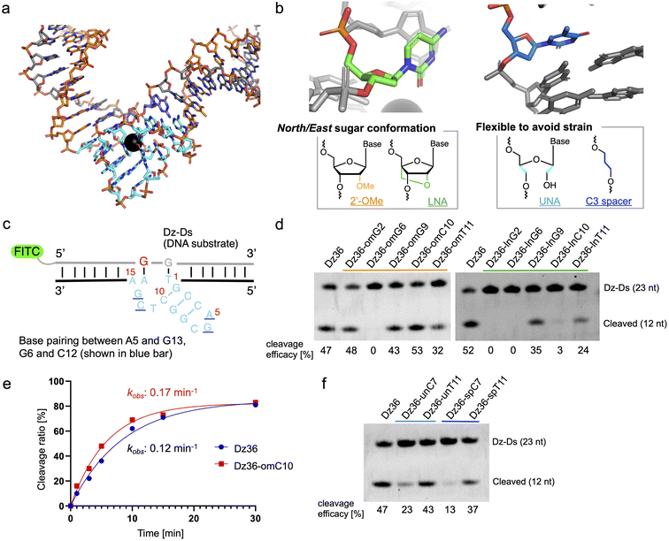
Results and discussion
Optimizing the catalytic core based on the X-ray crystal structure
The X-ray crystal structure of 8–17 DNAzyme, reported by a previous study, reflects the probable precatalytic form at high resolution (Fig. 1a, Protein Data Bank accession 5XM8).29 Based on the crystal structure, 8–17 DNAzyme hybridizes the DNA substrate via two binding arms (Fig. 1a, orange) and forms a catalytic core (15 nt) between the two arms (Fig. 1a, light blue). The catalytic core is a complex higher-order structure formed by base-pairing. Within the complex catalytic core of 8–17 DNAzyme, we focused on (i) nucleotides with a North/East sugar conformation (pseudorotation phase angle: 0°–108°) and (ii) nucleotides which do not perform base-pairing (Fig. 1b). It was expected that 2′-OMe RNA and locked nucleic acid (LNA), which induce a North sugar conformation, would be suitable for nucleotides with a North/East sugar conformation. For the modification of nucleotides which do not perform base-pairing, unlocked nucleic acid (UNA) and C3 spacer were selected to make the nucleotides flexible by eliminating the strain of sugar conformation. Such modifications have been reported to increase biostability against nucleases.30–33To evaluate the effectiveness of this crystal structure-based chemical modification, a 5′-fluorescein isothiocyanate (FITC) modified DNA substrate in which the cleavage site was substituted with RNA (hereafter denoted as “Dz-Ds”) was used for the cleavage reaction (Fig. 1c). The substrate sequence that we used was identical to that of the X-ray crystal structure of 5XM8, with a DNA complementary strand. We refer to the unmodified 8–17 DNAzyme sequence (36 nt) as Dz36. First, we performed a cleavage assay under multiple turnover conditions (Dz36![[thin space (1/6-em)]](https://www.rsc.org/images/entities/char_2009.gif) :
:![[thin space (1/6-em)]](https://www.rsc.org/images/entities/char_2009.gif) Dz-Ds = 1
Dz-Ds = 1![[thin space (1/6-em)]](https://www.rsc.org/images/entities/char_2009.gif) :
:![[thin space (1/6-em)]](https://www.rsc.org/images/entities/char_2009.gif) 10) where Dz36 could cleave ca. 50% of the substrate (under incubation at 37 °C for 10 min in physiological buffer containing 0.25 mM Pb(OAc)2; Pb(OAc)2 was used because the X-ray crystal structure was obtained using Pb2+ as the metal ion catalyst).
10) where Dz36 could cleave ca. 50% of the substrate (under incubation at 37 °C for 10 min in physiological buffer containing 0.25 mM Pb(OAc)2; Pb(OAc)2 was used because the X-ray crystal structure was obtained using Pb2+ as the metal ion catalyst).
The results of the cleavage assay are shown in Fig. 1d. As expected, by substituting the nucleotides with a North/East sugar conformation via 2′-OMe modification, DNAzyme activity was retained in most cases, except for Dz36-omG6, which was modified with 2′-OMe at the G6 position in the catalytic core (Fig. 1d, lane 3). The G6 position, the sixth nucleotide from the 5′ end in the catalytic core, is highly essential for stabilizing Pb2+, so even a small conformational change could be critical at this position.29 Nonetheless, four out of the five selected nucleotides with the 2′-OMe modification were well-tolerated. In addition, the cleavage efficacy was slightly improved (by ca. 13%) at the C10 position (Dz36-omC10), relative to that of Dz36. We further compared the time-dependency of the cleavage rates of Dz36 and Dz36-omC10 (Fig. 1f) and also evaluated Michaelis–Menten kinetics (ESI Fig. 1†). The Dz36-omC10 rate constant (kobs) was slightly higher, and the Michaelis–Menten constant (Km) was smaller than that of Dz36, revealing its relatively better affinity to Dz-Ds and higher substrate cleavage activity. In contrast, LNA modifications were not tolerated, and almost all the X8–17s exhibited diminished activity, except for Dz36-lnG9, modified with LNA at G9 position (Fig. 1d, right). To clarify this further, we calculated the sugar puckering of each nucleotide at the catalytic core, based on the X-ray crystal structure (ESI Table 2†).29 The selected nucleotides exhibited the North/East sugar conformation (pseudorotation phase angle: 0°–108°). LNA modification induced the North conformation more than 2′-OMe modification, by fixing the C2′–C4′ bond (ESI Table 2, lower panel†). This abnormally fixed each nucleotide with the North/East sugar conformation and thereby potentially impaired DNAzyme activity. It is also plausible that the LNA structure is conformationally unfavorable for interaction with Pb2+.
For UNA and C3 spacer modification of nucleotides that do not perform base-pairing, modified at T11 position retained its DNAzyme activity after modification (Fig. 1e, Dz36-unT11 [UNA] and Dz36-spT11 [C3 spacer]), indicating that the T11 position did not need a sugar structure and nucleobase. However, in a previous study, the complete deletion of T11 reduced DNAzyme activity by approximately 20-fold.29 Our results indicate the usefulness of replacing non-base-pairing nucleotides with a UNA or C3 spacer to increase biostability.
Moreover, to improve Dz36 biostability, we synthesized X8–17s with two points of modification in the catalytic core (ESI Fig. 2†); the X8–17s modified in this way retained their DNAzyme activity. However, for further analysis, we selected Dz36-omC10, which exhibited relatively higher DNAzyme activity (Fig. 1d and e).
Optimizing substrate-binding arms by following the general ASO wing design
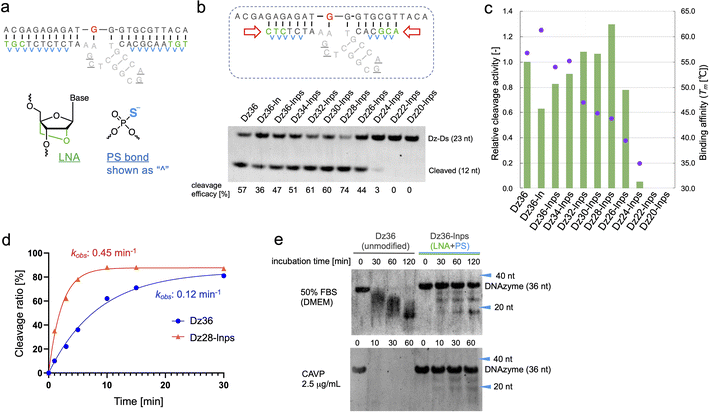
To improve Dz36 potency, we focused on its affinity for its substrate. For this purpose, we modified the binding arms of Dz36. We hypothesized that the role of the binding arms is similar to the role in ASOs, which bind to complementary RNA strands with suitable turnover and are stable in vivo. Therefore, chemical modifications reported in ASO design could be useful for DNAzymes.LNA is frequently applied to the 5′ and 3′ ends of ASOs, called the wing regions. LNA improves exonuclease resistance and binding affinity against the complementary strand.33 The phosphorothioate (PS) bond, a good alternative to the naturally occurring phosphodiester (PO) bond, is used in almost all linkages of ASO to improve its biostability against DNA nucleases.34 Similarly, Taylor et al.22 incorporated several PS bonds in the FR6_1 DNAzyme. Based on these, we applied LNA and PS bonds to the binding arms of Dz36 (Fig. 2a). As expected, the LNA and PS modified Dz36, hereafter referred to as “Dz36-lnps”, worked well and its activity was retained (Fig. 2b, lane 3), whereas LNA modification without PS (Dz36-ln) exhibited reduced activity relative to Dz36. Moreover, we presumed that adequate cleavage activity requires an optimal binding arm length, based on the cleavage mechanism (proper binding affinity and turnover against substrates). To test this hypothesis, arm length was investigated by shortening them one by one from both ends with Dz36-lnps (Fig. 2b, right). The shortened 28 nt derivative (“Dz28-lnps”) exhibited the highest cleavage activity (57% higher than that of Dz36-lnps).
To establish the connection between binding affinity and cleavage efficacy, the thermal stability, denoted by melting temperature (Tm) values, of X8–17s and the substrate were measured (Fig. 2c). The Tm values and corresponding cleavage activity levels were inversely correlated from Dz36-lnps to Dz28-lnps. These findings reveal that shortening the binding arms reduced affinity toward the substrate, resulting in efficient cleavage activity due to improved turnover. When the binding arm was shorter than 26 nt (“Dz26-lnps”), Tm was below 35 °C and cleavage activity was almost absent. It was hypothesized that Dz24-lnps, Dz22-lnps, and Dz20-lnps could not even bind to the substrate under normal physiological conditions (cleavage assay temperature: 37 °C). These results are congruent with those of a previous report suggesting that Tm should ideally be close to the working temperature.26 Moreover, the Tm for Dz36-ln was 5 °C higher than that for Dz36, which was also reflected in the cleavage activity, with Dz36-ln exhibiting lower activity than Dz36 and Dz36-lnps, owing to its insufficient turnover arising from its higher thermal stability. This could be explained by the fact that LNA modifications raise binding affinity, whereas PS modifications slightly reduce this affinity.35,36 Next, we performed kinetic studies to compare the unmodified Dz36 and Dz28-lnps, which exhibited the highest cleavage activity (Fig. 2d; ESI Fig. 1†). Notably, Dz28-lnps exhibited almost 4-fold higher kobs value than Dz36 in multiple-turnover assays. In addition, it was revealed that Dz28-lnps had >2-fold higher V0 and kcat than Dz36. These results indicate that turnover was substantially improved by adjusting binding affinity toward the substrate and that our strategy to shorten the binding arms was reasonable.
The biostability of Dz36-lnps, modified with LNA and PS, was assessed by comparing it with that of unmodified Dz36 (Fig. 2e). In the presence of 50% fetal bovine serum (FBS) or phosphodiesterase, Dz36-lnps exhibited much higher biostability than Dz36, as expected, indicating that these modifications worked well in terms of biostability. We further evaluated the stability of Dz28-lnps along with that of Dz36 and Dz36-lnps (ESI Fig. 3†) and found that Dz28-lnps also exhibited good biostability.
Confirming the effect of metal ions and the difference between DNA and RNA substrates
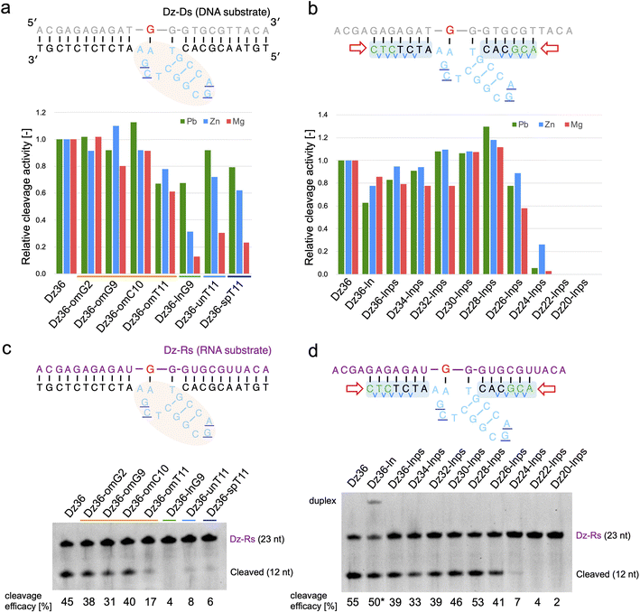
Based on our findings, we expected X8–17 with both modified catalytic cores and binding arms to be functional in living cells. Prior to evaluating RNA knockdown activity in cells, it was important to confirm whether our design could be applied to an RNA substrate and in the presence of Mg2+ or Zn2+, which reflect intracellular conditions.We first tested the effect of using different metal ions, whereby Pb2+ was replaced with Mg2+ or Zn2+ (Fig. 3a and b). When modifying the catalytic core, the difference in metal ion strongly affected X8–17 cleavage activity (Fig. 3a). The 2′-OMe-modified X8–17s exhibited good tolerance of the change in metal ion, whereas the other X8–17s exhibited reduced activity, especially under Mg2+ conditions. The differences in the impact of each modification might be due to differences in the catalytic core structure. 8–17 DNAzyme is known to folds into a more compact structure in the presence of Mg2+ or Zn2+ than in the presence of Pb2+.37,38 Therefore, X-ray crystal structure-based modification of DNAzymes might be limited the effectiveness when the structure is slightly altered by replacing with Mg2+ or Zn2+.
 | ||
| Fig. 3 Effects of metal ions on Dz36 modification. Metal ion dependency is illustrated for (a) catalytic core modifications and (b) binding arm modifications. Substrate-cleavage reactions were performed under standard condition with either 0.25 mM Pb(OAc)2, 2.5 mM ZnCl2, or 2.5 mM MgCl2. Cleavage activity was normalized by setting the activity of Dz36 to 1.0. The results for Pb2+ were analyzed using the data in Fig. 1d, f and 2b. The cleavage assay used an RNA substrate in the presence of 2.5 mM MgCl2. Modifications of (c) the catalytic core and (d) the binding arms were applied to the RNA substrate. Dz36-ln cleavage activity was used as the reference value (represented by *) because of its insufficient dissociation of the duplex. Dz-Rs: RNA substrate. | ||
In contrast, binding arm modification was well tolerated in the Mg2+ condition (Fig. 3b). The trends in cleavage activity were almost the same, and Dz28-lnps (at 28 nt, with a shorter binding arm) exhibited the best activity. From these results, it can be inferred that changing the metal ions strongly affected catalytic core modifications but did not affect binding arm modifications.
We next evaluated the effects of PS modification. “Dz36-ps_full” (i.e., Dz36 with an entirely PS modifications), exhibited significantly reduced activity under Zn2+ or Mg2+ conditions (ESI Fig. 4b†). PS bonds are known to be more hydrophobic than PO bonds. In addition, the catalytic core phosphate bonds all face outward (toward the aqueous media).29 We speculated that the catalytic core with PS bonds becomes unstable in aqueous media, especially with additional folding induced by Zn2+ or Mg2+. It is also possible that PS modification destabilized the electrostatic interaction between a coordinated metal ion and the 5′-PO bond of A5 (distance: 5.1 Å).29 In contrast, Dz36-ps, with PS modifications only in its binding arm, exhibited almost the same activity as Dz36 even under Mg2+ conditions (ESI Fig. 4b†). Based on these results, we applied PS modifications only to the binding arms and not to the catalytic core (Fig. 2a). Further investigations are warranted concerning PS modifications in the catalytic core.
To demonstrate X8–17 activity in cells, the RNA substrate, not the DNA substrate, should be cleaved. Hence, we performed the same experiments using the RNA substrate (Dz-Rs) in the presence of Mg2+ (Fig. 3c and d). The 2′-OMe modification of the catalytic core and binding arm length optimization were similarly effective for the RNA substrate. Introducing chemical modifications without reducing RNA-cleavage activity is expected to improve biostability and the possibility of intracellular RNA cleavage. Moreover, Tm values were measured; the cleavage activity and binding affinity were correlated for the Dz-Rs as for the Dz-Ds (ESI Fig. 5†).
Evaluation of endogenous RNA cleavage activity in cells
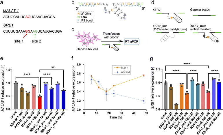
Finally, we investigated the target RNA cleavage activity of X8–17 inside cultured cells. A non-coding RNA metastasis-associated lung adenocarcinoma transcript 1 (MALAT-1), which is widely used for ASO studies,39 was selected as the model endogenous RNA. Dz36 preferentially cleaves a A–G dinucleotide to the same extent as a G–G dinucleotide;40 therefore, we designed X8–17 by targeting the A–G dinucleotide, which is located near the ASO binding site (Fig. 4a, shown in red).For the X8–17 targeting MALAT-1, we used 2′-OMe modification at C10 of the catalytic core, and applied LNA and 2′-OMe modifications to the binding arms incorporated with PS bonds (M34-1, Fig. 2c). It is possible that an unmodified region or only the PS-modified region would be recognized by RNase H, with its target RNA cleaved via the antisense effect. RNase H is known to recognize continuous DNA/RNA sequences.41,42 Therefore, 2′-OMe modifications were applied as alternate nucleotides to avoid RNase H recognition. In terms of its binding affinity, LNA modification increases Tm by ca. 4–6 °C per modification,35 whereas 2′-OMe modification increases it by ca. 0.5–1.5 °C.43 Hence, we changed the design described in Fig. 2a to include three 2′-OMe modifications instead of one LNA modification in the binding arm (Fig. 4b). We confirmed that the cleavage activity of these designs was comparable (M34-1, M34-2; ESI Fig. 6b†). Furthermore, the binding arm length of MALAT-1 targeting M34-1 and M34-2 (34 nt) was designed based on the calculated Tm value, which was close to that for Dz28-lnps, and was selected based on cleavage activity (ESI Fig. 6b†).
In the negative control (M34-1_inv), the sequence of the catalytic core was inverted, modifying the type of negative control used by Wang et al.20 However, Taylor et al.44 and Spitale et al.45 have revealed that a negative control inactivated via point mutations in the catalytic core is better than inversion of the catalytic core sequence because it causes minimal perturbation. Hence, we used another negative control sequence (M34-1_mut) inactivated by a two-point mutation of the catalytic core, which reduces Dz36 activity (Fig. 4d).29 Gel-electrophoresis analysis confirmed that neither negative control cleaved its substrate (ESI Fig. 6b†). In addition, M34-1 successfully exhibited sequence-specific cleavage of A–G dinucleotides against a 70 nt substrate of MALAT-1 partial RNA (M-Rs-2, ESI Fig. 6c†).
Intracellular knockdown assays were performed using Hepa1c1c7 cells. M34-1s and previously reported MALAT-1 targeting ASO (ASO-M) were transfected, followed by knockdown analysis using quantitative reverse transcription (qRT)-PCR (Fig. 4c). GAPDH was used as a housekeeping gene and MALAT-1 expression was corrected using the ΔΔCt method. M34-1 exhibited dose-responsive MALAT-1 knockdown with significant differences relative to the negative controls (Fig. 4e). These results indicate that the knockdown activity of M34-1 was DNAzyme-mediated. Meanwhile, we found that even the negative controls, M34-1_inv and M34-1_mut, exhibited some knockdown activity at 100 nM, implying the occurrence of RNase H-mediated knockdown. Therefore, the knockdown activity of M34-1 may involve the antisense effect of RNase H, along with DNAzyme-mediated knockdown.
Moreover, we evaluated the activity of M34-2, which had longer unmodified DNA sequences at the binding arms (ESI Fig. 7a and b†). Although M34-2 exhibited knockdown activity, this was almost the same as that of the corresponding negative control M34-2_inv. These results suggest that X8–17 has a potential antisense effect owing to its unmodified binding arms. In contrast, M34-1, with reduced RNase H recognition sites in the binding arms via LNA and 2′-OMe modifications, exhibited successful DNAzyme-mediated knockdown.
M34-1 activity became weaker with an increase in incubation time (Fig. 4f). This reflects the instability of X8–17 in biological fluids. Dz36-lnps, modified with LNA and PS bonds, exhibited significantly improved biostability relative to Dz36 (Fig. 2e). After 6 h in FBS only 30% of the Dz36-lnps remained, while after 24 h of incubation almost all of the Dz36-lnps had disappeared (ESI Fig. 8a and b†). This instability is due to the fact that the PS bonds were not applied to the catalytic core and that X8–17 is easily degraded by endonucleases. Further study is needed to obtain long-lasting X8–17-mediated knockdown activity in cells.
Next, we tried to establish the versatility of X8–17-mediated knockdown activity in cells. The scavenger receptor B1 (SRB1), which is also known as ASO-targeting RNA,46 was chosen as a coding RNA. We designed a 34 nt long X8–17 at the G–G and A–G dinucleotide sites around the ASO-targeting sequence (S34-1 and S34-2, Fig. 4a). We then confirmed the site-specific cleavage of them and the inactivity of the negative controls using gel-electrophoresis (ESI Fig. 9b and c†). The intracellular knockdown assay was performed in almost the same manner as in the case of MALAT-1; as the reference, SRB1 targeting ASO (ASO-S) (previously reported46) was assessed (Fig. 4g). Both S34-1 and S34-2 exhibited dose-dependent SRB1 knockdown activity. Remarkably, the negative controls with two-point mutation of the catalytic core exhibited negligible activity, indicating clearly that S34-1 and S34-2 exhibited DNAzyme-mediated knockdown without the antisense effect.
As demonstrated, X8–17s successfully exhibited DNAzyme-mediated knockdown activity for two kinds of endogenous RNA, MALAT-1 and SRB1, in cultured cells. The knockdown activity by X8–17s differed significantly from that of the negative controls that were prepared to distinguish DNAzyme-mediated cleavage from RNase H-mediated cleavage. Further, we experimentally confirmed that the degree of RNase H-mediated cleavage was almost the same for X8–17s and the negative controls (ESI Fig. 10†). In case of the negative controls for SRB1, S34-1_mut and S34-2_mut did not exhibit any intracellular knockdown activity. These results were inconsistent with those shown in ESI Fig. 10,† which reveals RNase H-mediated cleavage with S34-1_mut and S34-2_mut. This is probably because the human RNase H1 concentration (10 ng μL−1) used in the assays is greater than the intracellular concentration (0.5–2.5 ng μL−1),23 and the enzyme is possibly more reactive in buffer conditions than in crowded cellular conditions. To the best of our knowledge, this is the first study clearly demonstrating intracellular cleavage activity of 8–17 DNAzyme.
In summary, based on the X-ray crystal structure of the 8–17 DNAzyme, we effectively modified its catalytic core without reducing its activity. Further, ASO design enabled us to improve DNAzyme biostability and cleavage activity. The chemically modified X8–17s demonstrated endogenous RNA-knockdown activity in cultured cells. These findings therefore highlight the effectiveness of chemical modification at suitable sites in 8–17 DNAzyme. This approach can also be applied to other nucleic acid enzymes.
DNAzyme has great potential to effectively mediate allele-selective RNA knockdown.21,22 It is known that 8–17 DNAzyme preferentially cleaves the N–G dinucleotide (where “N” indicates all four nucleobases in the following order of preference: G > A > C > U).40 In contrast, the 10–23 DNAzyme preferentially cleaves G–U,20 and FR6_1, developed by Taylor et al.,22 cleaves almost all dinucleotides that have adenosine one nucleotide downstream. The X8–17 designed here thus broadens range of dinucleotides that can be cleaved intracellularly by DNAzyme.
Several potential limitations need to be considered. First, the RNA-knockdown activity of our X8–17s was ca. 10 times weaker than that of ASOs evaluated at the same time. The cleavage activity of M34-1, S34-1, and S34-2 was lower than that of the original Dz36 (Fig. 3c; ESI Fig. 6 and 9†). Consequently, optimization of each X8–17 sequence was necessary to approach the knockdown activity of ASOs. Second, X8–17 exhibited instability within 24 h post-transfection (Fig. 4f; ESI Fig. 11†). Therefore, to improve its biostability against nucleases, there is a need for catalytic core modifications, such as PS modifications, South conformation-induced modifications such as FANA, and North type modifications. Here, we focused on the North-type sugar conformations seen in 8–17 DNAzyme, as we have been researching ASO chemistry in which 2′-OMe, MOE, and LNA are commonly used. We suggest that highly modified DNAzymes developed in future could have great potential in vitro and even in vivo, thereby promoting future therapeutic modalities.
Methods
Materials
The oligonucleotide sequences used in the study are shown in ESI Table 1.† The DNA and RNA oligonucleotides (including DNA primers and complementary strands) were synthesized and purified by GeneDesign Inc. (Osaka, Japan) or Hokkaido System Science Co., Ltd (Sapporo, Japan).DNAzyme activity in vitro
Each DNAzyme was first annealed to its 5′-FITC labeled complementary strand (substrate) for 3 min at 90 °C in 100 mM Hepes buffer (pH 7.2) and gradually cooled to 26 °C for 2 h. DNAzyme cleavage assay was performed in 100 mM Hepes buffer (pH 7.3) containing 400 mM KCl, 100 mM NaCl, and 0.25 mM Pb(OAc)2 (or 2.5 mM ZnCl2, or 2.5 mM MgCl2) at 37 °C. The cleavage reaction was quenched by adding loading buffer (90% formamide, 40 mM EDTA, 0.01% xylene cyanol) at each time-point and was heated for 3 min at 95 °C. The samples were analyzed using 4 M urea and 15% PAGE in 0.5× TBE buffer. The FITC-labeled substrates were visualized using the iBright FL1500 imaging system (Thermo Fisher Scientific, Waltham, MA) and quantified using ImageJ 1.53 (National Institutes of Health, Bethesda, MD). For the DNA substrate (in which the cleavage site was substituted with an RNA substrate), the DNAzyme concentration was 10 nM and 25 nM in the case of the RNA substrate. In each case, 100 nM of substrate was used. kobs was calculated by fitting the amount of cleaved substrate as the percentage of the total substrate and the reaction time (min), using the Prism 9.5.0 (GraphPad Software, San Diego, CA) (eqn (1)):| Rt = R∞(1 − e−kobst) | (1) |
Sugar puckering assessment
Sugar puckering was calculated based on the reported X-ray crystal structure27 using Pymol 2.4.0 (Schrödinger, New York, NY). Pseudorotation phase angle (Pp) and νmax were calculated using eqn (2) and (3), respectively: | (2) |
νmax = ν2/cos![[thin space (1/6-em)]](https://www.rsc.org/images/entities/char_2009.gif) Pp Pp | (3) |
| ν0: C4′–O4′–C1′–C2′ |
| ν1: O4′–C1′–C2′–C3′ |
| ν2: C1′–C2′–C3′–C4′ |
| ν3: C2′–C3′–C4′–O4′ |
| ν4: C3′–C4′–O4′–C1′ |
T m analysis
T m was measured using the UV-1650PC and UV-1800 spectrometers (Shimadzu, Kyoto, Japan) equipped with a Tm analysis accessory. Each DNAzyme and its complementary DNA (acgagagagatgggtgcgttaca) or RNA (ACGAGAGAGAUgGGUGCGUUACA) strand (lower case: DNA, upper case: RNA) was dissolved in 100 mM Hepes buffer (pH 7.3) containing 400 mM KCl and 100 mM NaCl to a final concentration of 2 μM. Absorbance was measured at 260 nm from 20 to 95 °C at a scan rate of 0.5 °C min−1. Tm was obtained from the melting curve as the temperature of the half-dissociation of the formed duplexes.Biostability assay
Each DNAzyme (1 μM) was incubated in 50% FBS and Dulbecco's modified Eagle medium (DMEM) solution at 37 °C and was collected at each time-point (0.5, 1, 2, 6, and 24 h) by quenching with stop and loading buffers (90% formamide, 40 mM EDTA, and 0.01% xylene cyanol), followed by denaturing for 5 min at 95 °C. The samples were analyzed using 15% denaturing PAGE with urea, and the gels were stained using SYBR Gold (Thermo Fisher Scientific), as per the manufacturer′s instructions. The stained gels were visualized using the iBright FL1500 system. For the nuclease stability assay, each DNAzyme (1 μM) was dissolved in 50 mM Tris–HCl buffer (pH 8.0) containing 10 mM MgCl2 and 2.5 μg mL−1Crotalus adamanteus venom phosphodiesterase at 37 °C; the same procedure was followed as for the 50% FBS experiment.Cell culture and X8–17 transfection
Hepa1c1c7 cells (ATCC, Manassas, VA) were cultured in low-glucose DMEM (WAKO Chemicals, Osaka, Japan) containing 10% FBS and 1× antibiotic–antimycotic solution for cell culture and were maintained in a 5% CO2 incubator at 37 °C. For X8–17 transfection, the cells were seeded 1 day before transfection, at a density of 5000 to 12![[thin space (1/6-em)]](https://www.rsc.org/images/entities/char_2009.gif) 000 cells per well in Iwaki 96-well plates (AGC Techno Glass, Shizuoka, Japan), depending on the incubation time. After 24 h, the cells were transfected with X8–17s or ASOs, using Lipofectamine 3000, as per the manufacturer's instructions, and were further grown in high-glucose DMEM containing 10% FBS and 1× antibiotic–antimycotic solution. After transfection and incubation, the cells were harvested and used for assays.
000 cells per well in Iwaki 96-well plates (AGC Techno Glass, Shizuoka, Japan), depending on the incubation time. After 24 h, the cells were transfected with X8–17s or ASOs, using Lipofectamine 3000, as per the manufacturer's instructions, and were further grown in high-glucose DMEM containing 10% FBS and 1× antibiotic–antimycotic solution. After transfection and incubation, the cells were harvested and used for assays.
Quantification of MALAT-1 RNA
After X8–17 transfection for each time-period, total RNA was isolated from the samples using the SuperPrep II Cell Lysis Kit (Toyobo, Osaka, Japan), according to the manufacturer's instructions. Next, total RNA was reverse transcribed using the SuperPrep II RT Kit (Toyobo). For qRT-PCR, the obtained cDNA and specific primer sets were used, along with the PowerTrack SYBR Green Master Mix (Thermo Fisher Scientific). qRT-PCR was performed using StepOnePlus (Applied Biosystems; Thermo Fisher Scientific), and the amplification specificity of the PCR products was assessed by analyzing the melting temperature curve of the qRT-PCR products. The forward and reverse primers for MALAT-1 were 5′-ACATTCCTTGAGGTCGGCAA-3′ and 5′-CACCCGCAAAGGCCTACATA-3′, respectively. The housekeeping gene GAPDH was used as an internal control. The forward and reverse primers for GAPDH were 5′-TCACCACCATGGAGAAGGC-3′ and 5′-GCTAAGCAGTTGGTGGTGCA, respectively. The data were statistically analyzed using Prism 9.5.0 (GraphPad Software).Quantification of SRB1 RNA
After X8–17 transfection for each time-period, SRB1 RNA was assessed using the same procedure as for MALAT-1 RNA estimation. Next, the obtained cDNA and specific TaqMan primer sets were mixed with the TaqMan Fast Advanced Master Mix (Applied Biosystems; Thermo Fisher Scientific), followed by qRT-PCR. The TaqMan probes for GAPDH (Mm99999915_g1) and SRB1 (Mm00450234_m1) were purchased from Thermo Fisher Scientific. Data were statistically analyzed using Prism 9.5.0 (GraphPad Software).Initial velocity determination for Michaelis–Menten curve
The Michaelis–Menten curves for DNAzyme were determined by determining the initial velocity (V0 [nM min−1]). V0 was calculated from a linear fit of the DNAzyme-mediated cleavage reaction within the 10–25% cleavage range, using eqn (4): | (4) |
 | (5) |
 | (6) |
RNase H1 assays
Co-annealed 0.13 μM X8–17s and 0.13 μM long substrate (70 nt) was incubated with 0.01 μg μL−1 human RNase H1 (Abcam, Cambridge, UK) in 100 mM Hepes buffer (pH 7.3) containing 400 mM KCl, 100 mM NaCl, and 1 mM MgCl2 (1 h at 37 °C). The reactions were quenched by adding an excess of loading buffer (90% formamide, 40 mM EDTA, 0.01% xylene cyanol) and heating for 5 min at 95 °C. The samples were analyzed by gel electrophoresis.Data availability
The datasets supporting this article have been uploaded as part of the ESI.†Author contributions
The experiments were designed by K. C., T. Y., and S. O., and were performed by K. C. The manuscript was written by K. C. and T. Y. All authors have approved the final version of the manuscript.Conflicts of interest
The authors declare no competing interests.Acknowledgements
This work was supported by the Sasakawa Scientific Research Grant from The Japan Science Society (grant number: 2022-6019), the Japan Society for the Promotion of Science (JSPS) KAKENHI (grant number: 20K05748), and the Japan Agency for Medical Research and Development (AMED) (grant numbers: JP19am0401003, JP21ae0121022, JP21ae0121023, and JP21ae0121024).References
- R. R. Breaker and G. F. Joyce, A DNA enzyme that cleaves RNA, Chem. Biol., 1994, 1, 223–229 CrossRef CAS PubMed.
- J. Li and Y. Lu, A highly sensitive and selective catalytic DNA biosensor for lead ions, J. Am. Chem. Soc., 2000, 122, 10466–10467 CrossRef CAS.
- C. E. McGhee, et al., DNAzyme-based lithium-selective imaging reveals higher lithium accumulation in bipolar disorder patient-derived neurons, ACS Cent. Sci., 2021, 7, 1809–1820 CrossRef CAS PubMed.
- B. Cuenoud and J. W. Szostak, A DNA metalloenzyme with DNA ligase activity, Nature, 1995, 375, 611–614 CrossRef CAS PubMed.
- W. E. Purtha, R. L. Coppins, M. K. Smalley and S. K. Silverman, General deoxyribozyme-catalyzed synthesis of native 3′–5′ RNA linkages, J. Am. Chem. Soc., 2005, 127, 13124–13125 CrossRef CAS PubMed.
- S. W. Santoro and G. F. Joyce, A general purpose RNA-cleaving DNA enzyme, Proc. Natl. Acad. Sci. U. S. A., 1997, 94, 4262–4266 CrossRef CAS PubMed.
- Y. Wang, E. Liu, C. H. Lam and D. M. Perrin, A densely modified M2+-independent DNAzyme that cleaves RNA efficiently with multiple catalytic turnover, Chem. Sci., 2018, 9, 1813–1821 RSC.
- S. K. Silverman, Catalytic DNA: scope, applications, and biochemistry of deoxyribozymes, Trends Biochem. Sci., 2016, 41, 595–609 CrossRef CAS PubMed.
- R. Micura and C. Hobartner, Fundamental studies of functional nucleic acids: aptamers, riboswitches, ribozymes and DNAzymes, Chem. Soc. Rev., 2020, 49, 7331–7353 RSC.
- L. Ma and J. Liu, Catalytic nucleic acids: biochemistry, chemical biology, biosensors, and nanotechnology, iScience, 2020, 23, 100815 CrossRef CAS PubMed.
- S. T. Crooke, B. F. Baker, R. M. Crooke and X. H. Lian, Antisense technology: an overview and prospectus, Nat. Rev. Drug Discovery, 2021, 20, 427–453 CrossRef CAS PubMed.
- B. Hu, L. Zhong, Y. Weng, L. Peng, Y. Huang, Y. Zhao and X. J. Liang, Therapeutic siRNA: state of the art, Signal Transduction Targeted Ther., 2020, 5, 101 CrossRef CAS PubMed.
- M. B. Appaiahgari and S. Vrati, DNAzyme-mediated inhibition of Japanese encephalitis virus replication in mouse brain, Mol. Ther., 2007, 15, 1593–1599 CrossRef CAS PubMed.
- H. Cai, et al., DNAzyme targeting c-jun suppresses skin cancer growth, Sci. Transl. Med., 2012, 4, 139ra82 Search PubMed.
- H. Takahashi, H. Hamazaki, Y. Habu, M. Hayashi, T. Abe, N. Miyano-Kurosaki and H. Takaku, A new modified DNA enzyme that targets influenza virus A mRNA inhibits viral infection in cultured cells, FEBS Lett., 2004, 560, 69–74 CrossRef CAS PubMed.
- P. P. Gerber, M. J. Donde, N. J. Matheson and A. I. Taylor, XNAzymes targeting the SARS-CoV-2 genome inhibit viral infection, Nat. Commun., 2022, 13, 6716 CrossRef PubMed.
- E. A. Cho, et al., Safety and tolerability of an intratumorally injected DNAzyme, Dz13, in patients with nodular basal-cell carcinoma: a phase 1 first-in-human trial (DISCOVER), Lancet, 2013, 381, 1835–1843 CrossRef CAS PubMed.
- N. Krug, et al., Allergen-induced asthmatic responses modified by a GATA3-specific DNAzyme, N. Engl. J. Med., 2015, 372, 1987–1995 CrossRef PubMed.
- D. D. Young, M. O. Lively and A. Deiters, Activation and deactivation of DNAzyme and antisense function with light for the photochemical regulation of gene expression in mammalian cells, J. Am. Chem. Soc., 2010, 132, 6183–6193 CrossRef CAS PubMed.
- Y. Wang, K. Nguyen, R. C. Spitale and J. C. Chaput, A biologically stable DNAzyme that efficiently silences gene expression in cells, Nat. Chem., 2021, 13, 319–326 CrossRef CAS PubMed.
- K. Nguyen, Y. Wang, W. E. England, J. C. Chaput and R. C. Spitale, Allele-specific RNA knockdown with a biologically stable and catalytically efficient XNAzyme, J. Am. Chem. Soc., 2021, 143, 4519–4523 CrossRef CAS PubMed.
- A. I. Taylor, C. J. K. Wan, M. J. Donde, S. Y. Peak-Chew and P. Holliger, A modular XNAzyme cleaves long, structured RNAs under physiological conditions and enables allele-specific gene silencing, Nat. Chem., 2022, 14, 1295–1305 CrossRef CAS PubMed.
- K. Nguyen, T. N. Malik and J. C. Chaput, Chemical evolution of an autonomous DNAzyme with allele-specific gene silencing activity, Nat. Commun., 2023, 14, 2413 CrossRef CAS PubMed.
- W. Zhang, Y. Li, S. Du, Z. Chai and J. He, Activation of 8–17 DNAzyme with extra functional group at conserved residues is related to catalytic metal ion, Bioorg. Med. Chem. Lett., 2021, 48, 128234 CrossRef CAS PubMed.
- A. Peracchi, M. Bonaccio and M. Clerici, A mutational analysis of the 8–17 deoxyribozyme core, J. Mol. Biol., 2005, 352, 783–794 CrossRef CAS PubMed.
- S. Schubert, D. C. Gül, H. P. Grunert, H. Zeichhardt, V. A. Erdmann and J. Kurreck, RNA cleaving ‘10-23’ DNAzymes with enhanced stability and activity, Nucleic Acids Res., 2003, 31, 5982–5992 CrossRef CAS PubMed.
- A. A. Fokina, M. I. Meschaninova, T. Durfort, A. G. Venyaminova and J. C. Francois, Targeting insulin-like growth factor I with 10–23 DNAzymes: 2′-O-methyl modifications in the catalytic core enhance mRNA cleavage, Biochemistry, 2012, 51, 2181–2191 CrossRef CAS PubMed.
- J. Borggräfe, et al., Time-resolved structural analysis of an RNA-cleaving DNA catalyst, Nature, 2022, 601, 144–149 CrossRef PubMed.
- H. Liu, et al., Crystal structure of an RNA-cleaving DNAzyme, Nat. Commun., 2017, 8, 2006 CrossRef PubMed.
- D. Braasch and D. R. Corey, Locked nucleic acid (LNA): fine-tuning the recognition of DNA and RNA, Chem. Biol., 2001, 8, 1–17 CrossRef CAS PubMed.
- H. Inoue, Y. Hayase, A. Imura, S. Iwai, K. Miura and E. Ohtsuka, Synthesis and hybridization studies on two complementary nona(2′-O-methyl)ribonucleotides, Nucleic Acids Res., 1987, 15, 6131–6148 CrossRef CAS PubMed.
- A. V. Itkes, et al., A route to 2′,5′-oligoadenylates with increased stability towards phosphodiesterases, FEBS Lett., 1988, 236, 325–328 CrossRef CAS PubMed.
- K. M. Lennox, R. Owczarzy, D. M. Thomas, J. A. Walder and M. A. Behike, Improved performance of anti-miRNA oligonucleotides using a novel non-nucleotide modifier, Mol. Ther.--Nucleic Acids, 2013, 2, e117 CrossRef PubMed.
- S. T. Crooke and C. F. Bennett, Progress in antisense oligonucleotide therapeutics, Annu. Rev. Pharmacol. Toxicol., 1996, 36, 107–129 CrossRef CAS PubMed.
- S. Obika, et al., Stability and structural features of the duplexes containing nucleoside analogues with a fixed N-type conformation, 2′-O,4′-C-methyleneribonucleosides, Tetrahedron Lett., 1998, 39, 5401–5404 CrossRef CAS.
- C. A. Stein, C. Subasinghe, K. Shinozuka and J. S. Cohen, Physicochemical properties of phosphorothioate oligodeoxynucleotides, Nucleic Acids Res., 1988, 16, 3209–3221 CrossRef CAS PubMed.
- H. K. Kim, J. Liu, J. Li, N. Nagraj, M. Li, C. M.-B. Pavot and Y. Lu, Metal-dependent global folding and activity of the 8–17 DNAzyme studied by fluorescence resonance energy transfer, J. Am. Chem. Soc., 2007, 129, 6896–6902 CrossRef CAS PubMed.
- H. K. Kim, I. Rasnik, J. Liu, T. Ha and Y. Lu, Dissecting metal ion-dependent folding and catalysis of a single DNAzyme, Nat. Chem. Biol., 2007, 3, 763–768 CrossRef CAS PubMed.
- G. Hung, X. Xiao, R. Peralta, G. Bhattacharjee, S. Murray, D. Norris, S. Guo and B. P. Monia, Characterization of target mRNA reduction through in situ RNA hybridization in multiple organ systems following systemic antisense treatment in animals, Nucleic Acid Ther., 2013, 23, 369–378 CrossRef CAS PubMed.
- K. Schlosser, J. Gu, L. Sule and Y. Li, Sequence-function relationships provide new insight into the cleavage site selectivity of the 8–17 RNA-cleaving deoxyribozyme, Nucleic Acids Res., 2008, 36, 1472–1481 CrossRef CAS PubMed.
- W. F. Lima, H. Wu, J. G. Nichols, T. P. Prakash, V. Ravikumar and S. T. Crooke, Human RNase H uses one tryptophan and two lysines to position the enzyme at the 3′-DNA/5′-RNA terminus of the heteroduplex substrate, J. Biol. Chem., 2003, 278, 49860–49867 CrossRef CAS PubMed.
- M. Nowotny, S. A. Gaidamakov, R. Ghirlando, S. M. Cerritelli, R. J. Crouch and W. Yang, Structure of human RNase H complexed with an RNA/DNA hybrid: insight into HIV reverse transcription, Mol. Cell, 2007, 28, 264–276 CrossRef CAS PubMed.
- E. A. Lesnik, et al., Oligodeoxynucleotides containing 2′-O-modified adenosine: synthesis and effects on stability of DNA:RNA duplexes, Biochemistry, 1993, 32, 7832–7838 CrossRef CAS PubMed.
- A. I. Taylor and P. Holliger, On gene silencing by the X10-23 DNAzyme, Nat. Chem., 2022, 14, 855–858 CrossRef CAS PubMed.
- R. C. Spitale and J. C. Chaput, Reply to: On gene silencing by the X10-23 DNAzyme, Nat. Chem., 2022, 14, 859–861 CrossRef CAS PubMed.
- S. Murray, et al., TricycloDNA-modified oligo-2′-deoxyribonucleotides reduce scavenger receptor B1 mRNA in hepatic and extra-hepatic tissues—a comparative study of oligonucleotide length, design and chemistry, Nucleic Acids Res., 2012, 40, 6135–6143 CrossRef CAS PubMed.
Footnote |
| † Electronic supplementary information (ESI) available. See DOI: https://doi.org/10.1039/d3sc01928d |
| This journal is © The Royal Society of Chemistry 2023 |


