 Open Access Article
Creative Commons Attribution 3.0 Unported Licence
Correction: Improving time-resolution and sensitivity of in situ X-ray photoelectron spectroscopy of a powder catalyst by modulated excitation
M.
Roger
ab,
L.
Artiglia
*a,
A.
Boucly
a,
F.
Buttignol
ab,
M.
Agote-Arán
a,
J. A.
van Bokhoven
ac,
O.
Kröcher
ab and
D.
Ferri
*a
aPaul Scherrer Institut, Forschungsstrasse 111, CH-5232 Villigen PSI, Switzerland. E-mail: luca.artiglia@psi.ch; davide.ferri@psi.ch
bÉcole Polytechnique Fédérale de Lausanne (EPFL), Institute for Chemical Sciences and Engineering, Lausanne, Switzerland
cDepartment of Chemistry and Applied Biosciences, Institute for Chemical and Bioengineering, ETH Zurich, Zurich, Switzerland
First published on 25th September 2023
Abstract
Correction for ‘Improving time-resolution and sensitivity of in situ X-ray photoelectron spectroscopy of a powder catalyst by modulated excitation’ by M. Roger et al., Chem. Sci., 2023, 14, 7482–7491, https://doi.org/10.1039/D3SC01274C.
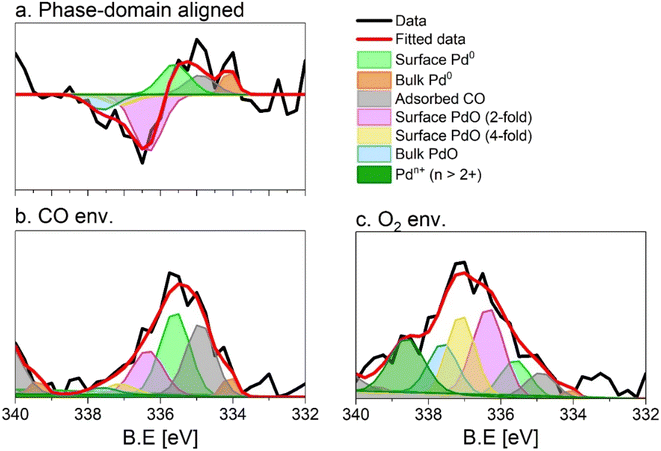
The originally published version of this article contained an incorrect figure and figure caption for Fig. 5, in which the labels ‘O2 env.’ and ‘CO env.’ in Fig. 5b and c, respectively, were inverted. The correct Fig. 5 and corresponding caption are displayed below. Two paragraphs that refer to Fig. 5 on page 7488 in the Results and discussion section under the sub-heading ‘Identification of species on 5 wt% Pd/Al2O3’ have also been updated to replace the original text.
The fit of the averaged spectrum obtained at 150 s under oxidising conditions is dominated by cationic Pd species (Fig. 5c and Table 2). The contribution of Pd0 is low (7%), while surface Pd0 is absent. The fraction of Pd0 with adsorbed CO (11%) is not negligible and is attributed to the Pd surface poisoning by CO due to the strong Pd–CO bond. The poisoning effect of CO on Pd sites is supported by the very low levels of CO2 all along the O2 half-period in the MS data (Fig. S3), after a less pronounced sharp peak than that detected in the CO half-period. The presence of the signals of surface and bulk Pd0 under oxidizing conditions suggests that the thickness of the oxide layer formed is smaller than the mean escape depth of photoelectrons and thus that metallic Pd is coated by a thin skin of oxide.
Under reducing conditions, the fit of the averaged spectrum at 150 s (Fig. 5b and Table 2) shows predominantly peaks corresponding to metallic Pd species as well as Pd0 with adsorbed CO together with minor contributions from bulk and surface PdO but no signal of Pdn+. The poor contribution from the surface Pd0 (4%) is justified by the presence of adsorbed CO molecules.
The Royal Society of Chemistry apologises for these errors and any consequent inconvenience to authors and readers.
| This journal is © The Royal Society of Chemistry 2023 |

