 Open Access Article
Creative Commons Attribution 3.0 Unported Licence
Correction: Intelligent antibacterial surface based on ionic liquid molecular brushes for bacterial killing and release
Lunqiang
Jin
ab,
Zhenqiang
Shi
a,
Xiang
Zhang
a,
Xiaoling
Liu
a,
Huiling
Li
a,
Jingxia
Wang
a,
Feng
Liang
*b,
Weifeng
Zhao
*a and
Changsheng
Zhao
*a
aCollege of Polymer Science and Engineering, The State Key Laboratory of Polymer Materials Engineering, Sichuan University, Chengdu, 610065, P. R. China. E-mail: zhaoscukth@163.com
bThe State Key Laboratory of Refractories and Metallurgy, Coal Conversion and New Carbon Materials Hubei Key Laboratory, School of Chemistry & Chemical Engineering, Wuhan University of Science and Technology, Wuhan 430081, P. R. China. E-mail: feng_liang@whu.edu.cn
First published on 9th August 2023
Abstract
Correction for ‘Intelligent antibacterial surface based on ionic liquid molecular brushes for bacterial killing and release’ by Lunqiang Jin et al., J. Mater. Chem. B, 2019, 7, 5520–5527, https://doi.org/10.1039/C9TB01199D.
The authors regret errors in Fig. 2b and c, 4c and 7c.
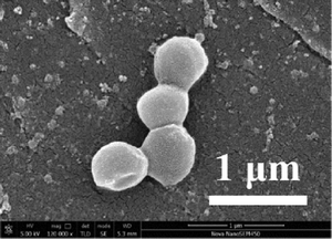
During the manuscript preparation, the SEM data in the left-hand panel in Fig. 2b, the right-hand panel in Fig. 2b and the top-right panel in Fig. 7c were edited to remove contaminants, the AFM data in the right hand panel in Fig. 2c was edited to remove shadows, and the EDS mapping images for C/N/O/S/Br, C and N in Fig. 4c are incorrect.
The authors became aware of the concerns and have repeated the experiments in triplicate.
An independent expert has viewed the corrected images and the replicated experimental raw data, and has concluded that they are consistent with the discussions and conclusions presented.
The corrected figures, with the original data, are shown below (it should be noted that the labels are unchanged).
 | ||
| Fig. 2 (b) The SEM image of the PES membrane and the SEM image of the IL(Br)/PDA@PES membrane. (c) AFM image of the IL(Br)/PDA@PES membrane (scale bar: 2 × 2 μm). | ||
The authors sincerely apologize for this mistake in the preparation of the article and apologize for any inconvenience caused.
The Royal Society of Chemistry apologises for these errors and any consequent inconvenience to authors and readers.
| This journal is © The Royal Society of Chemistry 2023 |


