Direct observation of split-mode exciton-polaritons in a single MoS2 nanotube†
A. I.
Galimov
 a,
D. R.
Kazanov
 a,
A. V.
Poshakinskiy
 a,
M. V.
Rakhlin
 a,
I. A.
Eliseyev
 a,
A. A.
Toropov
 a,
M.
Remškar
 b and
T. V.
Shubina
 *a
*a
aIoffe Institute, 26 Politekhnicheskaya, St. Petersburg, 194021, Russia. E-mail: shubina@beam.ioffe.ru
bJozef Stefan Institute, 39 Jamova cesta, Ljubljana, 1000, Slovenia
First published on 22nd April 2024
Abstract
A single nanotube synthesized from a transition metal dichalcogenide (TMDC) exhibits strong exciton resonances and, in addition, can support optical whispering gallery modes. This combination is promising for observing exciton-polaritons without an external cavity. However, traditional energy-momentum-resolved detection methods are unsuitable for this tiny object. Instead, we propose to use split optical modes in a twisted nanotube with the flattened cross-section, where a gradually decreasing gap between the opposite walls leads to a change in mode energy, similar to the effect of the barrier width on the eigenenergies in the double-well potential. Using micro-reflectance spectroscopy, we investigated the rich pattern of polariton branches in single MoS2 tubes with both variable and constant gaps. Observed Rabi splitting in the 40–60 meV range is comparable to that for a MoS2 monolayer in a microcavity. Our results, based on the polariton dispersion measurements and polariton dynamics analysis, present a single TMDC nanotube as a perfect polaritonic structure for nanophotonics.
New conceptsWe present the discovery of exciton-polaritons in flattened nanotubes of transition metal dichalcogenides (TMDC). These structures exhibit strong exciton resonances and can support whispering gallery modes in the nanotube wall. A flattened cross-section with gradually decreasing gap between opposite walls splits doubly degenerate whispering gallery modes into two optical modes of different symmetry and leads to a significant change in their energies. As the split mode approaches the exciton resonance, the formation of an exciton-polariton occurs in the strong coupling mode, which is confirmed by the anticrossing of polariton branches with Rabi splitting. This new approach provides an optical-mechanical method to control the interaction of excitonic resonances with an array of whispering gallery modes within a tube, in contrast to traditional energy-momentum-resolved detection methods. The use of micro-reflection spectroscopy allows demonstration of the rich pattern of polariton branches in a single MoS2 nanotube with a variable gap between the walls and discovery of the dispersionless polaritons with stable energy in a nanotube with a constant gap in an unchanged but flattened cross-section. Obtained results and agreement with theoretical and numerical calculations confirm the possibility of producing polaritons in the strong coupling regime in flattened nanotubes, making them promising candidates for nanophotonics. |
1 Introduction
Transition metal dichalcogenides (TMDCs), such as MoX2 and WX2 (X = S, Se), have high binding energy and giant oscillator strength of direct A and B excitons in both atomically thin and multilayer structures.1,2 This leads to a strong interaction between light and matter, resulting in the formation of an exciton-polariton (or polariton for short). The polariton, that is, a quantum superposition of an exciton and a photon mode, was observed in many semiconductor systems, being of practical importance for nanophotonics (threshold-less lasers3) and for quantum technologies (computing and simulators4,5). Of greatest interest are polaritons in microcavities, where excitons interact with the strongly confined electromagnetic field,6–8 which increases the interaction energy and makes it greater than the broadening of the bare modes. This situation, called the strong coupling regime, manifests itself as an anticrossing of the upper and lower polariton branches (UPB and LPB, respectively) in the dependence of the eigenenergies on the detuning between the exciton and cavity modes.9 The Rabi splitting ħΩR = 2g characterizes the frequency of oscillation between the photon and exciton states, where g is a coupling strength.A quasi-two-dimensional (2D) polariton is characteristic of a quantum well in a microcavity.10 Its extreme case is the polariton formed when a TMDC monolayer is placed into an external resonator.11–16 All studies of 2D polaritons have exploited the ability to tune the cavity mode energy either by varying the cavity length or by changing its in-plane wave vector k ≈ (ω/c)sin(θ), that is controlled by detection angle θ.17,18 For example, angle-resolved reflectivity spectroscopy revealed the strong coupling with the Rabi splitting of ∼46 meV in MoS2 monolayer inside the Bragg microcavity.11 Similar research of both reflection and photoluminescence (PL) demonstrated a strong coupling in WSe2 and WS2 monolayers in photonic crystals.14 Changing the cavity length in the open microcavity made it possible to observe polaritons with the Rabi splitting ∼20 meV in MoSe2 monolayers12 and ∼70 meV in WS2 monolayers.13
TMDC nanotubes (NTs) were synthesized shortly after carbon NTs19 in the early 1990s.20 They are usually multi-walled, have a length from hundreds of nanometers to several millimeters and a characteristic diameter from tens of nanometers to several micrometers.21,22 We will use the same designation “NT” for both micro- and nanotubes. Tube shapes can vary from perfectly cylindrical to almost ribbon-like, depending on the internal tensile stresses.23,24 In twisted tubes, in addition, the flattened cross-section rotates along the tube axis.25 Multi-walled NTs have an indirect band structure; however, resonances of direct excitons are observed up to room temperatures due to their giant oscillator strength and high recombination rate.26,27 While individual monolayers are unstable under ambient conditions and must be protected by hBN,28 monolayers scrolled in NT are naturally protected by outer layers.
Recently, many interesting phenomena have been discovered in NTs that are promising for wide application.25,29 Among them is the specific confinement of electromagnetic fields in NTs, leading to the appearance of whispering gallery modes (WGMs), the peaks of which modulate the emission spectra.30 The coexistence of strong direct excitons and pronounced optical modes is very promising for the creation of polaritons in the strong coupling regime without an external cavity. However, the application of the described above detection methods with energy-momentum resolution to NTs seems to be very problematic. Providing a non-zero wave vector k along the NT axis by changing the angle of incidence can hardly be combined with precise focusing on a single NT, especially in cryogenic measurements. It is also impossible to tune the optical mode energy by gradually reducing the diameter of the tube, as it is done for nanowires,31 since in conventional synthesized cylindrical NTs the cross-section is constant over a length of more than 50 μm.32
A reasonable solution seemed to be to observe the dispersion of polaritons by an indirect method from extinction spectra measured in ensembles of NTs of different diameters in the range 36–110 nm, separated by centrifugation.33 However, the fact that strong coupling can only be realized when the NT diameter exceeds 80 nm34 limits the capabilities of this method. Indisputable proof of the existence of exciton-polaritons would be their direct observation by reflectivity in a single NT, which is still missing.
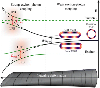
To carry out such an experiment, we propose a new concept that exploits a recently discovered phenomenon, namely, the splitting of optical modes in a NT with a flattened cross-section that features a gradual decrease in the gap between opposite walls along the NT.35Fig. 1 illustrates the principle of split-mode polariton formation. In a cylindrical NT, the WGM is doubly degenerate and has energy of ħω0. As the minor axis b of the NT cross-section decreases, the gap between the walls also decreases and the evanescent tails of the electric fields begin to overlap. By analogy with the electron states in the double-well potential,36 this leads to the splitting of the WGM into two non-degenerate even and odd modes, which are symmetric and antisymmetric with respect to the major axis. Their energies read ħωe,o = ħω0(1 ± tWGM), where the odd mode has higher energy. Note that the splitting increases exponentially as tWGM ∝ e−κrb, where κr is the decay constant of the evanescent tails (see ESI† for details). When an odd or even mode approaches the exciton resonance, in the case of strong coupling, an exciton-polariton is formed and Rabi splitting of UPB and LPB is observed.
This work presents studies of split-mode polaritons in a single MoS2 NT using micro-reflectance and micro-PL spectroscopy. It is shown that the energy of the split mode can vary over a wide range in a tube with a variable gap between the walls, which makes it possible to observe the transformation of purely optical modes into polariton branches demonstrating distinct anticrossing. On the contrary, dispersionless polaritons with stable energies are observed in NTs with a constant gap. In both cases, we register close Rabi splitting values of ∼40 meV for A-exciton-polariton and ∼60 meV for B-exciton-polariton. The observed PL dynamics is consistent with the formation of polaritons.
2 Results
2.1 Variable wall-gap nanotube
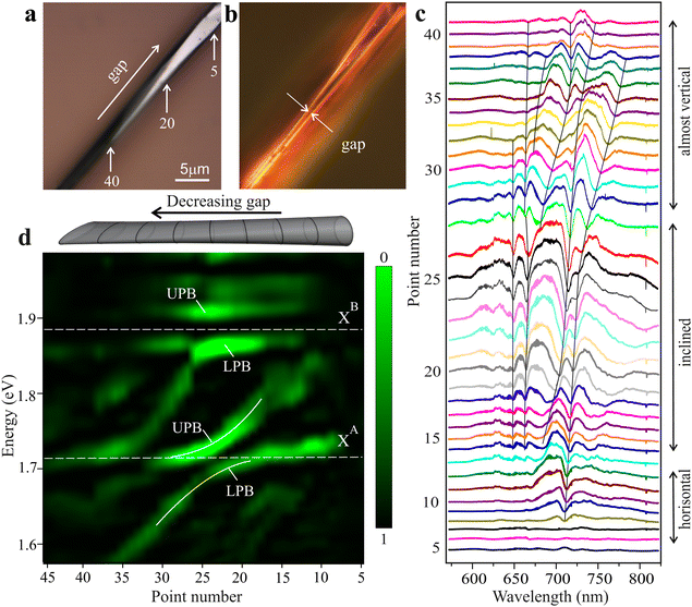
For the experimental study, we selected a twisted MoS2 NT with a total length of about one hundred microns, synthesized by a chemical vapor transfer reaction using iodine as a transport agent.25 A common property of multi-walled NTs is internal tensile strain, which arises due to the fact that each subsequent layer must be stretched relative to the previous one in order to maintain the crystalline structure. The tensile strain promotes flattening of the cross-section and, in addition, reduces the band gap due to the modification of the band structure,37 as occurs in atomically-thin layers when the conduction band minimum shifts downward.38–41 Using Raman studies (see the ESI†), we estimated the tensile strain values in the NT to be ≥1%, resulting in a red shift of A exciton towards 1.72–1.75 eV with B exciton being ∼140 meV higher in energy. New strain-induced positions of excitons in NTs are about 100 meV lower than in unstrained flakes obtained in the same synthesis process.27 In different regions, the NT has a different degree of strain which leads to different flattening and rotation of the cross-section, as well as to some change in the exciton energy. The cross-section circumference remains constant along the NT.Optical images of a part of the NT with a variable gap between the walls, shown in Fig. 2(a and b), were obtained in different registration modes: (a) bright-field, when light scattered perpendicularly from the surface is recorded; (b) dark-field with cross-polarization, when light scattered by inclined surfaces is recorded. The bright-field image shows the rotation of the cross-section along the NT, whereas the dark-field image shows a gradual decrease in the gap between the opposite walls of the NT. It takes place as the detection point number increases. At the narrowest point, the gap is comparable to the thickness of the tube walls of 50–100 nm.
Fig. 2 shows selected original (c) and processed (d) reflectance spectra measured along the NT axis as described in the Methods section, with a distance between the adjacent points of 1–1.5 μm. In Fig. 2(c), thin lines trace the main dips in the reflectance spectra. Based on the width of isolated peaks of optical modes, the quality factor of the NT cavity is estimated at approximately 500. Far from the exciton resonances, the dips are associated with the pure optical modes excited inside the NT walls. In the points no. 30–40, the modes (even, presumably) are observed up to the region of relative transparency (1.55 eV). The mode splitting is maximal here due to the narrowest gap. When the split modes are close to excitons in energy, which occurs in the inclined region (points no. 15–27), exciton-polaritons are formed. The reflectance in this region is approximately four times higher than in others. This is partly the result of strong exciton-photon coupling, and partly due to the dependence of the WGM mode brightness on the rotation angle of the NT cross-section.35
To get a clearer picture of the mode transformation, we removed hardware interference and background noise from the reflection spectra. In the demo display shown in Fig. 2(d), green areas correspond to dips down to zero in the original reflectance spectra. A clear anticrossing of LPB and UPB is observed when one of the split modes approaches exciton A. The other mode creates anticrossing branches at exciton B. The UPB at exciton B is less pronounced due to high absorption in the spectral region above exciton B.
In the region where the gap between the walls increases (points no. 5–15) and respectively the strain value becomes smaller, a blue shift of the A exciton resonance is observed, which affects the dispersion of polariton branches, bending them towards higher energy compared to the average. The experimentally recorded ħΩR = 2g between the LPB and UPB are found to be about 40 meV for exciton A (1.74 eV) and 60 meV for exciton B (1.88 eV), which has higher oscillator strength. These values indicate a strong coupling regime and provide a sufficient ratio of ≳3 to the width of the reflection dip to observe the full picture of polaritons using reflectance spectroscopy. In addition, the value of coupling strength g ensures that the LPB will be located below a dark exciton, which promotes the emission brightening.42,43
To analyze the evolution of optical modes leading to the formation of polariton, we consider a simplified theoretical model. In the weakly deformed NT, the electric field of WGMs polarized along the NT axis is described by E(e)m ∝![[thin space (1/6-em)]](https://www.rsc.org/images/entities/char_2009.gif) cos(mφ) and E(o)m ∝
cos(mφ) and E(o)m ∝![[thin space (1/6-em)]](https://www.rsc.org/images/entities/char_2009.gif) sin(mφ), where e and o denote even and odd modes, m is the angular number, and the angle ϕ is measured from the major axis of the flattened cross-section. If the light falls normally on the NT axis, the back-scattering intensity near the resonance of the even mode ω(m)e has the form
sin(mφ), where e and o denote even and odd modes, m is the angular number, and the angle ϕ is measured from the major axis of the flattened cross-section. If the light falls normally on the NT axis, the back-scattering intensity near the resonance of the even mode ω(m)e has the form
 | (1) |
![[thin space (1/6-em)]](https://www.rsc.org/images/entities/char_2009.gif) sin. The trigonometric function in the numerator originates from the convolution of the incident field with the field distribution of the mode. This leads to a change in the visibility of modes in the spectra when the cross-section rotates. (The details on the derivation are given in the ESI.†)
sin. The trigonometric function in the numerator originates from the convolution of the incident field with the field distribution of the mode. This leads to a change in the visibility of modes in the spectra when the cross-section rotates. (The details on the derivation are given in the ESI.†)
We performed numerical calculations to elucidate the evolution of modes taking into account exciton resonances in the strong coupling regime using Comsol Multiphysics as described in the Method section. The dielectric response of NT wall was taken in Drude–Lorentz form with two resonances of A and B excitons:
 | (2) |
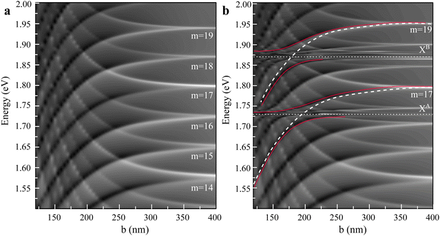
Fig. 3(a) shows the behavior of WGMs without interaction with exciton resonances. As the cross-section flattens, all even modes, curved downward, decrease their energy, and all odd modes, curved upward, increase energy. When the minor axis b of the cross-section becomes less than half the wavelength of the exciting light, the odd-mode resonances begin to quench. At a double-well potential,36 these antisymmetric modes have electric field in antiphase on opposite sides of the cross-section. Therefore, their coupling to the free waves is decreased as the gap between the walls narrows. In contrast, for even modes, which have electric field of the same phase on the opposite sides, the coupling to the free waves increases. This explains why only even, downward-curved modes are retained in the experimental spectra of highly flattened NTs.
The case of strong coupling between the optical modes and the excitons is shown in Fig. 3(b). In the absence of the cross-section deformation, WGMs shift their energies slightly near the exciton resonances due to the changes in the dielectric background. As the axis b decreases to 200–250 nm, stronger even modes reach exciton resonances from above and form exciton-polaritons. For clarity, we have highlighted with red lines the UPB and LPB originating from the WGMs with angular numbers m = 17 and m = 19. The anticrossing with ΩRabi of ∼35 meV and ∼55 meV for excitons A and B is quite close to the experimental results. The general picture of the calculated modes and their slopes also satisfactorily reproduce the experimental spectra.
2.2 Constant wall-gap nanotube
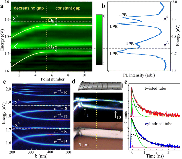
Now let us focus on another NT, which has an extended flat region with a small rotation angle and a constant gap between the walls (see Fig. 4). The map of the reflection spectra presented in Fig. 4(a) shows optical modes of constant energies along the flat region. They are grouped into pairs, with mode spacing in each pair very close to the Rabi splitting for A and B exciton-polaritons. PL spectra measured in this region exhibit four peaks that perfectly correspond to the modes in reflection (Fig. 4(c)). This picture corresponds to the dispersionless polaritons with constant energy, which were previously theoretically considered for TMDC monolayers.44 The parameters of these polaritons are stable along the large segment of the NT, where its geometry does not change.Calculations of optical modes for this case were carried out using almost the same model as described above, but assuming a close to zero cross-section rotation angle. We did not divide the NT into two regions, but gradually changed the value of b from 500 nm to 200 nm (Fig. 4). For the same circumference of ∼5.5 μm, the found optical modes have the angular numbers m as in Fig. 3(a). However, there is a remarkable difference due to the small rotation angle. At degrees less than 10°, only one of the two modes (even or odd) is excited, and this occurs in antiphase to each adjacent WGM pair.
Comparison of experimental and calculated pictures in Fig. 4(a and c) shows that the observed stability of mode energies and their separation can be realized only if the axis b exceeds 350 nm. Although the change in mode energies above this value is weak, it can certainly be resolved experimentally. Thus, the gap value is most likely constant and equals to approximately 400 nm. In another part of the NT, close to its kink, when the deformation increases and the gap is narrowed, the even modes begin to shift towards lower energies in both experimental and calculated spectra, as it occurs in the NT with the variable gap (Fig. 2). In the case of completely flattened NT with the shape of a ribbon, it is possible to form the polaritons on Fabry–Pérot modes, as it has been observed in thin WS2 flakes.45
In addition, we performed time-resolved photoluminescence (TRPL) measurements in the time domain of 10 ns as described in the Methods section. Extended PL decay curves for all peaks in Fig. 4(b) and the fitting procedure are given in the ESI.†Fig. 4(e) shows a part of the decay curve for the A-exciton LPB together with a curve measured at the same energy in a reference cylindrical NT with conventional WGMs. Both curves contain two components with characteristic decay times of less than 20 ps and about 200 ps, which are close, respectively, to the published lifetimes of the photon and exciton components of exciton-polariton radiative decay in a monolayer.46 In the tube under study, compared to the reference tube, a clear redistribution of intensity is observed in favor of the photonic component associated with the polariton formation.47 In particular, its contribution to the integral PL is at least 30 times higher than in the reference tube (see the ESI†). The reason is that as the cross-section of the tube gradually flattens, the resonance of the split WGM is adjusted to the exciton resonance, which leads to a strong coupling with the acceleration of radiative decay, akin to the Purcell effect.48 Such adjustment is impossible in a cylindrical tube with stable mode energies. The calculated dependence of the lifetime of optical modes on the degree of flattening is presented in the ESI.† Note that the third component with a decay time of about 1 ns is associated with emission of indirect excitons of close energy, which arise due to the band structure modification during tensile deformation.41,49,50 In the strong coupling regime, the presence of such excitons does not affect polaritons, since the LPB energy is lower than their energy.42,43
3 Conclusions
The discovery of anticrossing of polariton branches is usually considered as indisputable evidence of the existence of polaritons in the strong coupling regime. We were able to obtain this result using the change in the energy of split modes in NT with a variable gap between the walls in a flattened cross-section. This approach represents an optical-mechanical method for controlling the interaction of exciton resonances with a set of optical modes excited in NT.Another important discovery was the observation of polaritons with stable energy in a NT with a constant gap in an unchanged cross-section. The general characteristics of such dispersionless polaritons are in a good agreement with those found for exciton-polaritons in the NT with the variable gap, which excludes another explanation. Note that polaritons of this type were realized at a relatively large gap between NT walls (∼400 nm), which indicates the possibility of their excitation in almost cylindrical MoS2 NTs.
In both cases, the experimentally observed pattern of optical modes and their transformation into polariton branches are consistent with the proposed theoretical description. The simulation performed reproduces polaritons with Rabi splitting values close to experimental ones and makes it possible to reconstruct the geometric parameters of the NTs under study. Taken together, our experimental and theoretical results confirm the possibility of producing polaritons in the strong coupling regime in individual TMDC NTs, making them promising candidates for nanophotonics. The ability to tune the optical mode energy to exciton resonance and eliminate the negative influence of dark excitons will expand the scope of applications of TMDC NTs for sensoring and creating polariton lasers.
4 Methods
4.1 Optical studies
Optical measurements of MoS2 NTs were done at 8 K. The Si/SiO2 substrate with the NTs on its surface was mounted in the ST-500-Attocube (Janis) cryostat, containing the three-coordinate piezo-driver with an accuracy in positioning of ∼20 nm, located in the cold zone of the cryostat. This provides high mechanical stability and vibration isolation necessary for studying such tiny objects. For PL excitation, we used a semiconductor laser PILAS 405 nm (Advanced Laser Systems) with a 40 ps pulse duration and 100 MHz repetition rate. Focusing the laser light using a 100x apochromatic objective (Mitutoyo plan apochromat, NA = 0.7) provided spot size less than 1 μm, which corresponds to ∼6 W cm−2 (10 nW μm−2) power density on the sample surface. The PL signal was collected by the same objective in a confocal setup and directed onto the entrance slit of an SP-2500 spectrometer with cooled PyLoN CCD detector (Princeton Instruments). To measure the time resolved histogram of PL, a single-photon avalanche photodiode PDM 100 (Micro Photon Devices) with time resolution ∼40 ps was used. The time period between exciting pulses was 10 ns. For blocking the scattered laser radiation, a band pass interference filter was used.The reflection spectra along the NTs were measured on the same optical setup using a halogen lamp coupled to a single-mode optical fiber with subsequent collimation. In this mode, it was possible to achieve high spatial resolution when scanning the NTs. To highlight features, the reflection spectra were subjected to additional processing. First, the reflection spectrum was smoothed using a low-pass Fourier filter to get rid of unwanted noise. Secondly, the moving-average background was subtracted from the reflection spectrum, which made it possible to enhance the visibility of exciton and polariton reflection dips against the background of other effects.
Raman spectra were obtained in backscattering geometry at room temperature. For these investigations, we used a Horiba LabRAM HR Evo UV-VIS-NIR-Open spectrometer (Horiba, Lille, France) with confocal optics. Olympus MPLN100x (Olympus, Tokyo, Japan) objective lens (NA = 0.9) focused the laser beam into a spot ∼1 μm in diameter. As an excitation source, a Nd:YAG laser (Torus Laser Quantum, Stockport, UK) with λ = 532 nm was used. To avoid damage and heating of the NTs, the laser power on its surface was limited to 200 μW.
Optical images of the NTs in different polarizations were obtained using a Nikon ECLIPSE LV150 (Nikon Europe, Amsterdam, Netherlands) industrial microscope operating in bright-field and dark-field modes. The sample under study was illuminated using an LED source passed through a linear polarizer, while long-exposure detection was carried out in orthogonal polarization. This method made it possible to see features of NTs that were inaccessible with the conventional bright-field method.
4.2 Comsol multiphysics calculations
To simulate the reflection and PL map, we divided the computational domain into two parts, outside part with conventional refractive index of air, and inside NT domain with refractive index using Drude–Lorentz dispersion model with A and B exciton resonances. The exciton line broadening was chosen to be 5 meV to resolve the rich pattern of optical modes in the TMDC NT. We assumed a wave vector along the NT axis kz = 0, because kz ≠ 0 shifts the energies of optical modes in the NT much less than the flattening effects. To neglect reflection from computational domain boundaries, we added a boundary box with perfectly matched layer. To excite optical modes, we put a plane-wave source incident on the NT with polarization along its axis. To find the back scattered signal, we summed the reflected electric field far away from the NT along a line, while to find the PL signal we calculated the integral of the squared electromagnetic field inside the NT walls.Author contributions
All authors contributed to this article. A. I. G., M. V. R., I. A. E. and A. A. T. carried out optical measurements and analyzed data; D. R. K. and A. V. P. performed theoretical calculations; M. R. carried out the growth of nanotubes; T. V. S. proposed the physical model, wrote the final version, and supervised this research. All authors gave approval for the final version of the manuscript.Conflicts of interest
There are no conflicts to declare.Acknowledgements
This work was supported in part by the Russian Science Foundation project no. 23-12-00300 (A. I. G. - reflection study, T. V. S. - physical model). M. R. thanks the Slovenian Research and Innovation Agency (P1-0099) for funding. D. R. K. acknowledges the grant #SP5068.2022.5. We thank B. Borodin for transferring and positioning the nanotubes on the substrate and A. Veretennikov for his assistance in data processing.Notes and references
- B. L. Evans and P. A. Young, Proc. R. Soc. London, Ser. A, 1965, 284, 402–422 CAS.
- G. Wang, A. Chernikov, M. M. Glazov, T. F. Heinz, X. Marie, T. Amand and B. Urbaszek, Rev. Mod. Phys., 2018, 90, 021001 CrossRef CAS.
- A. Imamoglu, R. J. Ram, S. Pau and Y. Yamamoto, Phys. Rev. A: At., Mol., Opt. Phys., 1996, 53, 4250–4253 CrossRef CAS PubMed.
- S. Ghosh and T. C. H. Liew, npj Quantum Inf., 2020, 6, 16 CrossRef.
- N. Y. Kim and Y. Yamamoto, Exciton-Polariton Quantum Simulators, Springer International Publishing, Cham, 2017, pp. 91–121 Search PubMed.
- A. V. Kavokin, J. J. Baumberg, G. Malpeuch and F. P. Laussy, Microcavities, Oxford University Press, UK, 2008 Search PubMed.
- S. Luo, H. Zhou, L. Zhang and Z. Chen, Appl. Phys. Rev., 2023, 10, 011316 CAS.
- K. Wei, Q. Liu, Y. Tang, Y. Ye, Z. Xu and T. Jiang, Nat. Commun., 2023, 14, 5310 CrossRef CAS PubMed.
- C. Weisbuch, M. Nishioka, A. Ishikawa and Y. Arakawa, Phys. Rev. Lett., 1992, 69, 3314–3317 CrossRef CAS PubMed.
- V. D. Kulakovskii, A. I. Tartakovskii, D. N. Krizhanovskii, A. Armitage, J. S. Roberts and M. S. Skolnick, Phys.-Usp., 2000, 43, 853 CrossRef CAS.
- X. Liu, T. Galfsky, Z. Sun, F. Xia, E.-C. Lin, Y.-H. Lee, S. Kéna-Cohen and V. M. Menon, Nat. Photonics, 2015, 9, 30–34 CrossRef CAS.
- S. Dufferwiel, S. Schwarz, F. Withers, A. A. P. Trichet, F. Li, M. Sich, O. Del Pozo-Zamudio, C. Clark, A. Nalitov, D. D. Solnyshkov, G. Malpuech, K. S. Novoselov, J. M. Smith, M. S. Skolnick, D. N. Krizhanovskii and A. I. Tartakovskii, Nat. Commun., 2015, 6, 8579 CrossRef CAS PubMed.
- L. C. Flatten, Z. He, D. M. Coles, A. A. P. Trichet, A. W. Powell, R. A. Taylor, J. H. Warner and J. M. Smith, Sci. Rep., 2016, 6, 33134 CrossRef CAS PubMed.
- L. Zhang, R. Gogna, W. Burg, E. Tutuc and H. Deng, Nat. Commun., 2018, 9, 713 CrossRef PubMed.
- C. Schneider, M. M. Glazov, T. Korn, S. Höfling and B. Urbaszek, Nat. Commun., 2018, 9, 2695 CrossRef PubMed.
- L. Lackner, M. Dusel, O. A. Egorov, B. Han, H. Knopf, F. Filenberger, S. Schröder, K. Watanabe, T. Taniguchi, S. Tongay, C. Anton-Solanas, S. Höfling and C. Schneider, Nat. Commun., 2021, 12, 4933 CrossRef CAS PubMed.
- R. Houdré, C. Weisbuch, R. P. Stanley, U. Oesterle, P. Pellandini and M. Ilegems, Phys. Rev. Lett., 1994, 73, 2043–2046 CrossRef PubMed.
- D. S. Dovzhenko, S. V. Ryabchuk, Y. P. Rakovich and I. R. Nabiev, Nanoscale, 2018, 10, 3589–3605 RSC.
- S. Iijima, Nature, 1991, 354, 56–58 CrossRef CAS.
- R. Tenne, L. Margulis, M. Genut and G. Hodes, Nature, 1992, 360, 444–446 CrossRef CAS.
- M. Remškar, Z. Skraba, F. Cléton, R. Sanjinés and F. Lévy, Appl. Phys. Lett., 1996, 69, 351–353 CrossRef.
- M. Remškar, Adv. Mater., 2004, 16, 1497–1504 CrossRef.
- V. Kralj-Iglič, M. Remškar, G. Vidmar, M. Fošnarič and A. Iglič, Phys. Lett. A, 2002, 296, 151–155 CrossRef.
- A. Enyashin, S. Gemming and G. Seifert, Eur. Phys. J.: Spec. Top., 2007, 149, 103–125 Search PubMed.
- M. Rem
![[s with combining breve]](https://www.rsc.org/images/entities/char_0073_0306.gif) kar, A. K. Hüttel, T. V. Shubina, A. Seabaugh, S. Fathipour, R. Lawrowski and R. Schreiner, Isr. J. Chem., 2022, 62, e202100100 CrossRef.
kar, A. K. Hüttel, T. V. Shubina, A. Seabaugh, S. Fathipour, R. Lawrowski and R. Schreiner, Isr. J. Chem., 2022, 62, e202100100 CrossRef. - M. Selig, G. Berghäuser, A. Raja, P. Nagler, C. Schüller, T. F. Heinz, T. Korn, A. Chernikov, E. Malic and A. Knorr, Nat. Commun., 2016, 7, 13279 CrossRef CAS PubMed.
- T. V. Shubina, M. Remškar, V. Y. Davydov, K. G. Belyaev, A. A. Toropov and B. Gil, Ann. Phys., 2019, 531, 1800415 CrossRef.
- F. Cadiz, E. Courtade, C. Robert, G. Wang, Y. Shen, H. Cai, T. Taniguchi, K. Watanabe, H. Carrere, D. Lagarde, M. Manca, T. Amand, P. Renucci, S. Tongay, X. Marie and B. Urbaszek, Phys. Rev. X, 2017, 7, 021026 Search PubMed.
- J. L. Musfeldt, Y. Iwasa and R. Tenne, Phys. Today, 2020, 73, 42–48 CrossRef CAS.
- D. R. Kazanov, A. V. Poshakinskiy, V. Y. Davydov, A. N. Smirnov, I. A. Eliseyev, D. A. Kirilenko, M. Remškar, S. Fathipour, A. Mintairov, A. Seabaugh, B. Gil and T. V. Shubina, Appl. Phys. Lett., 2018, 113, 101106 CrossRef.
- L. Sun, Z. Chen, Q. Ren, K. Yu, L. Bai, W. Zhou, H. Xiong, Z. Q. Zhu and X. Shen, Phys. Rev. Lett., 2008, 100, 156403 CrossRef PubMed.
- D. Kazanov, M. Rakhlin, A. Poshakinskiy and T. Shubina, Nanomaterials, 2020, 10, 373 CrossRef CAS PubMed.
- L. Yadgarov, B. Višić, T. Abir, R. Tenne, A. Y. Polyakov, R. Levi, T. V. Dolgova, V. V. Zubyuk, A. A. Fedyanin, E. A. Goodilin, T. Ellenbogen, R. Tenne and D. Oron, Phys. Chem. Chem. Phys., 2018, 20, 20812–20820 RSC.
- S. S. Sinha, A. Zak, R. Rosentsveig, I. Pinkas, R. Tenne and L. Yadgarov, Small, 2020, 16, 1904390 CrossRef CAS PubMed.
- I. A. Eliseyev, B. R. Borodin, D. R. Kazanov, A. V. Poshakinskiy, M. Remškar, S. I. Pavlov, L. V. Kotova, P. A. Alekseev, A. V. Platonov, V. Y. Davydov and T. V. Shubina, Adv. Opt. Mater., 2023, 11, 2202782 CrossRef CAS.
- L. Landau and E. Lifshits, Quantum Mechanics: Non-Relativistic Theory, Elsevier Science, 1991, vol. 3 Search PubMed.
- M. Ghorbani-Asl, N. Zibouche, M. Wahiduzzaman, A. F. Oliveira, A. Kuc and T. Heine, Sci. Rep., 2013, 3, 2961 CrossRef PubMed.
- H. Peelaers and C. G. Van de Walle, Phys. Rev. B: Condens. Matter Mater. Phys., 2012, 86, 241401 CrossRef.
- H. J. Conley, B. Wang, J. I. Ziegler, R. F. Haglund Jr., S. T. Pantelides and K. I. Bolotin, Nano Lett., 2013, 13, 3626–3630 CrossRef CAS PubMed.
- D. Lloyd, X. Liu, J. W. Christopher, L. Cantley, A. Wadehra, B. L. Kim, B. B. Goldberg, A. K. Swan and J. S. Bunch, Nano Lett., 2016, 16, 5836–5841 CrossRef CAS PubMed.
- X. Han, J. Lin, J. Liu, N. Wang and D. Pan, J. Phys. Chem. C, 2019, 123, 14797–14802 CrossRef CAS.
- V. Shahnazaryan, V. Saroka, I. Shelykh, W. Barnes and M. Portnoi, ACS Photonics, 2019, 6, 904–914 CrossRef CAS.
- H. Shan, I. Iorsh, B. Han, C. Rupprecht, H. Knopf, F. Eilenberger, M. Esmann, K. Yumigeta, K. Watanabe, T. Taniguchi, S. Klembt, S. Höfling, S. Tongay, C. Antón-Solanas, I. A. Shelykh and C. Schneider, Nat. Commun., 2022, 13, 3001 CrossRef CAS PubMed.
- F. Alpeggiani, S.-H. Gong and L. Kuipers, Phys. Rev. B, 2018, 97, 205436 CrossRef CAS.
- Q. Wang, L. Sun, B. Zhang, C. Chen, X. Shen and W. Lu, Opt. Express, 2016, 24, 7151 CrossRef CAS PubMed.
- X. Liu, W. Bao, Q. Li, C. Ropp, Y. Wang and X. Zhang, Phys. Rev. Lett., 2017, 119, 027403 CrossRef PubMed.
- N. Lundt, N. S. Stoll, P. Nagler, A. Nalitov, S. Klembt, S. Betzold, J. Goddard, E. Frieling, A. V. Kavokin, C. Schüller, T. Korn, S. Höfling and C. Schneider, Phys. Rev. B, 2017, 96, 241403 CrossRef.
- E. Purcell, Phys. Rev., 1946, 69, 681 CrossRef.
- Y. Uchiyama, A. Kutana, K. Watanabe, T. Taniguchi, K. Kojima, T. Endo, Y. Miyata, H. Shinohara and R. Kitaura, npj 2D Mater. Appl., 2019, 3, 26 CrossRef.
- I. A. Eliseyev, A. I. Galimov, M. V. Rakhlin, E. A. Evropeitsev, A. A. Toropov, V. Y. Davydov, S. Thiele, J. Pezoldt and T. V. Shubina, Phys. Status Solidi RRL, 2021, 15, 2100263 CrossRef CAS.
Footnote |
| † Electronic supplementary information (ESI) available. See DOI: https://doi.org/10.1039/d4nh00052h |
| This journal is © The Royal Society of Chemistry 2024 |


