 Open Access Article
A new green fluorimetric micelle complexation approach for reduction of the consumed solvent and quantification of avapritinib in biological fluids†
Baher I. Salman *a,
Hany A. Batakoushyb,
Roshdy E. Sarayac,
Mohamed A. A. Abdel-Aald,
Adel Ehab Ibrahime,
Yasser F. Hassana,
Ahmed I. Hassana and
Ehab A. M. El-Shouraf
aPharmaceutical Analytical Chemistry Department, Faculty of Pharmacy, Al-Azhar University, Assiut Branch, Assiut, 71524, Egypt. E-mail: bahersalman@azhar.edu.eg; bahersalman2013@yahoo.com
bPharmaceutical Analytical Chemistry Department, Faculty of Pharmacy, Menoufia University, Shebin Elkom, 32511, Egypt
cPharmaceutical Analytical Chemistry Department, Faculty of Pharmacy, Port-Said University, Port Said 42511, Egypt
dDepartment of Pharmaceutical Chemistry, Faculty of Pharmacy, Al-Azhar University, Assiut Branch, Assiut, 71524, Egypt
eNatural and Medical Sciences Research Center, University of Nizwa, P.O. Box 33, Birkat Al Mauz, Nizwa 616, Oman
fDepartment of Clinical Pharmacy, Faculty of Pharmacy, Al-Azhar University, Assiut Branch, Assiut, 71524, Egypt
First published on 2nd April 2024
Abstract
Avapritinib (AVA) is the first medication authorized by the US-FDA in 2020 for the management of gastrointestinal stromal tumours (GISTs) that can't be treated by surgery. Cancer is among the most common causes of death worldwide and is the second most common cause of death after cardiovascular disease. Therefore, a quick, easy, sensitive, and straightforward fluorimetric approach was used to analyse AVA in pharmaceutical materials and blood plasma (pharmacokinetic). The suggested technique relies on 2% sodium dodecyl sulphate (SDS, pH 4) micellar system augmentation of the fluorescence of the tested drug. The technique demonstrated high relative fluorescence intensity (RFI) at 430 nm after excitation at 340 nm. Concentrations ranging from 20.0–400.0 ng mL−1 with a limit of quantitation of 9.47 ng mL−1 were used to obtain luminescence data for the studied medicine. In addition, the quantum yield of the AVA fluorescence was increased with the gradual addition of a surfactant at a concentration above its critical micellar level. This knowledge has been exploited to enhance the effectiveness of a spectrofluorometric technique for the estimation of AVA in human plasma (98.95 ± 1.22%) and uniformity tests with greenness assessments. The conditions for enhanced fluorescence were optimized and fully validated using US-FDA and International Conference on Harmonization (ICH) rules. This innovative strategy was expanded for AVA stability research in human plasma across various circumstances. This approach is an eco-friendly solution compared to traditional testing methods that use hazardous chemicals.
1. Introduction
The second leading cause of mortality worldwide, behind cardiovascular disorders, is cancer. This disease often results from malfunctions in the regulatory systems that control cell division and proliferation.1,2 Every year, 1.7 million new instances of cancer are diagnosed, and 600![[thin space (1/6-em)]](https://www.rsc.org/images/entities/char_2009.gif) 000 individuals pass away from cancer in the United States alone.2,3 The cornerstones to effective cancer treatment are decreasing mortality, improving survival, enhancing patient survival, rapid diagnosis, prompt and targeted treatments. Early cancer detection is crucial since it shortens the treatment process and lowers the overall cost of care. AVA (ESI Fig. S1†) is a tyrosine kinase inhibitor that is used to treat gastrointestinal stromal tumours (GISTs). It is important to monitor the levels of avapritinib in biological fluids to ensure that patients are receiving the correct dose and to prevent drug toxicity.4,5 Only three approaches for the AVA assay have been reported.6–8 Moreover, earlier techniques6,7 were not supported by bioanalytical validation studies, which are currently mandated by US-FDA recommendations, and required the use of an organic solvent, which increases environmental toxicity. Researchers' focus has recently shifted more towards green chemistry to decrease environmental pollution and enhance public health.9–12 Green chemistry alludes to the improvement of chemical merchandise and methods that reduce or do not involve the utilization or generation of perilous materials. Besides, green chemistry covers all aspects of a chemical product's life cycle, such as its creation, utilization, and last transfer.13 Many applications in the field of analysis have been made for the use of various surfactant types to increase the fluorescence of several medications. The molar absorptivity and/or fluorescence quantum yield of a given fluorophore solution frequently increases when a surfactant is added at a concentration greater than its critical micellar concentration.14–16
000 individuals pass away from cancer in the United States alone.2,3 The cornerstones to effective cancer treatment are decreasing mortality, improving survival, enhancing patient survival, rapid diagnosis, prompt and targeted treatments. Early cancer detection is crucial since it shortens the treatment process and lowers the overall cost of care. AVA (ESI Fig. S1†) is a tyrosine kinase inhibitor that is used to treat gastrointestinal stromal tumours (GISTs). It is important to monitor the levels of avapritinib in biological fluids to ensure that patients are receiving the correct dose and to prevent drug toxicity.4,5 Only three approaches for the AVA assay have been reported.6–8 Moreover, earlier techniques6,7 were not supported by bioanalytical validation studies, which are currently mandated by US-FDA recommendations, and required the use of an organic solvent, which increases environmental toxicity. Researchers' focus has recently shifted more towards green chemistry to decrease environmental pollution and enhance public health.9–12 Green chemistry alludes to the improvement of chemical merchandise and methods that reduce or do not involve the utilization or generation of perilous materials. Besides, green chemistry covers all aspects of a chemical product's life cycle, such as its creation, utilization, and last transfer.13 Many applications in the field of analysis have been made for the use of various surfactant types to increase the fluorescence of several medications. The molar absorptivity and/or fluorescence quantum yield of a given fluorophore solution frequently increases when a surfactant is added at a concentration greater than its critical micellar concentration.14–16
This technique has been used for the direct estimation of the anticancer drug AVA in biological fluids. This method has been applied to uniformity tests, pharmacokinetic studies, and greenness assessments. The use of this technique in drug analysis has several advantages, including high sensitivity, selectivity, and accuracy, as well as the potential to reduce the use of hazardous reagents and solvents in the analysis process. Overall, the green fluorimetric micelle complexation approach has shown great promise in drug analysis, particularly in the field of anticancer drug estimation in biological fluids.
2. Experiments
The chemical AVA (98.70%) was obtained from Shanghai Chuangsai Technology Co. (China), and Ayvakit® (100 mg per tablet) was purchased from the Egyptian local market. El Nasr Chemical Co. provided all other chemicals, such as tween 80 (2% in distilled water), methanol, ethanol, sodium dodecyl sulphate (2% SDS), acetone, 2% polyethylene glycol 400 (PEG 400), acetonitrile, 2% cyclo-dextrin, N,N′-dimethylformamide, phosphoric acid, boric acid, carboxy methyl cellulose (CMC 2%), acetic acid, HCl, and sodium hydroxide (Cairo, Egypt). In a 10 mL calibrated flask, 20 mg of AVA was dissolved in 10 mL of methanol to create a stock AVP solution (200 μg mL−1). Further dilution with ultrapure water was performed to obtain the working solutions.Ten Ayvakit® tablets (100 mg per tablet) were weighed, mixed well, and crushed into fine powder. Then, 5 mL of methanol was added to an equivalent amount of 20 mg of the drug. The solution was ultrasonically treated for approximately 10 minutes, followed by filtering and content completion with distilled water to 100 mL. For content uniformity, each tablet was individually analysed in the pharmaceutical dosage form according to the USP procedure.17
The results were obtained through a grating excitation and emission monochromator with slit widths set to 2 nm, and a 1 cm quartz cell was included in the Fluorescence Spectrometer FS-2 with serial No. 1304002 with firmware version 131![[thin space (1/6-em)]](https://www.rsc.org/images/entities/char_2009.gif) 024 (Sinco, Korea) apparatus. 3510 Jenwey digital pH meter (E.U.). Unicen 21 centrifuges speed (Spain).
024 (Sinco, Korea) apparatus. 3510 Jenwey digital pH meter (E.U.). Unicen 21 centrifuges speed (Spain).
The spiked human plasma was subjected to the following procedure: using specific amounts of the cited drug standard working solution (0.50–4.0 μg mL−1), 1.0 mL of the drug-free plasma was spiked. As a protein precipitator, one millilitre of acetonitrile was included in mixture,8 which was then diluted with distilled water to 10.0 mL and centrifuged at 4000 rpm for approximately 15 minutes. The analytical technique used one millilitre of the supernatant.
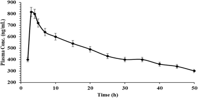
The pharmacokinetic study was performed and approved by the ethical committee of the international review board of Menoufia University, Egypt, with IRB approval no. 00437/2023. AVA pills (300.0 mg per tablet) were administered orally to ten healthy human subjects. At 1, 2, 4, 6, 8, 10, 15, 20, 25, 30, 35, 40, and 50 h, three millilitres of blood were taken intravenously into heparinized tubes.7 To extract the plasma, blood specimens were centrifuged at 4000 rpm for 30 min. A total of 1.0 mL of the plasma was combined with 1.0 mL of acetonitrile to act as a protein precipitate agent, and the supernatant was then separated through centrifugation at 4000 rpm for 15 minutes.
2.1. Fluorimetric calibration graph
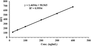
Several aliquots of AVA were placed into volumetric flasks with a volume of 10 mL to obtain the final concentration range (20.0–400.0 ng mL−1). Then, 1 mL of SDS (2% solution) was added, accompanied by the addition of 1.0 mL of phosphate buffer (pH 4). Ultrapure water was added to the flasks, which were then scanned at 430 nm.2.2. Stability study of AVA in human plasma
The stability of the pharmaceuticals under investigation in human plasma was examined using a variety of tests that used three concentrations (40.0, 100.0, and 350.0 ng mL−1) of low-quality control samples (LQC), medium-quality control samples (MQC), and high-quality control sample (HQC) respectively. These studies included three freeze–thaw cycles at −24 °C, a one-month long-term stabilization test, a 12 hours short-term stability experiment, a 6 hours post-preparative test procedure, and a 12 hours post-preparative steady-state experiment.3. Results and discussion
To create a new approach for the examination of AVA, this work attempted to determine the amount of the referenced drug in tablets and conduct a pharmacokinetic analysis using an enhanced emission band. Many approaches in the analysis field have been developed for the use of various surfactant types to increase the fluorescence of several medications. The molar absorptivity and/or fluorescence quantum yield of a given fluorophore solution is frequently increased when a surfactant is added at a concentration over its critical micellar concentration.14,15,18 This technique has been employed to enhance the effectiveness of spectroscopic techniques for several different compounds. Various types of media (beta-cyclodextrin, PEG 400, tween 80, and SDS) were used to investigate the luminescence characteristics of the drugs under investigation. When 2% SDS surfactant was added, the fluorescence intensity increased 6.8-fold more than that in aqueous solution Fig. 1.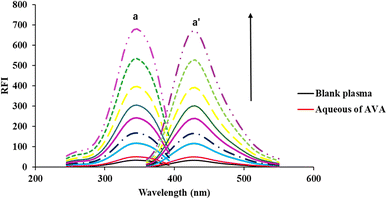 | ||
| Fig. 1 Fluorescence spectra (a,a′ excitation and emission) of AVA in the presence of 2% SDS at different concentrations (20.0, 50.0, 100.0, 150.0, 200.0, 300.0, 400.0 ng mL−1). | ||
As shown in Fig. 1, the micelle complexation of AVA with 2% SDS provided an emission wavelength of 430 nm (excitation of 340 nm) with a calibration range of 20.0–400.0 ng mL−1. The purpose of this research was to develop a straightforward, sensitive, trustworthy, selective, and affordable spectrofluorimetric technique for the evaluation of the purest forms of examined drugs, pharmaceutical preparations, biological fluids, and content uniformity. The green fluorimetric micelle complexation approach is fast and sensitive and has a low limit of detection. Its accuracy is comparable to that of traditional methods, making it an excellent alternative for the evaluation of avapritinib in biological fluids. Its application in the pharmaceutical industry has provided a solution to the need for a safer, more efficient, and more environmentally responsible approach to drug testing.
3.1. Optimization of the calibration graph conditions
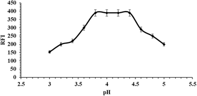
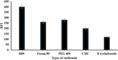
The factors that impact the reaction's advancement, sensitivity, and stability, as well as the properties of the fluorescence results, were thoroughly analysed and improved. Only these factors changed, while others remained constant. The examined parameters included pH, buffer type and quantity, surfactant quantity, and solvent dilution type. The selection of an appropriate pH for the reaction between the surfactant and the studied drug (AVA) was conducted using various buffers at different pH levels, as shown in Fig. S2.† The results showed that AVA fluorescence was enhanced with phosphate buffer at a pH of 4 ± 0.2; these findings are presented in Fig. 2. Additionally, the volume of phosphate buffer at pH 4 generated the highest fluorescence with 1.0 ± 0.25 mL Fig. S3.†The fluorescence of the examined medications was increased using a variety of surfactants, including 2% β-cyclodextrin, PEG 400, Tween 80, SDS, and CMC. It was observed SDS was the most sensitive surfactant with high fluorescence intensity using 1.0 ± 0.25 mL Fig. 3.
Compared with other solvents such as water, methanol, ethanol, acetonitrile, acetone, and DMF, distilled water was shown to be the best solvent for studying the influence of micelle production Fig. S4.†
3.2. Analysis validation of the fluorimetric approach
The proposed fluorometric technique was fully verified by the International Conference on Harmonization (ICH) and FDA recommendations.19,20| AAVA = 1.47C + 90.96, r2 = 0.9994 |
| Parameter | Results |
|---|---|
| a LOD: lower limit of detection; LOQ: lower limit of quantitation. | |
| λex (nm) | 340 |
| λem (nm) | 430 |
| Concentration range (ng mL−1) | 20.0–400.0 |
| Determination coefficient (r2) | 0.9994 |
| Slope | 1.47 |
| Intercept | 90.96 |
| SD the intercept (Sa) | 1.40 |
| LOD (ng mL−1) | 3.14 |
| LOQ (ng mL−1) | 9.47 |
| Sample number | Taken conc. (ng mL−1) | Found conc. (ng mL−1) | % Recoverya ± RSD |
|---|---|---|---|
| a Average of three determinations. RSD: Relative standard deviation. | |||
| 1 | 40.0 | 40.21 | 100.52 ± 0.33 |
| 2 | 80.0 | 82.33 | 102.90 ± 0.65 |
| 3 | 100.0 | 102.40 | 102.40 ± 0.87 |
| 4 | 200.0 | 203.00 | 101.50 ± 0.78 |
| 5 | 350.0 | 350.83 | 100.32 ± 0.60 |
| Intra-day precision | 50.0 | 50.24 | 100.48 ± 0.93 |
| 200.0 | 201.50 | 100.75 ± 0.72 | |
| 300.0 | 303.37 | 101.12 ± 0.98 | |
| Inter-day precision | 50.0 | 49.97 | 99.94 ± 1.07 |
| 200.0 | 200.20 | 100.10 ± 1.29 | |
| 300.0 | 298.80 | 99.60 ± 0.80 | |
| LQC 40.0 ng mL−1 | MQC 100.0 ng mL−1 | HQC 350.0 ng mL−1 | |
|---|---|---|---|
| a Data presented as recovery (%) ± SD (n = 5). | |||
| Three freeze–thaw cycle stability (−24 °C) | 97.51 ± 1.60 | 98.00 ± 1.33 | 97.72 ± 2.07 |
| Long-term stability (1 month at −24 °C) | 97.63 ± 1.74 | 98.12 ± 1.12 | 97.21 ± 2.19 |
| Short-term stability (12 h at −24 °C) | 98.97 ± 1.29 | 97.19 ± 1.55 | 98.10 ± 1.38 |
| Post-preparative stability (6 h at room temperature 25 °C) | 98.41 ± 1.30 | 97.66 ± 1.72 | 96.28 ± 1.47 |
| Post-preparative stability (12 h at room temperature 25 °C) | 96.88 ± 1.80 | 97.10 ± 1.22 | 97.18 ± 1.68 |
In addition, the selectivity of the proposed method was checked in human plasma using three concentrations, 50.0, 150.0, and 350.0 ng mL−1. As shown in Table 5, the results indicated the high selectivity of the proposed method in human plasma.
| Conc. (ng mL−1) | Intra-day assay (n = 6) | Inter-day assay (n = 18) | ||
|---|---|---|---|---|
| Accuracy (%) | Precision (CV %) | Accuracy (%) | Precision (CV %) | |
| 50.0 | 96.80 | 1.43 | 95.80 | 1.00 |
| 150.0 | 98.11 | 1.75 | 97.50 | 1.83 |
| 350.0 | 97.77 | 1.39 | 97.00 | 1.65 |
3.3. Applications of the inventive approach
AVA was evaluated in real and spiked human plasma utilizing the proposed method, which is a highly fluorogenic method with high affectability and selectivity. The recovery rate for the proposed strategy ranged from 95.33 ± 1.93 to 98.95 ± 1.22, utilizing six concentration levels inside the calibration run Table 6.The high affectability of the appropriate approach has been effectively applied to pharmacokinetic measurements, as shown in Fig. 5. The pharmacokinetics of AVA were evaluated in solid human volunteers, and the maximal plasma concentration (Cmax) was 817.13 ± 3.40 ng mL−1, tmax of 3.50 ± 0.50 hours, the half-life was 35.20 ± 4.11 hours, and the area under curve (AUC) was determined to be 15![[thin space (1/6-em)]](https://www.rsc.org/images/entities/char_2009.gif) 430.0 ± 104.90 ng h mL−1, as shown in Table 7. The impacts are reliable with already detailed approaches.8,22,23
430.0 ± 104.90 ng h mL−1, as shown in Table 7. The impacts are reliable with already detailed approaches.8,22,23
| Time (h) | Found conc. (ng mL−1) | Parameters | Results |
|---|---|---|---|
| 2 | 402.52 ± 1.50 | Cmax (ng mL−1) | 817.13 ± 3.40 |
| 3 | 817.73 ± 3.40 | tmax (h) | 3.5 ± 0.50 |
| 4 | 801.25 ± 1.67 | t½ (h) | 35.20 ± 4.11 |
| 5 | 703.20 ± 2.21 | AUC (ng h mL−1) | 15![[thin space (1/6-em)]](https://www.rsc.org/images/entities/char_2009.gif) 430.0 ± 104.90 430.0 ± 104.90 |
| 7 | 655.94 ± 1.11 | ||
| 10 | 612.10 ± 3.76 | ||
| 15 | 544.87 ± 2.60 | ||
| 20 | 492.70 ± 2.30 | ||
| 25 | 432.55 ± 0.75 | ||
| 30 | 399.40 ± 1.90 | ||
| 35 | 395.93 ± 2.16 | ||
| 40 | 393.21 ± 1.40 | ||
| 45 | 384.82 ± 1.22 | ||
| 50 | 297.50 ± 1.00 |
The endorsed strategy was effectively utilized to evaluate AVA within the pharmaceutical dose range of Ayvakit® (100 mg per tablet). The rate of recovery ± SD was 102.57 ± 0.45, with t-value and F-value equal to 1.21 and 2.43, respectively, compared with the detailed strategy.8
To ensure uniformity in dosage units, each prescription pill in a batch must have an active ingredient within the predetermined range of values listed on the label. The AVA method is perfect for checking content consistency because it requires less time and has a greater effect on greenness than traditional analysis techniques. Indeed, the proposed technique is very sensitive and allows fast and accurate measurement of the fluorescence intensity of a single-pellet extract. The testing procedure was performed according to USP rules Table S1.† 17
3.4. Evaluation of the greenness of the creative method
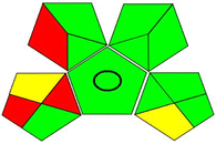
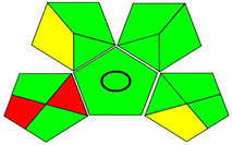
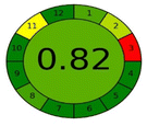
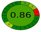
From the creation of energy, food, and clean drinking water to pharmaceuticals, household cleaners, personal care items, and a wide range of other products, chemistry has a role in almost every area of modern life. In this sense, the chemical sector makes a substantial contribution to increasing living standards by offering fertilizers and agrochemicals to increase the availability of food as well as improving nutrition, sanitation, and a wide range of treatments to improve quality of life and life expectancy.13,24 Green chemistry is a basic component of feasible chemistry and centres on making chemical responses and forms more ecologically invasive, considering variables such as molecular productivity, vitality proficiency, safe reactants, renewable assets, and contamination avoidance. Two analytical techniques were applied for measurements of the greenness effect of the proposed method (GAPI and agree methods).25–29The proposed method provides eco-friendly and high-greenness measurements that can be used for environmental applications Table 8.
4. Conclusion
This technique has been used for the direct estimation of the anticancer drug avapritinib in biological fluids (pharmacokinetic study) with (Cmax) equal to 817.13 ± 3.40 ng mL−1. The use of this technique in drug analysis has several advantages, including high sensitivity (LOQ equal to 9.47 ng mL−1), selectivity, and accuracy, as well as the potential to reduce the use of hazardous reagents and solvents in the analysis process.Ethical statement
All the experiments were conducted and approved according to the ethical committee of the international review board of Menoufia University, Egypt with IRB approval no. 00437/2023. The authors confirmed informed consent was obtained from all subjects participating in the experiments.Author contributions
Conceptualization, B. I. Salman, and R. E. Saraya; methodology, B. I. Salman, H. A. Batakoushy, E. A. M. El-Shoura, and R. E. Saraya.; software, A. I. Hassan., M. A. A. Abdel-Aal, Y. F. Hassan. and B. I. Salman; validation, B. I. Salman, A. E. Ibrahim, and E. A. M. El-Shoura; formal analysis, B. I. Salman and A. E. Ibrahim.; investigation, Y. F. Hassan, H. A. Batakoushy and B. I. Salman; resources, A. I. Hassan and B. I. Salman; data curation, A. E. Ibrahim and E. A. M. El-Shoura.; writing – original draft preparation, B. I. Salman; writing – review and editing, B. I. Salman, Y. F. Hassan, and A. E. Ibrahim; visualization, B. I. Salman; supervision, B. I. Salman; project administration B. I. Salman and E. A. M. El-Shoura; all authors have read and agreed to the published manuscript.Conflicts of interest
There are no conflicts of interest to declare.References
- M. C. Heinrich, R. L. Jones, M. von Mehren, P. Schöffski, C. Serrano, Y.-K. Kang, P. A. Cassier, O. Mir, F. Eskens, W. D. Tap, P. Rutkowski, S. P. Chawla, J. Trent, M. Tugnait, E. K. Evans, T. Lauz, T. Zhou, M. Roche, B. B. Wolf, S. Bauer and S. George, Lancet Oncol., 2020, 21, 935–946 CrossRef CAS PubMed.
- R. L. Siegel, K. D. Miller and A. Jemal, Ca-Cancer J. Clin., 2018, 68, 7–30 CrossRef PubMed.
- K. J. McHugh, L. Jing, A. M. Behrens, S. Jayawardena, W. Tang, M. Gao, R. Langer and A. Jaklenec, Adv. Mater., 2018, 30, 1706356 CrossRef PubMed.
- P. Bose and S. Verstovsek, Expert Rev. Hematol., 2021, 14, 687–696 CrossRef CAS PubMed.
- S. Dhillon, Drugs, 2020, 80, 433–439 CrossRef CAS PubMed.
- X. Xu, S. Luo, Q. Yang, Y. Wang, W. Li, G. Lin and R. Xu, Arabian J. Chem., 2021, 14, 103152 CrossRef CAS.
- T. G. Singh, J. B. Vadlakonda and V. R. M. Gupta, World J. Pharm. Pharm. Sci., 2020, 9, 1834–1840 CAS.
- R. E. Saraya, B. I. Salman, Y. F. Hassan, A. I. Hassan, S. A. Refaat and H. A. Batakoushy, Luminescence, 2023, 38, 1632–1638 CrossRef CAS PubMed.
- B. I. Salman, J. Fluoresc., 2023, 33, 1887–1896 CrossRef CAS PubMed.
- M. Sajid and J. Płotka-Wasylka, Talanta, 2022, 238, 123046 CrossRef CAS PubMed.
- M. S. Imam and M. M. Abdelrahman, Trends Environ. Anal. Chem., 2023, 38, e00202 CrossRef CAS.
- S. Armenta, S. Garrigues and M. de la Guardia, TrAC, Trends Anal. Chem., 2008, 27, 497–511 CrossRef CAS.
- J. Colberg, K. K. Hii and S. G. Koenig, ACS Sustain. Chem. Eng., 2022, 10, 8239–8241 CrossRef.
- M. A. Omar, M. A. Hammad and B. I. Salman, Luminescence, 2018, 33, 1226–1234 CrossRef CAS PubMed.
- S. F. Hammad, S. F. El-Malla and B. Z. El-Khateeb, Spectrochim. Acta, Part A, 2023, 291, 122317 CrossRef CAS PubMed.
- J. S. Nowick and J. S. Chen, J. Am. Chem. Soc., 1992, 114, 1107–1108 CrossRef CAS.
- United States Pharmacopeial Convention, (905) Uniformity of Dosage Units. Stage 6 Harmonization, 2011, vol. 3, pp. 4–6 Search PubMed.
- A. E. Ibrahim, R. E. Saraya, H. Saleh and M. Elhenawee, Heliyon, 2019, 5, e01518 CrossRef CAS PubMed.
- D. Zimmer, Bioanalysis, 2014, 6, 13–19 CrossRef CAS PubMed.
- S. K. Branch, J. Pharm. Biomed. Anal., 2005, 38, 798–805 CrossRef CAS PubMed.
- B. I. Salman, A. E. Ibrahim, S. El Deeb and R. E. Saraya, RSC Adv., 2022, 12, 16624–16631 RSC.
- S. Dhillon, Drugs, 2020, 80, 433–439 CrossRef CAS PubMed.
- J. Li, J. Gong, X. Wang, J. Li, Y. Li, C. QI, L. Shen, W. Ren, W. Zhao, Y. Wang, J. Hu, C. Ren, W. Song and J. Yang, J. Clin. Oncol., 2020, 38, e23526 Search PubMed.
- F. K. Algethami and M. Gamal, Microchem. J., 2023, 195, 109511 CrossRef CAS.
- F. Pena-Pereira, W. Wojnowski and M. Tobiszewski, Anal. Chem., 2020, 92, 10076–10082 CrossRef CAS PubMed.
- B. I. Salman, A. I. Hassan, Y. F. Hassan, R. E. Saraya and H. A. Batakoushy, J. Fluoresc., 2023, 33, 1101–1110 CrossRef CAS PubMed.
- B. I. Salman, BMC Chem., 2023, 17, 83 CrossRef CAS PubMed.
- J. Płotka-Wasylka, Talanta, 2018, 181, 204–209 CrossRef PubMed.
- M. Gamal, I. A. Naguib, D. S. Panda and F. F. Abdallah, Anal. Methods, 2021, 13, 369–380 RSC.
Footnote |
| † Electronic supplementary information (ESI) available. See DOI: https://doi.org/10.1039/d4ra01198h |
| This journal is © The Royal Society of Chemistry 2024 |

