 Open Access Article
Sugar mimics and their probable binding sites: design and synthesis of thiazole linked coumarin-piperazine hybrids as galectin-1 inhibitors†
Aaftaab Sethi ab,
Janish Kumarc,
Divya Vemulaa,
Divya Gadded,
Venu Tallad,
Insaf A. Qureshic and
Mallika Alvala
 *ae
*ae
aDepartment of Medicinal Chemistry, National Institute of Pharmaceutical Education and Research (NIPER), Hyderabad 500037, India. E-mail: mallikaalvala@yahoo.in
bLaboratory of Biomolecular Interactions and Transport, Department of Gene Expression, Institute of Molecular Biology and Biotechnology, Faculty of Biology, Adam Mickiewicz University, Uniwersytetu Poznanskiego 6, Poznan 61–614, Poland
cDepartment of Biotechnology & Bioinformatics, School of Life Sciences, University of Hyderabad, Hyderabad 500046, India
dDepartment of Pharmacology and Toxicology, National Institute of Pharmaceutical Education and Research (NIPER), Hyderabad 500037, India
eMARS Training Academy, Hyderabad, India
First published on 18th November 2024
Abstract
Sugar mimics are valuable tools in medicinal chemistry, offering the potential to overcome the limitations of carbohydrate inhibitors, such as poor pharmacokinetics and non-selectivity. In our continued efforts to develop heterocyclic galectin-1 inhibitors, we report the synthesis and characterization of thiazole-linked coumarin piperazine hybrids (10a–10i) as Gal-1 inhibitors. The compounds were characterized using 1H NMR, 13C NMR and HRMS. Among the synthesized molecules, four compounds demonstrated significant inhibitory activity, with more than 50% inhibition observed at a concentration of 20 μM in a Gal-1 enzyme assay. Fluorescence spectroscopy was further utilized to elucidate the binding constant for the synthesized compounds. 10g exhibited the highest affinity for Gal-1, with a binding constant (Ka) of 9.8 × 10⁴ M−1. To elucidate the mode of binding, we performed extensive computational analyses with 10g, including 1.2 μs all-atom molecular dynamics simulations coupled with a robust machine learning tool. Our findings indicate that 10g binds to the carbohydrate binding site of Gal-1, with the coumarin moiety playing a key role in binding interactions. Additionally, our study underscores the limitations of relying solely on docking scores for conformational selection and highlights the critical importance of performing multiple MD replicas to gain accurate insights.
1. Introduction
Galectins are a family of animal lectins characterized by their affinity for β-galactosides and notable sequence similarity in their carbohydrate-binding sites.1 Each galectin contains one or two highly conserved carbohydrate recognition domains (CRDs), typically composed of approximately 135 amino acids.2 These proteins are implicated in various pathological processes, including cancer and infections,3,4 among others. The first identified member of the galectin family, galectin-1 (Gal-1), has been demonstrated to play a significant role in several biological processes. Its involvement in tumour progression, inflammatory diseases, neurodegenerative disorders, immune escape and muscular dystrophies, underscores the importance of targeted therapeutic strategies. Specifically, enhancing the targeted overexpression or delivery of Gal-1 may be beneficial for treating certain inflammation-related conditions, while the development of selective Gal-1 inhibitors is essential for combating cancer progression.5 Gal-1 is a homodimer characterized by a β-sandwich fold, consisting of two antiparallel β-sheets – one with five (F1–F5) and the other with six (S1–S6) β-strands. The carbohydrate-binding site (CBS) of Gal-1 is located on the surface of the β-sheet formed by the S4–S6 strands (Fig. 1).6 The highly conserved amino acid residues involved in Gal-1 interactions include His44, Asn46, Arg48, Val59, Asn61, Trp68, Glu71, and Arg73.6 The specificity and affinity of galectin-1 for various carbohydrates are partially governed by a complex cooperative hydrogen bond network in addition to the CH–π interactions between the carbohydrate and residues with aromatic sidechains.7,8 Notably, computational studies have shown that the conserved tryptophan residue present within the CBS when mutated to alanine results in the complete rotation of the galactose ligand within the CBS due to the loss of stabilization.9 This has been further corroborated by another study which demonstrated that His52 and Trp68 are essential for sugar recognition prior to binding.7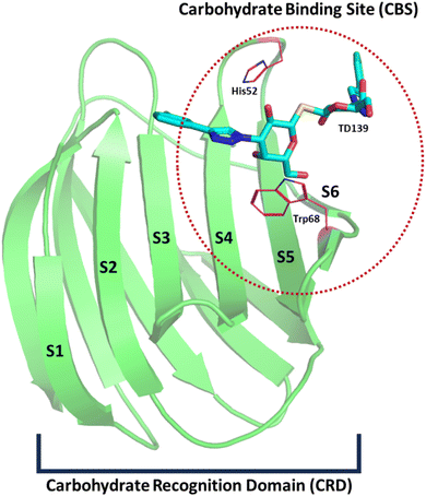 | ||
| Fig. 1 Depiction of galectin-1 (Gal-1) PDB ID: 4Y24 (green) bound to the co-crystal TD-139 (cyan). The structure represents a single chain of the Gal-1 dimer, with strands S1–S6 labelled. The carbohydrate-binding site (CBS) is circled with a red dotted line, located among strands S4–S6. The carbohydrate recognition domain (CRD) is highlighted by a black bracket. Key residues responsible for sugar recognition, His52 and Trp68, are shown in magenta. The loops have been smoothened for representational purposes. | ||
Most Gal-1 inhibitors reported so far are carbohydrates or their derivatives.8,10,11 However, these inhibitors present several challenges. The synthesis and characterization of carbohydrate-based inhibitors are complex, costly and often require extensive modifications of protecting groups.12 Additionally, they lack cell membrane permeability and selectivity.13 Recently, however, there has been a growing interest in heterocyclic compounds as Gal-1 inhibitors.13,14 These compounds have been extensively discussed in recent reviews on the topic.13,14 They offer a promising alternative due to their rich background in medicinal chemistry, allowing for easier development and optimization. Selected heterocyclic Gal-1 inhibitors along with their determined binding affinity values have been succinctly discussed in Fig. 2. Encouraged by these prior developments of heterocyclic Gal-1 inhibitors, coupled with the fact that hydrophobic/aromatic substitutions aid in enhancing binding to galectins,13,15 and reports of cyclic amino analogues acting as biologically active substitutions for the noviose (carbohydrate) moiety in novobiocin;16,17 we set out to develop thiazole linked coumarin-piperazine hybrids as potential Gal-1 inhibitors. Moreover, while numerous crystallographic structures have been resolved for carbohydrate-based Gal-1 inhibitors, which gives insight into their binding mode; studies pertaining to determination of binding site and the binding mode for heterocyclic inhibitors remains elusive. Thus, we also wanted to investigate and throw some light on the probable binding site of these compounds. This was explored by a comprehensive study utilizing all-atom molecular dynamic simulations.
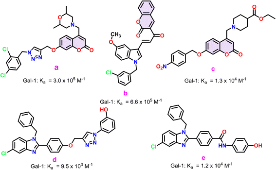 | ||
| Fig. 2 Select heterocyclic Gal-1 inhibitors (a–e) alongside their binding constants. For a comprehensive and up-to-date list of all known heterocyclic Gal-1 inhibitors, refer to recent review articles on the topic.13,14 | ||
2. Methods
2.1. Chemistry
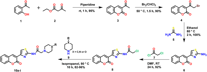
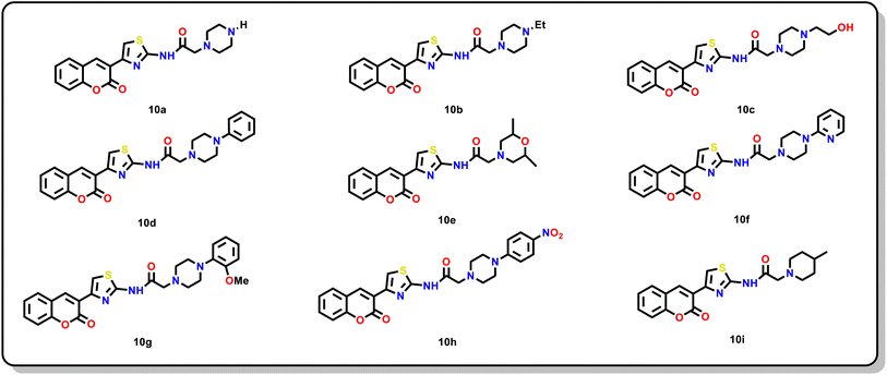
All starting materials, reagents and solvents used in this study were sourced from commercial suppliers. Analytical thin-layer chromatography (TLC) was conducted on pre-coated silica gel 60-F254 aluminium plates from MERCK. Visualization of the TLC plates was performed using UV light or in some instances, an iodine chamber. Melting points were measured with a Stuart® SMP30 melting point apparatus. Column chromatography was performed using silica gel (60–120 mesh) and compounds were eluted with a mixture of ethyl acetate and hexane. NMR spectra were recorded on Bruker 500 (500 MHz for 1H NMR and 125 MHz for 13C NMR) using DMSO or CDCl3 as solvent. Chemical shift was reported in parts per million (ppm) with respect to internal standard Tetra Methyl Silane (TMS). Coupling constants were quoted in Hertz (Hz). High-resolution mass spectra (HRMS) was obtained on Agilent Q-TOF-Mass Spectrometer 6540-UHD LC/HRMS operating at 70 eV using direct inlet.The target thiazole linked coumarin-piperazine hybrids (10a–i) were synthesized by a 5-step method (Scheme 1). One standout feature of the depicted scheme was that “no column chromatography purification” was required until the last step of the reaction. The products were obtained by simple purification. Initially, Knoevenagel condensation between aldehydic functionality of salicylaldehyde (1) and active methylene of ethyl acetoacetate (2) in presence of piperidine followed by cyclization gave us 3-acetyl coumarin (3) in high yields. Next, we performed electrophilic substitution on bromine to obtain bromoacetyl coumarin (4). Further, 4 was heated in ethanol in presence of thiourea (5) to afford cyclized product 2-amino thiazole coumarin (6). Subsequently, a nucleophilic substitution was performed by amino group of 6 on chloroacetyl chloride (7) to obtain 8. Finally, another nucleophilic substitution reaction was performed on 8 by the amine present in the cyclic amino ring (9) to give the final product (10a–i) in excellent yields. The final step of the reaction was optimized based on a previous report.18 All synthesized compounds (10a–i) were characterized by 1H-NMR, 13C-NMR and High-Resolution Mass Spectrometry (HRMS) spectral techniques [ESI†]. The characteristic thiazole proton peak was observed at > δ 8.5 ppm for all the compounds. The amide NH proton was highly acidic and it rapidly underwent deuterium exchange. Thus, it was not found for all the compounds. The protons associated with the saturated heterocyclic rings were observed in the range of δ 1–4 ppm. The characteristic CH2 protons next to the amide functionality were observed in the range of δ 3–4 ppm as a sharp singlet. The 13C NMR spectrum displayed the characteristic peak of carbonyl carbon of chalcone at > δ 160. The HRMS (ESI) of compounds showed corresponding [M + H]+ peaks based on their molecular weights.
2.2. Biology
2.3. Computational studies
The prediction of binding site using machine learning was carried using RoseTTAFold All-Atom,27 on neurosnap platform.28 Briefly, the sdf file for the TD-139 (co-crystal in PDB ID: 4Y24) and compound 10g were uploaded along with the Gal-1 sequence and the run was subsequently executed. The output pdb file generated was downloaded and analyzed using PyMOL.26
3. Results
3.1 Chemistry
3.1.2 1. N-(5-(2-Oxo-2H-chromen-3-yl)thiazol-2-yl)-2-(piperazin-1-yl)acetamide (10a). White solid; 95% yield; mp: 170–172 °C; 1H NMR (500 MHz, DMSO-d6) δ 8.64 (s, 1H), 8.03 (s, 1H), 7.87 (dd, J = 7.7, 1.2 Hz, 1H), 7.69–7.61 (m, 1H), 7.48 (d, J = 8.2 Hz, 1H), 7.41 (t, J = 7.5 Hz, 1H), 3.29 (s, 2H), 2.81–2.68 (m, 4H), 2.49–2.46 (m, 4H); 13C NMR (125 MHz, DMSO-d6) δ 169.23, 159.32, 157.50, 152.66, 142.98, 139.28, 132.79, 129.92, 125.56, 121.15, 119.75, 116.74, 115.08, 67.57, 61.13, 54.11. HRMS (ESI): m/z calc. for C18H18N4O3S, 371.1173 found (M + H)+ 371.1157.
3.1.2 2. 2-(4-Ethylpiperazin-1-yl)-N-(5-(2-oxo-2H-chromen-3-yl)thiazol-2-yl)acetamide (10b). White solid; 91% yield; mp: 161–163 °C; 1H NMR (500 MHz, CDCl3) δ 8.63 (s, 1H), 8.03 (s, 1H), 7.86 (dd, J = 7.7, 1.4 Hz, 1H), 7.65 (ddd, J = 8.6, 7.5, 1.5 Hz, 1H), 7.47 (d, J = 8.3 Hz, 1H), 7.41 (td, J = 7.6, 0.9 Hz, 1H), 2.56 (s, 4H), 2.43 (dd, J = 17.4, 13.2 Hz, 4H), 2.34 (dd, J = 14.3, 7.1 Hz, 2H), 1.00 (t, J = 7.2 Hz, 3H); 13C NMR (125 MHz, DMSO-d6) δ 169.38, 159.25, 157.63, 152.95, 142.54, 139.09, 132.40, 129.37, 125.26, 120.83, 119.55, 116.43, 114.80, 60.68, 53.12, 52.75, 52.06, 12.47; HRMS (ESI): m/z calc. for C20H22N4O3S, 398.1486 found (M + H)+ 399.1511.
3.1.2 3. 2-(4-(2-Hydroxyethyl)piperazin-1-yl)-N-(5-(2-oxo-2H-chromen-3-yl)thiazol-2-yl)acetamide (10c). White solid; 96% yield; mp: 184–186 °C; 1H NMR (500 MHz, DMSO-d6) δ 8.63 (s, 1H), 8.03 (s, 1H), 7.87 (dd, J = 7.8, 1.6 Hz, 1H), 7.66 (td, J = 8.3, 2.0 Hz, 1H), 7.48 (d, J = 8.2 Hz, 1H), 7.41 (t, J = 7.5 Hz, 1H), 4.39 (brs, 1H), 3.50 (t, J = 6.3 Hz, 2H), 3.31 (s, 2H), 2.55 (s, 4H), 2.47 (s, 4H), 2.44–2.28 (m, 2H); 13C NMR (125 MHz, DMSO-d6) δ 169.37, 159.26, 157.62, 152.95, 142.54, 139.10, 132.40, 129.38, 125.26, 120.83, 119.55, 116.43, 114.81, 60.69, 58.97, 53.56, 53.15, 53.09; HRMS (ESI): m/z calc. for C20H22N4O4S, 415.1435 found (M + H)+ 415.1466.
3.1.2 4. N-(5-(2-Oxo-2H-chromen-3-yl)thiazol-2-yl)-2-(4-phenylpiperazin-1-yl)acetamide (10d). White solid; 90% yield; mp: 199–201 °C; 1H NMR (500 MHz, DMSO-d6) δ 8.63 (s, 1H), 8.03 (s, 1H), 7.85 (dd, J = 7.8, 1.8 Hz, 2H), 7.65 (dd, J = 4.4, 3.2 Hz, 1H), 7.47 (d, J = 8.2 Hz, 1H), 7.41 (dd, J = 7.6, 3.8 Hz, 1H), 7.21 (dd, J = 6.7, 1.8 Hz, 2H), 6.95 (d, J = 8.5 Hz, 1H), 6.79 (t, J = 7.2 Hz, 1H), 3.41 (s, 2H), 3.25–3.04 (m, 4H), 2.81–2.56 (m, 4H); 13C NMR (125 MHz, DMSO-d6) δ 170.03, 159.64, 158.24, 153.09, 152.41, 142.55, 141.68, 139.65, 133.01, 129.95, 125.41, 122.93, 121.07, 120.62, 119.55, 118.38, 116.08, 60.94, 54.15, 51.24. HRMS (ESI): m/z calc. for C24H22N4O3S, 447.1486 found (M + H)+ 447.1514.
3.1.2 5. 2-(2,6-Dimethylmorpholino)-N-(5-(2-oxo-2H-chromen-3-yl)thiazol-2-yl)acetamide (10e). White solid; 95% yield; mp: 173–175 °C; 1H NMR (500 MHz, DMSO-d6) δ 12.02 (s, 1H), 8.63 (s, 1H), 8.03 (s, 1H), 7.86 (dd, J = 7.7, 1.4 Hz, 1H), 7.69–7.62 (m, 1H), 7.48 (d, J = 8.3 Hz, 1H), 7.44–7.39 (m, 1H), 3.63 (dtd, J = 8.3, 6.2, 2.0 Hz, 2H), 3.33 (s, 2H), 2.79 (d, J = 10.1 Hz, 2H), 1.91 (t, J = 10.7 Hz, 2H), 1.06 (d, J = 6.3 Hz, 6H); 13C NMR (125 MHz, DMSO-d6) δ 169.25, 159.24, 157.63, 152.94, 142.54, 139.06, 132.38, 129.34, 125.25, 120.84, 119.53, 116.42, 114.78, 71.42, 60.47, 59.06, 19.34; HRMS (ESI): m/z calc. for C20H21N3O4S, 400.1326 found (M + H)+ 400.1327.
3.1.2 6. N-(5-(2-Oxo-2H-chromen-3-yl)thiazol-2-yl)-2-(4-(pyridin-2-yl)piperazin-1-yl)acetamide (10f). White solid; 85% yield; mp: 235–238 °C; 1H NMR (500 MHz, DMSO-d6) δ 8.64 (s, 1H), 8.12 (dd, J = 4.9, 2.3 Hz, 1H), 8.04 (s, 1H), 7.86 (dd, J = 7.8, 1.8 Hz, 1H), 7.65 (td, J = 7.1, 3.6 Hz, 1H), 7.54 (ddd, J = 8.8, 7.0, 1.9 Hz, 1H), 7.48 (d, J = 8.1 Hz, 1H), 7.41 (dd, J = 9.6, 5.4 Hz, 1H), 6.84 (d, J = 8.5 Hz, 1H), 6.64 (dd, J = 7.1, 4.9 Hz, 1H), 3.63–3.43 (m, 4H), 3.40 (s, 2H), 2.77–2.55 (m, 4H); 13C NMR (125 MHz, DMSO-d6) δ 169.32, 159.47, 159.25, 157.66, 152.94, 148.03, 142.55, 139.07, 137.97, 132.39, 129.36, 125.26, 120.84, 119.54, 116.43, 114.82, 113.46, 107.58, 60.64, 52.80, 45.11; HRMS (ESI): m/z calc. for C23H21N5O3S, 448.1438 found (M + H)+ 448.1472.
3.1.2 7. 2-(4-(2-Methoxyphenyl)piperazin-1-yl)-N-(5-(2-oxo-2H-chromen-3-yl)thiazol-2-yl)acetamide (10g). White solid; 94% yield; mp: 210–212 °C; 1H NMR (500 MHz, DMSO-d6) δ 1H NMR (500 MHz, DMSO) δ 8.64 (s, 1H), 8.04 (s, 1H), 7.86 (dd, J = 7.7, 1.2 Hz, 1H), 7.68–7.63 (m, 1H), 7.48 (d, J = 8.2 Hz, 1H), 7.43–7.38 (m, 1H), 6.98–6.93 (m, 2H), 6.92–6.86 (m, 2H), 3.78 (s, 3H), 3.41 (s, 2H), 3.02 (s, 4H), 2.72 (s, 4H); 13C NMR (125 MHz, DMSO-d6) δ 169.38, 159.26, 157.65, 152.94, 152.49, 142.55, 141.68, 139.09, 132.39, 129.37, 125.26, 122.93, 121.31, 120.83, 119.55, 118.49, 116.43, 114.82, 112.41, 60.70, 55.81, 53.27, 50.53; HRMS (ESI): m/z calc. for C25H24N4O4S, 476.1592 found (M + H)+ 477.1591.
3.1.2 8. 2-(4-(4-Nitrophenyl)piperazin-1-yl)-N-(5-(2-oxo-2H-chromen-3-yl)thiazol-2-yl)acetamide (10h). Bright yellow solid; 82% yield; mp: 249–251 °C; 1H NMR (500 MHz, DMSO-d6) δ 8.62 (s, 1H), 8.11 (dd, J = 7.3, 2.2 Hz, 2H), 8.08–8.05 (m, 2H), 8.04 (s, 1H), 7.88–7.82 (m, 1H), 7.69–7.62 (m, 1H), 7.48 (d, J = 8.2 Hz, 1H), 7.43–7.38 (m, 1H), 3.71 (dd, J = 6.4, 4.3 Hz, 4H), 3.22 (dd, J = 6.2, 4.5 Hz, 4H); 13C NMR (125 MHz, DMSO-d6) δ 170.32, 160.37, 158.24, 153.55, 148.43, 143.27, 139.76, 137.89, 132.39, 129.79, 125.36, 121.89, 120.22, 116.95, 116.03, 114.19, 108.99, 61.01, 53.34, 47.24. HRMS (ESI): m/z calc. for C24H21N5O5S, 491.1337 found (M + H)+ 492.1364.
3.1.2 9. 2-(4-Methylpiperidin-1-yl)-N-(5-(2-oxo-2H-chromen-3-yl)thiazol-2-yl)acetamide (10i). White solid; 88% yield; mp: 168–170 °C; 1H NMR (500 MHz, DMSO-d6) δ 11.71 (s, 1H), 8.64 (s, 1H), 8.03 (s, 1H), 7.87 (dd, J = 7.7, 1.2 Hz, 1H), 7.66 (dd, J = 15.6, 1.5 Hz, 1H), 7.48 (d, J = 8.3 Hz, 1H), 7.41 (dd, J = 11.6, 4.2 Hz, 1H), 3.33 (s, 2H), 2.85 (d, J = 11.4 Hz, 2H), 2.17 (td, J = 11.5, 2.0 Hz, 2H), 1.59 (d, J = 11.2 Hz, 2H), 1.34 (ddd, J = 14.4, 12.4, 7.1 Hz, 1H), 1.21 (ddd, J = 15.3, 12.1, 3.6 Hz, 2H), 0.91 (d, J = 6.4 Hz, 3H); 13C NMR (125 MHz, DMSO-d6) δ 169.71, 159.26, 157.66, 152.94, 142.54, 139.10, 132.39, 129.37, 125.26, 120.82, 119.56, 116.43, 114.78, 61.17, 53.75, 34.37, 30.28, 22.28; HRMS (ESI): m/z calc. for C20H21N3O3S, 384.1377 found (M + H)+ 384.1406.
3.2. Biology
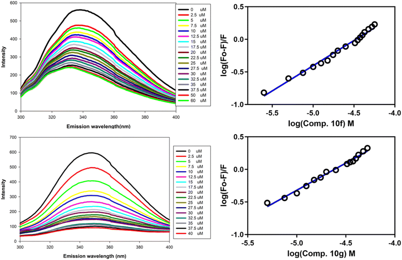
All synthesized compounds were initially screened using a Gal-1 enzymatic assay to assess their inhibitory potential (Table 1). Compounds that demonstrated >20% inhibition at a 20 μM concentration were further analysed for their affinity to Gal-1 using fluorescence spectroscopy. Among the tested compounds, 10f exhibited the highest inhibition in the enzymatic assay, achieving >80% inhibition. Compounds 10a, 10d, and 10g also showed significant inhibitory activity, with >50% inhibition. In contrast, compounds 10h and 10i exhibited <10% inhibition and thus were not pursued in subsequent analyses. To enumerate the binding constants of selected compounds (10f and 10g) through fluorescence spectroscopy, recombinant human Gal-1, Gal-3 and LdPase were purified to homogeneity using affinity chromatography. The fluorescence spectra for compounds 10f and 10g are displayed in Fig. 4, which revealed that compounds 10f and 10g had the highest affinity for Gal-1 (Table 1). Importantly, both compounds displayed significantly lower binding to Gal-3 compared to Gal-1, and no detectable binding to LdPase (Table 1). These studies indicate that compound 10g holds significant promise as a hit candidate suitable for further development.| Compound | % Inhibition (EA) | Gal-1 Ka (104 M−1) | Gal-3 Ka (10−6 M−1) | LdPasea |
|---|---|---|---|---|
| a NE – not evaluated, NB – no binding. | ||||
| 10a | 58.19 ± 1.12 | 8.8 | NE | NE |
| 10b | 25.93 ± 0.81 | 2.1 | NE | NE |
| 10c | 22.46 ± 1.67 | 4.1 | NE | NE |
| 10d | 56.73 ± 2.13 | 6.6 | NE | NE |
| 10e | 27.80 ± 0.76 | 6.5 | NE | NE |
| 10f | 80.57 ± 1.77 | 9.3 | 1.1 | NB |
| 10g | 58.19 ± 3.28 | 9.8 | 1.0 | NB |
| 10h | <10 | NE | NE | NE |
| 10i | <10 | NE | NE | NE |
3.3. Computational studies
To further validate our findings, we utilized the RoseTTAFold All-Atom,27 a robust machine learning model, to predict the binding site of compound 10g. Such machine learning models are now gained prominence are now at the forefront of drug discovery.39 Before applying RoseTTAFold All-Atom to 10g, we first validated the model by predicting the co-crystal pose, which achieved a remarkable RMSD of 1.037 Å compared to its crystallographic pose. For compound 10g, the model indicated a binding location within the CBS, consistent with the location predicted by pose 3 in our docking analysis. This alignment between the docking and machine learning predictions reinforced the potential accuracy of our identified binding sites.
4. Discussion
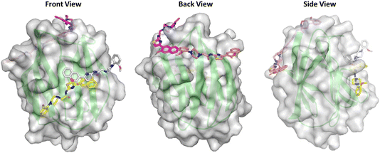
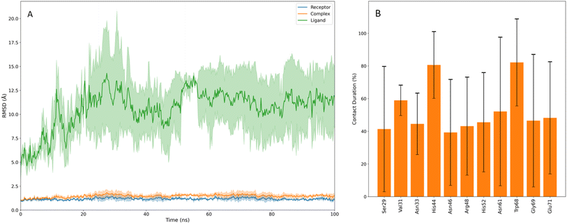
In this study, we synthesized and characterized a series of novel thiazole-linked coumarin piperazine derivatives, compounds 10a–10i, utilizing 1H NMR, 13C NMR, and HRMS. These compounds were subsequently evaluated for their inhibitory activity against Gal-1 (Table 1). Among the synthesized compounds, 10a, 10d, 10f, and 10g demonstrated significant inhibitory activity, with >50% inhibition at a concentration of 20 μM. Further analysis was carried out to determine the binding constants using fluorescence spectroscopy. It revealed that compound 10g exhibited the highest affinity for Gal-1, with a binding constant (Ka) of 9.8 × 10⁴ M−1. Further, it also suggested that both aromatic and aliphatic groups attached to the piperazine moiety are well-tolerated, though aromatic groups appear to have a higher affinity, as predicted in prior studies.13,15 Interestingly, compound 10h, which contains a para-nitro substituent, exhibited <10% inhibition of Gal-1. We hypothesize that the para-nitro group may introduce steric hindrance that disrupts the optimal fit of the coumarin moiety within the binding site, thereby reducing its inhibitory potential. This result highlights the importance of steric considerations in the design of effective Gal-1 inhibitors. Even more surprising was the finding that compound 10i, which shares significant structural similarity with compounds 10a and 10b, also showed <10% inhibition. This observation suggests that the presence of a hydrogen bond donor is critical in the absence of an aromatic moiety. This is further supported by the moderate inhibition observed for compounds 10b, 10c and the morpholine-containing 10e, which lack a hydrogen bond donor.Despite the development of numerous heterocyclic Gal-1 inhibitors, the precise mode of binding has remained largely unexplored. To address this, we employed a combination of molecular docking and RoseTTAFold All-Atom simulations to predict the potential binding modes of compound 10g. Four plausible binding poses were identified and subjected to MD simulations, with each pose being run for 100 ns in replicates of three to ensure the robustness of the findings. It was observed that in three of the four poses, compound 10g dissociated from the Gal-1 binding site and diffused into the bulk solvent, suggesting a lack of stable interactions. However, in pose 15, located within the CBS of Gal-1, compound 10g remained bound throughout the simulation, albeit with a high RMSD value. This elevated RMSD can primarily be attributed to the significant fluctuations of the piperazine moiety (Fig. 7), which failed to maintain consistent interactions with the protein. Nevertheless, the core coumarin-thiazole structure remained bound, interacting with key residues Trp68 and His52, which are crucial for sugar recognition within the binding site.7 These findings also underscore the importance of the hydrogen bond network that natural sugars typically exploit within the CBS of Gal-1. Our data suggest that the piperazine moiety, in its current form, does not adequately mimic these interactions, leading to the observed instability. Future inhibitor designs should focus on incorporating additional hydrogen bond donors or acceptors into the piperazine ring or similar scaffolds to better replicate the hydrogen bonding capabilities of sugars, thereby enhancing their affinity. Additionally, this study highlights the limitations of relying solely on docking scores to select conformations for further investigation. While pose 1 had the highest docking score, it was pose 15, with a least docking score among the four poses that was found to be stable during MD simulations. This discrepancy reinforces the need for comprehensive post-docking analyses, such as MD simulations, to validate and refine docking predictions. Moreover, the importance of conducting MD simulations in replicates was underscored by the observation that, even among the discarded poses, there was at least one run in which the compound remained stable. This variability suggests that relying on a single MD simulation could lead to misleading conclusions.
In summary, the synthesized thiazole-linked coumarin-piperazine hybrids were found to effectively inhibit Gal-1. The study provides significant insights into the binding mode of synthesized compounds within the Gal-1 binding site, particularly highlighting the significance of the coumarin moiety. The findings suggest that future development of heterocyclic Gal-1 inhibitors should prioritize the incorporation of functional groups capable of enhancing the hydrogen bonding network, akin to the hydroxy groups in natural sugars, while leveraging the stabilizing effect of the coumarin scaffold.
Data availability
The 1H NMR, 13C NMR spectra and HRMS data alongside RMSD plots for pose 1, 3 and 5 are provided in ESI.† MD trajectories, along with all necessary parameters, are publicly accessible via the Zenodo repository (10.5281/zenodo.13774410). Any additional data can be obtained from the authors upon through direct correspondence.Conflicts of interest
The authors declare that they have no conflict of interest.Acknowledgements
The authors would like to thank Prof. Jan Brezovsky for granting access to the internal cluster of Laboratory of Biomolecular Interactions and Transport for performing the MD simulations. A part of the computations was also performed at the Poznan Supercomputing and Networking Center.References
- H. Leffler, S. Carlsson, M. Hedlund, Y. Qian and F. Poirier, Introduction to Galectins, Glycoconjugate J., 2002, 19(7), 433–440, DOI:10.1023/B:GLYC.0000014072.34840.04.
- R. C. Hughes, Secretion of the Galectin Family of Mammalian Carbohydrate-Binding Proteins, Biochim. Biophys. Acta, Gen. Subj., 1999, 1473(1), 172–185, DOI:10.1016/S0304-4165(99)00177-4.
- K. V. Mariño, A. J. Cagnoni, D. O. Croci and G. A. Rabinovich, Targeting Galectin-Driven Regulatory Circuits in Cancer and Fibrosis, Nat. Rev. Drug Discovery, 2023, 22(4), 295–316, DOI:10.1038/s41573-023-00636-2.
- F.-T. Liu and S. R. Stowell, The Role of Galectins in Immunity and Infection, Nat. Rev. Immunol., 2023, 23(8), 479–494, DOI:10.1038/s41577-022-00829-7.
- I. Camby, M. Le Mercier, F. Lefranc and R. Kiss, Galectin-1: A Small Protein with Major Functions, Glycobiology, 2006, 16(11), 137R–157R, DOI:10.1093/glycob/cwl025.
- M. F. López-Lucendo, D. Solís, S. André, J. Hirabayashi, K. Kasai, H. Kaltner, H.-J. Gabius and A. Romero, Growth-Regulatory Human Galectin-1: Crystallographic Characterisation of the Structural Changes Induced by Single-Site Mutations and Their Impact on the Thermodynamics of Ligand Binding, J. Mol. Biol., 2004, 343(4), 957–970, DOI:10.1016/j.jmb.2004.08.078.
- I. Echeverria and L. M. Amzel, Disaccharide Binding to Galectin-1: Free Energy Calculations and Molecular Recognition Mechanism, Biophys. J., 2011, 100(9), 2283–2292, DOI:10.1016/j.bpj.2011.03.032.
- C. P. Modenutti, J. I. B. Capurro, S. Di Lella and M. A. Martí, The Structural Biology of Galectin-Ligand Recognition: Current Advances in Modeling Tools, Protein Engineering, and Inhibitor Design, Front. Chem., 2019, 7, 823, DOI:10.3389/fchem.2019.00823.
- C. Meynier, F. Guerlesquin and P. Roche, Computational Studies of Human Galectin-1: Role of Conserved Tryptophan Residue in Stacking Interaction with Carbohydrate Ligands, J. Biomol. Struct. Dyn., 2009, 27(1), 49–57, DOI:10.1080/07391102.2009.10507295.
- H. Blanchard, K. Bum-Erdene, M. H. Bohari and X. Yu, Galectin-1 Inhibitors and Their Potential Therapeutic Applications: A Patent Review, Expert Opin. Ther. Pat., 2016, 26(5), 537–554, DOI:10.1517/13543776.2016.1163338.
- A. Sethi, S. Sanam, R. Alvala and M. Alvala, An Updated Patent Review of Galectin-1 and Galectin-3 Inhibitors and Their Potential Therapeutic Applications (2016–Present), Expert Opin. Ther. Pat., 2021, 31(8), 709–721, DOI:10.1080/13543776.2021.1903430.
- A. Sethi, K. Sasikala, P. Jakkula, D. Gadde, S. Sanam, I. A. Qureshi, V. Talla and M. Alvala, Design, Synthesis and Computational Studies Involving Indole-Coumarin Hybrids as Galectin-1 Inhibitors, Chem. Pap., 2021, 75(6), 2791–2805, DOI:10.1007/s11696-021-01534-w.
- A. Sethi, S. Sanam and M. Alvala, Non-Carbohydrate Strategies to Inhibit Lectin Proteins with Special Emphasis on Galectins, Eur. J. Med. Chem., 2021, 222, 113561, DOI:10.1016/j.ejmech.2021.113561.
- S. G. Nerella, Non-Carbohydrate Galectin-1 Inhibitors as Promising Anticancer Agents: Design Strategies, Structure Activity Relationship and Mechanistic Insights, Eur. J. Med. Chem. Rep., 2024, 11, 100170, DOI:10.1016/j.ejmcr.2024.100170.
- A. Sethi, S. Sanam and M. Alvala, New Clues Arising from Hunt of Saccharides Binding to Galectin 3 via 3D QSAR and Docking Studies, Inform. Med. Unlocked., 2020, 21, 100411, DOI:10.1016/j.imu.2020.100411.
- H. Zhao, B. Reddy Kusuma and B. S. J. Blagg, Synthesis and Evaluation of Noviose Replacements on Novobiocin That Manifest Antiproliferative Activity, ACS Med. Chem. Lett., 2010, 1(7), 311–315, DOI:10.1021/ml100070r.
- G. Garg, H. Zhao and B. S. J. Blagg, Design, Synthesis, and Biological Evaluation of Ring-Constrained Novobiocin Analogues as Hsp90 C-Terminal Inhibitors, ACS Med. Chem. Lett., 2015, 6(2), 204–209, DOI:10.1021/ml5004475.
- A. Sethi and M. Alvala, β-CD/Lewis Acid Dual Catalytic System for Efficient Synthesis of Pyranopyrazoles in Aqueous Environment, Lett. Org. Chem., 2023, 20(7), 585–595, DOI:10.2174/1570178620666221122121021.
- C. A. Prato, J. Carabelli, V. Cattaneo, O. Campetella and M. V. Tribulatti, Purification of Recombinant Galectins Expressed in Bacteria, STAR Protoc., 2020, 1(3), 100204, DOI:10.1016/j.xpro.2020.100204.
- J. Kumar, Jyotisha, R. Qureshi, P. Jagruthi, M. Arifuddin and I. A. Qureshi, Discovery of 8-Hydroxy-2-Quinoline Carbaldehyde Derivatives as Inhibitors for M1 Aminopeptidase of Leishmania Donovani, Int. J. Biol. Macromol., 2024, 279, 135105, DOI:10.1016/j.ijbiomac.2024.135105.
- R. Anandakrishnan, B. Aguilar and A. V. Onufriev, H++ 3.0: Automating pK Prediction and the Preparation of Biomolecular Structures for Atomistic Molecular Modeling and Simulations, Nucleic Acids Res., 2012, 40(W1), W537–W541, DOI:10.1093/nar/gks375.
- J. C. Gordon, J. B. Myers, T. Folta, V. Shoja, L. S. Heath and A. Onufriev, H++: A Server for Estimating p Ka s and Adding Missing Hydrogens to Macromolecules, Nucleic Acids Res., 2005, 33(suppl_2), W368–W371, DOI:10.1093/nar/gki464.
- X. Pan, H. Wang, C. Li, J. Z. H. Zhang and C. Ji, MolGpka: A Web Server for Small Molecule pKa Prediction Using a Graph-Convolutional Neural Network, J. Chem. Inf. Model., 2021, 61(7), 3159–3165, DOI:10.1021/acs.jcim.1c00075.
- N. M. O'Boyle, M. Banck, C. A. James, C. Morley, T. Vandermeersch and G. R. Hutchison, Open Babel: An Open Chemical Toolbox, J. Cheminf., 2011, 3(1), 33, DOI:10.1186/1758-2946-3-33.
- J. Eberhardt, D. Santos-Martins, A. F. Tillack and S. Forli, AutoDock Vina 1.2.0: New Docking Methods, Expanded Force Field, and Python Bindings, J. Chem. Inf. Model., 2021, 61(8), 3891–3898, DOI:10.1021/acs.jcim.1c00203.
- PyMOL | pymol.org, https://www.pymol.org/, accessed 2024-08-24.
- R. Krishna, J. Wang, W. Ahern, P. Sturmfels, P. Venkatesh, I. Kalvet, G. R. Lee, F. S. Morey-Burrows, I. Anishchenko, I. R. Humphreys, R. McHugh, D. Vafeados, X. Li, G. A. Sutherland, A. Hitchcock, C. N. Hunter, A. Kang, E. Brackenbrough, A. K. Bera, M. Baek, F. DiMaio and D. Baker, Generalized Biomolecular Modeling and Design with RoseTTAFold All-Atom, Science, 2024, 0(0), eadl2528, DOI:10.1126/science.adl2528.
- Neurosnap – Computational Biology, Simplified, https://neurosnap.ai/?gad_source=1%26gclid=CjwKCAjwiaa2BhAiEiwAQBgyHiATHtuQEi4rMTV-3JynNQGb1e1Y__o6I8Tecfzha5nCnizXLhoUBhoC2poQAvD_BwE, accessed 2024-08-24.
- C. Tian, K. Kasavajhala, K. A. A. Belfon, L. Raguette, H. Huang, A. N. Migues, J. Bickel, Y. Wang, J. Pincay, Q. Wu and C. Simmerling, ff19SB: Amino-Acid-Specific Protein Backbone Parameters Trained against Quantum Mechanics Energy Surfaces in Solution, J. Chem. Theory Comput., 2020, 16(1), 528–552, DOI:10.1021/acs.jctc.9b00591.
- D. A. Case, H. M. Aktulga, K. Belfon, D. S. Cerutti, G. A. Cisneros, W. D. Vinicius, N. Forouzesh, T. J. Giese, A. W. Gotz, H. Gohlke, S. Izadi, K. Kasavajhala, M. C. Kaymak, E. King, T. Kurtzman, T.-S. Lee, P. Li, J. Liu, T. Luchko, R. Luo, M. Manathunga, M. R. Machado, H. M. Nguyen, K. A. O’Hearn, A. V. Onufriev, F. Pan, S. Pantano, R. Qi, A. Rahnamoun, A. Risheh, S. Schott-Verdugo, A. Shajan, J. Swails, J. Wang, H. Wei, X. Wu, Y. Wu, S. Zhang, S. Zhao, Q. Zhu, T. E. Cheatham III, D. R. Roe, A. Roitberg, C. Simmerling, D. M. York, M. C. Nagan and K. M. Merz Jr, J. Chem. Inf. Model, 2023, 63(20), 6183–6191, DOI:10.1021/acs.jcim.3c01153.
- D. J. Sindhikara, N. Yoshida and F. Hirata, Placevent: An Algorithm for Prediction of Explicit Solvent Atom Distribution—Application to HIV-1 Protease and F-ATP Synthase, J. Comput. Chem., 2012, 33(18), 1536–1543, DOI:10.1002/jcc.22984.
- Three-Dimensional Molecular Theory of Solvation Coupled with Molecular Dynamics in Amber | Journal of Chemical Theory and Computation, https://pubs.acs.org/doi/10.1021/ct900460m, accessed 2024-08-24 Search PubMed.
- C. W. Hopkins, S. Le Grand, R. C. Walker and A. E. Roitberg, Long-Time-Step Molecular Dynamics through Hydrogen Mass Repartitioning, J. Chem. Theory Comput., 2015, 11(4), 1864–1874, DOI:10.1021/ct5010406.
- M. Scheurer, P. Rodenkirch, M. Siggel, R. C. Bernardi, K. Schulten, E. Tajkhorshid and T. PyC. Rudack, Rapid, Customizable, and Visual Analysis of Noncovalent Interactions in MD Simulations, Biophys. J., 2018, 114(3), 577–583, DOI:10.1016/j.bpj.2017.12.003.
- D. E. V. Pires, T. L. Blundell and D. B. Ascher, pkCSM: Predicting Small-Molecule Pharmacokinetic and Toxicity Properties Using Graph-Based Signatures, J. Med. Chem., 2015, 58(9), 4066–4072, DOI:10.1021/acs.jmedchem.5b00104.
- L. Fu, S. Shi, J. Yi, N. Wang, Y. He, Z. Wu, J. Peng, Y. Deng, W. Wang, C. Wu, A. Lyu, X. Zeng, W. Zhao, T. Hou and D. Cao, ADMETlab 3.0: An Updated Comprehensive Online ADMET Prediction Platform Enhanced with Broader Coverage, Improved Performance, API Functionality and Decision Support, Nucleic Acids Res., 2024, 52(W1), W422–W431, DOI:10.1093/nar/gkae236.
- S. N. Rao, M. S. Head, A. Kulkarni and J. M. LaLonde, Validation Studies of the Site-Directed Docking Program LibDock, J. Chem. Inf. Model., 2007, 47(6), 2159–2171, DOI:10.1021/ci6004299.
- R. P. M. Dings, M. C. Miller, I. Nesmelova, L. Astorgues-Xerri, N. Kumar, M. Serova, X. Chen, E. Raymond, T. R. Hoye and K. H. Mayo, Antitumor Agent Calixarene 0118 Targets Human Galectin-1 as an Allosteric Inhibitor of Carbohydrate Binding, J. Med. Chem., 2012, 55(11), 5121–5129, DOI:10.1021/jm300014q.
- A. Sethi and B. Rathi, Artificial Intelligence in Drug Discovery: A Mirage or an Oasis?, Drug Discovery Today, 2024, 29(6), 103994, DOI:10.1016/j.drudis.2024.103994.
- B. Knapp, L. Ospina and C. M. Deane, Avoiding False Positive Conclusions in Molecular Simulation: The Importance of Replicas, J. Chem. Theory Comput., 2018, 14(12), 6127–6138, DOI:10.1021/acs.jctc.8b00391.
Footnote |
| † Electronic supplementary information (ESI) available. See DOI: https://doi.org/10.1039/d4ra06715k |
| This journal is © The Royal Society of Chemistry 2024 |

