DOI:
10.1039/D5AY00555H
(Minireview)
Anal. Methods, 2025,
17, 4482-4495
Research progress on regulation strategies for surface-enhanced Raman spectroscopy
Received
2nd April 2025
, Accepted 25th April 2025
First published on 2nd May 2025
Abstract
As a highly sensitive analytical technology, surface enhanced Raman spectroscopy (SERS) based on localized surface plasmon resonance has been widely explored in the field of environment monitoring, food safety, material identification and biomedicine. In the field of biosensing, the design of sensing models, the regulation of enhancement factors (EFs), and the stability of detection results have always been crucial research keys. Progress in these areas has continuously expanded the application scope of SERS technology and improved the feasibility of its application. Among them, the regulation of EFs through physical enhancement and chemical enhancement is a crucial point in improving the performance of SERS. Starting from the physicochemical mechanism, this review discusses the relevant influencing parameters and then summarizes the latest regulation strategies based on the above theory, as well as special regulation methods such as E-SERS. A diverse array of regulation strategies underpinned by the SERS enhancement mechanism have been effectively harnessed to amplify the EF of the SERS system. These include a wide spectrum of metal nanostructures based on the electromagnetic mechanism (EM), as well as regulation approaches predicated on the chemical mechanism (CM), such as energy-level manipulation, defect engineering, and material coupling. In addition, it encompasses specialized regulation methods such as analyte pre-concentration. This article focuses on summarizing the principal regulation approaches that have significantly impacted SERS enhancement in recent years, complemented by specialized regulation methods, with the hope of facilitating smoother progress in future work related to SERS.
1. Introduction
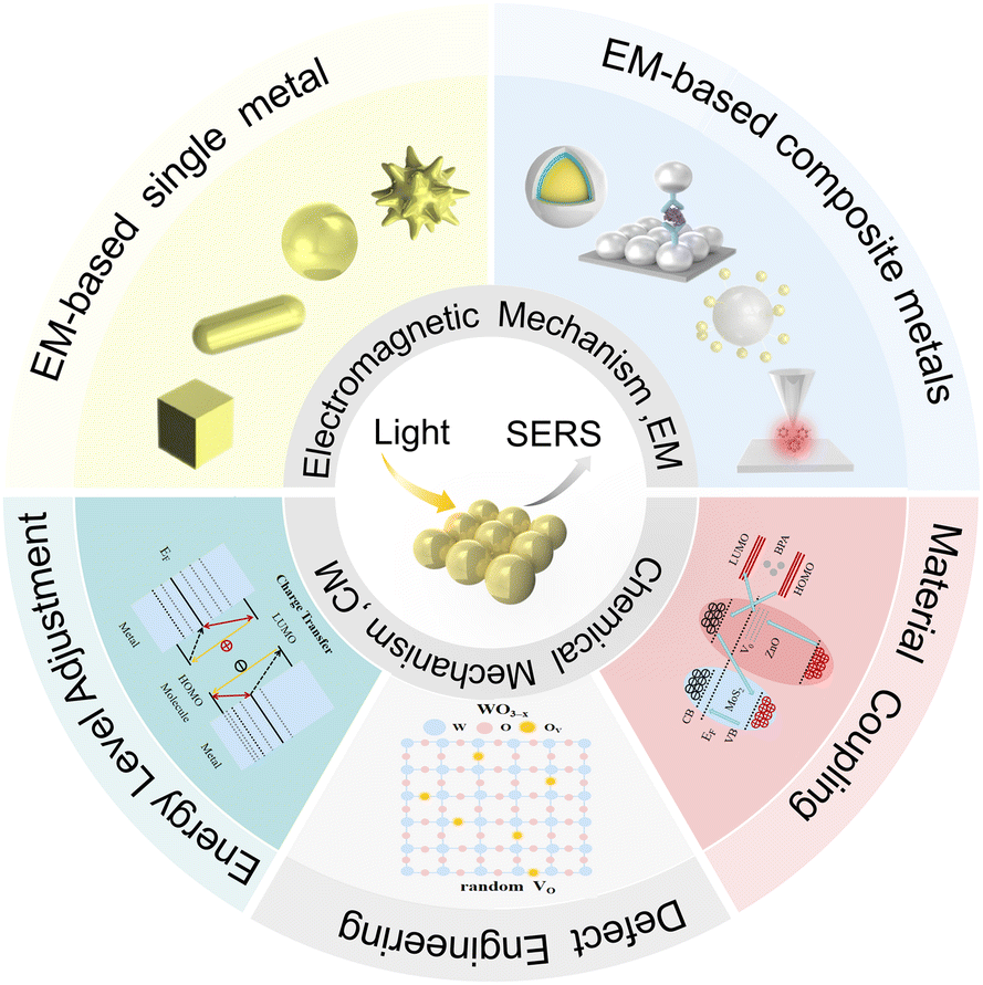
Surface enhanced Raman spectroscopy (SERS) is a vibrational spectroscopy technique that inherits the advantages of spontaneous Raman spectroscopy in molecular “fingerprint” detection while overcoming the limitation of its insufficient detection sensitivity. Spontaneous Raman spectroscopy can easily obtain rich “fingerprint” information on material structures,1 but the spontaneous Raman signal is weak, with a Raman cross section as low as 10−30 cm2 sr−1, which limits its wide application.2 SERS stimulates the localized surface plasmon resonance (LSPR) effect through the interaction between light and nanometal surfaces, thereby enhancing the Raman signal of target molecules adsorbed on or very close to the metal surface, which can achieve single molecule detection and further realize rapid and non-destructive qualitative and quantitative detection.3 SERS-based methods offer unique advantages over other analytical methods. For example, SERS is more suitable for long-term monitoring than fluorescence because of its resistance to photodegradation and photobleaching. The detection sensitivity of SERS is much higher and less susceptible to interference from water compared to that of IR spectroscopy.4 The advanced SERS technology, combined with the rapid development of nanofabrication technology in recent years, has been applied in the fields of biomedicine,5,6 environmental monitoring,7,8 food safety, 9,10 and material identification.11–14 In the field of biosensing, current research mainly focuses on the design of sensing models, the regulation of enhancement factors (EFs), and the stability of detection results.4,15,16 Among them, the regulation of EFs through electromagnetic enhancement and chemical enhancement is a crucial point in improving the performance of SERS. Many studies explore the influence of substrate materials, structures, sizes and shapes on the SERS enhancement effect,17–19 and also rely on adjusting energy levels to improve substrate performance.20 Some studies are aimed at special regulation methods that contribute to the SERS Enhancement Factor (SERS EF).21,22 Some existing excellent SERS reviews mainly demonstrate the specific applications of SERS technology, such as the construction of sensing models,16 immunoassays 23 and multimodal technology,24 providing good guidance for researchers. Varying from them, this review mainly focuses on the progress of the latest SERS regulation methods. We first introduce two enhancement mechanisms of SERS, including the electromagnetic mechanism (EM) and chemical mechanism (CM), and especially highlight the key factors affecting these two mechanisms. Then, we review the main ways of electromagnetic enhancement, including single metal and composite metal. Similarly, we also summarize the main ways of chemical enhancement, including energy level adjustment, defect engineering and material coupling, and supplement them with special regulation that affects SERS enhancement, including analyte enrichment and E-SERS. Finally, we look forward to the regulation of SERS enhancement. The regulation methods for SERS enhancement are sorted out and summarized (Fig. 1).
 |
| | Fig. 1 Regulation methods for surface-enhanced Raman spectroscopy. | |
2. SERS enhancement mechanism
SERS was initially observed by the Fleischmann team in 1974 through the study of the enhanced Raman scattering of pyridine adsorbed on rough silver electrodes.20 In 1977, the theory of the SERS mechanism, which has gained widespread acceptance within the scientific community, emerged.25 These are EM and CM, which dictate the extent of the SERS enhancement effect.26 Since then, researchers have delved further into the enhancement mechanism of SERS, both theoretically and experimentally. Among these, the most significant contribution of the EM stems from surface plasmon resonance (SPR), which is the collective oscillation effect of delocalized conductive electrons in metals under the incident light field. This requires a high density of free electrons in the visible and near-infrared regions. When the light frequency matches the electron oscillation frequency, SPR can be highly localized to a local location, and this is referred to as LSPR. When two rough metallic surfaces approach each other, a “hot-spot” region is formed, capable of markedly amplifying the signal. The CM is primarily attributable to the change in polarizability of substances adsorbed on the rough metallic surface, encompassing phenomena such as metal-molecule charge transfer (CT) and interactions at surface complex active sites (Fig. 2).27,28 Recent research confirms that the CM provides a limited contribution to SERS enhancement (typically <103), while the electromagnetic enhancement factor (EM EF) linked to the EM exhibits a significantly wider range (102–1012).29,30 Hence, the contribution of the EM is generally far more substantial than that of the CM. However, in reality, within the SERS system, since the EM and CM coexist simultaneously, the analysis of the EF attributed to the EM and CM cannot be entirely disentangled. In order to further explore the methods of regulating SERS enhancement, the parameters affecting EM and CM are discussed and summarized here.
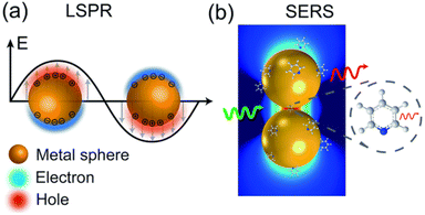 |
| | Fig. 2 Schematic diagrams of the (a) LSPR effect and (b) molecular polarization change.2 | |
2.1. Electromagnetic enhancement (EM)
The EM in SERS is primarily attributable to the intrinsic plasmon resonance effect of individual metal nanoparticles (NPs) and the pronounced “hot-spot” effect that arises in the interstices between metal nanostructures. Within the framework of classical electromagnetic theory, which posits polarizable Raman dipoles, and under a simplified theoretical model that disregards the majority of intricate tensor and orientation-related effects, the instantaneous Raman process within the SERS system can be partitioned into the excitation and emission phases of Raman dipoles.31
Specifically, if the initial electric field E0 and the incident laser frequency ωL are given, a Raman dipole P0 can be induced, and the spontaneous Raman intensity |P0|2 is proportional to it.
During the emission process, the dipole emits at the Raman shift frequency ωR. Under the influence of the electromagnetic effect, the local electric field generated is much larger than the initial electric field, and the local field enhancement factor MLoc(ωL) can be obtained, which is defined as eqn (1):
| |  | (1) |
At this time, there is also an enhancement factor MRad(ωR) generated by dipole emission at ωR. Due to optical reciprocity, it can be approximated as: MLoc(ωR). At this point, it is no longer the spontaneous Raman intensity but the SERS intensity, which is proportional to MLoc(ωR) and |P|2, affected by the change in polarizability. Consequently, the theoretical EM EF can be described as a fourth-power law,29 as shown in eqn (2):
| |  | (2) |
This involves the matching of LSPR and excitation wavelength, as well as the red shift and resonance mismatch of the Raman signal. When the wavelength of incident light matches the LSPR peak of NPs, the excitation enhancement reaches its maximum. However, since the Raman scattering signal itself will be red-shifted relative to the excitation wavelength, if the LSPR peak is strictly matched with the excitation wavelength, the red-shifted Raman signal may deviate from the resonance range of LSPR, resulting in a decrease in the enhancement effect. Therefore, it is necessary to maximize the signal enhancement by regulating the relative position of LSPR and excitation wavelength. For example, by adjusting the size and shape of NPs, the LSPR peak is slightly red-shifted to the excitation wavelength, or an excitation wavelength that is slightly blue-shifted to the LSPR peak is selected to ensure that the red-shifted Raman signal is still within the enhancement range of LSPR.32
2.2. Chemical enhancement (CM)
CM affects SERS intensity by changing the polarizability of materials directly adsorbed or just close to the surface, mainly including three aspects of enhancement: (1) non-resonant enhancement (Chemical-Bonding (CB) enhancement) due to chemical bonding between the analyte and substrate; (2) resonant enhancement (Surface Complex (SC) enhancement) due to the formation of surface complexes between adsorbed molecules and surface adsorbed atoms; (3) quasi-resonant enhancement of photoinduced charge transfer (Photon-Induced Charge-Transfer (PICT) enhancement) of molecule-metal by excitation light, among which PICT enhancement is the most studied.33,34
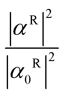
If we still base our analysis on the classical electromagnetic theory of polarizable Raman dipoles discussed in 2.1, we can obtain the theoretical ratio of Raman polarizability describing the chemical enhancement factor (CM EF) of the system, as shown in eqn (3):
| |  | (3) |
where α
0R is the system Raman polarizability and α
R is the Raman polarizability including various chemical effects when the electric dipole is emitted.
29
Combined with the above theoretical formulas, their differences are further described. CB enhancement is due to partial charge transfer caused by the formation of chemical bonds, the system polarizability does not change significantly, and the highest occupied molecular orbital (HOMO) and the lowest unoccupied molecular orbital (LUMO) of the molecule are broadened. SC enhancement is due to the formation of surface complexes by atomic clusters composed of partially positively charged metal atoms, partially negatively charged molecules and electrolyte anions on the surface. Different HOMOs and LUMOs can reach resonance under visible light excitation, and the system polarizability changes significantly. PICT enhancement mainly depends on whether the energy difference between the Fermi level of the metal and the HOMO or LUMO of the molecule matches the energy of the excitation light. When matching, CT from molecule to metal or from metal to molecule will occur, and the change in the system polarizability is mainly reflected in the CT state of the system.
3. Main regulation methods for SERS enhancement
In order to continuously improve the SERS enhancement effect and finally enhance its sensitivity in actual detection, researchers have used the SERS enhancement mechanism as a theoretical guide and tried different methods to improve EM and CM. Although in theory EM and CM always exist simultaneously in the SERS system, they can be roughly divided into the following two aspects according to the different regulating subjects.
3.1. Main regulatory methods based on EM enhancement
EM contributes a major part of the SERS EF, so many related studies are carried out in depth horizontally with the direction of regulating EM. It can be inferred from the previous discussion that MLoc(ωL) is the key parameter for regulating EM, and many related studies are carried out from this. For example, Ma et al. compared and explored in detail the effects of geometric parameters such as dielectric function on the local field enhancement factor of gold–dielectric–silver (GDS) nanotubes and silver–dielectric–gold (SDG) nanotubes based on quasi-static theory. Finally, it was concluded that SDG nanotubes can obtain a stronger MLoc(ωL) than GDS nanotubes when the thickness of the dielectric layer is small and the surrounding dielectric function is high, thereby improving EM enhancement.35 Jeremy et al. developed special picocavity models in which the maximum EF of the hyperlocal hot spots can be 100 times larger than the nanocavities supporting them, reaching ∼1012.36 This result shows a significant enhancement and illustrates the importance of the structure for EM enhancement. Next, EM enhancement regulation methods based on single metal and composite metal structures are introduced.
3.1.1. EM enhancement based on single metal structures.
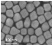
The pivotal strategy for modulating the enhanced EM in SERS hinges on the utilization and refinement of materials, the fabrication of diverse substrate architectures and dimensions, with the primary emphasis placed on generating a greater number of hot-spot regions, more intense LSPR, and achieving a higher degree of Raman enhancement. Spontaneous SERS substrates typically employ noble metals such as gold (Au), silver (Ag), and copper (Cu),37,38 which exhibit robust SPR effects under visible and near-infrared light excitation, and generally possess an EF exceeding 105. These advantageous properties have made noble metals widely favored for SERS substrates.39,40 Au, known for its stable chemical properties, has been leveraged in various configurations. Yue et al. constructed a series of size-dependent Au nanoparticle (AuNP) models and calculated the squares of the EM key influencing parameter values corresponding to different models. This work effectively enhanced the evaluation of SERS performance.41 Chen and colleagues utilized a thin silica shell and an Au nanosphere (AuNS) monolayer to create a smooth AuNS monolayer material (Fig. 3a) Leveraging the “hot-spot” effect, this material enables the detection of 10 ng mL−1 prostate-specific antigen (PSA) and 1 ng mL−1 carcinoembryonic antigen (CEA), which can be effectively employed for cancer diagnosis.42 To further improve the sensing performance, Huang et al. proposed a strategy to enhance the interfacial mass transfer rate of the target, self-assembled AuNP bridge arrays, which exhibited fast substance transfer and high sensitivity43 (Fig. 3b). To break through the limitation of difficult SERS tag preparation, Cardellini et al. proposed a method for the spontaneous binding of AuNPs on lipid vesicles, resulting in several orders of magnitude enhancement of their Raman signal44 (Fig. 3c). Saleh et al. transformed semi-crystalline CuNPs with sharp geometric features,45 with an EF of up to 2.1 × 10⁵ (Fig. 3d). However, Cu is chemically unstable in air. To solve this problem, Fang et al. prevented CuNPs from oxidation by oxygen plasma irradiation.46 Since Ag typically boasts a higher EF than Au and Cu, it also ranks as a highly popular substrate for traditional SERS applications. Zhang et al. proposed a strategy to construct AgNP reinforced hydrogels on a PNIPAM membrane, with a detection concentration of the probe molecules CV and 4-ATP solution as low as 10−9 M (ref. 47) (Fig. 3e). Huo et al. prepared a Ag-NPs@BNC SERS substrate based on AgNPs, which has high sensitivity and spectral reproducibility (Fig. 3f).48
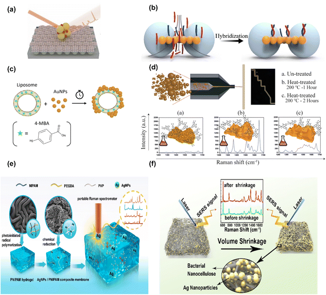 |
| | Fig. 3 Substrate structures based on different single metal enhanced EM: (a) smooth AuNS monolayer structure,42 (b) AuNP-bridge SERS sensor,43 (c) self-assembly of AuNPs on liposomes with different membrane rigidities,44 and (d) CuNP SERS sensors.45 (e) Schematic illustration of the synthesis and SERS detection process of the flexible AgNPs/PNIPAM hydrogel membrane and47 (f) AgNPs@bacterial nanocellulose SERS substrate.48 | |
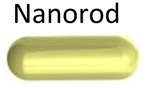
Beyond considering the characteristics of different single metals, the impact of various shapes and structures of the same metal on EM enhancement also varies.49 Yan et al. synthesized an Au-coated polystyrene nanosphere with an EF of approximately 3.57 × 107.50 Pardehkhorram and colleagues designed a Au nanorod (AuNR) probe for biological detection in complex culture media.51 Zhou et al. synthesized a structurally adjustable Au nanocube (AuNC) monolayer with an analytical enhancement factor of 1.24 × 108.52 Zhu and co-workers utilized non-polar solvents to synthesize symmetrical Au nanostars (AuNSTs) with a specific number of tips and successfully red-shifted the LSPR peak from 850 nm to 1880 nm.53 Relatively speaking, the nanostar morphology can generate a more pronounced LSPR effect. Table 1 summarizes the types, structures, and characteristics of the aforementioned four plasmonic nanostructures. In general, AuNSs have a single plasma mode, the LSPR peak red-shifts as the size of the sphere increases, and the optimal size is about 10–20 nm.50,54 AuNRs support both plasmon modes, with ELoc concentrated at both ends of the nanorods being more than 103 times higher than that of spheres.51,55 AuNCs can excite higher-order plasmon wave modes, enabling tuning of plasmon resonances and electromagnetic field enhancement.52,56,57 AuNSTs have core-and tip-related plasmon hybrid modes, and the LSPR is mainly confined to the tip.53,58
Table 1 Comparison table of types, structures and characteristics of single metal plasmonic nanoparticles
| Types |
SEM/TEM |
Plasmonic properties |
Ref. |

|

|
Single plasma mode |
50, 54 and 59 |
| LSPR peak redshift; the optimal size of AuNSs is about 10–20 nm |

|

|
In both plasma modes |
51 and 55 |
|
E
Loc at both ends of the nanorod is more than 103 times higher than that of the sphere |

|

|
Higher-order plasma wave mode |
52, 56 and 57 |
| Tuning plasmon resonance |
| Tuning the electromagnetic field enhancement |

|

|
Core and tip associated plasma hybrid patterns |
53 and 58 |
| The LSPR is mainly confined to the tip |
3.1.2. EM enhancement based on composite metal structures.
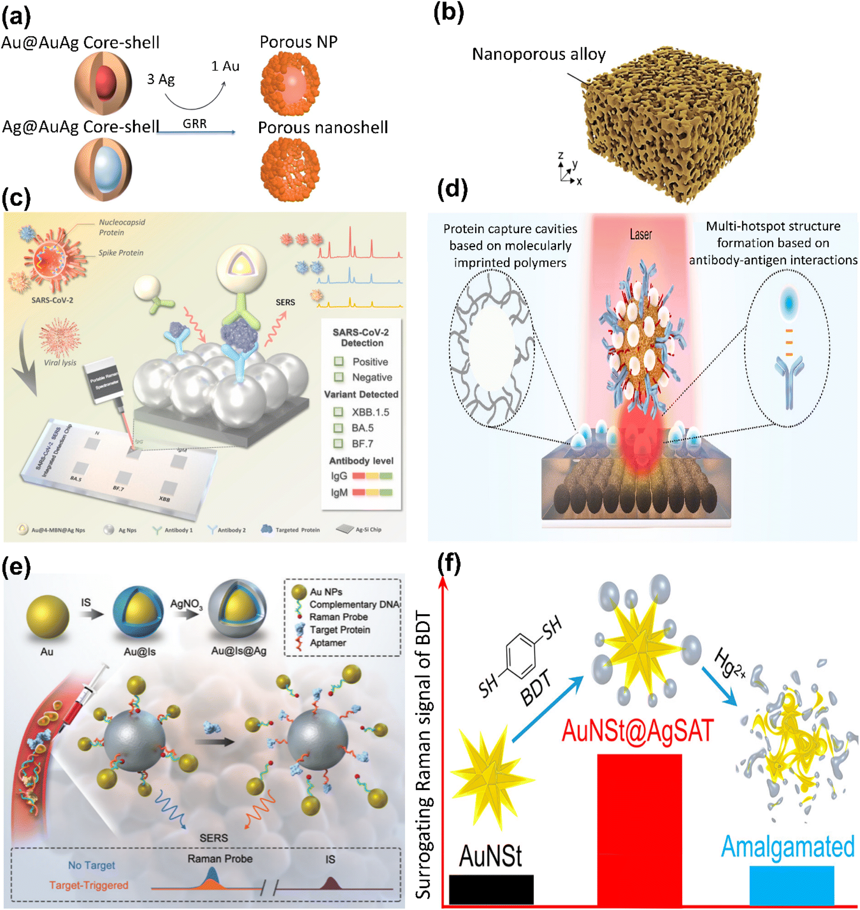
In addition to exploring the SERS performance of the aforementioned single noble metals, bimetallic nanostructures, mixed multimetallic composites, and metal–semiconductor hybrid nanomaterials have been proposed to enhance the SERS signal,60,61 particularly in certain metal nanogap structures,62–64 sandwich structures,65,66 satellite-like structures,67,68 and tip-enhanced structures.69,70 Ye's group innovatively proposed a gap-enhanced Raman tag (GERT), which utilizes the gap junction of the GERT to generate a large number of hot-spot regions, thereby amplifying Raman scattering on a large scale to provide high-sensitivity biological imaging and successfully ablate micro-tumors. Moreover, core–shell structures and porous structures are also typical examples of metal nanogap structures.71,72 For instance (Fig. 4a), Wang and colleagues synthesized a nanostructured Raman tag using an electro-substitution reaction. Porous core–shell Au-AgNPs, with Au or Ag as the core, exhibit strong LSPR coupling due to their rough surface and adjustable hollowness. Porous NPs possess a high density of hot-spot regions, which allows for fine-tuned regulation of their plasmonic properties and a red-shift of the plasmon peak to the near-infrared region. At the single-particle level, they demonstrate a Raman enhancement that is 68 times higher than that of AuNSs and can be employed as a quantitative monitor for drug release.73 La et al. prepared three-dimensional nanoporous structure composed of an alloy based on gold (Fig. 4b). By controlling the composition, they achieved a high surface area and a strong local electric field. In terms of plasmonic molecule detection, this structure is capable of detecting Aβ (β-amyloid protein) with a concentration as low as approximately 1 fM, and the SERS EF is 10 orders of magnitude higher than that of single-layer AuNPs.74 However, the concave curvature of the nanogaps in these structures is not conducive to SERS enhancement, which can be solved by changing the core shape and other strategies. For example, Zhao et al. prepared a highly uniform Au/Ag/Au monolayer based on Au nanotriangles, which has abundant nanogaps and a strong electromagnetic enhancement effect.75 And Fan et al. fabricated three types of intra-gap NPs using nanorods, bipyramids, and nanospheres as cores, among which the intra-gap NPs based on nanorods had the best SERS performance.62
 |
| | Fig. 4 Various substrate structures based on composite metal-enhanced EM: (a) Galvanic displacement reactions on Au@Au/Ag and Ag@Au/Ag to produce porous shells with solid (porous NPs) or hollow interiors (porous nanoshells), respectively,73 (b) schematic diagram of 3D nanoporous alloys based on Au, Ag, Cu, and Al,74 (c) schematic diagram of antigen/antibody detection using a multifunctional SARS-CoV-2 SERS sensor chip,76 (d) the immune-like sandwich multiple hotspot SERS biosensor,77 (e) schematic diagram of PSA detection using an aptamer-assisted SERS sensing platform,78 and (f) Au nanostar@Ag satellite nanostructure sensor.79 | |
The sandwich nanostructure (metal nanomaterial–target–metal nanomaterial combined with a probe) is one of the most commonly used models for biosensing applications. Our group constructed a multifunctional SARS-CoV-2 sandwich-structured sensor chip (Fig. 4c).76 This antibody-based high-affinity sensor is capable of detecting six types of SARS-CoV-2 proteins, including the S and N proteins of the Omicron variants (VoC-BA.5, VoC-XBB.1.5, and VoC-BF.7). Zhang et al. constructed a SERS-based sandwich multi-hotspot biosensor for trace detection of NDKA in serum with a detection limit as low as 0.25 pg mL−1 (Fig. 4d).77 Diverging from the SERS immunoassay based on the sandwich structure, our group developed a core-satellite-structured SERS sensing model assembled by NPs surrounded by multiple NPs (Fig. 4e).78 This model can generate a sensitive response to the SERS signal through the change in the distribution of external NPs triggered by the target and detect prostate-specific antigen (PSA) using the recognition-release mechanism, with a dynamic detection range of 10−2–10−15 mg mL−1. The Au nanostar@Ag satellite nanostructure (AuNSt@AgSAT) designed by Matthew can detect Hg2+ concentrations as low as 0.1 ppb, which is much lower than the MRL of 2 ppb in current detection methods (Fig. 4f).79
In addition to the structures mentioned above, tip-enhanced structures are also widely employed. For example, Au nanopyramids (Au NPy) are SERS substrates with pronounced tips and tunable shape parameters. Ding et al. utilized AuNPs to form macroscopic two-dimensional superlattices (2D SLs) and enhanced SERS performance by adjusting the tip arrangement. They found that the tip spacing of the tip-on-tip superlattice (tip-on-tip SL) was 3.6 nm, and the SERS EF reached 1.95 × 108.80Table 2 provides some examples of the aforementioned common composite metal structures. In conclusion, the LSPR band of the metal nanogap structure can be tuned by changing the relative size between the shell and the core.73,81 Sandwich structures use two metal substrates to achieve double amplification of signals.76,82 Core-satellite structures use a target-triggered dynamic response to achieve simultaneous target capture and signal amplification.78,83 The tip-enhanced structure is characterized by the LSPR at its tip, generating a high-intensity near-field electromagnetic field.80,84,85
Table 2 Some examples of common composite metal structures
| Types |
SEM/TEM |
Plasmonic properties |
Ref. |

|

|
The LSPR band can be tuned by changing the relative size between the shell and the core |
73 and 81 |

|

|
Using two metal substrates to achieve double amplification of signals |
76, 77 and 82 |

|

|
Target-triggered dynamic response achieves simultaneous target capture and signal amplification |
78 and 83 |

|

|
The LSPR of the metal tip generates a high-intensity near-field electromagnetic field |
80, 84 and 85 |
3.2. Main regulatory methods based on CM enhancement
The main regulation methods based on CM enhancement are mainly applied to non-plasma materials, especially semiconductor materials. Due to the complexity of the CM, the changes in molecular properties cannot be easily determined in a short time. Factors such as orientation effects and tensor properties will affect CM, and there is currently no comprehensive and systematic explanation for this. This makes the regulation based on CM enhancement more promising in the common case where the magnitude of CM enhancement is not as obvious as EM. There is no doubt that enhancing the sensitivity of SERS on non-noble metal surfaces is an urgent issue that needs to be addressed. At present, the regulation of CM enhancement mainly focuses on three aspects: energy-level modulation, defect engineering, and material coupling.86,87
3.2.1. CM enhancement based on the energy level adjustment strategy.
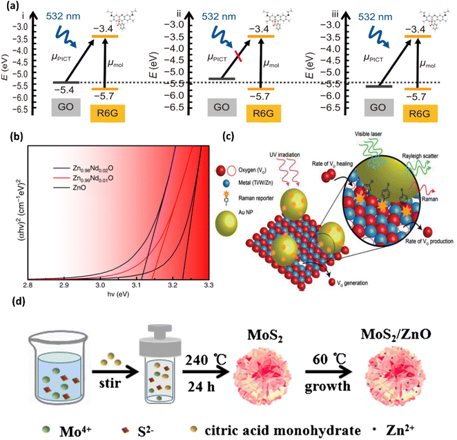
The energy level adjustment strategy is one of the mainstream methods to regulate chemical enhancement of non-plasma materials such as semiconductors. It is crucial to realize the precise tuning of energy levels, and Tang et al. prepared single-layer binary TMD alloys. They maximized the SERS efficiency of the target molecules by tuning the energy levels of the SERS substrate, and continuously tuned the energy levels of these alloys over a wide range to meet the requirements of specific target molecules by constructing CT resonance channels on demand.88 Zhang et al. prepared semi-metallic phases (1T’) WS2 and MoS2, optimized the band alignment by regulating the crystalline phase of two-dimensional transition metal disulfides (TMDs), achieved a higher CT rate, and increased the SERS EF to 4.6 × 108.89 Liang et al. developed a two-dimensional (2D) borocarbonate (BCN) SERS platform by altering the carbon atomic ratio. This platform can adjust the CB level to align with the LUMO of the target molecule, which is utilized to enhance the Raman signal for detecting specific molecules with suitable band structures.90 However, this regulatory approach can only effectively detect probe molecules with specific band structures, and further optimization of the system is required. Shao et al. prepared a SERS substrate composed of graphene oxide-coupled ferroelectric PMN-PT (PMN-PT@GO) to facilitate CT, thereby achieving optimal SERS signals for different molecules (R6G, CV, MB, and PNTP) with diverse band structures on the same substrate.91Fig. 5a presents a schematic diagram of the band structure of PMN-PT@GO (Ps−)/R6G and the electronic transition process under 532 nm laser excitation.
 |
| | Fig. 5 Various regulation methods for enhancing CM: (a) schematic diagram of the band structure of PMN-PT@GO (Ps−)/R6G and the electronic transition process under 532 nm laser excitation,91 (b) spectra of the optical energy band gap of ZnO and Zn1−xNdxO,92 (c) schematic diagram of the PIERS technique for probing vacancy dynamics,94 and (d) schematic diagram of the low-cost synthesis procedure for obtaining MoS2/ZnO MC.97 | |
3.2.2. CM enhancement based on the defect engineering strategy.
In addition, employing defect engineering (i.e., introducing defects into semiconductor substrates) to facilitate CT is also a prevalent method. From a mechanistic standpoint, CT can separate positive and negative charges, augment the polarizability of materials, and thus boost the Raman scattering cross-section. In other words, promoting the interfacial CT process is a crucial step in enhancing the chemical enhancement effect. One such approach is chemical doping. For instance, Yang et al. achieved Nd doping in ZnO, which augmented the surface defect count, enhanced light absorption characteristics, facilitated interband charge transitions, and reduced the SERS background (Fig. 5b).92 In addition, new doping materials have also attracted the attention of researchers. Lin et al. synthesized a new type of 3D amorphous nitrogen-doped carbon (NDC) nanocages (NCs) via a simple template-based method, and successfully established three distinct nitrogen atom doping configurations (graphitic-N, pyrrolic-N, and pyridinic-N) within the NDC NCs, thereby increasing valence electrons and expanding the electronic active region. The SERS EF value for the methylene blue (MB) molecule can reach 5.52 × 106.93 Another method of introducing defects is to induce the formation of oxygen vacancies (OVs) on the substrate surface. As illustrated in (Fig. 5c), Glass et al. investigated the formation and healing kinetics of surface active oxygen vacancies (Vo) in typical metal oxide semiconductors (i.e., TiO2, WO3, and ZnO) during pre-irradiation. The generation of these atomic-scale defects enhances the Raman transition in defective semiconductors due to PICT, with the most vacancies being generated.94 Fan et al. utilized oxygen-vacancy-rich substrates. OVs create surface defect levels between the CB and valence band (VB) of the substrate, thereby enhancing the SERS activity of the substrate.95
3.2.3. CM enhancement based on the material coupling strategy.
The SERS performance of most semiconductor-based substrates is not remarkable and exhibits poor sensitivity for molecular trace detection. However, Yu et al. demonstrated that the SERS intensity of the hybrid structure is higher than that of the single component. They developed a CuO/TiO2 p-n heterojunction as a new SERS substrate for amplifying the Raman signal of 4-mercaptobenzoic acid (4-MBA) molecules, which can improve the photogenerated carrier separation efficiency due to the strong interfacial coupling in the heterojunction interface, and the EF of this substrate is 8.87 × 106.96 The work of Quan et al. achieved a higher SERS EF. They fabricated a MoS2/ZnO microcrystal (MC) composite semiconductor SERS substrate and constructed a semiconductor heterojunction to significantly enhance PICT. The EF when detecting R6G molecules was approximately 3.45 × 10⁷ (Fig. 5d).97
4. Special regulation methods for SERS enhancement
The abovementioned SERS EF regulation methods primarily focus on employing diverse approaches to generate as many hot-spot regions as possible to bolster the LSPR effect, thereby amplifying the EM. Additionally, it involves altering the polarizability of materials by facilitating CT to achieve more pronounced CM. This section mainly supplements some special regulation methods for SERS enhancement. One such method is the analyte enrichment method, which controls the controllable contact between the SERS-active substrate and the analyte or increases the analyte concentration to enhance its impact on the SERS effect. The other is the electrical regulation (E-SERS) strategy, which generates additional electric fields through pyroelectric,98 piezoelectric,99 triboelectric,100 and other means to attain a stronger LSPR effect and CT. The E-SERS discussed here mainly focuses on the integration of pyroelectric materials with SERS substrates. These special regulation methods can simultaneously enhance both the EM and CM.101 The summary of special regulation methods for SERS enhancement is depicted in Fig. 6.
 |
| | Fig. 6 Special regulation method for SERS enhancement. | |
4.1. Special regulation based on analyte enrichment methods
Analyte enrichment methods can facilitate greater contact between analytes and hot-spot regions. By leveraging the positive correlation between the number of molecules on the SERS substrate surface and the SERS intensity, Fan et al. constructed a hierarchical micromotor (HSM) featuring an Au/SiO2/Fe plasma nanostructure on the outer wall. The hot-spot regions located on the outer wall, driven by an external rotating magnetic field, enriched analytes in the fluid through dense motion, achieving SERS sensing with an EF of approximately 3.39 × 107.102 Yang et al. also fabricated a mobile hotspot SERS sensor by growing Au nanospikes on the surface of magnetic microspheres (Fig. 7a), to enable controllable contact and separation of analytes.103 They maximized contact with target molecules by creating dynamic hotspots, which can overcome the limitations of the fixed positions of traditional hotspots. Hydrophobic surface enrichment methods have also been employed to effectively aggregate analytes on substrates. Mangalassery et al. fabricated a novel plasma-active laser-induced periodic surface structure (LIPSS). The hydrophobicity of the substrate can enrich analytes without diffusion, thereby immobilizing molecules near the surface of the nanostructure to achieve Raman signal enhancement, with a SERS EF of 2 × 10⁹. However, this hydrophobic surface enrichment method is predominantly applied to noble metal substrates, which are costly and time-consuming. This has prompted researchers to explore lower-cost materials.104 Lan et al. utilized 2D vanadium carbide (V4C3 and V2C) MXenes, which possess good conductivity and strong adsorption capabilities, as SERS-active substrates (Fig. 7b), to achieve ultra-fast molecular enrichment (within 2 minutes). They employed this new material to offer novel insights into molecular enrichment methods at the non-metal level.105 However, various flexible SERS substrates prepared from substrates such as polydimethylsiloxane (PDMS),106 filter paper (FP),107 textiles,108etc. have also presented unique advantages in recent years compared to the abovementioned conventional rigid substrates that are still insufficiently expressive in terms of analyte enrichment. Furthermore, physical tweezers (such as optical tweezers,109 acoustic tweezers,110 electric tweezers,111etc.) are also utilized to pre-concentrate analytes. Jin et al. proposed multifunctional droplet electrostatic tweezers (DEST) (Fig. 7c), which are used for remote and non-invasive manipulation of droplets based on electrostatic induction, eliminating the need for the complex electrode patterns used in traditional electrical manipulation. The entire process takes less than 20 seconds.112
 |
| | Fig. 7 Special regulation methods based on molecular enrichment strategies or E-SERS: (a) mobile hotspot SERS sensor based on Au nanospikes grown on the surface of magnetic microspheres,103 (b) flexible 2D vanadium carbide MXene substrate with ultra-fast molecular enrichment function,105 (c) schematic diagram of the multifunctional droplet electrostatic tweezers (DEST) platform,112 and (d) SERS enhancement mechanism of PMN-PT/AgNPs.115 | |
4.2. Special regulation based on the E-SERS strategy
The pyroelectric effect is a phenomenon that releases charges on its surface due to changes in spontaneous polarization strength caused by temperature fluctuations, and thermal energy can be converted into electrical energy through the pyroelectric effect. There are several studies introducing the pyroelectric effect as an active regulation strategy into SERS substrates. The pyroelectric-based strategy in the SERS toolkit can introduce additional electromagnetic fields into the system. The integration of metal substrates with pyroelectric materials can enhance the substrate electron density and facilitate charge transfer, thereby modulating both the electromagnetic and chemical enhancement aspects of SERS. Li et al. synthesized a vibration-free pyroelectric NG driven by light illumination to enhance the local thermal charge density, and the total electric field induced by the dual effect of pyroelectric and equipartitioned excitations could effectively enhance SERS activity, realizing the dual application of industrial chemical catalysis and in situ detection of SERS in the reaction process.113 You et al. fabricated Ag–BiFeO3/CNF plasma metal/ferroelectric hybrid SERS substrates, which can efficiently convert photoinduced thermal energy into pyroelectric charges, modulate the electron density of Ag, and thus amplify the electromagnetic field at the “hot-spot” regions.114 They believe that the signal enhancement is mainly due to the electromagnetic field upregulation effect. To further probe the impact of the combination of pyroelectric and plasmonic materials on the SERS signal, Shao et al. combined the pyroelectric material PMN-PT with AgNPs (Fig. 7d) to create a SERS composite substrate (PMN-PT/AgNP). When a positive or negative pyroelectric potential was applied, the SERS signal was further amplified by more than two orders of magnitude.115 This work is expected to further accelerate the application of plasma metal nanoparticle-based pyroelectric materials in fields such as optical sensors.
5. Summary and outlook
This review initially introduces SERS technology and its theoretical enhancement mechanisms, including EM and CM. The ongoing refinement of these theories provides robust theoretical guidance for efforts aimed at enhancing the SERS EF based on these two primary mechanisms. The ever-advancing nanofabrication technology offers strong technical support for the SERS substrate, which is the main contributor to the EF. Coupled with the inherent advantages of SERS technology, it showcases the boundless potential of SERS. Traditional noble metals, mixed multimetals, metal–semiconductor hybrid nanomaterials, etc., have been proposed to boost the SERS signal, particularly in structures such as core–shell configurations, porous structures, metal nanocavity composites, sandwich structures, gap structures, and nanostars. The utilization and refinement of these materials and structures, along with adjustments in size and shape, have significantly impacted the EM. The work on regulating CM involves modulating the SERS intensity by altering the polarizability of substances directly adsorbed or in close proximity to the surface. This area of research encompasses energy-level modulation of non-plasmonic substrates (e.g., semiconductors), defect engineering, and material coupling. Finally, we conclude our discussion on regulation methods by supplementing the use of analyte enrichment methods and E-SERS strategies to amplify the impact on the SERS effect. When it comes to analyte enrichment methods, in addition to the methods mentioned above, the introduction of antibodies, aptamers or DNA to form specific immune complexes can overcome the interference of environmental interferents and produce higher selectivity. These three target recognition and capture schemes can improve the binding affinity of SERS materials. Obviously, the above work has played a crucial role in improving the SERS EF. However, the SERS technology still faces many challenges in practical applications: (1) in CM research, a more comprehensive and systematic understanding of the CM is needed to maximize its regulatory effects; (2) SERS has been shown to have single-molecule sensitivity. However, there are challenges in achieving single-molecule detection sensitivity in complex biological systems, which requires SERS substrates with a high density of “hot spots” and homogeneous SERS enhancement; (3) although the analyte enrichment strategies mentioned above can reduce the interference of non-target analytes to some extent, there's still a lot of room for improvement in terms of selectivity. Specifically, when using SERS technology for direct detection, only analytes with high affinity for the SERS substrate used can enhance the Raman signal, which is detrimental to the occupancy of the SERS active site by certain analyte molecules; (4) the spatial and temporal resolution of SERS technology needs to be improved. Since biological systems (e.g., cellular metabolism or pathogen infection) are highly dynamic, many critical processes occur on millisecond or even shorter time scales. The insufficient temporal resolution of conventional SERS techniques may result in missing the capture of these transient changes, and thus there is an increasing demand for nanospatial resolution using Raman spectroscopy; (5) practical applications, such as clinical diagnosis, place obvious demands on the development of highly reproducible SERS sensors for use. In terms of material structure enhancement, although nanostructures (e.g., nanostars) exhibit high energy efficiency (EF) for SERS applications, the reproducibility of their preparation methods remains a challenge. The future should be devoted to (1) enhancing the sensitivity of SERS detection in real complex environments; (2) for real sample detection, new surface chemical engineering should be designed to minimize the nonspecific adsorption of biofluids; (3) further improving the temporal and spatial resolution of SERS systems; and (4) developing more stable and reproducible preparation methods to ensure the homogeneity and consistency of nanostructures; (5) in addition, future studies can explore more diverse multi-component composite structures, such as combining noble metal nanostructures with 2D materials, metal–organic frameworks (MOFs), and other materials, in order to fully utilize the advantages of each component and achieve superior SERS performance. These advances will effectively enhance the SERS effect and promote the steady development of the field.
Data availability
No new data were generated or analysed as part of this review.
Author contributions
Yangmin Wu: investigation, writing – original draft. Shuohong Weng: investigation. Tingyin Wang: visualization, project administration. Kien Voon Kong: project administration. Duo Lin: project administration, writing – review & editing, funding acquisition, supervision.
Conflicts of interest
The authors declare that they have no known competing financial interests or personal relationships that could have appeared to influence the work reported in this paper.
Acknowledgements
This research was financially supported by the National Natural Science Foundation of China (No. 12374405) and Provincial Science Foundation for Distinguished Young Scholars of Fujian (No. 2024J010024).
Notes and references
- Q.-F. He, Y.-J. Zhang, Z.-L. Yang, J.-C. Dong, X.-M. Lin and J.-F. Li, Chin. J. Chem., 2023, 41, 355–369 CrossRef CAS
 .
.
- A. I. Pérez-Jiménez, D. Lyu, Z. Lu, G. Liu and B. Ren, Chem. Sci., 2020, 11, 4563–4577 Search PubMed .
- H. Zhang, L. Yang, M. Zhang, H. Wei, L. Tong, H. Xu and Z. Li, Nano Lett., 2024, 24, 11116–11123 CrossRef CAS PubMed .
- C. Zong, M. Xu, L.-J. Xu, T. Wei, X. Ma, X.-S. Zheng, R. Hu and B. Ren, Chem. Rev., 2018, 118, 4946–4980 Search PubMed .
- Y. Liu, M. Li, H. Liu, C. Kang and C. J. T. Wang, Theranostics, 2024, 14, 1966–1981 Search PubMed .
- J. Zhang, Y. Weng, Y. Liu, N. Wang, S. Feng, S. Qiu and D. Lin, J. Photochem. Photobiol., B, 2024, 257, 112968 CrossRef CAS PubMed .
- J. Wang, Y. Zheng, X. Wang, X. Zhou, Y. Qiu, W. Qin, X. ShenTu, S. Wang, X. Yu and Z. Ye, Sci. Total Environ., 2024, 912, 169440 CrossRef CAS PubMed .
- L. Liu, T. Gao, Q. P. Zhao, Z. K. Xue and Y. Z. Wu, Opt. Laser Technol., 2025, 181, 111827 CrossRef CAS .
- H. Kim, B. T. Trinh, K. H. Kim, J. Moon, H. Kang, K. Jo, R. Akter, J. Jeong, E. K. Lim, J. Jung, H. S. Choi, H. G. Park, O. S. Kwon, I. Yoon and T. Kang, Biosens. Bioelectron., 2021, 179, 111827 Search PubMed .
- H. Pu, Q. Ouyang and D. Sun, Technology, Trends Food Sci. Technol., 2024, 147, 104416 Search PubMed .
- M. Arabi, A. Ostovan, Z. Zhang, Y. Wang, R. Mei, L. Fu, X. Wang, J. Ma and L. Chen, Biosens. Bioelectron, 2021, 174, 112825 CrossRef CAS PubMed .
- M. Arabi, A. Ostovan, Y. Wang, R. Mei, L. Fu, J. Li, X. Wang and L. Chen, Nat. Commun., 2022, 13, 5757 CrossRef CAS .
- A. Ostovan, M. Arabi, Y. Wang, J. Li, B. Li, X. Wang and L. J. A. M. Chen, Adv. Mater., 2022, 34, 2203154 CrossRef CAS PubMed .
- X. Chen, A. Ostovan, M. Arabi, Y. Wang, L. Chen and J. Li, Anal. Chem., 2024, 96, 6417–6425 CrossRef CAS PubMed .
- M. Fan, Y. Weng, Y. Liu, Y. Lu, L. Xu, J. Ye, D. Lin, S. Qiu, S. J. L. Feng and P. Reviews, Laser Photonics Rev., 2024, 18, 2301072 Search PubMed .
- D. Cialla-May, A. Bonifacio, T. Bocklitz, A. Markin, N. Markina, S. Fornasaro, A. Dwivedi, T. Dib, E. Farnesi and C. Liu, Chem. Soc. Rev., 2024, 53, 8957–8979 RSC .
- J. P. Li, Y. Feng, L. Liang, F. Liao, W. X. Huang, K. G. Li, G. L. Cui and Z. W. Zuo, ACS Appl. Mater. Interfaces, 2024, 16, 35771–35780 CrossRef CAS PubMed .
- G. Q. Fang, X. Lin, X. Liang, J. L. Wu, W. Xu, W. L. J. Hasi and B. Dong, Small, 2022, 18, 2204588 CrossRef CAS .
- Z. Huang, Y. Chen, L. Xu, J. Peng and P. Liu, ACS Appl. Nano Mater., 2024, 7, 25099–25106 Search PubMed .
- L. Zhou, L. Pusey-Nazzaro, G. H. Ren, L. G. Chen, L. Y. Liu, W. T. Zhang, L. Yang, J. Zhou and J. G. Han, ACS Nano, 2022, 16, 577–587 CrossRef CAS PubMed .
- Y. Wu, T. Sun, M. Shao, C. Ji, C. Li, C. Zhang and Z. Li, Laser Photonics Rev., 2024, 19, 2401152 CrossRef .
- Y. Xu, Z. Li, Y. Liao, J. Wang, T. Zhang, X. Liu and Y. Zhang, ACS sensors, 2024, 9, 849–859 CrossRef CAS PubMed .
- Z. Wang, S. Zong, L. Wu, D. Zhu and Y. Cui, Chem. Rev., 2017, 117, 7910–7963 CrossRef CAS .
- E. X. Tan, Q.-Z. Zhong, J. R. Ting Chen, Y. X. Leong, G. K. Leon, C. T. Tran, I. Y. Phang and X. Y. Ling, ACS Nano, 2024, 18, 32315–32334 CrossRef CAS .
- M. Fleischmann, P. J. Hendra and A. J. McQuillan, Chem. Phys. Lett., 1974, 26, 163–166 Search PubMed .
- M. G. Albrecht and J. A. Creighton, J. Am. Chem. Soc., 1977, 99, 5215–5217 CrossRef CAS .
- S. Y. Ding, E. M. You, Z. Q. Tian and M. Moskovits, Chem. Soc. Rev., 2017, 46, 4042–4076 RSC .
-
L. Jensen, Presented in Part at the International Conference of Computational Methods in Sciences and Engineering 2009 (ICCMSE 2009), 2012 Search PubMed .
- E. C. Le Ru and B. Auguié, ACS Nano, 2024, 18, 9773–9783 CrossRef CAS PubMed .
- S.-Y. Ding, J. Yi, J.-F. Li, B. Ren, D.-Y. Wu, R. Panneerselvam and Z.-Q. Tian, Nat. Rev. Mater., 2016, 1, 1–16 Search PubMed .
- E. C. Le Ru and P. G. Etchegoin, MRS Bull., 2013, 38, 631–640 CrossRef .
-
J. Aizpurua and R. Hillenbrand, in Plasmonics: from Basics to Advanced Topics, Springer, 2012, vol. 167, pp. 151–176 Search PubMed .
- A. Otto, Appl. Surf. Sci., 1980, 6, 309–355 CrossRef CAS .
- R. Graham D Fau - Goodacre and R. Goodacre, Chem. Soc. Rev., 2008, 37, 883–884 RSC .
- Y. W. Ma, Z. W. Wu, J. Li, J. B. Hu, X. C. Yin, M. F. Yi and L. H. Zhang, Plasmonics, 2023, 18, 623–633 CrossRef CAS .
- J. J. Baumberg, Nano Lett., 2022, 22, 5859–5865 CrossRef CAS PubMed .
- M. Chen, J. Zhang, X. Zhu, Z. Liu, J. Huang, X. Jiang, F. Fu, Z. Lin and Y. Dong, Interfaces, ACS Appl. Mater. Interfaces, 2022, 14, 26216–26224 CrossRef CAS PubMed .
- D. Wang, L. Bao, H. Li, X. Guo, W. Liu, X. Wang, X. Hou and B. J. N. He, Nanoscale, 2022, 14, 6212–6219 RSC .
- X. L. Zheng, Z. W. Ye, Z. Akmal, C. He, J. L. Zhang and L. Z. Wang, Chem. Soc. Rev., 2024, 53, 656–683 Search PubMed .
- K.-W. Liu, P.-Y. Sie, J.-Y. Huang, H.-Y. Chen, Y.-L. Chen, Y.-C. Lin and M.-Y. Liao, Anal. Chim. Acta, 2024, 1329, 343189 Search PubMed .
- X. Yue, S. Yan, T. Gao, S. Pu, H. Tang, X. Pei, Z. Tian, X. Wang, B. Ren and G. Liu, Anal. Chem., 2024, 96, 17517–17525 CrossRef CAS PubMed .
- X. Chen, A. Cui, M. He, M. Yan, X. Zhang, J. Ruan and S. Yang, Nano Lett., 2023, 23, 6736–6743 CrossRef CAS PubMed .
- X. Huang, W. Zhao, X. Chen, J. Li, H. Ye, C. Li, X. Yin, X. Zhou, X. Qiao and Z. Xue, J. Am. Chem. Soc., 2022, 144, 17533–17539 CrossRef CAS PubMed .
- J. Cardellini, C. Dallari, I. De Santis, L. Riccio, C. Ceni, A. Morrone, M. Calamai, F. S. Pavone, C. Credi and C. J. N. C. Montis, Nat. Commun., 2024, 15, 7975 CrossRef CAS PubMed .
- S. Aghajani, A. Accardo and M. Tichem, ACS Appl. Nano Mater., 2020, 3, 5665–5675 CrossRef CAS .
- Y. Fang, B. Xu, S. Wang, H. Liu, J. Wang and M. J. N. Si, Nanoscale, 2024, 16, 9748–9753 RSC .
- T. Zhang, X. Li, D. Liu, J. An, M. Zhang, J. hua Li and C. Jiang, Chem. Eng. J., 2024, 494, 153082 CrossRef CAS .
- D. Huo, B. Chen, G. Meng, Z. Huang, M. Li and Y. Lei, Interfaces, ACS Appl. Mater. Interfaces, 2020, 12, 50713–50720 CrossRef CAS PubMed .
- C. Wang, X.-P. Zhao, Q.-Y. Xu, X.-G. Nie, M. R. Younis, W.-Y. Liu and X.-H. Xia, ACS Appl. Nano Mater., 2018, 1, 5805–5811 CrossRef CAS .
- S. Yan, H. Tang, J. Sun, C. Zhu, Q. Pan, B. Chen and G. Meng, Adv. Opt. Mater., 2024, 12, 2302010 CrossRef CAS .
- R. Pardehkhorram, F. A. Alshawawreh, V. R. Gonçales, N. A. Lee, R. D. Tilley and J. J. Gooding, Anal. Chem., 2021, 93, 12954–12965 CrossRef CAS PubMed .
- Z.-R. Zhou, W.-X. Liu, Z.-Y. Yao, Z.-X. Zheng, L. Ma, X.-B. Chen and S.-J. Ding, ACS Appl. Nano Mater., 2024, 7, 17009–17016 CrossRef CAS .
- D. Zhu, Y. Liu, M. Liu, X. Liu, P. N. Prasad and M. T. Swihart, J. Mater. Chem. B, 2020, 8, 5491–5499 RSC .
- S. A. Maier and H. A. Atwater, J. Appl. Phys., 2005, 98, 011101 CrossRef .
- E. Hao and G. C. Schatz, J. Chem. Phys., 2004, 120, 357–366 Search PubMed .
- J.-E. Park, Y. Lee and J.-M. Nam, Nano Lett., 2018, 18, 6475–6482 CrossRef CAS PubMed .
- M. Tavakkoli Yaraki and Y. N. Tan, Sens Int., 2020, 1, 100049 CrossRef .
- F. Hao, C. L. Nehl, J. H. Hafner and P. Nordlander, Nano Lett., 2007, 7, 729–732 CrossRef CAS PubMed .
- T. Zhang, X. Li, C. Li, W. Cai and Y. Li, Chem. Mater., 2021, 33, 2593–2603 CrossRef CAS .
- J. Dong, W. Zhou, C. Yang, H. Wu, Q. Han, C. Zhang, W. Gao, X. Yan and M. Sun, ACS Appl. Mater. Interfaces, 2023, 15, 28840–28848 CrossRef CAS PubMed .
- Y. Xie, C. Chen, C. Zhang, L. Xu, Z. Li, W. Ren, X. Xu, Y. Ren, J. Lin and A. Wu, Nano Today, 2024, 54, 102140 CrossRef CAS .
- S. Fan, B. T. Scarpitti, Z. Luo, A. E. Smith, J. Ye and Z. D. Schultz, Nano Res., 2024, 17, 8415–8423 CrossRef CAS PubMed .
- F. Xu, F. Ma, Z. Ding, L. Xiao, X. Zhang, Q. Lu, G. Lu and D. L. Kaplan, ACS Appl. Mater. Interfaces, 2019, 11, 42896–42903 CrossRef CAS PubMed .
- J. Song, J. Song, W. Nam, W. Nam, W. Zhou and W. Zhou, Adv. Mater. Technol., 2019, 4, 1800689 CrossRef .
- R. Guo, J. R. Wang, W. S. Zhao, S. C. Cui, S. H. Qian, Q. X. Chen, X. Li, Y. Liu and Q. Zhang, Talanta, 2024, 269, 125466 CrossRef CAS PubMed .
- W. Gao, T. Wang, C. Zhu, P. Sha, P. Dong and X. J. T. Wu, Talanta, 2022, 236, 122824 Search PubMed .
- G.-F. Wu, J. Zhu, G.-J. Weng, J.-J. Li and J.-W. Zhao, Talanta, 2025, 289, 127742 CrossRef CAS PubMed .
- W.-B. Wang, J.-J. Li, G.-J. Weng, J. Zhu, Y.-B. Guo and J.-W. Zhao, J. Colloid Interface Sci., 2023, 647, 81–92 CrossRef CAS PubMed .
- X. X. Qi, Y. Q. Cheng, R. R. Xu, X. T. Li, Z. W. Zhang, L. Y. Chen, Y. F. Shao, Z. H. Gao and M. J. Zhu, Anal. Chim. Acta., 2023, 1280, 341872 CrossRef CAS PubMed .
- F. Schuknecht, K. Kolataj, M. Steinberger, T. Liedl and T. Lohmueller, Nat. Commun., 2023, 14, 42943–42947 Search PubMed .
- T. Wu, H. Zheng, Y. C. Kou, X. Y. Su, N. R. Kadasala, M. Gao, L. Chen, D. L. Han, Y. Liu and J. H. Yang, Microsyst. Nanoeng., 2021, 7, 23 Search PubMed .
- M. Li, X. He, C. Wu, L. Wang, X. Zhang, X. Gong, X. Zeng and Y. Huang, ACS Sensors, 2024, 9, 979–987 CrossRef CAS PubMed .
- L. Wang, S. Patskovsky, B. Gauthier-Soumis and M. Meunier, Small, 2022, 18, 2105209 CrossRef CAS PubMed .
- J. A. La, H. Lee, D. Kim, H. Ko and T. Kang, Nano Lett., 2024, 24, 7025–7032 CrossRef CAS PubMed .
- Y.-X. Zhao, X. Liang, Y.-L. Chen, Y.-T. Chen, L. Ma, S.-J. Ding, X.-B. Chen and Q.-Q. Wang, Anal. Chem., 2024, 96, 8416–8423 CrossRef CAS PubMed .
- Y. Liu, H. J. Weng, Z. W. Chen, M. Zong, S. B. Fang, Z. L. Wang, S. H. He, Y. M. Wu, J. Z. Lin, S. Y. Feng and D. Lin, Biosens. Bioelectron., 2025, 271, 117015 Search PubMed .
- X. Zhang, T. Gan, Z. Xu, H. Zhang, D. Wang, X. Zhao, Y. Huang, Q. Liu, B. Fu, Z. Dai, P. Li and W. Xu, Talanta, 2024, 271, 125630 Search PubMed .
- Q. Wu, G. Chen, S. Qiu, S. Feng and D. Lin, Nanoscale, 2021, 13, 7574–7582 RSC .
- M. G. Ellis, U. Pant, J. Lou-Franco, N. Logan and C. Cao, ACS Appl. Nano Mater., 2023, 6, 10431–10440 Search PubMed .
- W. K. Ding, Y. Xia, H. Y. Song, T. T. Li, D. Yang and A. G. Dong, Angew. Chem., Int. Ed., 2024, 63, e202401945 CrossRef CAS PubMed .
- G. Awiaz, J. Lin and A. A.-O. Wu, Exploration, 2022, 3, 20220072 CrossRef PubMed .
- A. Pollap and P. Świt, Int. J. Mol. Sci., 2022, 23, 4740 CrossRef CAS PubMed .
- X. Luo, X. Zhao, G. Q. Wallace, M.-H. n. Brunet, K. J. Wilkinson, P. Wu, C. Cai, C. G. Bazuin and J.-F. Masson, ACS Appl. Mater. Interfaces, 2021, 13, 6545–6556 Search PubMed .
- X. Wang, S.-C. Huang, T.-X. Huang, H.-S. Su, J.-H. Zhong, Z.-C. Zeng, M.-H. Li and B. Ren, Chem. Soc. Rev., 2017, 46, 4020–4041 RSC .
- X. Tang, Q. Hao, X. Hou, L. Lan, M. Li, L. Yao, X. Zhao, Z. Ni, X. Fan and T. Qiu, Adv. Mater., 2024, 36, 2312348 CrossRef CAS PubMed .
- Y. Wang, M. Zhang, H. Ma, H. Su, A. Li, W. Ruan and B. Zhao, ACS Appl. Mater. Interfaces, 2021, 13, 35038–35045 CrossRef CAS PubMed .
- Y. Zhou, Q. Gu, T. Qiu, X. He, J. Chen, R. Qi, R. Huang, T. Zheng and Y. Tian, Angew. Chem., 2021, 133, 26464–26471 Search PubMed .
- X. Tang, X. Fan, J. Zhou, S. Wang, M. Li, X. Hou, K. Jiang, Z. Ni, B. Zhao, Q. Hao and T. Qiu, Nano Lett., 2023, 23, 7037–7045 Search PubMed .
- Y. Zhang, Z. Shi, H. Cui, Q. Xia, F. Liu, Z. Wang, J. Wang, H. Fan, C. Shu, B. Chen, H. Li, Z. Lai, Z. Luo, W. Zheng, L. Wang and Z. Huang, Nano Lett., 2024, 24, 14293–14301 CrossRef CAS PubMed .
- C. Liang, Z.-A. Lu, M. Zheng, M. Chen, Y. Zhang, B. Zhang, J. Zhang and P. Xu, Nano Lett., 2022, 22, 6590–6598 CrossRef CAS PubMed .
- M. Shao, C. Ji, J. Tan, B. Du, X. Zhao, J. Yu, B. Man, K. Xu, C. Zhang and Z. Li, Institute of Materials and Clean Energy, Opto-Electron. Adv., 2023, 6, 230094 CAS .
- S. Yang, J. Yao, Y. Quan, M. Hu, R. Su, M. Gao, D. Han and J. Yang, Light: Sci. Appl., 2020, 9, 117 CrossRef CAS PubMed .
- J. Lin, D. Zhang, J. Yu, T. Pan, X. Wu, T. Chen, C. Gao, C. Chen, X. Wang and A. Wu, Anal. Chem., 2023, 95, 4671–4681 Search PubMed .
- D. Glass, E. Cortés, S. Ben-Jaber, T. Brick, W. J. Peveler, C. S. Blackman, C. R. Howle, R. Quesada-Cabrera, I. P. Parkin and S. A. Maier, Advanced Science, 2019, 6, 1901841 CrossRef CAS PubMed .
- X. Fan, M. Li, Q. Hao, M. Zhu, X. Hou, H. Huang, L. Ma, O. G. Schmidt and T. Qiu, Adv. Mater. Interfaces, 2019, 6, 1901133 CrossRef CAS .
- D. Yu, L. Xu, H. Zhang, J. Li, W. Wang, L. Yang, X. Jiang and B. Zhao, Chin. Chem. Lett., 2023, 34, 107771 CrossRef CAS .
- Y. Quan, J. Yao, S. Yang, L. Chen, J. Li, Y. Liu, J. Lang, H. Shen, Y. Wang, Y. Wang, J. Yang and M. Gao, Microchim. Acta, 2019, 186, 593 CrossRef PubMed .
- B. Q. Du, Y. L. Liu, J. B. Tan, Z. N. Wang, C. Ji, M. R. Shao, X. F. Zhao, J. Yu, S. Z. Jiang, C. Zhang, B. Y. Man and Z. Li, ACS Sensors, 2024, 9, 502–513 CrossRef CAS PubMed .
- M. Sun, K. Chen, H. M. Hu, P. Chen, M. D. Yang, H. O. Zhou and H. Xuan, Microchem. J., 2024, 207, 112225 CrossRef CAS .
- H. T. Li, H. Dai, Y. H. Zhang, Y. H. Lee, C. S. L. Koh, G. C. Phan-Quang, W. S. Tong, Y. Zhang, H. Gao, X. Y. Ling and Q. An, Nano Energy, 2019, 64, 103959 CrossRef CAS .
- H. Li, H. Yin, H. Dai, H. K. Lee, Y. Cui, F. Sun, Y. Zhang and Q. An, Nano Energy, 2022, 92, 106737 CrossRef CAS .
- X. C. Fan, Q. Hao, M. Z. Li, X. Y. Zhang, X. Z. Yang, Y. F. Mei and T. Qiu, ACS Appl. Mater. Interfaces, 2020, 12, 28783–28791 Search PubMed .
- T. Yang, J. Zhou, Y. Wang, B. Fan, J. Qiao, L. Chen, X. Wang, L. Guo, H. Yang and Q. Li, Small, 2024, 20, e2405193 CrossRef PubMed .
- S. Mangalassery, N. Chaudhary and S. R. G. Naraharisetty, Surf. Interfaces, 2023, 42, 103454 CrossRef CAS .
- L. L. Lan, X. C. Fan, S. B. Yu, J. Gao, C. Y. Zhao, Q. Hao and T. Qiu, ACS Appl. Mater. Interfaces, 2022, 14, 40427–40436 CrossRef CAS PubMed .
- D. Xia, P. Jiang, Z. Cai, R. Zhou, B. Tu, N. Gao, G. Chang, H. He and Y. He, Microchim. Acta, 2022, 189, 232 CrossRef CAS PubMed .
- D. Xu, W. Su, H. Lu, Y. Luo, T. Yi, J. Wu, H. Wu, C. Yin and B. Chen, Phys. Chem. Chem. Phys., 2022, 24, 12036–12042 RSC .
- J. Wu, J. Xi, H. Chen, S. Li, L. Zhang, P. Li and W. Wu, Carbohydr. Polym., 2022, 277, 118890 CrossRef CAS PubMed .
- L. D. Shang, P. Liang, L. Xu, Y. Xue, K. X. Liu, Y. T. Wang, X. D. Bao, F. Y. Chen, H. Peng, Y. Wang, J. Ju and B. Li, Anal. Chem., 2023, 96, 248–255 Search PubMed .
- N. J. Hao, P. Z. Liu, H. Bachman, Z. C. Pei, P. R. Zhang, J. Rufo, Z. Y. Wang, S. G. Zhao and T. J. Huang, ACS Nano, 2020, 14, 6150–6163 CrossRef CAS PubMed .
- X. Y. Wang, Y. Q. Zhang, J. H. Yu, X. Xie, R. P. Deng, C. J. Min and X. C. Yuan, ACS Nano, 2022, 16, 18621–18629 Search PubMed .
- Y. K. Jin, W. H. Xu, H. H. Zhang, R. R. Li, J. Sun, S. Y. Yang, M. J. Liu, H. Y. Mao and Z. A. K. Wang, Proc. Natl. Acad. Sci. U. S. A., 2022, 119, e2105459119 CrossRef CAS PubMed .
- C. Li, S. Xu, J. Yu, Z. Li, W. Li, J. Wang, A. Liu, B. Man, S. Yang and C. Zhang, Nano Energy, 2021, 81, 105585 CrossRef CAS .
- D. T. You, R. Wang, J. W. Xie, L. Liu, K. W. Li, X. L. Han, T. Guo and C. X. Xu, J. Mater. Chem. A, 2022, 10, 14078–14089 RSC .
- M. R. Shao, D. Liu, J. X. Lu, X. F. Zhao, J. Yu, C. Zhang, B. Y. Man, H. Pan and Z. Li, Nanoscale horiz., 2023, 8, 948–957 RSC .
|
| This journal is © The Royal Society of Chemistry 2025 |
Click here to see how this site uses Cookies. View our privacy policy here. 
 b and
Duo
Lin
b and
Duo
Lin