
 Open Access Article
 This Open Access Article is licensed under a Creative Commons Attribution-Non Commercial 3.0 Unported Licence
Enhancing hand hygiene compliance through the long-lasting antimicrobial effects of nitric oxide-releasing hand sanitizer gel†
Manjyot Kaur
Chug
,
Gabrielle
Aluisio
,
Cole
Bousquet
,
Mark
Garren
 ,
Yun
Qian
,
Joseph H.
Campbell
and
Elizabeth J.
Brisbois
*
,
Yun
Qian
,
Joseph H.
Campbell
and
Elizabeth J.
Brisbois
*
School of Chemical, Materials, and Biomedical Engineering, University of Georgia, 302 East Campus Rd., Athens, GA 30602, USA. E-mail: ejbrisbois@uga.edu; Tel: +1 706-542-1243
First published on 3rd June 2025
Abstract
Effective hand hygiene is crucial for reducing the transmission of disease-causing pathogens. While alcohol-based hand sanitizers have become popular, their increased usage during the COVID-19 pandemic raised concerns about their short-lived activity and potential side effects. The increased application of hand sanitizers and harmful side effects has necessitated an effective alternative with prolonged and enhanced antimicrobial properties which could result in a reduced number of sanitizer applications. To address these issues and improve antimicrobial efficacy, this study developed a nitric oxide (NO)-releasing hand sanitizer (NORel) gel enriched with other antimicrobial and moisturizing ingredients like ethanol, tea tree oil, and glycerin. The NORel gel underwent comprehensive analysis, including assessments of pH for 60 d, rheology, NO release, cytocompatibility, and in vitro and ex vivo antimicrobial effectiveness on rabbit skin proving its ability to eliminate over 97% of bacteria and fungi, including antibiotic-resistant strains. One NORel gel formulation, NORel2, demonstrated antimicrobial activity comparable to a commercial alcohol-based gel containing 62% ethyl alcohol, achieving a reduction of more than 5 logs in S. aureus bacteria on a rabbit skin model. Additionally, the NORel gel significantly outperformed the commercial alcohol gel by maintaining its antimicrobial efficacy on infected rabbit skin, showing a persistent activity with a 1.6-log reduction in viable S. aureus 2 h after application. This research introduces a biocompatible NO-releasing gel with superior antimicrobial properties compared to common alcohol-based sanitizers, making it an effective hand hygiene solution to reduce infections, especially in high-risk environments.
1. Introduction
Infectious diseases have become an enduring challenge, affecting individuals and communities worldwide. The burden of infectious diseases encompasses strains on healthcare infrastructure, economic losses, and disruption of daily life. In hospitals, healthcare-associated infections (HCAIs) occur when the patient does not present with infection upon admittance. According to the US Centers for Disease Control and Prevention (CDC), approximately 1.7 million hospitalized patients each year contract HCAIs while receiving medical treatment. Alarmingly, over 98![[thin space (1/6-em)]](https://www.rsc.org/images/entities/char_2009.gif) 000 of these patients, equating to one in 17 cases, succumb to their HCAIs.1 These infections can lead to severe bodily infections including bloodstream, urinary tract, surgical site, and ventilator-associated pneumonia, which prolong the duration of hospital stays, increase morbidity and mortality, and may necessitate additional diagnostic and therapeutic interventions.1,2
000 of these patients, equating to one in 17 cases, succumb to their HCAIs.1 These infections can lead to severe bodily infections including bloodstream, urinary tract, surgical site, and ventilator-associated pneumonia, which prolong the duration of hospital stays, increase morbidity and mortality, and may necessitate additional diagnostic and therapeutic interventions.1,2
Infectious microorganisms have multiple pathways by which an individual can encounter them. These pathways can be broadly categorized as direct or indirect (Fig. 1A). Direct sources involve close contact with infected individuals, and transmission can occur through airborne means, or through direct physical contact with an infected person. Indirect sources, on the other hand, pertain to situations where microorganisms can be transferred through contact with contaminated surfaces or objects, like doorknobs, countertops, or mobile phones, which can serve as reservoirs for these pathogens, causing their transmission when touched. Understanding these potential sources of infection is crucial for implementing effective infection control measures, such as regular hand hygiene, to mitigate the risk of disease transmission and protect public health.
 | ||
| Fig. 1 (A) Various sources of hand contamination. (B) Structural composition of bacteria (C) a detailed, step-by-step guide to the methodology for creating NO-releasing hand sanitizer (NORel) gel. | ||
Effective hand hygiene is one of the most straightforward and scientifically validated methods for reducing the transmission of infectious agents. Numerous studies have indicated that implementing basic infection-control measures, such as regular hand sanitization using alcohol-based hand rubs, can play a pivotal role in preventing HCAIs.3–5 When executed correctly and consistently, hand hygiene significantly reduces the risk of healthcare-associated infections, community-acquired infections, the spread of contagious diseases, and healthcare costs associated with treating HCAIs. The World Health Organization (WHO) advocates that effective hand hygiene is the single most important practice to prevent and control HCAIs.1,6,7
Hand hygiene can be achieved through handwashing with soap and water or using hand sanitizers. While handwashing remains the gold standard, it is not always feasible in various settings, such as healthcare facilities, public transportation, or during emergencies. In such situations, hand sanitizers are a convenient and effective alternative due to their portability and ease of use. Conventional alcohol-based hand sanitizers primarily rely on ethanol or isopropyl alcohol, typically in concentrations ranging from 60% to 95%, as their only active ingredient. These sanitizers have proven highly effective against a broad spectrum of microorganisms due to the ability of ethanol to infiltrate microbial membranes (Fig. 1B), denature proteins and impede microbial growth.8,9 However, their efficacy depends on several factors, including the type and concentration of active ingredients, application technique, and contact time. Additionally, their rapid evaporation upon application limits their residual activity, necessitating frequent reapplication to maintain protection. Frequent use of hand sanitizers with alcohol has been shown to cause skin dryness, irritation, acute toxicity, and other dermatological side effects.10
This rapid vaporization is facilitated by the heat from the user's skin. Once all of the alcohol has vaporized, the skin's surface is largely devoid of pathogens. However, this sanitized surface is vulnerable to rapid recolonization by airborne or surface-dwelling microorganisms, which can breed in the nutrient-rich environment that results from cells killed by the sanitizer. As a result, there exists a brief sanitization window, typically lasting only 1 to 2 min after the initial application, during which the sanitization effect remains effective.11 Beyond this window, the initial application loses its relevance in maintaining a pathogen-free surface.12 In situations requiring prolonged protection, such as in hospital-based settings, the limited persistent activity of hand sanitizers poses challenges.
Researchers have explored various natural and synthetic antimicrobial ingredients in combination with alcohol to mitigate the limitations observed in commercial hand sanitizers. Emerging hand sanitizer technologies incorporate essential oils, aloe vera, benzalkonium chloride, etc. to prevent skin dehydration and help stabilize the product to increase its biocidal activity.13–15 One technology gaining traction is the use of hydrogels in hand sanitizers. Hydrogels possess many desirable qualities for hand sanitization, including pleasant hand-feel, ease of delivery, and reduced risk of spillage due to higher viscosity than liquid preparations.16 However, it is important to note that many of these studies primarily aim to improve the antimicrobial effects and reduce the toxicity of hand sanitizer gels. To the best of our knowledge, there is no existing report that not only strives to meet antimicrobial standards comparable to alcohol-based gels but also emphasizes the long-term antimicrobial action of these sanitizer gels. An ideal hand sanitizer should maintain its efficacy for extended periods, reducing the need for frequent reapplication and lowering the risk of infection from various pathogens.
Nitric oxide (NO) is a natural gasotransmitter in the human body with diverse biological functions. It plays a crucial role in the body's defense against pathogens and contributes to various physiological processes, making it a promising candidate for infection control, wound healing, vasodilation, and other therapeutic applications.17–19 The gaseous nature and short half-life of NO allows it to rapidly penetrate a wide range of microbial species, including bacteria, viruses, and fungi, regardless of their structural composition, without inducing resistance.20 Given NO's highly reactive and unstable nature in physiological conditions, researchers have focused on designing stable NO donors like S-nitroso-N-acetylpenicillamine (SNAP) and S-nitrosoglutathione (GSNO) to emulate biological functions of NO externally for prolonged periods.21–23 These donors can be readily incorporated into various substrates (e.g., polymers, gels, wound dressings) to achieve the therapeutic benefits of NO, which also include wound healing, blood flow regulation, and preventing blood clotting.24,25 While some NO-releasing technologies have made strides in the fight against infections, the opportunity to apply these principles to hand sanitizer products is an exciting frontier that remains largely unexplored.
Inspired by the benefits of NO, this study developed a no-rinse NO-releasing hand sanitizer gel (NORel) designed to provide enduring antimicrobial advantages to combat HCAIs. The NORel gel is comprised of NO donor SNAP in addition to ethanol, water, and carbomer, enriched with tea tree oil and glycerin for enhanced antimicrobial properties and skin protection (Fig. 1C). Following the preparation of NORel hand sanitizer gel, the comprehensive assessment of all prepared formulations was conducted in terms of pH measurement, rheological behavior, NO release kinetics, and gel stability. The killing efficiency of NORel gels was performed against different microorganisms S. aureus, E. coli, a clinically isolated antibiotic-resistant strain of Methicillin-resistant S. aureus, and C. albicans yeast. In a more challenging study, the persistence activity of NORel gel was evaluated using an explanted rabbit skin model and compared to a commercially available alcohol-based hand sanitizer gel. Finally, the biocompatibility of gels was conducted against 3T3 mouse fibroblast cells to assess the safety of the prepared NORel gel. This study is the first to report the combination of NO with conventional hand sanitizer ingredients designed for topical application. The NORel gel is anticipated to be a valuable addition to clinical settings and hospitals, where the risk of infection transmission is high. The development of NORel is expected to provide prolonged protection, surpassing the efficacy of conventional alcohol-based hand sanitizers in terms of antimicrobial potency, persistence, and safety.
2. Materials and methods
2.1. Materials
N-Acetyl-D-penicillamine (NAP), sodium nitrite, L-cysteine, sodium chloride, potassium chloride, sodium phosphate dibasic, potassium phosphate monobasic, copper(II) chloride, tea tree oil, Luria–Bertani (LB) broth with agar (Lennox), yeast media and agar, ethylenediaminetetraacetic acid (EDTA), phosphate-buffered saline (PBS) (10 mM, pH 7.4), and glycerin were purchased from Sigma-Aldrich (St Louis, MO, USA). Carbomer 940 (Carbopol 940), triethylamine (TEA), and ethyl alcohol (100%) were obtained from VWR (Radnor, PA, USA). Methanol, hydrochloric acid, and sulfuric acid were obtained from Fisher Scientific (Hampton, NH). The bacterial strains S. aureus ATCC 6538, E. coli ATCC 25922, C. albicans ATCC MYA 4441, and 3T3 mouse fibroblast cells for cell compatibility experiments were purchased from the American Type Cultural Collection (ATCC). The clinical isolate of antibiotic-resistant bacterial strain Methicillin-resistant S. aureus (CDC AR-1003) was obtained from the Centers for Disease Control and Prevention (CDC, Atlanta, GA). Dulbecco's modified Eagle's medium (DMEM), fetal bovine serum (FBS), and penicillin–streptomycin (P/S, 5000 U mL−1) were purchased from VWR (Radnor, PA). 3T3 (ATCC CRL-2522) human fibroblast cells for cell compatibility experiments were obtained from the American Type Culture Collection (ATCC, Manassas, VA). All of the liquid buffers and media were sterilized prior to biological studies using autoclave sterilization cycles with saturated steam at 121 °C under 15 psi pressure for 30 min.2.2. Synthesis of NO donor S-nitroso-N-acetylpenicillamine (SNAP)
The protocol for synthesizing the NO donor S-nitroso-N-acetylpenicillamine (SNAP) was adapted from a previous publication with slight modifications.22 Briefly, the precursor NAP was dissolved in a mixture of two parts water and three parts methanol. Next, 0.7 M of H2SO4 and 1.6 M of HCl were added to the solution, and sodium nitrite, pre-dissolved in water, was added drop by drop. The mixture was stirred for 10 min at room temperature (RT, ∼23 °C). To facilitate the formation of SNAP, the mixture was then crystallized on ice under continuous nitrogen purging for 8 h, all while being shielded from ambient light. After the 8 h incubation period, a filtration setup was prepared using Whatman cellulose filter paper in a Buchner funnel connected to a vacuum suction. The SNAP crystals were collected on the filter paper through suction filtration. Following collection, the SNAP crystals were rinsed with ice-cold deionized water and left in a vacuum desiccator overnight for drying. Care was taken to protect the samples from exposure to light throughout the entire process. The purity of the synthesized SNAP crystals, >90%, was confirmed using chemiluminescence NOA and a UV-vis calibration curve, with SNAP exhibiting a characteristic absorption band at 340 and 590 nm.2.3. Preparation of NO-releasing (NORel) hand sanitizer gel
Hand sanitizer gels were prepared by initially dissolving carbomer (105 mg) at room temperature (RT) into rapidly agitated ethanol (12 mL). Subsequently, DI water (7.12 mL) was added, and the mixture was stirred for 15 min at 200 rpm to achieve homogenous dispersion. After ensuring homogeneity, tea tree oil (40 μL) and glycerin (600 μL) were introduced into the blend. To include NO-releasing properties, S-nitroso-N-acetylpenicillamine (SNAP), a tertiary S-nitrosothiol (RSNO) NO donor, was incorporated into the formulations. To demonstrate the gel's adaptability, two distinct concentrations of SNAP were integrated into it. NORel1 and NORel2 hand sanitizer gels were formulated by adding 10.5 mg (10 wt%) and 31.5 mg (30 wt%) of SNAP, respectively, followed by additional stirring until SNAP dissolved fully and the liquid mixture became a uniform green color. Finally, triethylamine (TEA, 139.4 μL) was included for its coagulation and neutralizing properties, which transformed the liquid mixture into a viscous gel. A corresponding control without SNAP was prepared as a negative control for the study and labeled as the control gel. The resulting gels were transferred into 20 mL glass vials with caps for further characterization.2.4. Characterization of NORel gel
2.5. NO release kinetics
2.6. In vitro cytocompatibility screening
000 cells per cm2 in 24-well plates. The extraluminal space was supplemented with 600 μL of complete media with 400 μL aliquoted in the intraluminal side. Cells were grown for 24 h prior to treatment.
 | (1) |
Final data is reported as the mean percent viability ± SD for each formulation (n = 4 independent treatments per formulation).
2.7. In vitro antimicrobial activity of NORel gel
 | (2) |
2.8. Ex vivo analysis of NORel gel
To evaluate the efficacy of NO-releasing (NORel) hand sanitizer gels, the rabbit skin samples were taken out of the freezer at −20 °C and soaked in Deionized water to thaw the specimen. Once thawed, the samples were cut into fifteen 8 mm diameter circles using a biopsy punch and placed into a 24-well plate. To disinfect the specimens, a 1000 μL of diluted chlorhexidine (CHXD) disinfectant was added to each well. After 15 min, the CHXD was washed out by two 1000 μL aliquots of sterilized PBS. The samples were soaked in fresh PBS for 2–3 h to fully remove the effect of disinfectant. The samples were then removed and briefly rinsed again in fresh PBS and transferred into a new 24-well plate for testing.
 | (3) |
 | (4) |
2.9 Statistics
All the findings reported in this study are derived from a minimum sample size of n ≥ 3. Data is reported as mean ± standard deviations (SD) unless stated otherwise. To evaluate the statistical significance among various sample types, a one-way ANOVA was employed and a significance level of p < 0.05 was established as the threshold for determining statistical differences between the test and the control groups.3. Results and discussion
3.1. pH analysis and stability of NORel
The pH of a hand sanitizer is crucial for its effectiveness against harmful microorganisms like bacteria and viruses. Most hand sanitizers are formulated within a specific pH range to optimize their antimicrobial activity. Deviating from this range can reduce the sanitizer's pathogen-killing capability. Moreover, hand sanitizers come into direct contact with the skin, and an improper pH can lead to skin irritation, dryness, or redness.26 Maintaining a skin-friendly pH level is essential for user comfort.27 The pH of a hand sanitizer also affects the stability of its active ingredients, such as alcohol-based agents.26 Extreme pH conditions can degrade these ingredients, reducing their effectiveness and shelf life.In the context of NORel gels, pH analysis was performed upon the initial formulation of the gel and the addition of SNAP. The pH values for the 0%, NORel1, and NORel2 gels were 8.04 ± 0.42, 7.96 ± 0.48, and 7.40 ± 0.16, respectively (Fig. 2A). Notably, the incorporation of SNAP led to a reduction in the pH level of the NORel gel, attributable to the acidic nature of the SNAP moiety with the carboxylic acid group.22 However, the pH of the gel remained within the safe range for skin application, posing no risk of skin imbalances.28
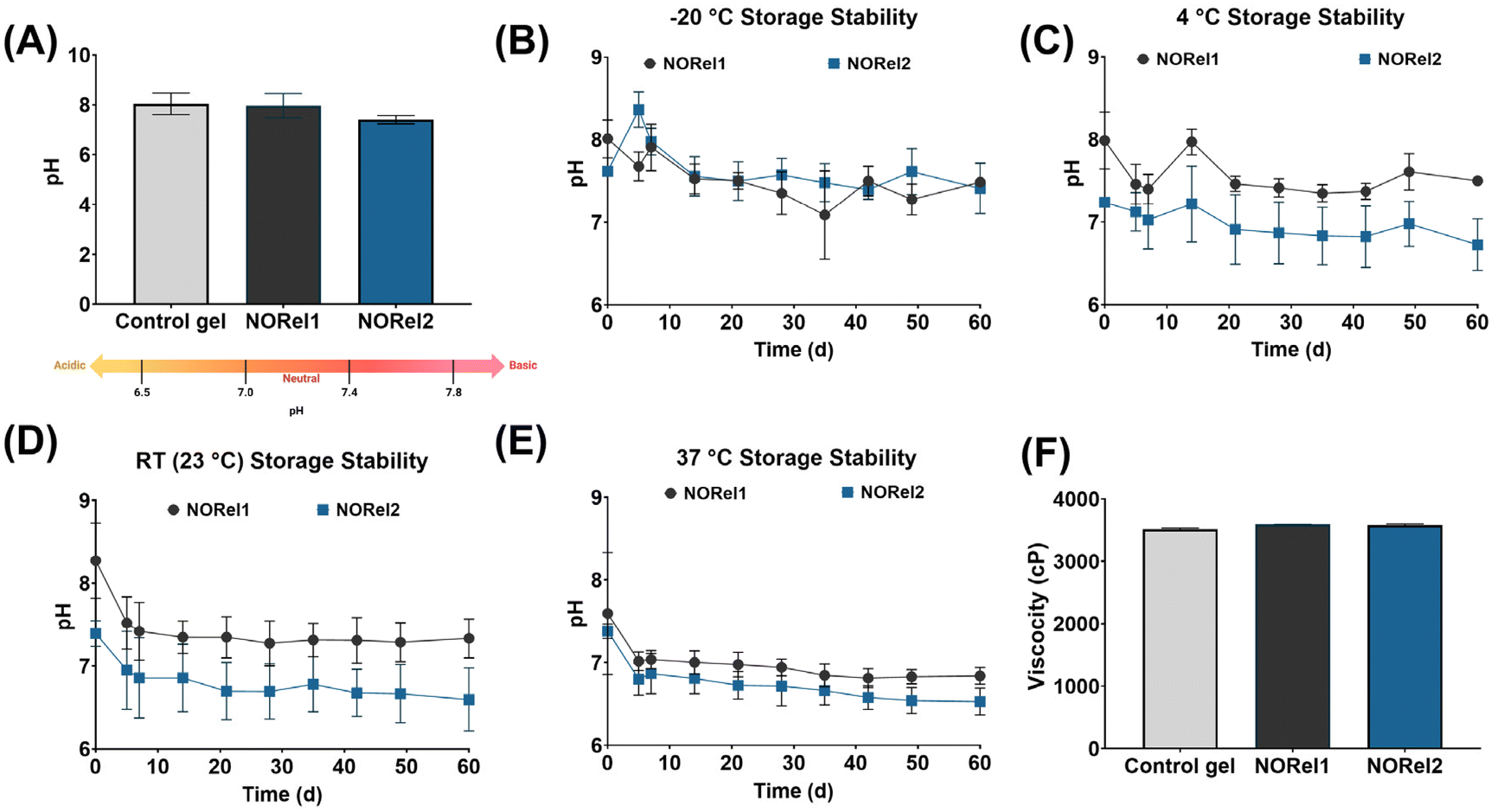 | ||
| Fig. 2 (A) pH analysis of NORel hand sanitizer gel. pH stability of NORel gels at (B) −20 °C freezer (C) 4 °C fridge (D) room temperature (23 °C) and (E) 37 °C (physiological temperature), up to 60 d of storage. (F) Average viscosity measurements of NORel and control gels. Data represents mean ± SD, n = 3. | ||
To determine the optimal storage conditions for preserving the stability of carbomer and SNAP, NORel1, and NORel2 gels were subjected to storage at various temperatures (−20 °C, 4 °C, room temperature (∼23 °C), and 37 °C) for a duration of 60 d (Fig. 2C–E). The pH measurements presented revealed that all prepared formulations maintained a pH within the safe and skin-friendly range of 6.5–8.5 throughout the storage period at all temperatures, demonstrating their suitability for skin application.29–31 This pH range ensures that NORel is well-tolerated by the skin, minimizing the risk of irritation or dryness. Moreover, it's worth noting that hand sanitizers are most effective in killing germs within this pH range of 6.5–8.5, and significant deviations from it can compromise their efficacy. In summary, these findings affirm the long-term suitability of NORel formulations for skin use for at least 60 d.
3.2. Viscosity of gels
Viscosity is a key parameter in assessing the overall quality and performance of a hand sanitizer.32 Proper viscosity ensures that the sanitizer can be easily applied to the hands without being too runny or too thick, which could affect user experience and compliance. Hand sanitizers with the right viscosity are easier to dispense and spread evenly across the hands. This is crucial for ensuring complete coverage and effective sanitization, especially in healthcare settings and during disease outbreaks. The rheological properties and viscosity characteristics in response to the addition of SNAP were assessed using a Brookfield Viscometer to investigate the impact of gel components on NORel's rheological properties. All hand sanitizer gels (0% Control, NORel1, and NORel2) (Fig. S1†), regardless of the amount of SNAP added, initially exhibited a viscosity of approximately 3500 cP at 1 rpm speed (Fig. S2†). However, as the speed of the rheometer increased, the viscosity dropped significantly to below 100 cP, reaching as low as 36 cP at 100 rpm. The onset of this viscosity reduction occurred at 12 rpm, indicating a shear-thinning property where viscosity rapidly decreases under shear force. The initial viscosity at 1 rpm showed slight variations depending on the SNAP percentage in the formulation. Specifically, control, NORel1, and NORel2 had initial viscosities of 3516 ± 20, 3596 ± 1, and 3583 ± 20 cP, respectively, suggesting potential viscosity effects attributed to SNAP addition (Fig. 2F). The addition of SNAP increased viscosity, likely due to its carboxylic acid group. Overall, the viscosity measurements obtained in this study are consistent with earlier published findings that utilized carbomer and triethylamine in the creation of phenolic gels, which reported viscosity measurements within the range of 3405–4604 cP.33In this study, carbomer, a cross-linked polyacrylic acid polymer, was used as a thickening agent to create a three-dimensional network that can expand upon hydration in aqueous solutions.34 However, its maximum viscosity occurs at a pH of approximately 6.5–7.5 and decreases at a pH ≥ 9, rendering it unstable under alkaline conditions.35,36 To address this, triethylamine was employed as a neutralizing agent to stabilize carbomers in NORel gels. Hydrogen bonds, which are attractive forces between molecules, have a profound impact on the gel's properties. In this case, glycerin played a crucial role by facilitating the formation of hydrogen bonds among the gel's molecules, thereby enhancing its structural integrity and viscosity. Additionally, the SNAP moiety, with its carboxylic acid and secondary amine groups, played a dual role in the gelation process. Firstly, it contributed to the neutralization of carboxylic acids, akin to TEA. Secondly, SNAP participated in the formation of hydrogen bonds, similar to glycerin.37 These combined effects resulted in an increased quantity of hydrogen bonds within the gel. Consequently, when higher concentrations of SNAP were introduced into the gel, the number of hydrogen bonds formed increased, leading to augmented gel strength and viscosity. This intricate interplay of carbomer, TEA, glycerin, and SNAP collectively contributed to the stability, consistency, and overall performance of the gel. These findings align with previous research on Pluronic F127 where the viscosity of gel increased due to the addition of RSNO donor.38 With these adjustable physical properties, further investigations into NO release were pursued.
3.3. Nitric oxide release kinetics
In the pursuit of robust infection control measures, the role of NO has gained considerable attention for its potential. Nonetheless, the intrinsic instability of NO donors in solution has presented a substantial hurdle, limiting the ability to achieve sustained and efficacious levels of NO. To overcome this, inventive strategies have emerged, centered on the use of polymeric matrices, that can stabilize and prolong the release of NO from materials.39 These strategies offer a protective shield to NO donors, preserving NO and prolonging its release characteristics that enable consistent, regulated, and highly efficient release of NO. For this reason, a robust NO donor SNAP was used to formulate the hand sanitizer gel. In the presence of heat, light, and metal ions, the thiol bond is cleaved and can readily release NO from the donor (Fig. 3A). The NO release from NORel hand sanitizer gel was characterized using a chemiluminescence method (Fig. 3B). The study aimed to assess the adaptability of NORel gel by varying the SNAP content and its impact on NO release under physiologically relevant conditions. Specifically, NORel1 and NORel2 gels were subjected to NO release testing within an amber sample cell maintained at a temperature of 37 °C. The results demonstrated that, on average, NORel2 gel exhibited the highest NO release rates during the entire 24 h study period. The concentration of SNAP within the gel significantly influenced the general trends in NO release. Notably, at 0, 8, and 24 h, NORel1 gel released 0.78 ± 0.10, 0.30 ± 0.05, and 0.21 ± 0.07 × 10⁻¹⁰ mol min−1 mg−1 of NO, respectively, while NORel2 released 1.18 ± 0.29, 0.80 ± 0.37, and 0.53 ± 0.05 × 10⁻¹⁰ mol min−1 mg−1 of NO, respectively (Fig. 3B). Over time, the levels of NO declined as SNAP decomposed in the gel. Prior studies have examined NO release from hydrogels composed of Pluronic F127, alginate, gelatin, and similar materials for antibacterial purposes that showed comparable levels of NO from hydrogels over 24 h.38,40 However, the combination of traditional hand sanitizer components with NO represents a novel approach that has not been explored before. In summary, both NORel1 and NORel2 gels have demonstrated their ability to release NO under physiological conditions, indicating the increase in NO release by altering the NO donor amount within the gel. | ||
| Fig. 3 (A) Chemical structure of NO donor S-nitroso-N-acteylpenicillamine (SNAP) non-covalently dispersed with other traditional hand sanitizer ingredients to generate NO-releasing hand sanitizer. NO donor can be catalyzed via heat, light or metal ions to achieve real-time NO release. (B) Nitric oxide release analysis from NORel gels tested using chemiluminescence method at physiological conditions (37 °C). (C) UV-vis spectra of SNAP recorded at 250–600 nm wavelength at various concentrations in PBS–EDTA. Storage stability of NORel after 28 d of storage at (D) −20 °C, (E) 4 °C, (F) room temperature (23 °C) and (G) 37 °C. The retention of NO in NORel gels was measured using the absorbance peak of SNAP at 340 nm using UV-vis spectroscopy normalized to day 0. (H) Cytocompatibility of NORel tested towards 3T3 mouse fibroblast cells relative to cell control in a 24 h cell viability assay using MTT cell viability kit. All data are presented as mean ± SD (n ≥ 3). | ||
These outcomes validate the feasibility and effectiveness of NORel hand sanitizer gel as a proof-of-concept. Importantly, it should be noted that the duration, quantity, and extent of NO release can be further tailored to specific situational needs by changing the NO donor concentration, varying the type of NO donor, or even changing the gel composition. The enhanced NO release exhibited by NORel gels, in combination with other antimicrobial agents like ethanol and tea tree oil, holds therapeutic potential, particularly in combatting the proliferation of opportunistic pathogens. This innovative approach could have significant implications in the field of infection control and prevention.
3.4. SNAP stability in NORel gel
The stability of SNAP in alcohol-based formulations, in conjunction with carbomer, under various thermal conditions is not well understood. This lack of understanding could potentially affect the shelf-life of NORel gels. From a commercial perspective, it is crucial to determine the optimal storage conditions for these gels, considering potential clinical applications. To investigate the stability, NORel1 and NORel2 gels were subjected to different temperatures (−20 °C, 4 °C, room temperature (∼23 °C), and 37 °C) to simulate real-world storage conditions and the percent of NO (%NO) remaining in the gels was monitored over 28 d, relative to NO donor SNAP present in fresh gels on day 0. The absorption spectra of SNAP at 340 nm wavelength were used to analyze SNAP concentrations in gel formulations by dissolving 100 mg of gel in 1 mL of 10 mM PBS (pH 7.4) with 100 μM EDTA. A standard curve of SNAP in PBS–EDTA was plotted to quantify the amount of SNAP in the gel (Fig. 3C). The molar extinction coefficient of SNAP in PBS–EDTA at room temperature was found to be 1025 M−1 cm−1 at 340 nm.The results from the study (Tables S1 and S2†) indicated high stability at −20 °C, with >90% SNAP remaining in both gels after 28 d of storage (Fig. 3D). At 4 °C, NORel1 gel remained stable (97.14%), while NORel2 gel had ∼65% SNAP remaining after 4 weeks of storage (Fig. 3E). Both gels showed reduced stability at higher temperatures, with NORel1 and NORel2 gels having 58.54% and 27.60% SNAP remaining at room temperature (Fig. 3F) and 44.01% and 25.20% SNAP remaining at 37 °C, respectively (Fig. 3G). These findings align with prior research, suggesting that RSNOs exhibit superior stability at lower temperatures substantially better than cysteine-based NO-donors with short half-lives and limited stability even at −20 °C.41,42 RSNOs can release NO more rapidly at higher temperatures due to heat-mediated catalysis of the NO donor SNAP, particularly at physiological temperatures.22 The observed decrease in stability of NORel2 gel over 28 d of storage with a higher concentration of SNAP, can be linked to the autocatalytic decomposition of RSNOs in a concentration-dependent manner.43 It can be hypothesized that the increased SNAP content in NORel2 gel might have led to a greater self-catalytic effect within the gel, subsequently accelerating the catalytic process from SNAP in NORel2 gel compared to the lower SNAP content in NORel1 gel. Although this phenomenon is less pronounced within the initial 24 h testing period with NOA, it becomes increasingly noticeable during prolonged testing. Overall, the findings from this study highlight that NORel gels exhibit optimal stability and potency when stored at lower temperatures, which is advantageous for their potential clinical applications. It is worth highlighting that the incorporation of NO into conventional hand sanitizer agents offers significant advantages in augmenting the antimicrobial effectiveness of the gel. This gel holds great potential for use in clinical settings to combat infectious pathogens, including antibiotic-resistant strains that can lead to severe infections. The SNAP degradation was more pronounced during storage at room temperature and 37 °C compared to the refrigerated temperatures, which may result in sub-optimal NO release after long-term storage. For potential commercial application of the NORel gel in the future, additional work would be required to enhance the storage stability of the gel at room temperature or other elevated temperature conditions.
3.5. Biocompatibility of gel
NO-releasing hand sanitizer gels were further evaluated for biocompatibility using an indirect contact cytocompatibility evaluation model with NIH/3T3 mouse fibroblast cells using our previously reported method.38 The NIH/3T3 mouse fibroblast cell line is a standardized cell line often used for evaluation of the biocompatibility of medical devices in accordance with ISO 10993-5 standards.44 In the present studies, the gel sanitizers contained several active ingredients – SNAP, tea tree oil, glycerin, carbomer, triethylamine, and ethanol, which may trigger a secondary cytotoxic effect from exposure. Therefore, it was critical to evaluate the relative cytotoxicity of these formulations against both untreated and commercial alternatives for comparison purposes.As shown in Fig. 3H, no statistically significant difference was observed in cellular viability between the control gel formulation and the commercial alcohol-based gel alternative, suggesting acceptable lack of cytotoxicity of the gel in absence of NO release. In contrast, both the NORel1 and NORel2 formulations demonstrated an upward trend in cellular proliferation with respect to the alcohol alternative (p < 0.05), while the NORel1 gel formulation also demonstrated improved viability compared to the gel control. The increase in viability with the NORel1 and NORel2 gel formulations is likely attributable to presence of NO donor SNAP, which has previously shown a proliferative effect for fibroblast cells in several previous NO-releasing materials.45–47 By promoting fibroblast proliferation, the NO-releasing gels offer a key benefit over the control and commercial gels in performing better against bacteria while concurrently developing cytoprotective effects ideal for topical application.
3.6. Antibacterial efficacy of NORel hand sanitizer
The broad-spectrum antimicrobial efficacy of NORel gel was assessed against four microbes frequently linked to hospital-acquired infections: S. aureus, E. coli, C. albicans, and a clinical isolate of methicillin-resistant S. aureus (MRSA) using microbial-killing assay. The antimicrobial effectiveness test demonstrated that the prepared formulations of NORel gels had significant antimicrobial activity against Gram-positive, and Gram-negative bacteria and C. albicans yeast (Fig. 4A–D). The killing efficiencies of each sanitizer gel are reported in Table S3.† The control gel, without SNAP, individually led to >70% reduction in pathogen viability compared to untreated control, resulting in log reductions of 0.81 ± 0.04, 1.05 ± 0.12, 1.25 ± 0.10, and 0.56 ± 0.12 against S. aureus, E. coli, MRSA, and C. albicans, respectively. This can be attributed to the antimicrobial efficacy of the ethanol and tea tree oil in the gel. The addition of SNAP to gel significantly enhanced its antimicrobial potential, with NORel1 gel achieving log reductions of 1.97 ± 0.04, 1.98 ± 0.09, 1.71 ± 0.04, and 1.04 ± 0.35 against all microbial strains under evaluation.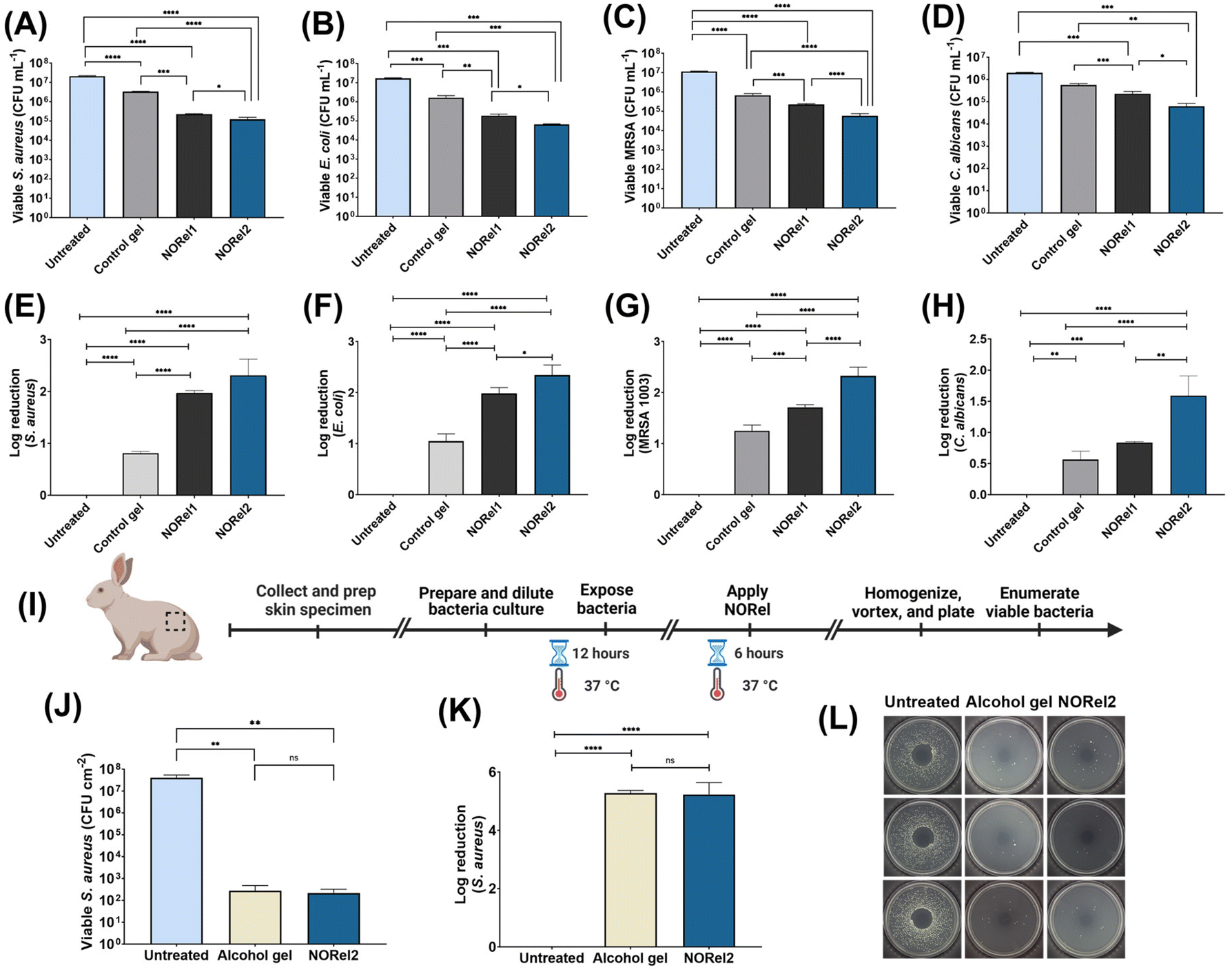 | ||
| Fig. 4 Antimicrobial activity of NORel hand sanitizer gel calculated as log of colony forming unit (CFU) mL−1 against (A) S. aureus, (B) E. coli, (C) methicillin-resistant S. aureus (MRSA), and (D) C. albicans after 6 h exposure. Corresponding log reduction in microbial viability after expsoure to NORel and controls for (E) S. aureus, (F) E. coli, (G) methicillin-resistant S. aureus (MRSA), and (H) C. albicans, plotted with respect to untreated bacteria control. (I) Experimental design used to evaluate the antimicrobial activity of NORel vs. control alcohol gel using an ex vivo rabbit skin model. (J) Ex vivo disinfection of an infected rabbit skin using commercial alcohol gel and NORel. (K) Corresponding log reduction in bacterial viability after exposure to S. aureus bacteria. (L) Representative images of LB agar plates with viable S. aureus bacteria CFU after 6 h of exposure to NORel and commercial alcohol gel in the ex vivo rabbit disinfection model. All data are represented as mean ± SD (n ≥ 3). | ||
Notably, NORel2 gel, containing higher amounts of SNAP, exhibited the highest efficacy among all formulations, achieving log reductions of 2.30 ± 0.27, 2.34 ± 0.17, 2.32 ± 0.15, and 1.59 ± 0.28 against S. aureus, E. coli, MRSA, and C. albicans, respectively, compared to untreated control (Fig. 4E–H). These findings are consistent with the NO release levels observed in Fig. 3B, where higher levels of NO release correlated positively with the amount of SNAP incorporated into the gel. N-Acetylpenicillamine, the synthetic precursor to SNAP, does not produce any antibacterial effect, which has led to the conclusion in literature that the bactericidal effect of SNAP is solely a result of the NO released from the gel.48 While numerous antibiotics focus on specific pathways within microorganisms, NO utilizes a multifaceted approach to deactivate microorganisms. It disrupts essential proteins, damages DNA, and affects cell membranes, among other actions.49–52 This comprehensive strategy enables NO to exhibit broad-spectrum antimicrobial properties, effective against various bacteria, fungi, and viruses. This characteristic of NO has been consistently proven in prior research, and the outcomes observed with this innovative material are in line with findings from previously published studies.21,53
3.7. Disinfection efficacy of NORel gel using ex vivo infected rabbit skin model
The antimicrobial effectiveness of NORel gel was further assessed through an ex vivo animal study using rabbit skin specimens. The explanted skin specimens from euthanized rabbits were exposed to S. aureus bacteria for 12 h to replicate real-world conditions, allowing bacteria to adhere to the skin. Subsequently, NORel2 gel, chosen due to its superior NO release and in vitro antimicrobial performance compared to NORel1, was applied and left to incubate on the skin for 6 h to enable NO action against the bacteria (Fig. 4I). In this ex vivo study, NORel2 gel's performance was compared to that of a commercial alcohol-based gel, which served as a secondary test group. Both NORel2 and the alcohol-based gel were assessed against a bacterial control group, which consisted of infected skin samples with no active antimicrobial treatment. Results from this investigation revealed that NORel2 gel exhibited similar antimicrobial effects to the commercial alcohol-based gel containing 62% ethyl alcohol as its active component. Notably, both the commercial alcohol and NORel2 gel demonstrated exceptional bacterial reduction, with >99.99% bacterial eradication (Fig. 4J) and reductions of 5.28 ± 0.07 and 5.23 ± 0.33 log in bacterial viability compared to the untreated control, respectively (Fig. 4K and L). Similar animal models have been employed in previous studies to demonstrate the antimicrobial potential of gels on skin.54 Various NO-releasing gels have been developed, serving as versatile platforms for angiogenesis, antimicrobial purposes, wound healing therapies, and formulations in the form of topical lotions.55,56 As an endogenous regulatory molecule, NO serves multiple advantageous roles, including providing localized immunity against infectious pathogens.Until recently, the progress of topical NO treatments was impeded by difficulties in securely storing and effectively delivering NO to infection or inflammation sites. Fortunately, with the advancement of stable NO donors, it has become possible to achieve long-term NO release for various biomedical applications. This study's results underscore that NORel gel preserves its antimicrobial effectiveness when applied to the skin, expanding its applicability beyond liquid-based solutions.
3.8. Persistent activity of NORel gel using ex vivo rabbit skin model
The effectiveness of hand sanitizer gels largely relies on their active components, typically alcohol-based compounds like ethanol. These ingredients function by disrupting bacterial cell membranes, effectively eliminating the bacteria. Although alcohol-based hand sanitizers are successful in eliminating many types of bacteria and viruses, they face certain limitations, including limited residual activity, a short lifespan upon application, and the potential for skin irritation and dryness with excessive usage. To address these drawbacks, this study introduced a combination of ethanol with other antimicrobial agents, such as NO and tea tree oil, as well as moisturizers and skin conditioners. This strategy aimed to prolong the duration of antimicrobial action and enhance the overall effectiveness of the gel while minimizing skin dryness and irritation (Fig. 5A). | ||
| Fig. 5 (A) Schematic illustration of difference between standard alcohol containing sanitizer vs. NO-releasing hand sanitizer gel (NORel). Alcohol containing gels with no secondary antimicrobial action evaporate quickly and lack in continuous activity overtime. NO releasing gel with other active ingredients can effectively kill and also exhibit persistent antimicrobial activity over an extended period of time. (B) Experimental design used to evaluate the persistence activity of NORel vs. control alcohol gel using a rabbit skin model with 2 h exposure to the test gel. (C) Validation of long-term effectiveness of NORel on rabbit skin specimen tested against S. aureus bacteria. (D) Log reduction in bacteria viability 2 h after gel exposure. (E) Schematic illustration of mechanism by which NO denatures bacteria. All data is presented as mean ± SD for n ≥ 3. | ||
To assess the endurance and sustained effects of NORel gel in comparison to commercially available alcohol-based sanitizer gel, a similar infection model employing rabbit skin specimens was utilized as described in Section 4.7. However, in this scenario, the sanitizers were applied to the pristine skin specimens first and allowed to incubate at 37 °C for 2 h to evaluate the persistence of the gel's antimicrobial activity post-application (Fig. 5B). Following the incubation period, S. aureus bacteria were introduced and allowed to incubate with both NORel and a commercial alcohol-based gel for an additional 6 h at 37 °C. Viable bacteria remaining on the skin were then extracted and quantified using a plate counting method.
As anticipated, the commercial alcohol-based gel had minimal effect on bacterial viability and proved ineffective in eradicating the bacteria 2 h after application (Fig. 5C). The results revealed that the alcohol gel only resulted in a modest 0.33 ± 0.03 log reduction in bacterial viability (p > 0.05). This outcome can be a result of alcohol's evaporation from the skin samples within 2 h of application. Consequently, when the skin samples were exposed to bacteria, there was no antimicrobial agent present to impede bacterial growth. Conversely, NORel2 gel, enriched with tea tree oil, ethanol, glycerin, and carbomer, displayed significant antimicrobial activity even hours following application. Notably, NORel2 gel demonstrated a 1.66 ± 0.22 log reduction in viable S. aureus bacteria compared to the untreated control and a 1.33 ± 0.39 log reduction compared to the commercial alcohol-based gel (Fig. 5D). The persistent activity of NORel hand sanitizer can primarily be attributed to the continuous release of NO from the NORel gel. The gel-like consistency of NORel allows it to create a protective layer on the skin, enabling the release of NO at physiological temperatures. As a result, it maintains a microbe-free environment for a more extended period compared to alcohol gel, which has a limited duration of activity. It is expected that NORel's persistent activity will reduce the frequency of gel applications, thereby mitigating issues such as dryness and irritation often caused by repeated exposure to alcohol on the skin.
Beyond its antimicrobial action (Fig. 5E), NO's vasodilatory, anti-inflammatory, and wound-healing properties can offer biocompatible and therapeutic benefits. This persistence of NORel is particularly valuable in healthcare settings and other high-risk environments where continuous protection against pathogens is crucial, making it a superior alternative to existing alcohol-based gels. In summary, this study underscores the potential of synergizing NO with other antimicrobial agents such as ethanol and tea tree oil to create an innovative and potent gel. This approach effectively addresses the multifaceted challenges posed by antibiotic-resistant strains and provides enduring protection against a diverse array of pathogens. These findings constitute a valuable contribution to the ongoing endeavors aimed at combating antibiotic resistance and enhancing infection control within healthcare settings and beyond.
4. Conclusions
While alcohol-based formulations have become ubiquitous in our daily lives, the growing demand for hand sanitizers has raised pertinent questions regarding their effectiveness and potential drawbacks. Alcohol-based sanitizers can offer immediate protection; however, they often fall short in providing prolonged defense, necessitating frequent reapplication. Additionally, repeated exposure to alcohol can lead to skin issues like dryness and irritation. To address these concerns, this study formulated a nitric oxide-releasing (NORel) hand sanitizer gel using antimicrobial agents (NO donor, ethanol, and tea tree oil) and moisturizers (glycerin). The NORel gel maintained a neutral pH (∼7) and stability under various storage conditions for at least 60 d, indicating the effectiveness of its antimicrobial and moisturizing ingredients. The viscosity of the gel was found to be around 3500 cP matching the standards for sanitizer gels. Chemiluminescence tests demonstrated the adjustable release of NO from the gel by varying the NO donor concentration. NORel1 and NORel2 gels released NO at physiologically relevant levels for at least 24 h and all gel variants exhibited biocompatibility, confirming their safe nature. NORel gels effectively eliminated bacteria and fungi, including antibiotic-resistant MRSA, with over 97% efficiency compared to untreated controls. Their effectiveness was also confirmed on an infected rabbit skin model that showed more than 99.99% efficacy against S. aureus bacteria. Furthermore, NORel gels demonstrated persistent antimicrobial effects even 2 h after application, in contrast to commercial alcohol-based gels, which showed no significant impact due to their short-lived action and rapid evaporation. This research presents a biocompatible NO-releasing gel with superior antimicrobial properties compared to alcohol-based sanitizers, offering an effective hand hygiene solution, especially in high-risk environments. The NORel gel is expected to provide extended protection, eliminate antibiotic-resistant pathogens, and mitigate skin-related side effects.Data availability
The data supporting this article have been included as part of the ESI.† Data will be made available upon a reasonable request.Conflicts of interest
Dr Elizabeth J. Brisbois is a co-founder of Nytricx, Inc., a startup company which is involved in exploring possibilities of using nitric oxide-releasing materials for medical applications.Acknowledgements
This work was supported by the National Institute of Health through the funds received under NIH R01HL151473. Graphics were created by the authors using the BioRender.com software.References
- M. Haque, M. Sartelli, J. McKimm and M. Abu Bakar, Infect. Drug Resist., 2018, 11, 2321–2333 CrossRef PubMed.
- Preventing Health Care-Associated Infections, ed. A. S. Collins, Agency for Healthcare Research and Quality, Rockville (MD), 2008 Search PubMed.
- H. Saito, K. Inoue, J. Ditai, B. Wanume, J. Abeso, J. Balyejussa and A. Weeks, Antimicrob. Resist. Infect. Control, 2017, 6, 1–12 Search PubMed.
- F. Tusabe, J. Nanyondo, M. J. Lozier, M. Kesande, O. Tumuhairwe, M. Watsisi, F. Twinomugisha, A. Medley, J. Mutoro and M. Lamorde, Am. J. Trop. Med. Hyg., 2023, 109, 191 Search PubMed.
- E. L. Larson, J. Cimiotti, J. Haas, M. Parides, M. Nesin, P. Della-Latta and L. Saiman, Arch. Pediatr. Adolesc. Med., 2005, 159, 377–383 Search PubMed.
- E. Tikhomirov, Chemioterapia, 1987, 6, 148–151 Search PubMed.
- D. Pittet, B. Allegranzi, H. Sax, S. Dharan, C. L. Pessoa-Silva, L. Donaldson and J. M. Boyce, Lancet Infect. Dis., 2006, 6, 641–652 CrossRef PubMed.
- A. P. Golin, D. Choi and A. Ghahary, Am. J. Infect. Control, 2020, 48, 1062–1067 Search PubMed.
- S. Huffer, M. E. Clark, J. C. Ning, H. W. Blanch and D. S. Clark, Appl. Environ. Microbiol., 2011, 77, 6400–6408 CrossRef CAS PubMed.
- D. W. Lachenmeier, J. Occup. Med. Toxicol., 2008, 3, 26 CrossRef PubMed.
- T. Saha, P. Khadka and S. C. Das, GERMS, 2021, 11, 408 CrossRef CAS PubMed.
- Q. Lin, J. Y. Lim, K. Xue, P. Y. M. Yew, C. Owh, P. L. Chee and X. J. Loh, VIEW, 2020, 1, e16 Search PubMed.
- N. A. Gold, T. M. Mirza and U. Avva, Alcohol Sanitizer, In: StatPearls [Internet], StatPearls Publishing, Treasure Island (FL), 2023.
- V. M. Jain, G. N. Karibasappa, A. S. Dodamani, V. K. Prashanth and G. V. Mali, Dent. Res. J., 2016, 13, 424 CrossRef PubMed.
- D. Pirgal, J. K. S. Javalkar, N. Suma, S. Murthy, S. Guha and L. Gayathri, J. Pharm. Negat. Results, 2022, 3951–3960 CAS.
- C. Villa and E. Russo, Materials, 2021, 14, 1577 CrossRef CAS PubMed.
- S. Kumar, R. K. Singh and T. R. Bhardwaj, Biomed. Pharmacother., 2017, 85, 182–201 CrossRef CAS PubMed.
- M. R. Garren, M. Ashcraft, Y. Qian, M. Douglass, E. J. Brisbois and H. Handa, Appl. Mater. Today, 2021, 22, 100887 Search PubMed.
- W. H. Poh and S. A. Rice, Molecules, 2022, 27, 674 Search PubMed.
- B. J. Privett, A. D. Broadnax, S. J. Bauman, D. A. Riccio and M. H. Schoenfisch, Nitric Oxide, 2012, 26, 169–173 CrossRef CAS PubMed.
- M. K. Chug and E. J. Brisbois, ACS Mater. Au, 2022, 2, 525–551 CrossRef CAS PubMed.
- I. Chipinda and R. H. Simoyi, J. Phys. Chem. B, 2006, 110, 5052–5061 CrossRef CAS PubMed.
- K. A. Broniowska, A. R. Diers and N. Hogg, Biochim. Biophys. Acta, Gen. Subj., 2013, 1830, 3173–3181 CrossRef CAS PubMed.
- C. Opländer, A. Römer, A. Paunel-Görgülü, T. Fritsch, E. Van Faassen, M. Mürtz, A. Bozkurt, G. Grieb, P. Fuchs and N. Pallua, Clin. Pharmacol. Ther., 2012, 91, 1074–1082 CrossRef PubMed.
- H. Baldwin, D. Blanco, C. McKeever, N. Paz, Y. N. Vasquez, J. Quiring, C. Enloe, E. De León and N. Stasko, J. Clin. Aesthetic Dermatol., 2016, 9, 12 Search PubMed.
- F. Fallica, C. Leonardi, V. Toscano, D. Santonocito, P. Leonardi and C. Puglia, Pharmaceutics, 2021, 13, 571 Search PubMed.
- J. Boyce and D. Pittet, Antimicrob. Resist. Infect. Control, 2024, 13, 49 Search PubMed.
- S. M. Ali and G. Yosipovitch, Acta Derm.-Venereol., 2013, 93, 261–267 CrossRef PubMed.
- S. M. Ali and G. Yosipovitch, Acta Derm.-Venereol., 2013, 93, 261–267 CrossRef PubMed.
- S. Surini, N. Amirtha and D. Lestari, Int. J. Appl. Pharm., 2018, 10, 216 CrossRef CAS.
- S. A. Al-Suwayeh, E. I. Taha, F. M. Al-Qahtani, M. O. Ahmed and M. M. Badran, Sci. World J., 2014, 2014, 127495 Search PubMed.
- Y. Ma, J. Yi, J. Ma, H. Yu, L. Luo, W. Wu, L. Jin, Q. Yang, T. Lou, D. Sun and M. Cao, Toxics, 2023, 11, 687 CrossRef CAS PubMed.
- S. Nurman, R. Yulia, Irmayanti, E. Noor and T. Candra Sunarti, Sci. Pharm., 2019, 87, 32 CrossRef CAS.
- R. Hamed, T. Al Baraghthi, A. Z. Alkilani and R. Abu-Huwaij, J. Pharm. Innovation, 2016, 11, 339–351 CrossRef.
- S. Sommatis, M. C. Capillo, C. Maccario, R. Rauso, E. D'Este, M. Herrera, M. Castiglioni, R. Mocchi and N. Zerbinati, Gels, 2023, 9, 108 CrossRef CAS PubMed.
- M. T. Islam, N. Rodríguez-Hornedo, S. Ciotti and C. Ackermann, Pharm. Res., 2004, 21, 1192–1199 CrossRef CAS PubMed.
- A. N. Lyapunov, E. P. Bezuglaya, N. A. Lyapunov and I. A. Kirilyuk, Pharm. Chem. J., 2015, 49, 639–644 CrossRef CAS.
- L. M. Estes Bright, M. R. S. Garren, M. Ashcraft, A. Kumar, H. Husain, E. J. Brisbois and H. Handa, ACS Appl. Mater. Interfaces, 2022, 14, 21916–21930 CrossRef CAS PubMed.
- M. K. Chug, N. Crutchfield, M. Garren, H. Handa and E. J. Brisbois, ACS Appl. Bio Mater., 2024, 7, 2993–3004 CrossRef CAS PubMed.
- S. Ghalei, M. Douglass and H. Handa, ACS Biomater. Sci. Eng., 2021, 8, 273–283 CrossRef PubMed.
- W. R. Mathews and S. W. Kerr, J. Pharmacol. Exp. Ther., 1993, 267, 1529–1537 CrossRef CAS PubMed.
- R. Kumar, H. Massoumi, M. K. Chug and E. J. Brisbois, ACS Appl. Mater. Interfaces, 2021, 13, 25813–25824 CrossRef CAS PubMed.
- L. Grossi, P. C. Montevecchi and S. Strazzari, J. Am. Chem. Soc., 2001, 123, 4853–4854 CrossRef CAS PubMed.
- International Organization for Standardization (ISO), Biological evaluation of medical devices – Part 5: Tests for in vitro cytotoxicity, 3rd edn, ISO 10993-5:2009, 2009, https://www.iso.org/standard/36406.html.
- M. Garren, P. Maffe, A. Melvin, L. Griffin, S. Wilson, M. Douglass, M. Reynolds and H. Handa, ACS Appl. Mater. Interfaces, 2021, 13, 56931–56943 CrossRef CAS PubMed.
- M. Douglass, S. Hopkins, M. K. Chug, G. Kim, M. R. Garren, M. Ashcraft, D. T. Nguyen, N. Tayag, H. Handa and E. J. Brisbois, ACS Appl. Mater. Interfaces, 2021, 13, 52425–52434 CrossRef CAS PubMed.
- S. Ghalei, S. Hopkins, M. Douglass, M. Garren, A. Mondal and H. Handa, J. Colloid Interface Sci., 2021, 590, 277–289 CrossRef CAS PubMed.
- Y. Wo, Z. Li, A. Colletta, J. Wu, C. Xi, A. J. Matzger, E. J. Brisbois, R. H. Bartlett and M. E. Meyerhoff, Composites, Part B, 2017, 121, 23–33 CrossRef CAS PubMed.
- D. A. Wink and J. B. Mitchell, Free Radical Biol. Med., 1998, 25, 434–456 CrossRef CAS PubMed.
- F. C. Fang, J. Clin. Invest., 1997, 99, 2818–2825 CrossRef CAS PubMed.
- F. C. Fang, Nat. Rev. Microbiol., 2004, 2, 820 CrossRef CAS PubMed.
- D. O. Schairer, J. S. Chouake, J. D. Nosanchuk and A. J. Friedman, Virulence, 2012, 3, 271–279 CrossRef PubMed.
- L. M. Estes Bright, M. K. Chug, S. Thompson, M. Brooks, E. J. Brisbois and H. Handa, J. Biomed. Mater. Res., Part B, 2024, 112, e35442 CrossRef CAS PubMed.
- B. More, S. Sakharwade, S. Tembhurne and D. Sakarkar, Int. J. Toxicol. Appl. Pharmacol., 2013, 3, 10–13 Search PubMed.
- Y. Liu, S. Guo, S. Wei, H. Wang, Y. Liu, A. V. Moscoso, Z. Ribkovskaia, T. Lazarova, S. Riesinger and D. P. Orgill, Plast. Reconstr. Regener. Surg., 2022, 24–33 CAS.
- J. P. Yapor, J. L. Gordon, C. N. Henderson and M. M. Reynolds, RSC Adv., 2019, 9, 21873–21880 RSC.
Footnote |
| † Electronic supplementary information (ESI) available. See DOI: https://doi.org/10.1039/d5bm00359h |
| This journal is © The Royal Society of Chemistry 2025 |