Rounding up Rh nanoparticles for ultraviolet plasmonic sensing†
Yikai
Xu

Key Laboratory for Advanced Materials and Feringa Nobel Prize Scientist Joint Research Center, Frontiers Science Center for Materiobiology and Dynamic Chemistry, School of Chemistry and Molecular Engineering, East China University of Science and Technology, Shanghai 200237, P. R. China. E-mail: yikaixu@ecust.edu.cn
Abstract
This article highlights the recent work of D. M. Arboleda and V. Amendola et al. (Nanoscale Horiz., 2025, 10, 336–348, https://doi.org/10.1039/D4NH00449C) on the synthesis of rhodium nanospheres for ultraviolet and visible plasmonics.
Localized surface plasmon resonance (LSPR) occurs due to surface-localized, oscillating hot electrons, generated by the interaction between light and plasmonic nanomaterials. Currently, the most widely used plasmonic materials are Au and Ag, which exhibit LSPR in the visible and near infrared region.1 If ultraviolet plasmonic materials were standardized, this would open up new possibilities in fields including LSPR catalysis and surface-enhanced Raman spectroscopy (SERS).
Recent work by researchers from the University of Padova reported the synthesis of spherical Rh colloidal nanoparticles which produce well-defined LSPR in the ultraviolet region. Obtaining spherical Rh nanoparticles is challenging due to the preferred FCC crystal structure of Rh. Previous methods for the synthesis of spherical Rh nanoparticles were limited to the creation of particles with diameters within 7 nm, which only showed a weak plasmonic response. As shown here in Fig. 1A–C, the researchers used laser ablation to create spherical Rh nanoparticles with diameters ranging between 20–45 nm. Moreover, since this approach of generating nanoparticles did not involve the use of any chemical ligands, this left the surface of the product particles accessible for the adsorption of ligand molecules which could be used to enhance colloidal stability or to provide additional functionality. To demonstrate this, the researchers functionalized the surface of the Rh nanoparticles with mercaptopropionic acid which interacted with metal ions, such as Cd(II), to induce agglomeration and in turn a change in the LSPR properties of the Rh colloid (Fig. 1D–E). As shown in Fig. 1F, the extent of agglomeration depended on the concentration of the metal ion, which allowed quantitative analysis of the concentration of metal ions in solution to be achieved. The surface accessibility of the Rh nanoparticles is also useful in facilitating the adsorption of analyte molecules for SERS analysis. By taking advantage of this property, the researchers demonstrated SERS detection of dyes, thiols and DNA using laser irradiation at 458 nm (Fig. 1G).
 | ||
| Fig. 1 (A) Schematic illustration of the synthesis of spherical Rh nanoparticles via laser ablation. (B) and (C) Transmission microscopy image of Rh nanoparticles ca. 45 nm in diameter and their LSPR response. (D) Schematic illustration showing mercaptopropionic (MPA) functionalized Rh particles acting as LSPR optical sensors for metal ions. (E) and (F) The optical response of MPA-Rh colloids at different Cd(II) concentrations and for different metal ions in water. (G) SERS spectra of DNA, benzenethiol (BT), nitrothiolphenol (NTP), crystal violet (CV) and rhodamine B (RB), obtained using Rh nanoparticles as the enhancing substrate. Reproduced from https://doi.org/10.1039/D4NH00449C with permission from the Royal Society of Chemistry. | ||
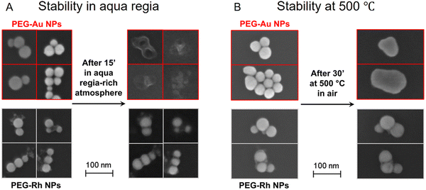
Another useful feature of the Rh nanoparticles is their improved stability in harsh conditions compared to Au and Ag nanoparticles, which for example could open new possibilities in operando SERS studies of catalytic processes that take place in pyrolysis. As shown in Fig. 2A, the Rh nanoparticles retained their structure when treated with aqua regia, while Au nanoparticles were dissolved completely. Similarly, the Rh nanoparticles were found to be stable when heated to 500 °C, while Au nanoparticles melted at this temperature (Fig. 2B).
 | ||
| Fig. 2 Scanning electron microscopy images of thiolated polyethylene glycol (PEG-SH) functionalized Rh and Au nanoparticles (NPs) before and after being treated with aqua regia (A), or 500 °C air (B). Reproduced from https://doi.org/10.1039/D4NH00449C with permission from the Royal Society of Chemistry. | ||
In summary, spherical Rh colloidal nanoparticles with diameters ranging from 20–45 nm were created via laser ablation. Compared to conventional Au and Ag plasmonic nanoparticles, the Rh nanoparticles shown in this work exhibited well-defined LSPR in the ultraviolet region and high stability under harsh experimental conditions. This unique combination of properties broadens the application of plasmonics and provides the tools for performing operando SERS studies in harsh conditions in which traditional plasmonic materials fail.
Acknowledgements
The author acknowledges the Young Scientists Fund of the National Natural Science Foundation of China (22402060) for support.References
- K. Kant, R. Beeram, Y. Cao, P. S. S. dos Santos, L. González-Cabaleiro, D. García-Lojo, H. Guo, Y. Joung, S. Kothadiya, M. Lafuente, Y. X. Leong, Y. Liu, Y. Liu, S. S. B. Moram, S. Mahasivam, S. Maniappan, D. Quesada-González, D. Raj, P. Weerathunge, X. Xia, Q. Yu, S. Abalde-Cela, R. A. Alvarez-Puebla, R. Bardhan, V. Bansal, J. Choo, L. C. C. Coelho, J. M. M. M. de Almeida, S. Gómez-Graña, M. Grzelczak, P. Herves, J. Kumar, T. Lohmueller, A. Merkoçi, J. L. Montaño-Priede, X. Y. Ling, R. Mallada, J. Pérez-Juste, M. P. Pina, S. Singamaneni, V. R. Soma, M. Sun, L. Tian, J. Wang, L. Polavarapu and I. P. Santos, Nanoscale Horiz., 2024, 9, 2085–2166 RSC.
Footnote |
| † This content was originally published on our blogs platform at https://blogs.rsc.org/nh/2024/12/18/rounding-up-rh-nanoparticles-for-ultraviolet-plasmonic-sensing/. |
| This journal is © The Royal Society of Chemistry 2025 |

