 Open Access Article
Stable self-powered X-ray detection with a low detection limit using a green halide hybrid perovskite ferroelectric crystal†
Yueying
Wang
a,
Qianwen
Guan
b,
Zeng-Kui
Zhu
*a,
Huang
Ye
b,
Hang
Li
b,
Ying
Zeng
a,
Panpan
Yu
a,
Huawei
Yang
a,
Wenhui
Wu
a and
Junhua
Luo
 *ab
*ab
aCollege of Chemistry and Materials, School of Chemical Engineering, Key Laboratory of Fluorine and Silicon for Energy Materials and Chemistry of Ministry of Education, Jiangxi Normal University Nanchang, Jiangxi 330022, P. R. China
bState Key Laboratory of Structural Chemistry Fujian Institute of Research on the Structure of Matter, Chinese Academy of Sciences, Fuzhou, Fujian 350002, P. R. China. E-mail: zkzhu@jxnu.edu.cn; jhluo@fjirsm.ac.cn
First published on 7th February 2025
Abstract
Lead halide hybrid perovskite ferroelectrics show great potential in the field of self-powered X-ray detection due to their excellent X-ray absorption, high carrier mobility, large carrier lifetime, and interesting ferroelectricity. Nonetheless, the toxicity of lead raises concerns regarding safety for humans and the environment, which limits their practical applicability. Herein, we successfully realized stable self-powered X-ray detection with a low detection limit using a lead-free halide hybrid perovskite ferroelectric crystal, [H2mdap]BiBr5 (1, H2mdap = N-methyl-1,3-diaminopropanium), driven by the switchable spontaneous polarization (Ps). Specifically, a remarkable switchable ferroelectric-photovoltaic (FE-PV) effect and excellent open-circuit photovoltage under X-ray irradiation endow 1 with a self-powered detection capability. Strikingly, the 1 detector shows a relatively high sensitivity of 79.0 μC Gy−1 cm−2 under 22 keV X-rays and achieves a low detection limit of 28 nGy s−1 at zero bias, much lower than that of the regular medical diagnosis (∼5.5 μGy s−1). Additionally, 1 also shows good operational stability, which may benefit from a stable structure and high activation energy (Ea). This study successfully demonstrates self-powered X-ray detection in 1D lead-free ferroelectric materials, which opens up new possibilities for safe and stable X-ray detection.
Introduction
The transformation of X-ray radiation into electrical signals using direct X-ray detectors holds significant value in areas such as wearable dosimeters, medical diagnostics, security checks, and scientific investigation.1–4 However, most X-ray detectors operate at high external electric fields, often leading to operational instability, high energy consumption, and bulky overall circuitry. Recently, self-powered X-ray detectors without the requirement for external electric fields have attracted interest.5–9 In conventional strategies, photogenerated carriers are separated and transmitted through p–n heterojunctions and p–i–n devices with a built-in electric field to enable self-powered detection.10–13 However, these approaches are plagued by issues such as interface engineering and complicated experimental procedures. Comparatively, as a much simpler technology, ferroelectric materials exhibit the ferroelectric-photovoltaic (FE-PV) effect directed by intrinsic spontaneous polarization (Ps), which can separate and transmit photogenerated carriers independently and make them well-suited for developing next-generation radiation devices with X-ray detection at 0 V bias.14,15 Recently, lead halide perovskite ferroelectrics have demonstrated considerable potential as X-ray detectors, thanks to their excellent X-ray absorption, large carrier mobility-lifetime (μτ) products, low fabrication cost, and interesting ferroelectricity.16–18 For instance, the (CH3OC3H9N)2CsPb2Br7 ferroelectric single crystal (SC) shows a considerable sensitivity under zero bias;19 the (NPA)2(EA)2Pb3Br10 ferroelectric SC presents a detection limit of 83.4 nGy s−1 at 0 V bias.20 Despite great progress, the toxicity of lead to some extent has prevented their practical applications toward green and sustainable competitors. Taking lead toxicity issues into account, the development of novel lead-free hybrid perovskite ferroelectrics is imperative for self-powered X-ray detection.It is worth noting that bismuth halide perovskites (BHPs) are lead-free and environmentally friendly, have excellent phase stability, and possess good photophysical properties. Indeed, a wide variety of BHPs are available for self-powered X-ray detection that performs impressively as exemplified by (R-MPA)4AgBiI8 (85 nGy s−1), (4-AMP)BiI5 (482 nGy s−1), etc.21–25 However, investigations on BHP ferroelectric materials have rarely been conducted in the X-ray region for self-powered detection, with only one example, (HDA)BiI5, which shows a high sensitivity of 170.7 μC Gy−1 cm−2 and a low detection limit of 266 nGy s−1 at 0 V bias, to date.26 Furthermore, the issue of ion migration poses a significant challenge for perovskite materials, particularly in detector applications where it leads to current drift and increases the noise current.27–29 Experimental evidence has illustrated that low-dimensional perovskites effectively suppress ion migration.30,31 Therefore, it is a rewarding frontier to exploit self-powered X-ray detection in low-dimensional BHP ferroelectrics.
Herein, we successfully realized self-powered X-ray detection with a low detection limit based on “green” BHP ferroelectrics. Specifically, exploiting the FE-PV effect of the BHP ferroelectric, [H2mdap]BiBr5 (1, H2mdap = N-methyl-1,3-diaminopropanium), presents a remarkable open-circuit photovoltage of 0.50 V along its polar axis. Moreover, a large μτ product value (3.2 × 10⁻⁴ cm2 V−1) under X-ray irradiation verifies its remarkable charge transport ability. Specifically, benefiting from these merits, the 1 SC detector exhibits outstanding self-powered X-ray detection performance with relatively high sensitivity and a low detection limit of 28 nGy s−1 at zero bias, which is lower than those of most Bi-based X-ray detector materials and also over 196-fold lower than the value of the typical medical diagnosis (5.5 μGy s−1). 1 also shows a low dark current drift (Idrift) of 4.66 × 10⁻⁷ nA cm−1 s−1 V−1, exhibiting good operational stability. These results highlight the way to dramatically decrease the detection limit of self-powered BHP-based X-ray detectors.
Results and discussion
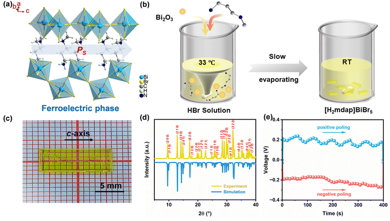
The reported BHP ferroelectric 1 was synthesized from stoichiometric Bi2O3 and H2mdap in a saturated hydrobromic acid solution32 and adopted the noncentrosymmetric orthorhombic space group Pna21 in the ferroelectric phase (Fig. 1a) and centrosymmetric space group Pnma in the paraelectric phase (Fig. S1, ESI†), which belongs to one of the reported 88 species of ferroelectric phase transitions deduced through the Aizu notation of mmmFmm2. Its basic 1D, ABX5-type perovskite structure consists of an inorganic zigzag chain formed by {BiBr5}n and the [H2mdap]2+ cations are located in the inorganic framework. A pair of amino groups in [H2mdap]2+ cations are tightly linked with the inorganic structure, creating sturdy N–H⋯Br hydrogen bonds (Fig. S2, ESI†), which strengthens the stability of the structure.33Large yellow crystals (i.e., 13 × 5 × 0.5 mm3) were obtained via a gradual process of solvent evaporation (Fig. 1b and c). The purity of the samples was confirmed through powder X-ray diffraction (Fig. 1d). As shown in Fig. S3 (ESI),† the test result shows that 1 is environmentally stable after being stored in ambient air for 1 month and 3 months. The thermogravimetric analysis shows that 1 also has ultrahigh thermal stability up to 580 K (Fig. S4, ESI†). Moreover, we measured the P–E hysteresis loop along the crystallographic c-axis direction, and the Ps value of 1 reaches ∼3.1 μC cm−2 (Fig. S5, ESI†). Additionally, ferroelectric materials are distinguished by their ability to reversibly alter the orientation of the Ps by applying an external electric field, thereby providing a practical means for controlling the Ps-based photovoltaic currents. To explore the link between ferroelectricity and self-powered behaviors, we realized the reversal of the optical voltage direction through the poling electric field's directional change. When exposed to 404 nm light, the clear Ps-linked FE-PV effect of switchable open-circuit optical voltage (VOC) is noticeable with its value of ∼0.20 V (Fig. 1e). The changeable VOC signal reveals a complex connection between the FE-PV effect and the inherent electric field in ferroelectrics, aiding in the segregation and transfer of photon-generated carriers, thereby hinting at self-powered detection capabilities.
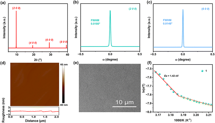
Moreover, the crystal's quality is pivotal in determining X-ray detection performance. The X-ray scan of the top facet of the 1 SC shows only (h 0 0) diffraction peaks (Fig. 2a), confirming its well-oriented single-crystalline lattice and good crystallinity. A more detailed rocking-curve analysis shows that a very narrow full width at half maximum (FWHM) of only 0.0180° and 0.0148° for the (2 0 0) and (6 0 0) peaks is obtained (Fig. 2b and c), lower than those of many reported X-ray detectors, including high-quality MA3Bi2I9 (0.024° at the (0 0 6) peak)34 and MAPbI3 (0.0414° at the (2 0 0) peak).35 Additionally, the atomic force microscope (AFM) measurement (Fig. 2d) and the scanning electron microscope (SEM) measurement (Fig. 2d) were conducted on the 1's (h 0 0) plane which shows that 1 has a very smooth and flat morphology, further confirming the high crystal quality, prone to photogenerated carriers' transport in direct X-ray detectors.
Furthermore, based on the I–V curves ranging from −4 to 4 V, a relatively high bulk resistivity (ρ) is calculated to be 2.53 × 10¹⁰ Ω cm−1 (Fig. S6, ESI†), which is comparable to those of many lead-free halide perovskites, such as (MA)3Bi2I9 (3.74 × 10¹⁰ Ω cm−1),34 (I–C4H8NH3)4AgBiI8 (3.04 × 10¹⁰ Ω cm−1),36 and commercial CdZnTe (1010 Ω cm−1),37 with its orders of magnitude greater than those of 3D lead halide perovskite SCs (107–108 Ω cm−1).38,39 This high resistivity would permit an extremely low dark current, which is crucial for stable high-performance X-ray detection.40 We also measured the variable temperature conductivity to extract the activation energy Ea based on the Nernst–Einstein relation:41
 | (1) |
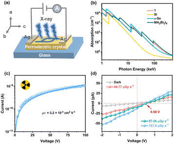
What's more, self-powered X-ray detection was executed on the 1 detector along the polar axis (c-axis) as demonstrated in Fig. 3a (Fig. S7, ESI†). To accurately determine the X-ray photon absorption capacity of 1, the absorption spectra of 1 as a function of photon energies were obtained using the photo cross-section database.43 In Fig. 3b, the absorption coefficient of 1 is shown to be higher than that of Si and comparable to that of traditional semiconductors α-Se and the MA3Bi2I9 SC, indicating its good X-ray attenuation ability, which guarantees 1 to be applied in direct X-ray detection. Specifically, for 50 keV hard X-ray photons, a 1.5 mm-thick 1 SC can absorb ≈ 92% of incident photons, much better than the ratio (≈15%) for Si (Fig. S8, ESI†), hence enabling the charge carriers to be collected more easily. Furthermore, efficient charge collection is also crucial for a high-performance detector, which is determined by the high μτ product according to the standard Hecht's equation:44
 | (2) |
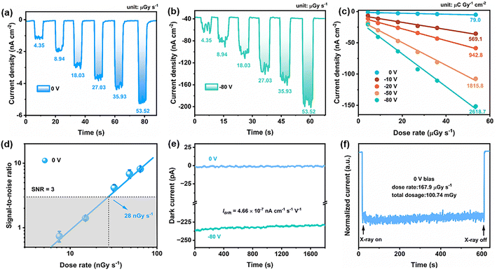
As a result, we test the X-ray detection performance of 1. Here, an Amptek Mini-X2 X-ray tube with a silver target (maximum power 4 W) is used as the X-ray source. The X-ray energy is up to 50 keV and the peak intensity is at 22 keV (Fig. S9a, ESI†). As shown in Fig. 4a, owing to the significant FE-PV effect induced by the spontaneous electric polarization, the 1 detector exhibits its excellent response to X-ray irradiation even at 0 V bias, with a fitting sensitivity of 79.0 μC Gy−1 cm−2 (Fig. 4c), four times higher than that of the commercial α-Se film detector (20 μC Gy−1 cm−2 under 2000 V) and higher than those of many reported self-powered X-ray detectors, including (4-AMP)BiI5 (66.84 μC Gy−1 cm−2),22 (R-PPA)BiI5 (31 μC Gy−1 cm−2),48etc. To the best of our knowledge, 1 successfully realizes the self-powered X-ray detection in 1D BHP ferroelectrics, showing its potential for future practical application. Furthermore, with the increase of the applied bias, the photocurrent density increases almost linearly with increasing X-ray dose rates (Fig. 4b and S10, ESI†). Additionally, the sensitivity can reach the highest value of 2618.7 μC Gy−1 cm−2 at −80 V bias, which surpasses those of most 1D X-ray detectors (Table S1, ESI†), showing that it's a promising material for X-ray detection. Moreover, such a high value exceeds the theoretical sensitivity of 24.6 and 2.1 μC Gy−1 cm−2 at a maximum X-ray energy of 50 keV and a peak intensity of 22 keV, respectively.49 This is likely due to photoconductive gain, a widespread phenomenon, and is beneficial for increasing the sensitivity of X-ray detectors.40,50–52
In addition, under 0 V bias, the SNR rates for 1 were further calculated. According to the International Union of Pure and Applied Chemistry (IUPAC), the dose rate that generates an SNR of three is regarded as the lowest detection limit.401 still has a good photoresponse at a low dose of 4.35 μGy s−1, according to the plot of the photocurrent density versus the dose ratio, suggesting its ability to detect weak X-rays. Therefore, we measured its photocurrent at a lower X-ray dose rate (Fig. S11, ESI†). At 0 V bias, a low detection limit (28 nGy s−1) of 1 is 196 times lower than the regular medical diagnosis of 5.5 μGy s−1 (Fig. 4d).53 The detection limit of 1 under −10 to −80 V was also calculated with detection limits of 749, 1989, 2384, and 3511 nGy s−1, respectively (Fig. S12, ESI†), all of which exceed the value at 0 V bias, showing that the detection limit can be greatly reduced in self-powered mode. Then, to evaluate the operating stability of 1, the Idrift under −80 V bias is further analyzed (Fig. 4e). The value of Idrift (4.66 × 10⁻⁷ nA cm−1 s−1 V−1) is lower than that of 1D (4-AMP)BiI5 (about 1.51 × 10⁻⁵ nA cm−1 s−1 V−1, 50 V),22 2D (R-MPA)4AgBiI8 (about 10−3 nA cm−1 s−1 V−1, 50 V),21 and 3D perovskites, including MAPbI3 (about 10−4 nA cm−1 s−1 V−1, 100 V cm−1)54 and MAPbBr3 (about 10−3 nA cm−1 s−1 V−1, 20 V cm−1),52 showing its good operational stability, probably owing to the 1D inorganic framework and the strong hydrogen bonds in 1. Then, the high dose stability of 1 was investigated, showing that 1 retained stability at total X-ray doses of 100.74 mGy (Fig. 4f), thereby highlighting its relatively high radiation stability.
Conclusions
In summary, we have successfully explored the ferroelectric in the class of 1D BHPs, [H2mdap]BiBr5 (1), toward relatively “green” self-powered X-ray detection. Exploiting the FE-PV effect, the 1 device demonstrates remarkable sensitivity. It achieves a low detection limit of 28 nGy s−1 at zero bias, surpassing most Bi-based X-ray detector materials and the typical medical diagnosis (∼5.5 μGy s−1). Furthermore, the 1 SC detector shows a low Idrift of 4.66 × 10⁻⁷ nA cm−1 s−1 V−1, exhibiting good operational stability, which may benefit from a stable structure and high activation energy (Ea, 1.42 eV). This study introduces a novel approach to preparing self-powered X-ray detectors based on the FE-PV effect of ferroelectric SC materials. In the future, it will continue to develop on this basis, such as the manufacture of BHP-based ferroelectric thin-film devices for X-ray detection.Data availability
The data supporting the findings of this study are available on request from the corresponding author, [Junhua Luo], upon reasonable request.Author contributions
Y. Wang prepared the samples and wrote the manuscript. H. Yang, W. Wu, and Y. Wang investigated the photoelectric properties. Q. Guan, H. Ye, H. Li, Y. Zeng, and P. Yu provided suggestions for the project. Z.-K. Zhu and J. Luo designed and directed this project. All the authors discussed and commented on the manuscript.Conflicts of interest
There are no conflicts to declare.Acknowledgements
This work was financially supported by the National Natural Science Foundation of China (22435005, 22193042, 22201284, 22305105, 22405108, 22175177, 22125110, and U21A2069), the Key Research Program of Frontier Sciences of the Chinese Academy of Sciences (ZDBS-LY-SLH024), the National Key Research and Development Program of China (2019YFA0210402), the Natural Science Foundation of Fujian Province (2023J05076), the Natural Science Foundation of Jiangxi Province (20224BAB213003 and 20232BAB213020), the Jiangxi Provincial Education Department Science and Technology Research Foundation (GJJ2200384), and the Graduate Innovation Fund Project of Jiangxi Provincial Department of Education (YC2024-S266).Notes and references
- H. Li, C. f. Wang, Q. f. Luo, C. Ma, J. Zhang, R. Zhao, T. Yang, Y. Du, X. Chen, T. Li, X. Liu, X. Song, Y. Yang, Z. Yang, S. Liu, Y. Zhang and K. Zhao, Adv. Funct. Mater., 2024, 34, 2407693 CrossRef CAS.
- Y. Li, Y. Lei, H. Wang and Z. Jin, Nano-Micro Lett., 2023, 15, 128 CrossRef CAS PubMed.
- Y. Wu, J. Feng, Z. Yang, Y. Liu and S. Liu, Adv. Sci., 2023, 10, 2205536 CrossRef CAS PubMed.
- X. He, Y. Deng, D. Ouyang, N. Zhang, J. Wang, A. A. Murthy, I. Spanopoulos, S. M. Islam, Q. Tu, G. Xing, Y. Li, V. P. Dravid and T. Zhai, Chem. Rev., 2023, 123, 1207–1261 CrossRef CAS PubMed.
- J. H. Heo, J. K. Park, Y. M. Yang, D. S. Lee and S. H. Im, iScience, 2021, 24, 102927 CrossRef CAS PubMed.
- H. Li, X. Liu, T. Yang, C. Ma, Y. Du, P. Xu, L. Zhang, X. Song, Q. Cui, S. Zhao, Z. Yang, S. Liu, S. Jin and K. Zhao, ACS Energy Lett., 2024, 9, 64–74 CrossRef CAS.
- N. Li, Y. Li, S. Xie, J. Wu, N. Liu, Y. Yu, Q. Lin, Y. Liu, S. Yang, G. Lian, Y. Fang, D. Yang, Z. Chen and X. Tao, Angew. Chem., Int. Ed., 2023, 62, e202302435 CrossRef CAS PubMed.
- J. Tan, X. Gao, X. Huang, P. Wangyang, H. Sun, D. Yang and T. Zeng, J. Mater. Sci.: Mater. Electron., 2023, 34, 1199 CrossRef CAS.
- Z.-K. Zhu, J. Wu, P. Yu, Y. Zeng, R. Li, Q. Guan, H. Dai, G. Chen, H. Yang, X. Liu, L. Li, C. Ji and J. Luo, Adv. Funct. Mater., 2024, 34, 2409857 CrossRef CAS.
- X. Zhang, T. Zhu, C. Ji, Y. Yao and J. Luo, J. Am. Chem. Soc., 2021, 143, 20802–20810 CrossRef CAS PubMed.
- J. Yan, F. Gao, Y. Tian, Y. Li, W. Gong, S. Wang, H. Zhu and L. Li, Adv. Opt. Mater., 2022, 10, 2200449 CrossRef CAS.
- M. Girolami, F. Matteocci, S. Pettinato, V. Serpente, E. Bolli, B. Paci, A. Generosi, S. Salvatori, A. D. Carlo and D. M. Trucchi, Nano-Micro Lett., 2024, 16, 182 CrossRef CAS PubMed.
- H. Tsai, F. Liu, S. Shrestha, K. Fernando, S. Tretiak, B. Scott, D. T. Vo, J. Strzalka and W. Nie, Sci. Adv., 2020, 6, eaay0815 CrossRef CAS PubMed.
- A. B. Swain, M. Rath, P. P. Biswas, M. S. R. Rao and P. Murugavel, APL Mater., 2019, 7, 011106 CrossRef.
- Y. Peng, X. Liu, Z. Sun, C. Ji, L. Li, Z. Wu, S. Wang, Y. Yao, M. Hong and J. Luo, Angew. Chem., Int. Ed., 2020, 59, 3933–3937 CrossRef CAS PubMed.
- C. Ji, S. Wang, Y. Wang, H. Chen, L. Li, Z. Sun, Y. Sui, S. Wang and J. Luo, Adv. Funct. Mater., 2020, 30, 1905529 CrossRef CAS.
- Z. Zhu, H. Chen, B. Zhao, W. Huang, Q. Lin, X. Yu and Y. Li, Appl. Phys. Lett., 2023, 122, 163301 CrossRef CAS.
- Y. Jiang, C. Zhang, Z.-K. Zhu, J. Wu, P. Yu, Y. Zeng, H. Ye, H. Dai, R. Li, Q. Guan, G. Chen, H. Yang and J. Luo, Angew. Chem., Int. Ed., 2024, 63, e202407305 CrossRef CAS PubMed.
- C. Ji, Y. Li, X. Liu, Y. Wang, T. Zhu, Q. Chen, L. Li, S. Wang and J. Luo, Angew. Chem., Int. Ed., 2021, 60, 20970–20976 CrossRef CAS PubMed.
- Y. Ma, W. Li, Y. Liu, W. Guo, H. Xu, S. Han, L. Tang, Q. Fan, J. Luo and Z. Sun, ACS Cent. Sci., 2023, 9, 2350–2357 CrossRef CAS PubMed.
- J. Wu, S. You, P. Yu, Q. Guan, Z.-K. Zhu, Z. Li, C. Qu, H. Zhong, L. Li and J. Luo, ACS Energy Lett., 2023, 8, 2809–2816 CrossRef CAS.
- S. You, P. Yu, T. Zhu, C. Lin, J. Wu, Z.-K. Zhu, C. Zhang, Z. Li, C. Ji and J. Luo, Adv. Funct. Mater., 2024, 34, 2310916 CrossRef CAS.
- Z.-K. Zhu, T. Zhu, S. You, P. Yu, J. Wu, Y. Zeng, Q. Guan, Z. Li, C. Qu, H. Zhong, L. Li and J. Luo, Small, 2023, 20, 2307454 CrossRef PubMed.
- W. Guo, H. Xu, Q. Fan, P. Zhu, Y. Ma, Y. Liu, X. Zeng, J. Luo and Z. Sun, Adv. Opt. Mater., 2024, 12, 2303291 CrossRef CAS.
- D. Fu, Y. Ma, Z. Chen, Z. Hou and J. Luo, Adv. Opt. Mater., 2024, 12, 2400193 CrossRef CAS.
- D. Fu, Y. Ma, S. Wu, L. Pan, Q. Wang, R. Zhao, X. M. Zhang and J. Luo, InfoMat, 2024, 6, e12602 CrossRef CAS.
- X. Xiao, J. Dai, Y. Fang, J. Zhao, X. Zheng, S. Tang, P. N. Rudd, X. C. Zeng and J. Huang, ACS Energy Lett., 2018, 3, 684–688 CrossRef CAS.
- Y. Zhang, Y. Liu, Z. Xu, Z. Yang and S. Liu, Small, 2020, 16, 2003145 CrossRef CAS PubMed.
- B. Zhang, Z. Xu, C. Ma, H. Li, Y. Liu, L. Gao, J. Zhang, J. You and S. Liu, Adv. Funct. Mater., 2022, 32, 2110392 CrossRef CAS.
- Y. Yuan and J. Huang, Acc. Chem. Res., 2016, 49, 286–293 CrossRef CAS PubMed.
- Y. Lin, Y. Bai, Y. Fang, Q. Wang, Y. Deng and J. Huang, ACS Energy Lett., 2017, 2, 1571–1572 CrossRef CAS.
- Q. Chen, H. Jiang, Y. Fan, Z. Li, H. Ye, Y. Yao, S. Chen, C. Ji, S. Zhang and J. Luo, Adv. Funct. Mater., 2023, 33, 2213964 CrossRef CAS.
- N. K. Tailor, A. Mahapatra, A. Kalam, M. Pandey, P. Yadav and S. Satapathi, Phys. Rev. Mater., 2022, 6, 045401 CrossRef CAS.
- Y. Liu, Z. Xu, Z. Yang, Y. Zhang, J. Cui, Y. He, H. Ye, K. Zhao, H. Sun, R. Lu, M. Liu, M. G. Kanatzidis and S. Liu, Matter, 2020, 3, 180–196 CrossRef.
- J. Wu, L. Wang, A. Feng, S. Yang, N. Li, X. Jiang, N. Liu, S. Xie, X. Guo, Y. Fang, Z. Chen, D. Yang and X. Tao, Adv. Funct. Mater., 2022, 32, 2109149 CrossRef CAS.
- Z. Xu, H. Wu, D. Li, W. Wu, L. Li and J. Luo, J. Mater. Chem. C, 2021, 9, 13157–13161 RSC.
- K. Kim, S. Kim, J. Hong, J. Lee, T. Hong, A. E. Bolotnikov, G. S. Camarda and R. B. James, J. Appl. Phys., 2015, 117, 145702 CrossRef.
- G. Maculan, A. D. Sheikh, A. L. Abdelhady, M. I. Saidaminov, M. A. Haque, B. Murali, E. Alarousu, O. F. Mohammed, T. Wu and O. M. Bakr, J. Phys. Chem. Lett., 2015, 6, 3781–3786 CrossRef CAS PubMed.
- D. Shi, V. Adinolfi, R. Comin, M. Yuan, E. Alarousu, A. Buin, Y. Chen, S. Hoogland, A. Rothenberger, K. Katsiev, Y. Losovyj, X. Zhang, P. A. Dowben, O. F. Mohammed, E. H. Sargent and O. M. Bakr, Science, 2015, 347, 519–522 CrossRef CAS PubMed.
- W. Pan, H. Wu, J. Luo, Z. Deng, C. Ge, C. Chen, X. Jiang, W.-J. Yin, G. Niu, L. Zhu, L. Yin, Y. Zhou, Q. Xie, X. Ke, M. Sui and J. Tang, Nat. Photonics, 2017, 11, 726–732 CrossRef CAS.
- S. Yang, S. Chen, E. Mosconi, Y. Fang, X. Xiao, C. Wang, Y. Zhou, Z. Yu, J. Zhao, Y. Gao, F. D. Angelis and J. Huang, Science, 2019, 365, 473–478 CrossRef CAS PubMed.
- S. Meloni, T. Moehl, W. Tress, M. Franckevicius, M. Saliba, Y. H. Lee, P. Gao, M. K. Nazeeruddin, S. M. Zakeeruddin, U. Rothlisberger and M. Graetzel, Nat. Commun., 2016, 7, 10334 CrossRef CAS PubMed.
- Y. Huang, L. Qiao, Y. Jiang, T. He, R. Long, F. Yang, L. Wang, X. Lei, M. Yuan and J. Chen, Angew. Chem., Int. Ed., 2019, 58, 17834–17842 CrossRef CAS PubMed.
- Y. C. Kim, K. H. Kim, D.-Y. Son, D.-N. Jeong, J.-Y. Seo, Y. S. Choi, I. T. Han, S. Y. Lee and N.-G. Park, Nature, 2017, 550, 87–91 CrossRef CAS PubMed.
- X. Dong, T. Chen, J. Liang, L. Wang, H. Wu, Z. Xu, J. Luo and L.-N. Li, Chin. J. Struct. Chem., 2024, 43, 100256 CrossRef CAS.
- C. Ma, H. Li, M. Chen, Y. Liu, K. Zhao and S. Liu, Adv. Funct. Mater., 2022, 32, 2202160 CrossRef CAS.
- Y. Zhou, J. Chen, O. M. Bakr and O. F. Mohammed, ACS Energy Lett., 2021, 6, 739–768 CrossRef CAS.
- S. You, Z.-K. Zhu, S. Dai, J. Wu, Q. Guan, T. Zhu, P. Yu, C. Chen, Q. Chen and J. Luo, Adv. Funct. Mater., 2023, 33, 2303523 CrossRef CAS.
- W. Pan, Y. He, W. Li, L. Liu, K. Guo, J. Zhang, C. Wang, B. Li, H. Huang, J. Zhang, B. Yang and H. Wei, Nat. Commun., 2024, 15, 257 CrossRef CAS PubMed.
- K. Guo, W. Li, Y. He, X. Feng, J. Song, W. Pan, W. Qu, B. Yang and H. Wei, Angew. Chem., Int. Ed., 2023, 62, e202303445 CrossRef CAS PubMed.
- W.-G. Li, X.-D. Wang, Y.-H. Huang and D.-B. Kuang, Adv. Mater., 2023, 35, 2210878 CrossRef CAS PubMed.
- W. Wei, Y. Zhang, Q. Xu, H. Wei, Y. Fang, Q. Wang, Y. Deng, T. Li, A. Gruverman, L. Cao and J. Huang, Nat. Photonics, 2017, 11, 315–321 CrossRef CAS.
- H. Wei and J. Huang, Nat. Commun., 2019, 10, 1066 CrossRef PubMed.
- S. Yakunin, D. N. Dirin, Y. Shynkarenko, V. Morad, I. Cherniukh, O. Nazarenko, D. Kreil, T. Nauser and M. V. Kovalenko, Nat. Photonics, 2016, 10, 585–589 CrossRef CAS.
Footnote |
| † Electronic supplementary information (ESI) available. See DOI: https://doi.org/10.1039/d4sc06049k |
| This journal is © The Royal Society of Chemistry 2025 |


