Minimising the payload solvent exposed hydrophobic surface area optimises the antibody-drug conjugate properties†
Adrian D.
Hobson
 *a,
Haizhong
Zhu
b,
Wei
Qiu
b,
Russell A.
Judge
 b and
Kenton
Longenecker‡
b
aAbbVie Bioresearch Center, 381 Plantation Street, Worcester, Massachusetts 01605, USA. E-mail: adrian.hobson@abbvie.com
bAbbVie Inc., 1 North Waukegan Road, North Chicago, IL 60064, USA
First published on 9th January 2024
Abstract
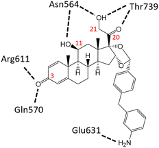
Glucocorticoid receptor modulators (GRMs) are an established and successful compound class for the treatment of multiple diseases. In addition, they are an area of high interest as payloads for antibody-drug conjugate s(ADCs) in both immunology and oncology. Solving the crystal structure of compound 2, the GRM payload from ABBV-3373 and ABBV-154, in the ligand binding domain of the glucocorticoid receptor (GR) revealed key information to facilitate optimal ADC payload design. All four critical H-bonds between the oxygen functional groups on the hexadecahydro-1H-cyclopenta[a]phenanthrene ring system of the small molecule and protein were shown to be made (carbonyl at C3 to Gln570 and Arg611 and Asn564, carbonyl at C20 to Thr739, hydroxyl at C21 to Asn 564 and Thr739). In addition, an extra H-bond between the linker attachment site on compound 2, the aniline in the biaryl region, was observed. Confirmation of the stereochemistry of the acetal in compound 2 as (R) was established. Finally, the importance of minimising the exposed hydrophobic surface area of a payload to reduce the negative impact on the properties of resulting ADCs, like aggregation, was rationalised by comparison of (R)-acetal compound 2 and (S)-acetal compound 3.
Introduction
Antibody-drug conjugates (ADCs) are now an established therapeutic modality in the oncology field with more than a dozen approved.1 Of these oncology ADCs brentuximab vedotin2 established key design elements for ADCs including a protease cleavable linker3 to release the payload in the lysosome and the self-immolative group para-amino benzylic alcohol which attaches via a carbamate to a suitable basic nitrogen on the cytotoxic payload monomethyl auristatin E (MMAE).Encouraged by this success in oncology other therapeutic areas have pursued ADCs most notably in the immunology field. Probably the most successful class of immunology small molecule drugs are glucocorticoids which have more than 70 years of successful drug discovery activity.4 Unfortunately, despite being highly functionalised glucocorticoids, exemplified by prednisolone5 (Fig. 1a) do not have a suitable nitrogen atom for linker attachment. Of the four oxygen functional groups the carbonyl at C3 via hydrazone attachment like in gemtuzumab ozogamicin6 and the hydroxyl at C21 via ester attachment like in many steroid prodrugs7 offer the best options for attachment.
 | ||
| Fig. 1 Marketed steroids a) prednisolone, b) dexamethasone, c) ciclesonide (prodrug), d) des-ciclesonide. | ||
To enable ADCs with a glucocorticoid payload, linker attachment to the C21 hydroxyl of dexamethasone8,9 (Fig. 1b) has been explored using both ester10 and carbonate.11 However, for an immunology (iADC) the stable attachment of the payload to the antibody to avoid premature loss of the payload is critical. Concerned that the instability of esters and carbonates in vivo would result in the premature loss of payload a glucocorticoid with a suitable nitrogen to facilitate stable linker attachment was pursued. Comprehensive SAR studies have identified the key structural features of glucocorticoids as the C3 carbonyl, C11 hydroxyl, C20 carbonyl and C21 hydroxyl12,13 and an ADC payload that retained these functional groups was desired. Ciclesonide14 (Fig. 1c) differs from dexamethasone in having an acetal at C16–C17 and is the C21 isobutyryl ester prodrug of the active desisobutyryl ciclesonide, more commonly known as des-ciclesonide (Fig. 1d) with crystallographic studies on the small molecule reported.15,16
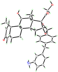
Structural information is a powerful tool for medicinal chemists and was used to guide the design of a glucocorticoid receptor modulator (GRM) ADC payload as there is a wealth of structural information of multiple steroidal small molecules bound in the ligand binding domain of the glucocorticoid receptor (GR).17 Structures of both dexamethasone (RSC Protein Data Bank entry 4UDC, https://www.rcsb.org/structure/4udc) and des-ciclesonide (RSC Protein Data Bank entry 4UDD, https://www.rcsb.org/structure/4udd) bound in the ligand binding domain of the glucocorticoid receptor have been solved.18 Modified analogues of des-ciclesonide that introduced a nitrogen from the cyclohexyl region of the compound were modelled and prioritised analogues synthesised and tested. This identified compound 1 (Fig. 2a) as a promising compound19 with the (R)-stereochemistry of the acetal being confirmed by X-ray crystallography (Cambridge Structural Database entry ZAZTAZ, https://dx.doi.org/10.5517/ccdc.csd.cc29yr89).20 Further SAR to add an attachment point for the dipeptide linker identified biaryl aniline compound 2 (ref. 21) which advanced to the clinic as the payload on ABBV-3373.22 Confirmation of the (R)-stereochemistry of the acetal in compound 2 was achieved by solving the small molecule X-ray structure (Cambridge Structural Database entry DIQQUT, https://dx.doi.org/10.5517/ccdc.csd.cc2gxcpg).23 The crystal was a colourless prism with dimensions 0.20 × 0.18 × 0.12 mm3 and the symmetry of the crystal structure was assigned the orthorhombic space group P2(1)2(1)2(1).
 | ||
| Fig. 2 GRM compounds a) mono aryl anisole compound 1, b) biaryl aniline with (R)-acetal compound 2, c) biaryl aniline with (S)-acetal compound 3. | ||
Three ADCs, ABBV-3373,24 ABBV-154,25,26 and ABBV-319 (ref. 27) from two therapeutic areas are in multiple clinical trials highlighting the importance of this class of GRM. As a result, the structure of compound 2 bound in GR was solved to provide structural information to guide medicinal chemistry efforts, identify new H-bond interactions and confirm the (R)-acetal stereochemistry.
Materials and methods
Protein expression and purification
Human glucocorticoid receptor (residues 528–777) with three mutations (V571M, F602S, C638D) was cloned into the expression vector pGEX4T1 with a thrombin-cleavable, N-terminal 8His-GST tag. The plasmid was transformed into E. coli. BL21(DE3), grown at 37 °C in TB medium to OD600 2–3 in the presence of 20 μM compound 2, then protein expression induced with 0.5 mM IPTG at 18 °C for 16 hours.Cell pellets were resuspended in lysis buffer (20 mM Tris, 300 mM NaCl, 5 mM Imidazole, 0.5 mM TCEP, pH 8.0) and lysed by processing once with an EmulsiFlex-C50 homogenizer (Avestin). Cellular debris was pelleted by centrifugation at 30![[thin space (1/6-em)]](https://www.rsc.org/images/entities/char_2009.gif) 000×g for 30
000×g for 30![[thin space (1/6-em)]](https://www.rsc.org/images/entities/char_2009.gif) min. The supernatant was batch-bound to Talon resin, followed by washing the resin with the lysis buffer. The protein was eluted with elution buffer (20 mM Tris, 300 mM NaCl, 300 mM Imidazole, 0.5 mM TCEP, pH 8.0). Eluted protein was mixed with thrombin and dialyzed at 4 °C (20 mM Tris, 200 mM NaCl, 0.25 mM TCEP, pH 8.0) for 16 hours to remove the imidazole. The protein was further purified by size exclusion on a HiLoad Superdex 200 16/600 column equilibrated with 20 mM Tris, 150 mM NaCl, 0.5 mM TCEP, pH 8.0. After that, 5 molar folds of compound 2 was added to the protein solution and the mixture was clarified with centrifugation and concentrated to 13 mg mL−1. The 3-molar fold of co-activator peptide (KENALLRYLLDKDD) was added to the concentrated protein to form the final complex for crystallisation.
min. The supernatant was batch-bound to Talon resin, followed by washing the resin with the lysis buffer. The protein was eluted with elution buffer (20 mM Tris, 300 mM NaCl, 300 mM Imidazole, 0.5 mM TCEP, pH 8.0). Eluted protein was mixed with thrombin and dialyzed at 4 °C (20 mM Tris, 200 mM NaCl, 0.25 mM TCEP, pH 8.0) for 16 hours to remove the imidazole. The protein was further purified by size exclusion on a HiLoad Superdex 200 16/600 column equilibrated with 20 mM Tris, 150 mM NaCl, 0.5 mM TCEP, pH 8.0. After that, 5 molar folds of compound 2 was added to the protein solution and the mixture was clarified with centrifugation and concentrated to 13 mg mL−1. The 3-molar fold of co-activator peptide (KENALLRYLLDKDD) was added to the concentrated protein to form the final complex for crystallisation.
Crystallisation and data collection
The ternary complex of GR, compound 2 and co-activator peptide was crystallised using sitting drop vapor diffusion method. More specifically, 100 nL of protein complex was mixed with 100 nL of crystallisation reagents and incubated over 80 μL of reservoir solutions of 24% (w/v) PEG400, 200 mM ammonium acetate, 100 mM sodium citrate pH 5.5 at 23 °C. The stacking thin plate crystals were initially found after 3 weeks, and they grew to their full size after 3 months. Plate crystals were separated, and flash frozen into liquid nitrogen using reservoir solutions as the cryo-protectant. Diffraction data were collected at a temperature of 100 K using IMCA-CAT beamline 17-ID at Argonne National Laboratory. Data collection statistics are summarised in Table 1.| Crystallographic parameter | PDB 8VKZ | |
|---|---|---|
| a Parentheses refers to values in highest resolution shell. | ||
| Data collection | Space group | P21 |
| Cell dimensions | a, b, c (Å) | 38.51, 141.65, 47.90 |
| α, β, χ (°) | 90.0, 93.46, 90.0 | |
| Resolution (Å) | 70.83–2.13 (2.33–2.13)a | |
| R pim | 0.064 (0.529) | |
| I/σI | 8.4 (1.5) | |
| Completeness (%) | Spherical | 56.7 (12.0) |
| Ellipsoidal | 89.6 (60.6) | |
| Redundancy | 3.4 (3.4) | |
| CC(1/2) | 0.99 (0.53) | |
| Refinement | Resolution (Å) | 70.8–2.13 |
| No. reflections | 16![[thin space (1/6-em)]](https://www.rsc.org/images/entities/char_2009.gif) 146 (808) 146 (808) |
|
| R work/Rfree (%) | 18.70/24.52 | |
| No. atoms | Protein | 4063 |
| Ligand/ion | 84 | |
| Water | 97 | |
| B-factors (Å2) | Protein | 42.89 |
| Ligand/ion | 38.02 | |
| Water | 39.35 | |
| R.m.s. deviations | Bond lengths (Å) | 0.008 |
| Bond angles (°) | 0.90 | |
| Ramachandran values (%) | Favoured | 97.1 |
| Allowed | 2.7 | |
| Outliers | 0.2 | |
Results and discussion
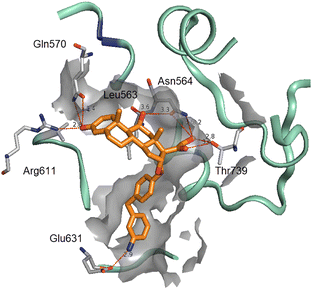
The crystal structure of the GR ligand binding domain complex was determined with a co-activator peptide and the ADC payload compound 2 to study the binding mode of the compound.28 The molecular packing exhibited a swapped dimer arrangement, where the monomers adopted similar conformations, and the co-activator peptide was visible as a short helix. The compound bound in the enclosed ligand pocket, where the four rings of the glucocorticoid scaffold formed the four expected H-bond interactions (Fig. 4). The pose of compound 2 core aligns closely with the common features of a smaller compound desisobutyryl ciclesonide (RSC Protein Data Bank entry 4UDD, https://www.rcsb.org/structure/4udd). By contrast, compound 2 features a biaryl extension, where the linked diphenyl extends through the cavity towards the ligand entry portal.29 The compound apparently destabilises the protein loop at the portal containing residues 634–636, which are disordered in the current structure, and the aniline is positioned at the opening and makes a H-bond interaction with Glu631. The stereochemistry of the acetal was confirmed as (R) and the V-shape of the compound is clearly visible. Distances for the H-bonds between compound 2 and GR are listed in Table 2 and depicted in Fig. 5.| Compound 2 | Amino acid | Functional group | H-bond distance/Å | |
|---|---|---|---|---|
| Functional group | Position | |||
| Carbonyl | C3 | Gln570 | NH2 | 3.4 |
| Carbonyl | C3 | Arg611 | NH2 | 2.8 |
| Hydroxyl | C11 | Asn564 | C![[double bond, length as m-dash]](https://www.rsc.org/images/entities/char_e001.gif) O O |
3.3 |
| Carbonyl | C20 | Thr739 | OH | 3.0 |
| Hydroxyl | C21 | Asn564 | NH2 | 3.2 |
| Hydroxyl | C21 | Thr739 | OH | 2.8 |
| Aniline | Biaryl | Glu631 | C![[double bond, length as m-dash]](https://www.rsc.org/images/entities/char_e001.gif) O O |
2.9 |
ADCs on mouse anti-TNF with equivalent DAR were prepared with both compound 2 and compound 3 as the payload using the same linker enabling any differences in the properties of the two ADCs to be directly attributed to the payload. Both the aggregation level and hydrophobicity of the ADCs were shown to differ greatly. The mouse anti-TNF ADC with compound 3 was more highly aggregated (4%) and had a longer retention time by hydrophobic interaction chromatography (HIC) of 4.51 minutes for the DAR4 peak. This contrasted with the mouse anti-TNF ADC with compound 2 that had lower aggregation (0.5%) and a shorter retention time by HIC of 4.28 minutes for the DAR4 peak.30 Aggregation and retention time by HIC are both used as measurements of the hydrophobicity of an ADC. This data clearly showed that compound 3 had a more negative impact on the hydrophobicity and drug-like properties of the ADC than compound 2.
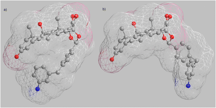
Having observed the V-shape of (R)-acetal compound 2 in both the small molecule crystal structure and the protein crystal structure of compound 2 bound in GR it was rationalised that this minimised the solvent exposed surface area compared to (S)-acetal compound 3 and resulted in the lower aggregation of ADCs with compound 2 as their payload. Energy minimised conformations of both compound 2 and compound 3 were generated in Chem3D and are depicted in Fig. 6 with their solvent exposed surface shown as a wire mesh coloured by atom.
Using 3D Methods in Pipeline Biovia three surface areas were calculated on the energy minimised conformations of compound 2 and compound 3. For calculation of the polar solvent accessible surface area N, O, P, S are considered as polar atoms along with any hydrogens attached to them and any atom with a formal charge.
The calculated data (Table 3) supported the hypothesis of ADC aggregation being impacted by the exposed hydrophobic surface area of a payload. While the polar solvent accessible surface area, the area capable of enabling aqueous solubility, for compound 2 and compound 3 was similar (216.5 and 224.3 Å respectively), the solvent accessible surface areas differed significantly. Compound 2 had a solvent accessible surface area of 718.9 Å, almost 100 Å less than that of compound 3 (814.7 Å). Similarly, the solvent accessible volume of compound 2 at 646.4 Å was substantially lower than for compound 3 (736.1 Å). Considering this data it is clear that while both compounds have similar solubility driving polar areas compound 2 has less hydrophobic surface area to solvate than compound 3. This significant finding identifies that a key parameter for ADC payload design is to maximise the exposed hydrophilic surface and probably more importantly to minimise the exposed hydrophobic surface of payload.
| ID | Polar solvent accessible surface area/Å | Solvent accessible surface area/Å | Solvent accessible volume/Å |
|---|---|---|---|
| 2 | 216.5 | 718.9 | 646.4 |
| 3 | 224.3 | 814.7 | 736.1 |
Identification of the H-bond interaction made between aniline and Glu631 provided a second important guide for ADC payload design. That is, introduction of nitrogen to the payload should not just be considered as a location to attach the linker. Moreover, whenever possible SAR and structural information should be used to propose possible additional interactions to drive both the potency and selectivity of the payload for its target.
Conclusion
Steroids are a major class of pharmaceuticals in multiple therapeutic areas. Following the clinical success of oncology ADCs with cytotoxic payloads, the use of GRM as an ADC payload is an area of high interest. While extensive guidelines for small molecule design exist, there is a dearth for ADC payloads. For example, following conjugation to an antibody the payload has a disproportionately large impact on the properties of the resulting ADC.The crystal structure of compound 2 bound in GR showed that all four of the oxygen functional groups on the hexadecahydro-1H-cyclopenta[a]phenanthrene ring system of the small molecule made their predicted H-bond interactions with the GR protein:
1. Carbonyl at C3 to Gln570.
2. Carbonyl at C3 to Arg611.
3. Hydroxyl at C11 to Asn564.
4. Carbonyl at C20 to Thr739.
5. Hydroxyl at C21 to Asn564.
6. Hydroxyl at C21 to Thr739.
In addition, a new H-bond interaction between the aniline in the biaryl region of compound 2 and Glu631 was observed. Typically, a nitrogen is incorporated to a compound of interest as an ADC payload to facilitate dipeptide linker attachment. However, identification of this new interaction provides a significant guide to ADC payload design. Incorporation of nitrogen should not just be seen as a method for linker attachment, but that SAR and structural information must be used to enhance payload design and introduce additional interactions with the target to drive both potency and selectivity of new analogues.
One of the goals of this crystallisation was to confirm the stereochemistry of the acetal as the (R)-isomer. Not only was this stereochemistry verified as (R) it also confirmed the importance of minimising the hydrophobic surface area of a payload. Inspection of Fig. 3–6 immediately emphasises the V-shape of compound 2 which dramatically reduces the exposed hydrophobic surface area thereby reducing impact on the drug-like properties of resulting ADCs. As such a key design parameter for ADC payloads is proposed that is to maximise the exposed hydrophilic surface and probably more importantly to minimise the exposed hydrophobic surface of payload.
 | ||
| Fig. 4 Structural pose of compound 2 (orange) in the GR binding pocket with key residues highlighted (PDB 8VKZ). | ||
 | ||
| Fig. 6 Solvent exposed surface area shown as a wire mesh coloured by atom for energy minimised conformations of a) (R)-acetal compound 2, b) (S)-acetal compound 3. | ||
PDB codes
Fig. 3 compound 2 in ligand binding domain of GR – PDB code 8VKZ, https://www.rcsb.org/structure/8VKZ. Structure for GR with compound 2 has been deposited in the RSC Protein Data Bank and the atomic coordinates released.Abbreviations
| ADC | Antibody-drug conjugate |
| BrAc | Bromoacetamide |
| DAR | Drug to antibody ratio |
| H-bond | Hydrogen bond |
| GR | Glucocorticoid receptor |
| GRM | Glucocorticoid receptor modulator |
| HIC | Hydrophobic interaction chromatography |
| MMAE | Monomethyl auristatin E |
| PDB | Protein data bank |
| SAR | Structure–activity relationship |
| TNF | Tumour necrosis factor |
Author contributions
The manuscript was written through contributions of all authors. All authors have given approval to the final version of the manuscript.Conflicts of interest
There are no conflicts to declare.Acknowledgements
Authors ADH, HZ, WQ, and RAJ are employees of AbbVie. KL was an employee of AbbVie at the time of the study. The design, study conduct, and financial support for this research were provided by AbbVie. AbbVie participated in the interpretation of data, review, and approval of the publication. Use of the IMCA-CAT beamline 17-ID (or 17-BM) at the Advanced Photon Source was supported by the companies of the Industrial Macromolecular Crystallography Association through a contract with Hauptman-Woodward Medical Research Institute (no additional funding to disclose). Use of the Advanced Photon Source was supported by the U.S. Department of Energy, Office of Science, Office of Basic Energy Sciences, under Contract No. DE-AC02-06CH11357 (no additional funding to disclose).References
- C. Dumontet, J. M. Reichert, P. D. Senter, J. M. Lambert and A. Beck, Antibody–drug conjugates come of age in oncology, Nat. Rev. Drug Discovery, 2023, 22, 641–661, DOI:10.1038/s41573-023-00709-2.
- P. D. Senter and E. L. Sievers, The discovery and development of brentuximab vedotin for use in relapsed Hodgkin lymphoma and systemic anaplastic large cell lymphoma, Nat. Biotechnol., 2012, 30, 631–637, DOI:10.1038/nbt.2289.
- F. M. de Groot, L. W. van Berkom and H. W. Scheeren, Synthesis and biological evaluation of 2′-carbamate-linked and 2′-carbonate-linked prodrugs of paclitaxel: selective activation by the tumor-associated protease plasmin, J. Med. Chem., 2000, 43, 3093–3102, DOI:10.1021/jm0009078.
- A. Hobson, The Medicinal Chemistry of Glucocorticoid Receptor Modulators, SpringerBriefs in Molecular Science, Springer, Cham, 2023, ISBN 978–3–031-28732-9, DOI:10.1007/978-3-031-28732-9.
- H. L. Herzog, A. Nobile, S. Tolksdorf, W. Charney, E. B. Hershberg and P. L. Perlman, New antiarthritic steroids, Science, 1955, 121, 176, DOI:10.1126/science.121.3136.176.
- P. R. Hamann, L. M. Hinman, I. Hollander, C. F. Beyer, D. Lindh, R. Holcomb, W. Hallett, H. R. Tsou, J. Upeslacis, D. Shochat, A. Mountain, D. A. Flowers and I. Bernstein, Gemtuzumab ozogamicin, a potent and selective anti-CD33 antibody-calicheamicin conjugate for treatment of acute myeloid leukemia, Bioconjugate Chem., 2002, 13, 47–58, DOI:10.1021/bc010021y.
- F. M. Corticosteroid, Carboxylic Acid Esters, in Bioactive Carboxylic Compound Classes: Pharmaceuticals and Agrochemicals, ed. C. Lamberth and J. Dinges, Wiley, 2016, pp. 245–267, DOI:10.1002/9783527693931.ch18.
- G. E. Arth, J. Fried, D. B. R. Johnston, D. R. Hoff, L. H. Sarett, R. H. Silber, H. C. Stoerk and C. A. Winter, 16-Methylated steroids. II. 16α-Methyl analogs of cortisone, a new group of anti-inflammatory steroids. 9α-Halo derivatives, J. Am. Chem. Soc., 1958, 80, 3161–3163, DOI:10.1021/ja01545a063.
- J. J. Bunim, R. L. Black, L. Lutwak, R. E. Peterson and G. D. Whedon, Studies on dexamethasone, a new synthetic steroid, in rheumatoid arthritis: a preliminary report; adrenal cortical, metabolic and early clinical effects, Arthritis Rheum., 1958, 1, 313–331, DOI:10.1002/art.1780010404.
- M. Everts, R. J. Kok, S. A. Asgeirsdottir, B. N. Melgert, T. J. Moolenaar, G. A. Koning, M. J. van Luyn, D. K. Meijer and G. Molema, Selective intracellular delivery of dexamethasone into activated endothelial cells using an E-selectin-directed immunoconjugate, J. Immunol., 2002, 168, 883–889, DOI:10.4049/jimmunol.168.2.883.
- J. H. Graversen, P. Svendsen, F. Dagnaes-Hansen, J. Dal, G. Anton, A. Etzerodt, M. D. Petersen, P. A. Christensen, H. J. Moller and S. K. Moestrup, Targeting the hemoglobin scavenger receptor CD163 in macrophages highly increases the anti-inflammatory potency of dexamethasone, Mol. Ther., 2012, 20, 1550–1558, DOI:10.1038/mt.2012.103.
- B. Shroot, J. C. Caron and M. Ponec, Glucocorticoid specific binding: structure-activity relationships, Br. J. Dermatol., 1982, 107, 30–34, DOI:10.1111/j.1365-2133.1982.tb01028.x.
- N. Bodor, A. J. Harget and E. W. Phillips, Structure-activity relationships in the antiinflammatory steroids: a pattern-recognition approach, J. Med. Chem., 1983, 26, 318–328, DOI:10.1021/jm00357a003.
- N. A. Reynolds and L. J. Scott, Ciclesonide, Drugs, 2004, 64, 511–519, DOI:10.2165/00003495-200464050-00005.
- M. P. Feth, J. Volz, U. Hess, E. Sturm and R. P. Hummel, Physicochemical, crystallographic, thermal, and spectroscopic behavior of crystalline and X-ray amorphous ciclesonide, J. Pharm. Sci., 2008, 97, 3765–3780, DOI:10.1002/jps.21223.
- L. Zhou, Q. Yin, S. Du, H. Hao, Y. Li, M. Liu and B. Hou, Crystal structure, thermal crystal form transformation, desolvation process and desolvation kinetics of two novel solvates of ciclesonide, RSC Adv., 2016, 6, 51037–51045, 10.1039/C6RA08351J.
- A. Hobson, Appendix A, X-Ray Structures, in The Medicinal Chemistry of Glucocorticoid Receptor Modulators, SpringerBriefs in Molecular Science, Springer, Cham, 2023, DOI:10.1007/978-3-031-28732-9_3.
- K. Edman, A. Hosseini, M. K. Bjursell, A. Aagaard, L. Wissler, A. Gunnarsson, T. Kaminski, C. Köhler, S. Bäckström, T. J. Jensen, A. Cavallin, U. Karlsson, E. Nilsson, D. Lecina, R. Takahashi, C. Grebner, S. Geschwindner, M. Lepistö, A. C. Hogner and V. Guallar, Ligand Binding Mechanism in Steroid Receptors: From Conserved Plasticity to Differential Evolutionary Constraints, Structure, 2015, 23, 2280–2290, DOI:10.1016/j.str.2015.09.012.
- A. D. Hobson, M. J. McPherson, W. Waegell, C. A. Goess, R. H. Stoffel, X. Li, J. Zhou, Z. Wang, Y. Yu, A. Hernandez Jr, S. H. Bryant, S. L. Mathieu, A. K. Bischoff, J. Fitzgibbons, M. Pawlikowska, S. Puthenveetil, L. C. Santora, L. Wang, L. Wang, C. C. Marvin, M. E. Hayes, A. Shrestha, K. A. Sarris and B. Li, Design and Development of Glucocorticoid Receptor Modulator Agonists as Immunology Antibody-Drug Conjugate (iADC) Payloads, J. Med. Chem., 2022, 65, 4500–4533, DOI:10.1021/acs.jmedchem.1c02099.
- A. D. Hobson, M. J. McPherson, W. Waegell, C. A. Goess, R. H. Stoffel, X. Li, J. Zhou, Z. Wang, Y. Yu, A. Hernandez Jr, S. H. Bryant, S. L. Mathieu, A. K. Bischoff, J. Fitzgibbons, M. Pawlikowska, S. Puthenveetil, L. C. Santora, L. Wang, L. Wang, C. C. Marvin, M. E. Hayes, A. Shrestha, K. A. Sarris and B. Li, CCDC 2143751: Experimental Crystal Structure Determination, 2022, DOI:10.5517/ccdc.csd.cc29yr89.
- M. J. McPherson, A. D. Hobson, M. E. Hayes, C. C. Marvin, D. Schmidt, W. Waegell, C. Goess, J. Z. Oh, A. Hernandez Jr and J. T. Randolph, Preparation of glucocorticoid receptor agonist and immunoconjugates thereof, US Pat., 10668167, 2020 Search PubMed.
- R. D'Cunha, H. Kupper, D. Arikan, W. Zhao, D. Carter, J. Blaes, M. Ruzek and Y. Pang, A first-in-human study of the novel immunology antibody-drug conjugate, ABBV-3373, in healthy participants, Br. J. Clin. Pharmacol., 2023, 1–11, DOI:10.1111/bcp.15888.
- A. D. Hobson, M. J. McPherson, M. E. Hayes, C. Goess, X. Li, J. Zhou, Z. Wang, Y. Yu, J. Yang, L. Sun, Q. Zhang, P. Qu, S. Yang, A. Hernandez Jr, S. H. Bryant, S. L. Mathieu, A. K. Bischoff, J. Fitzgibbons, L. C. Santora, L. Wang, L. Wang, M. M. Fettis, X. Li, C. C. Marvin, Z. Wang, M. V. Patel, D. L. Schmidt, T. Li, J. T. Randolph, R. F. Henry, C. Graff, Y. Tian, A. L. Aguirre and A. Shrestha, CCDC 2291386: Experimental Crystal Structure Determination, 2023, DOI:10.5517/ccdc.csd.cc2gxcpg.
- A. D. Hobson, J. Xu, C. C. Marvin, M. J. McPherson, M. Hollmann, M. Gattner, K. Dzeyk, H. Sarvaiya, M. M. Fettis, A. K. Bichoff, L. Wang, L. Wang, J. Fitzgibbons, P. Salomon, A. Hernandez, Y. Jia, C. A. Goess, S. L. Mathieu and L. C. Santora, Optimization of Drug-Linker to Enable Long Term Storage of Antibody Dug Conjugate for Subcutaneous Dosing, J. Med. Chem., 2023, 66, 9161–9173, DOI:10.1021/acs.jmedchem.3c00794.
- A. D. Hobson, J. Xu, D. Welch, C. C. Marvin, M. J. McPherson, B. Gates, X. Liao, M. Hollmann, M. Gattner, K. Dzeyk, H. Sarvaiya, V. Shenoy, M. M. Fettis, A. K. Bichoff, L. Wang, L. C. Santora, L. Wang, J. Fitzgibbons, P. Salomon, A. Hernandez, Y. Jia, C. A. Goess, S. L. Mathieu, S. H. Bryant, M. E. Larsen, B. Cui and Y. Tuan, Discovery of ABBV-154, an anti-TNF Glucocorticoid Receptor Modulator Immunology Antibody Drug Conjugate (iADC), J. Med. Chem., 2023, 66, 12544–12558, DOI:10.1021/acs.jmedchem.3c01174.
- A. D. Hobson, M. J. McPherson, W. Waegell, C. Goess, A. Hernandez Jr, L. Wang, L. Wang, C. C. Marvin and L. C. Santora, Glucocorticoid receptor agonist and immunoconjugates thereof, US Pat., 10772970, 2020 Search PubMed.
- A Study to Assess the Adverse Events, Change in Disease Activity, and How Intravenously Infused ABBV-319 Moves Through the Bodies of Adult Participants With Relapsed or Refractory (R/R) Diffuse Large B-cell Lymphoma (DLBCL), Follicular Lymphoma (FL), or Chronic Lymphocytic Leukemia (CLL), https://clinicaltrials.gov/ct2/show/NCT05512390.
- R. K. Bledsoe, V. G. Montana, T. B. Stanley, C. J. Delves, C. J. Apolito, D. D. McKee, T. G. Consler, D. J. Parks, E. L. Stewart, T. M. Willson, M. H. Lambert, J. T. Moore, K. H. Pearce and H. E. Xu, Crystal structure of the glucocorticoid receptor ligand binding domain reveals a novel mode of receptor dimerization and coactivator recognition, Cell, 2002, 110, 93–105, DOI:10.1016/s0092-8674(02)00817-6.
- K. Edman, A. Hosseini, M. K. Bjursell, A. Aagaard, L. Wissler, A. Gunnarsson, T. Kaminski, C. Köhler, S. Bäckström, T. J. Jensen, A. Cavallin, U. Karlsson, E. Nilsson, D. Lecina, R. Takahashi, C. Grebner, S. Geschwindner, M. Lepistö, A. C. Hogner and V. Guallar, Ligand Binding Mechanism in Steroid Receptors: From Conserved Plasticity to Differential Evolutionary Constraints, Structure, 2015, 23, 2280–2290, DOI:10.1016/j.str.2015.09.012.
- A. D. Hobson, M. J. McPherson, M. E. Hayes, C. Goess, X. Li, J. Zhou, Z. Wang, Y. Yu, J. Yang, L. Sun, Q. Zhang, P. Qu, S. Yang, A. Hernandez Jr, S. H. Bryant, S. L. Mathieu, A. K. Bischoff, J. Fitzgibbons, L. C. Santora, L. Wang, L. Wang, M. M. Fettis, X. Li, C. C. Marvin, Z. Wang, M. V. Patel, D. L. Schmidt, T. Li, J. T. Randolph, R. F. Henry, C. Graff, Y. Tian, A. L. Aguirre and A. Shrestha, Discovery of ABBV-3373, an Anti-TNF Glucocorticoid Receptor Modulator Immunology Antibody-drug Conjugate, J. Med. Chem., 2022, 65, 15893–15934, DOI:10.1021/acs.jmedchem.2c01579.
Footnotes |
| † Electronic supplementary information (ESI) available. CCDC 2291386. For crystallographic data in CIF or other electronic format see DOI: https://doi.org/10.1039/d3md00540b |
| ‡ Former AbbVie employee. |
| This journal is © The Royal Society of Chemistry 2024 |


