
 Open Access Article
 This Open Access Article is licensed under a Creative Commons Attribution-Non Commercial 3.0 Unported Licence
Conductive and piezoelectric biomaterials: a comprehensive review of load-bearing soft tissue repair
Wenjie
Wu†
a,
Huihui
Yang†
b,
Tingting
Li
c,
Yang
Xie
c,
Guoyou
Huang
 c and
Weiwei
Zhang
*a
c and
Weiwei
Zhang
*a
aSchool of Mathematics and Physics, China University of Geosciences, Wuhan 430074, P.R. China. E-mail: zhangweiwei@cug.edu.cn
bNephrology Department, Wuhan Children's Hospital (Wuhan Maternal and Child Healthcare Center), Tongji Medical College, Huazhong University of Science & Technology, Wuhan, China
cDepartment of Engineering Mechanics, School of Civil Engineering, Wuhan University, Wuhan 430072, P.R. China
First published on 30th May 2025
Abstract
Load-bearing soft tissues, such as tendons, cartilage, and ligaments, withstand substantial mechanical stress and are susceptible to injury, particularly in athletes. The increasing prevalence of these injuries poses a significant challenge, exacerbated by the limitations of traditional treatments, which often lead to lengthy recovery periods and a high risk of recurrence. In recent years, researchers have harnessed the electrical properties of conductive and piezoelectric biomaterials to address challenges in load-bearing soft tissue engineering. These materials facilitate electrical stimulation and/or enable the monitoring of biomechanical properties during motion, with the aim of advancing load-bearing soft tissue regeneration and repair. This review explores the roles and mechanisms of electrical cues in load-bearing soft tissues, highlighting the development and application of two primary types of biomaterials—conductive and piezoelectric materials—in electro-biomechanical sensing and stimulation therapies for load-bearing soft tissue engineering.
 Wenjie Wu | Wenjie Wu is a graduate student at China University of Geosciences (Wuhan), under the supervision of Dr Weiwei Zhang and Guoyou Huang. His research focuses on electroactive hydrogels, load-bearing soft tissue engineering, and multifunctional biological monitoring systems. |
 Weiwei Zhang | Weiwei Zhang received a Ph.D. in Physics from Xi'an Jiaotong University in 2018. Afterward, she worked as a postdoctoral fellow in Prof. Fan Xia's research group at the Faculty of Materials Science and Chemistry, China University of Geosciences (Wuhan). She later joined the School of Mathematics and Physics at China University of Geosciences (Wuhan). Her current research focuses on advanced nanostructures, biomaterials, and engineering technologies for biomedical applications. |
1 Introduction
Load-bearing soft tissues in the body primarily perform mechanical functions and endure significant tensile or compressive stress. For example, tendons and ligaments primarily bear tensile stress, while cartilage withstands compressive stress.1 These tissues, mainly composed of collagen and water, have strength ranging from 2 to 100 MPa.2,3 Globally, over 4 million new cases of tendon, ligament, and cartilage injuries are reported each year. These injuries have a profound impact on human health, leading to a decreased quality of life, reduced social productivity, and higher medical costs.4 Chronic injuries in tendons and ligaments involve collagen structure destruction, increased non-collagen extracellular matrix (ECM), and neovascularization.5,6 Cartilage injuries, on the other hand, involve chondrocyte degeneration and type II collagen degradation.7 Treatments focus on pain relief through anti-inflammatory drugs, mechanical loading, and autologous growth factors. Acute injuries, such as tears and detachments, are typically treated with biocompatible sutures and grafts (metal, autologous, or synthetic) to promote tissue repair and regeneration.8–10 Cartilage treatment may involve partial excision and repair.11 However, these methods often result in calcium deposition, inflammation, and secondary tearing, with surgical outcomes showing a high rate of retearing (20–94%).12,13 Scar tissue from postoperative repair also hinders full functional recovery.14–16 Moreover, the prolonged treatment durations and incomplete recovery outcomes have resulted in substantial losses, particularly for athletes. Thus, there remains an urgent need for tissue engineering technologies to improve the healing of load-bearing soft tissues.Mechanical loads and electrical stimulation (ES) have been proven to promote the repair of load-bearing soft tissues.2,17 Mechanical loading significantly impacts collagen synthesis, gene expression, development, and tissue regeneration.18 Mechanically sensitive membrane proteins such as integrins and force-sensitive ion channels like Piezo1 mediate the transmission of mechanical stress signals between cells and the ECM. Piezo1 directly senses mechanical stress, activating ion channels during cell membrane deformation and allowing cations like calcium to enter the cytoplasm, thereby influencing tissue regeneration and repair.19 Mechanical loading also induces the release of cytokines that influence tissue repair. For instance, stretching can release and activate transforming growth factor-beta (TGF-β) from the ECM, which reduces tendon cell proliferation, promotes collagen expression at the gene protein level, and increases scleraxis (Scx) expression. Meanwhile, interleukin-1 beta (IL-1β) can trigger synthetic metabolic reactions in adjacent tendon cells.20,21 At present, many in vitro loading models have been developed to study the mechanobiological responses of load-bearing soft tissues. These studies have greatly enhanced our understanding of the mechanical environment and its regulation, offering valuable insights and tools that accelerate tissue repair. However, electrical factors are also crucial in tissue engineering, and offer unique advantages in motion monitoring, real-time sensing, and precise, non-invasive loading.
Most biopolymers, including collagen, peptides, and cellulose, are piezoelectric, generating a potential when mechanical stress acts on tissues.22 This occurs due to the asymmetric displacement of charges in the molecular structure caused by varying degrees of polarity bonds during the loading process, resulting in polarization changes. Collagen, the main component of the ECM in load-bearing soft tissues, exhibits electric dipoles due to the asymmetric electronegativity difference between carboxyl and amino groups.23 When mechanical loads are applied to soft tissues, the electrical response of hydrated collagen structures (such as tendons, ligaments, and cartilage) is primarily dominated by strain-induced ion fluid flow, with the current transmitted through the matrix or collagen fibers. The direction of applied ES significantly influences the arrangement and redirection of soft tissue cells, allowing randomly distributed cells to align according to the direction of the applied ES.24 Furthermore, the intensity of ES can induce cell migration within a certain range without causing significant cell damage or affecting the phenotype or differentiation potential of cells.25 Appropriate ES can also increase cell proliferation and induce cell differentiation.26,27 Recently, many scientists have favored the production of conductive or piezoelectric composite biomaterial scaffolds. These scaffolds can generate an electric potential by conducting the voltage applied by external electrodes or the self-main force–electricity coupling of materials. This allows for the study of the electromechanical microenvironment and regenerative repair process of load-bearing soft tissues. Additionally, these scaffolds with sensing function can monitor the motion of load-bearing soft tissues during the repair process.28,29 During rehabilitation, the mechanical deformation of these tissues is converted into digital electrical signals, which are transmitted to an app. The app analyzes changes in mechanical properties throughout the recovery process based on the output data, enabling more informed rehabilitation decisions for future treatment.
In this review, we will begin by introducing the electrical microenvironment of load-bearing soft tissues. Then, we will discuss the research progress of both conductive and piezoelectric biomaterials, focusing on their roles in sensing monitoring and ES within load-bearing soft tissue engineering. Finally, we will address the challenges and future perspectives in this field. We hope that this review will offer valuable insights for the design and development of innovative biomaterials and technologies in load-bearing soft tissue engineering.
2 Electrical microenvironment of load-bearing soft tissues
2.1 The role of electrical cues in load-bearing soft tissues
Bioelectricity refers to endogenous electrical signals generated by ion channels, pumps, and synapses on the plasma membrane, playing a crucial regulatory role in human life.30 Specifically, voltage-operated calcium channels (VOCCs) allow the influx of extracellular calcium in response to changes in membrane potential, forming the basis of bioelectric signals. Under stress compression, piezoelectric molecules in the cell membrane are activated, selectively allowing cations (such as calcium) to enter the cytoplasm, resulting in a potential difference across the membrane (Fig. 1).19 In addition, the ECM of load-bearing soft tissues, mainly composed of collagen, can also generate electrical potential. Collagen can generate electrical pulses under mechanical stimuli due to electric dipoles originating from asymmetric electronegativity differences between carbon and amino groups.31 In collagen, the electronegativity difference between carboxyl and amine functional groups forms an inherent electric dipole moment along the backbone of the triple helix. Tension generated during exercise is transmitted through the collagen structure, resulting in the generation of electric charges (Fig. 1). The electrical response of hydrated collagen structures (such as tendons, ligaments, and cartilage) is primarily dominated by strain-induced ion fluid flow, with the current transmitted through the matrix or collagen fibers. The piezoelectric properties of collagen are influenced by internal environmental factors such as temperature and pH, with the maximum piezoelectric coefficient reaching 0.079 pC N−1.32 The electrical signals generated by collagen may be a significant factor in maintaining the homeostasis of load-bearing soft tissues.33,34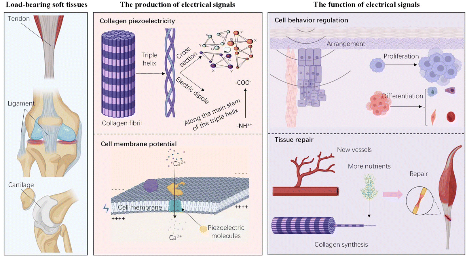 | ||
| Fig. 1 The electrical cues in load-bearing soft tissues. On the left are examples of load-bearing soft tissues, including tendons, ligaments, and cartilage. In the center, two pathways for generating electrical signals within the tissue microenvironment are shown: the collagen piezoelectrical potential, produced by electric dipoles along the main stem of the triple helix structure, and the endogenous electrical signals generated by ion channels on the cell membrane. On the right, the functional effects of electrical signals are highlighted, influencing cell behaviors such as alignment, proliferation, and differentiation, as well as promoting tissue repair through processes like neovascularization and collagen synthesis (figures created with BioRender— https://www.biorender.com/). | ||
Bioelectric signals are crucial for various functions in human cells and tissues, influencing cell migration, growth, and tissue formation.35,36 They provide specific guidance for embryonic development and tissue remodeling after injury.37 These signals are essential for morphogenesis, maintaining the developmental morphology, and enabling cell replacement after injury.38 ES influences the cell arrangement according to the electric field's direction.2 Research shows that an optimal cell arrangement occurs at intensities below 10 V cm−1, although cell activity remains relatively low.24 Additionally, ES between 0.1 V cm−1 and 12 V cm−1 can induce cell migration without causing significant damage or affecting cell phenotype or differentiation potential. Continuous stimulation below 1 V cm−1 can increase cell proliferation, and higher intensities above 100 V cm−1 can also promote cell proliferation through short (less than 1 ms), single stimulations.26 However, the extremely high intensity is significantly greater than the membrane potential needed to regulate ion channels controlling intracellular calcium signaling, which may lead to cell death.39 Despite this, there has been limited research on the specific parameters of ES in load-bearing soft tissues, and the parameters for tendons and ligaments remain unclear. In contrast, some parameters for cartilage therapy have been explored. For example, in a rabbit cartilage defect model, Han et al.40 used ultrasound to drive the mechanical vibration of piezoelectric nanoparticles, generating ES with an open circuit voltage of 451 mV and a short circuit current of about 17 μA. This treatment achieved considerable recovery after 15-minute stimulations, twice a week, for two months. Furthermore, ES has been shown to induce cell differentiation, such as osteogenic differentiation in bone marrow stromal cells (BMSCs) and MC3T3-E1 cells.26 Recent studies by Ge et al.41 and Wu et al.42 have shown that ES increases the number of capillaries and fibroblasts in load-bearing soft tissues within a short period. This stimulation also increases the required nutrients, significantly enhancing collagen synthesis, tissue rehydration, and scar tissue repair. Ultimately, it promotes the healing of tissue microstructure and tissue regeneration (Fig. 1). These properties make ES a valuable tool in load-bearing soft tissue engineering.
Animal studies have shown that the repair outcomes of load-bearing soft tissues under ES are superior to those achieved through natural healing without electrical intervention.41,43 Specifically, rats subjected to ES exhibit greater movement distances and expanded activity areas, reflecting improved functional recovery. The biomechanical properties of the Achilles tendon are also significantly enhanced, with tendon length approaching normal levels and notable increases in elastic modulus and load to failure. Structurally, the repaired tissue shows increased collagen deposition and more orderly collagen fiber alignment. Additionally, there is reduced scar formation, resulting in higher-quality tissue repair. Macroscopic observations also reveal that tissues treated with ES appear cleaner and whiter, with minimal blood vessels and defects, compared to other groups.
2.2 Working mechanisms of electrical cues
From a mechanistic perspective, both the piezoelectric effect—which generates electrical signals transmitted to various biochemical reactions within cells through piezoelectric-sensitive structures44—and externally applied electrical stimuli influence calcium ion concentration, electrical signal pathways, and the expression of specific genes in load-bearing soft tissues (Fig. 2). Firstly, ES can activate voltage-gated calcium channels, leading to an influx of calcium ions into cells and an increase in intracellular calcium concentration. Wu et al.42 suggest that this influx transports calcium to the mitochondria, accumulating in the mitochondrial matrix and activating the dynamin-related protein 1 and mitochondrial rho GTPase of the outer membrane 1 signaling pathways. This activation promotes oxidative phosphorylation of tendon stem cells, enhancing ATP synthesis and regulating tendon stem cell differentiation. Additionally, several research groups have proposed that Ca2+ activates the calmodulin/calcineurin/nuclear factor of activated T-cell signaling pathway, promoting osteogenic differentiation of rBMSCs.28,45,46 Secondly, Wu et al.42 found that ES induces cells to transform into a spindle shape, which may lead to upregulation of tendon-related gene expression, indicating that this morphological change promotes tendon gene differentiation. Thirdly, studies by Ge et al.,41 Liu et al.47 and Yague et al.48 indicate that ES activates signaling pathways such as extracellular signal-regulated kinase/mitogen-activated protein kinase (ERK/MAPK), vascular endothelial growth factor (VEGF), nuclear factor kappa-B, interleukin 17 (IL-17), and TGF-β. The ERK/MAPK pathway increases the expression of Scx and tenascin C. VEGF promotes tendon regeneration by reducing adipocyte accumulation and enhancing vascularization.49 Targeting the IKK β/NF Kappa B pathway enhances tendon–bone healing.50 IL-17 cytokines and receptors amplify tendon inflammation, activating intracellular signaling pathways of inflammatory genes.51 The TGF-β pathway enhances the secretion of TGF-β, promoting cartilage regeneration.47 Finally, Lai et al.52 report that piezoelectric stimulation impacts cellular metabolic activity, causing cells to secrete proteases and produce ECM proteins, thereby altering cell behavior and promoting tissue regeneration. Concurrently, ES can reduce alkaline phosphatase (ALP) activity and inhibit ECM mineralization.52 In summary, these mechanisms collectively highlight the potential of ES as a powerful tool in load-bearing soft tissue engineering, offering diverse pathways to enhance tissue repair and regeneration. | ||
| Fig. 2 Working mechanism of electrical cues. This illustrates the influx of calcium ions and related signaling pathways triggered by electrical signals, which influence gene expression and cell behavior. Abbreviations: ALP, alkaline phosphatase; ECM, extracellular matrix; VOCC, voltage-operated calcium channel; NFAT, nuclear factor of activated T-cell; CaM, calmodulin; CaN, calcineurin; MIRO-1, mitochondrial rho GTPase of the outer membrane 1; DRP-1, dynamin-related protein 1; R1, receptor 1; VEGF, vascular endothelial growth factor; VEGFR, vascular endothelial growth factor receptor; TGF-β, transforming growth factor beta; IL-17, interleukin 17; IL-17RA, interleukin 17 receptor A; NF-κB, nuclear factor kappa-B; ERK/MAPK, extracellular signal-regulated kinase/mitogen-activated protein kinase; shc, an adaptor protein; and Ras, a signaling switch protein (figures created with BioRender—https://www.biorender.com/). | ||
3 Conductive biomaterials for load-bearing soft tissue engineering
3.1 Brief introduction on conductive biomaterials
Conductive biomaterials, which are conductive materials with a degree of biocompatibility, can uniformly transmit exogenous ES to cultured cells in vitro, and can also uniformly transmit both exogenous and endogenous ES along the bioactive scaffold after implantation in vivo. Concurrently, conductive biomaterials also facilitate the reconstruction of electrical signaling pathways at the site of tissue injury. Relevant studies have demonstrated that, even in the absence of external ES, conductive polymers can accelerate tissue repair following implantation.53 Moreover, conductive materials with stress sensitivity can quickly and accurately adjust their conductivity based on the magnitude of stress applied to the scaffold, making them useful for in vivo monitoring of load-bearing soft tissues.Common conductive biomaterials include conductive polymers such as conjugated polypyrrole (PPy), polyaniline (PANi), and poly(3,4-ethylenedioxythiophene) (PEDOT); carbon-based materials such as graphene, diamond, carbon nanotubes and nanowires; as well as metal-based materials such as gold nanoparticles, silver nanowires (AgNWs) and liquid metals.54,55 PPy is one of the most extensively studied conductive polymers due to its high conductivity, good chemical stability in air and water, and good biocompatibility. However, polymerized PPy is not easily soluble, which limits its processability.56 The advantages of PANi include good conductivity, low cost, and good stability. However, PANi has poor processability, and there are reports that it may lead to chronic inflammation after implantation.57 PEDOT, a derivative of polythiophene, offers good conductivity and biocompatibility, as well as ideal chemical, electrical, and environmental stability. Poor solubility is a key challenge when using PEDOT, so it is usually combined with polystyrenesulfonates (PSS) to address this issue.58 Carbon based materials, such as graphene, have become a hot research topic in conductive materials due to their high conductivity, excellent mechanical properties, stability in water environments, ease of chemical modification, large surface area, and excellent performance in thermal and light conduction. The inherent conductivity of graphene allows it to establish electrical connections with cells, potentially regulating the electrophysiological properties. Additionally, non-covalent interactions between graphene and biomolecules, such as π–π stacking and hydrogen bonding, may promote cell differentiation.59 There are also ion-conductive polymers such as poly(ether sulfone) (SPES) and poly(phenylene oxide) (SPPO), which conduct charges through counter-ion flow in physiological environments, providing high conductivity with continuous conduction of stable currents.60–62 Metal-based conductive materials are well-suited for bioelectronic devices, particularly in applications that require precise monitoring and regulation of biological functions, due to their high conductivity and mechanical strength. In addition, transition metal carbides, nitrides, or carbonitrides (MXenes) represent a novel class of two-dimensional materials that exhibit high electrical conductivity, hydrophilicity, excellent mechanical strength, and partial biodegradability. They also possess antibacterial properties similar to those of AgNWs. These properties endow MXenes with significant potential for applications in tissue engineering. For instance, Fu et al.63 demonstrated that functionalized MXenes could effectively promote cell adhesion, proliferation, and osteogenic differentiation in bone repair studies. However, concerns regarding their cytotoxicity, particularly long-term in vivo toxicity, remain unsolved. Moreover, most of these materials lack biodegradability. Future efforts should focus on developing more biodegradable conductive biomaterials and conducting in-depth investigations into their potential toxicity, including the toxicity of their degradation products.
In recent years, scientists have extensively explored the use of conductive biomaterials combined with hydrogels to create composite scaffolds, which are being studied for their potential in the regeneration, repair and motion detection of load-bearing soft tissues (Fig. 3).64–67 Hydrogels are widely used in tissue engineering due to their ECM-mimicking structure, good biocompatibility, and tunable chemical and physical properties.68–70 The realization of conductive hydrogels depends on the introduction of conductive materials into the hydrogel matrix and their integration into the three-dimensional interconnected network, allowing for successful interaction between the conductive components and the hydrogel polymer chain. At present, conductive hydrogels are commonly classified based on their functional properties. Elastic conductive hybrid hydrogels, known for their excellent elastic mechanical properties, are widely used in electronic skin, artificial muscles, nerve prosthesis, and wearable or implantable biosensors.71,72 Ionic conductive hydrogels, with their high conductivity, have been extensively utilized in health testing, biological interfaces, and wearable devices over the past decades.73–75 Additionally, double network, interpenetrating network, and nanocomposite conductive hydrogels, which boast excellent mechanical properties such as tear resistance and fatigue resistance, are widely applied in tissue engineering.76–79
 | ||
| Fig. 3 Examples of conductive biomaterials and their applications in load-bearing soft tissue engineering. Abbreviations: PANi, polyaniline; PPy, polypyrrole; PEDOT, polyethylene dioxythiophene; AgNWs, silver nanowires; MWCNTs, multi-walled carbon nanotubes; PVA, polyvinyl alcohol; CNF, cellulose nanofibers; PSS, polystyrene sulfonate; GelMA, methacrylated gelatin; PAM, polyacrylamide; PAA, polyacrylic acid; and PU, polyurethane (figures created with BioRender—https://www.biorender.com/). | ||
3.2 Conductive biomaterials in electro-biomechanical sensing
The resistance of certain strain-sensitive conductive biomaterials can change in real time in response to the magnitude of the applied strain. For instance, the electronic structural properties and electron transport pathways of carbon nanotubes are susceptible to strain. When subjected to mechanical forces, the resulting deformations can lead to changes in their electrical conductivity. This characteristic endows them with promising potential for application in bioelectronic sensing and monitoring devices (Fig. 3). Below, we will introduce recent research on conductive biomaterials for electro-biomechanical sensing in load-bearing soft tissues, mainly including PEDOT, AgNWs, liquid metals, and PPy.PEDOT possesses high conductivity, biocompatibility, and low cytotoxicity, but its low solubility often necessitates its combination with PSS. Li et al.80 reported on a high-strength, tough conductive hydrogel with anti-swelling ability, biocompatibility, and anisotropy. In their study, PEDOT:PSS served as the conductive component, while polyvinyl alcohol (PVA) and cellulose nanofibers (CNF) formed the biocompatible polymer matrix. They demonstrated the potential of this conductive hydrogel as a multifunctional implantable electro-biomechanical sensor for monitoring tendon repair. However, in an Achilles tendon recovery experiment in mice, the in vivo responses observed were limited to one and two weeks, which is insufficient to capture the chronic inflammation or tissue reactions that may arise from long-term implantation. The expansion of hydrogels typically decreases their mechanical strength and exerts pressure on adjacent tissues,81–83 making the combination of CNF with PVA advantageous. PVA exhibits high water absorption and significant protein resistance, while the –COOH in CNF provides fast and effective rehydration, which enhances the mechanical properties and water content of the anisotropic conductive hydrogel. At the same time, CNF and PVA are highly crosslinked through hydrogen bonds, which can further improve the cohesion and stability of hydrogels, thus maintaining the anti-swelling ability of hydrogels. By installing the hydrogel in a knee joint model to simulate ligament function and monitoring the resistance change (ΔR/R0) caused by the deformation during knee joint bending, they could track the joint's bending state in real time. However, it may encounter issues such as signal interference, equipment stability, and data interpretation in practical applications.
PPy is highly conductive and biocompatible, and can interact with cells and tissues without causing a significant immune response. It can regulate cell adhesion, migration, protein secretion, and DNA synthesis.84 Hu et al.85 used PPy as a conductive component to prepare gelatin methacrylate (GelMA) and polyacrylamide (PAM) double-network conductive hydrogels through photo-crosslinking. This hydrogel exhibits good electrochemical activity and a stable current response, with its resistance fluctuating instantaneously with external loading and unloading. This fast response capability and high sensitivity make it ideal for implantable biomedical devices and fast-response actuators. However, the study only observed short-term cellular behavior and did not assess tissue responses following long-term implantation. Notably, in material design, introducing the catechol derivative dopamine (DA) as a dopant can improve the water dispersion of PPy by synergically regulating the formation of PPy nanomaterials.83,86 This facilitates the formation of a uniform conductive network, thereby enhancing the electrical conductivity of the material. Additionally, numerous hydrogen bond interactions are formed, enhancing the elastic properties of the double-network hydrogels.
Double network hydrogels are widely studied for their significantly enhanced mechanical properties and good tear resistance.76,78,79 The combination of the double network and ionic liquid (IL) facilitates the mechanical properties of conductive hydrogels, especially tear resistance and modulus, by forming hydrogen bonds and ionic coordination between polymer networks. Recently, Hu et al.65 prepared a konjac glucomannan (KGM) and PAM dual-network hydrogel with an IL as the conducting component. This hydrogel demonstrates high sensitivity in monitoring human movements, such as finger flexing, ankle joint, frowning, and throat movements. Compared to other conductive biomaterials, ILs have a more prominent strain response ability.87 However, the feasibility of in vivo monitoring and the long-term stability of the material remain unverified. Additionally, the mechanical strength of the fabricated material is significantly lower than that required for load-bearing soft tissues, and this strength may be further compromised under aqueous conditions.
Silver has the strongest electrical conductivity among metals. Due to its excellent ductility, biocompatibility, and antibacterial ability, silver is widely used in biomedicine, electronic devices, and other fields.88,89 Recently, Wu et al.90 prepared a conductive hydrogel with an electrical conductivity of up to 8571 S cm−1 using silver metal. Its strain-sensitive conduction mechanism relies on the modulus difference between the outer silver layer and the inner hydrogel during stretching. The difference creates a stress concentration area in the silver layer. As strain increases, microcracks expand to release accumulated stress, causing a continuous increase in resistance. To further improve the mechanical properties and biocompatibility of PVA hydrogels, non-toxic, natural, biodegradable, oxygen-rich lignosulfonate (LS) can be doped into PVA hydrogels.91 Their hydrogel strain sensor can detect micro-vibrations and various human activities, but it still lacks validation of its effectiveness in vivo.
Introducing rigid fillers into a hydrogel base can lead to crack propagation during deformation, affecting the material's mechanical properties and conductive stability. To address this issue, Wang et al.92 introduced a eutectic gallium indium alloy (EGaIn) into conductive PVA-AgNW hydrogels. EGaIn is a low-melting liquid metal with a melting point of about 16 °C, remaining liquid at room temperature. It has high electrical conductivity (3.4 × 10⁶ S m−1) and environmental stability. During deformation, the liquid metal can deform with the base polymer, preventing crack growth and improving the hydrogel's mechanical properties.93,94 Additionally, gallium can form coordination bonds with PVA, further enhancing the hydrogel's mechanical properties.95 The combination of AgNWs, EGaIn and PVA results in a composite with high electrical conductivity, and exceptional toughness. Notably, these materials can be combined through various physical crosslinks during preparation, avoiding irreversible covalent chemical crosslinking. At high temperatures, these reversible physical interactions are destroyed, allowing for the reversible unwinding of PVA segments, making the hydrogel recyclable. The design of these functions and features is worth emulating. However, the overall strain insensitivity of the material limits its application in electro-biomechanical sensing monitoring for load-bearing soft tissues.
Conductive biomaterials based on hydrogels are increasing in popularity in load-bearing soft tissue research, making the selection of conductive fillers particularly important. Some rigid fillers cannot deform during the stretching of the hydrogel, making it difficult to form conductive paths. To be suitable for implantable applications such as tendons, ligaments, and cartilage, conductive biomaterials should have excellent mechanical properties, anti-swelling performance, a biomimetic anisotropic structure, and basic bearing capacity. However, the biomimetic properties of the aforementioned conductive biomaterials still fall significantly short of those of natural load-bearing soft tissues. This highlights the need for further exploration of additional fabrication methods and reinforcement strategies. Additionally, implantable electronic sensing devices may require a long-term, stable, and safe power supply, yet current studies have not addressed power supply solutions. Nanoscale self-powered generators and piezoelectric materials may hold promise for addressing this issue. Finally, while the sensing functionality offers a novel approach for monitoring tendon repair, translating these sensing signals into meaningful clinical guidance and rehabilitation strategies remains a significant challenge in practical applications.
3.3 Conductive biomaterials in electrical stimulation therapy
The feasibility of using conductive biomaterials to promote load-bearing soft tissue repair through ES is becoming increasingly evident (Fig. 3). Conductive scaffolds must possess sufficient mechanical strength to ensure stable electrical conductivity and avoid breaking under stress. Additionally, the scaffold's modulus (stiffness and elasticity) can impact cell adhesion, spreading, and proliferation,96 so it must be designed within the optimal range for load-bearing soft tissue regeneration and comfort. Commonly used materials include carbon-based biomaterials, such as reduced GO (rGO) and carbon nanotubes, due to their stable electrical conductivity and biocompatibility. However, this non-metallic conductive biomaterial does not seem to exhibit stable and high conductivity in hydrogels.rGo is a nanostructure created by introducing functional groups, such as hydroxyl and carboxyl groups, onto graphene sheets through chemical methods and specific reduction reactions. Notably, rGO can be biodegradable under certain conditions.97 However, the introduction of oxygen-containing functional groups during oxidation disrupts the electronic structure of graphene, leading to lower electrical conductivity compared to that of the original graphene.98 Guo et al.99 designed a conductive fiber scaffold composed of rGo and PEDOT, demonstrating good cellular compatibility and high conductivity (2.52 S cm−1). This scaffold not only supports strong cell adhesion, facilitating effective cell communication, but can also be biodegraded by horseradish peroxidase (HRP). However, in the regeneration of load-bearing soft tissues, the degradation rate of the scaffold materials must be well-matched with the rate of tissue regeneration. If the material does not degrade within an appropriate timeframe, it may hinder the integration and functional recovery of the newly formed tissue. This issue could be potentially circumvented if the material is used as an auxiliary patch for ES conduction.41 Additionally, while rGO exhibits good biocompatibility and a certain degree of degradability, its complete degradation in the in vivo environment remains a significant challenge. Their cell experiments were specifically tailored for neural cell culture. In the realm of load-bearing soft tissue regeneration, in vitro experimental outcomes often fail to fully capture the complexity of the in vivo environment. This discrepancy introduces uncertainties regarding the applicability of their findings to load-bearing soft tissue regeneration. Nevertheless, the robust mechanical properties of their materials in vitro offer valuable insights that could potentially be leveraged for this purpose. Notably, the scaffold can be integrated with a self-powered triboelectric nanogenerator (TENG) to serve as an efficient and stable charge transfer platform for cell ES. They proposed a solution for power supply, which, despite the need for further investigation into its safety, has contributed to the practical clinical translation and application of the material.
Multi-walled carbon nanotubes (MWCNTs) are nanoscale tubular structures composed of multiple layers of carbon atoms. Their introduction significantly enhances the mechanical strength, toughness, and biocompatibility of composites, making them suitable for durable and reliable implants and stents. MWCNTs also offer excellent electrical conductivity, which is essential for biomedical applications involving ES or signal transmission.100 Zenoozi et al.101 used MWCNTs as conductive fillers to create a polyurethane (PU)/polyacrylic acid (PAA) conductive scaffold with a semi-interpenetrating polymer network structure. PU, known for its range of properties from thermosetting materials to soft elastomers, offers excellent mechanical properties such as tensile strength, toughness, wear resistance, and biocompatibility, but has poor hydrophilicity affecting cell adhesion.102 Conversely, PAA is highly hydrophilic and improves cell adhesion.103,104 Combining PU and PAA in a semi-interpenetrating network leverages the advantages of both materials, addressing the issue of cell adhesion.105 The PU/PAA/MWCNT composite is suitable for biomedical implants, including artificial blood vessels, cartilage, tendons, and ligament replacements. It facilitates electrical transmission to tissue cells and serves as a pathway for ES to promote tissue regeneration. Their work provides valuable insights into the load-bearing soft tissue regeneration. However, despite the promising biocompatibility demonstrated in vitro, the cell types evaluated in the experiments were relatively limited, and the lack of in vivo validation introduces uncertainties regarding its practical applicability. Moreover, when considered as a substitute for artificial tendons or ligaments, the mechanical properties of this composite material still fall short of those of native tissues.
Implantable conductive hydrogels for load-bearing soft tissues must balance both conductivity and mechanical properties. However, these two factors are often in conflict during preparation, leading to complex fabrication processes and limited research.106,107 Additionally, achieving ES of load-bearing soft tissues through conductive biomaterials is less convenient compared to that of piezoelectric materials, as conductive materials require external electrodes or TENG motors for power supply. In the aforementioned study, the TENG demonstrated remarkable self-powered ES capabilities, generating a stable current of 30 μA from human motion, with the advantages of portability and low cost. However, its high output voltage may require additional circuitry for regulation, potentially limiting its application in more complex biomedical scenarios. The design of the material structure and shape, the uniform conduction of an electrical current, and the safety concerns related to the power supply are significant challenges in the development of conductive biomaterials.
4 Piezoelectric biomaterials for load-bearing soft tissue engineering
4.1 Brief introduction on piezoelectric biomaterials
Piezoelectric biomaterials, which convert mechanical energy into electrical signals, hold significant promise for both electro-biomechanical sensing and ES therapy in load-bearing soft tissues. Piezoelectric materials generate an electric potential due to their asymmetric crystal structure. When subjected to mechanical force, the uneven movement of positive and negative charges causes macroscopic polarization and generates an electric potential. The linear conversion between mechanical energy and electrical energy is represented by the equation:108D = [d]T + [εt]E, where D is the electrical displacement, [d] is the direct piezoelectric effect matrix, T is a constant stress field, [εt] is the transpose of the permittivity matrix, and E is the electrical field strength. Currently, piezoelectric biomaterials are classified into three categories: inorganic, organic, and composite piezoelectric biomaterials.Inorganic piezoelectric biomaterials mainly include piezoelectric nanobiomaterials based on ceramic nanoparticles, such as barium titanate (BaTiO3/BTO),109 potassium sodium niobate (K0.5Na0.5NbO3),110 zinc oxide (ZnO),111 boron nitride (BN)112 and piezoelectric single crystal black phosphorus.113 These materials, especially BaTiO3 and ZnO nanoparticles, typically exhibit excellent piezoelectric and mechanical properties, and they also demonstrated excellent biocompatibility and low cytotoxicity in the experiment. In addition, piezoelectric nanoparticles exhibit multifunctionality; for instance, BaTiO3 nanoparticles can generate reactive oxygen species, thereby demonstrating antibacterial properties. Organic piezoelectric biomaterials mainly include artificial piezoelectric polymers, such as polyvinylidene fluoride (PVDF),114 poly-β-hydroxybutyrate (PHB),115 and polylactic acid (PLLA), which are commonly fabricated into nanofiber-based biomaterials via electrospinning,116 as well as natural piezoelectric polymers, such as chitosan,117 collagen118 and peptides.119 These organic materials generally exhibit good flexibility and plasticity. It is worth noting that when natural organic piezoelectric materials are used in clinical trials, the issue of immunogenicity should be taken into consideration. To date, various methods have been developed to reduce immunogenicity. For instance, methods to reduce the antigenicity of collagen mainly include enzymatic treatment and cryopreservation at ultra-low temperatures.120 Furthermore, under certain conditions, recombinant collagen may serve as an effective substitute for native collagen.121
However, single-component piezoelectric biomaterials have several drawbacks. Most lack biological activity, which severely limits their ability to regenerate tissues. For example, although BaTiO3 and PVDF are not cytotoxic, their poor biological activity is a significant limitation. To address this, bioactive ions such as Ca2+, Mn2+, and Li+ are often doped to improve biological activity. Additionally, piezoelectric polymers can be modified physically or chemically on the surface, such as by chemically crosslinking a layer of functional proteins to make the polymer film more hydrophilic and biologically active.122 Another issue is that most piezoelectric biomaterials, like PVDF and ZnO, cannot degrade in vivo, necessitating secondary surgeries for removal, which causes further trauma. To address this, researchers have explored using PLLA, a biodegradable polymer made from renewable resources like corn starch, which degrades into lactic acid within the body. PLLA is used in biomedical applications such as absorbable sutures, drug delivery systems, and temporary implants.123,124 However, PLLA's piezoelectricity is significantly lower than that of PVDF, making it less feasible on its own for certain applications. To enhance its piezoelectric response, PLLA can be combined with nanoparticles, such as incorporating rGO as demonstrated by Lai et al.52 Moreover, most piezoelectric materials do not fully meet the requirements for tissue engineering, often lacking suitable mechanical properties, biocompatibility, or biodegradability. For instance, BTO films, while being elastic and mechanically strong, are far inferior to human tissues, particularly under long-term cyclic loading or complex mechanical environments.
Composite piezoelectric biomaterials combine the advantages of inorganic and organic piezoelectric materials. Particularly, hydrogel-based piezoelectric composites can mimic the mechanical environment of natural load-bearing soft tissues, exhibiting excellent mechanical properties, piezoelectricity, electrical output stability, and biocompatibility. Scientists have developed piezoelectric scaffolds using strong or double network hydrogels combined with nanoparticles like piezoelectric BaTiO3 and copper calcium titanate (CCTO) (Fig. 4). To enhance the hydrophilicity and cell adhesion for improved biocompatibility, natural biological derivatives such as dopamine, hydroxyapatite, and chondroitin sulfate, are often incorporated into these scaffolds.125,126 Detailed discussions on the specific compositions and applications of composite piezoelectric biomaterials will be provided in subsequent sections.
 | ||
| Fig. 4 Examples of piezoelectric biomaterials and their applications in load-bearing soft tissue engineering. Abbreviations: PLLA, polylactic acid; PVDF, polyvinylidene fluoride; SA, sodium alginate; HA, hydroxyapatite; CCTO, copper calcium titanate; OCS, chondroitin sulfate oxide; PETRR, 2,3-butanediol (2,3-BDO), sebacic acid (SeA), succinic acid (SuA), 1,3-propanediol (1,3-PDO), and isopropanol; TrFE, trifluoroethylene; PDA, polydopamine; rGo, reduced graphene oxide; and ABTO, aminated barium titanate (figures created with BioRender—https://www.biorender.com/). | ||
4.2 Piezoelectric biomaterials in electro-biomechanical sensing
Due to their unique electromechanical coupling ability, piezoelectric biomaterials are widely used in biosensing, tissue engineering, energy harvesting, and drug delivery.127,128 In the field of electro-biomechanical sensing for load-bearing soft tissues, it is crucial to develop biocompatible and durable piezoelectric materials with high voltage electrical performance.129–132 Currently, the mainstream piezoelectric biomaterials for electro-biomechanical sensing are mainly PVDF and BaTiO3 (Fig. 4). Below, we will introduce relevant scientific research progress.BaTiO3 nanoparticles possess high piezoelectric properties, dielectric constants, and excellent biocompatibility, making them ideal for biomedical applications.133 Hydrogels incorporating BaTiO3 nanoparticles can convert mechanical signals into electrical outputs, allowing for real-time wireless monitoring of human physiological signals. For example, Fu et al.125 reported a BaTiO3-based piezoelectric hydrogel capable of stably monitoring human gestures, elbow flexion, knee flexion, and plantar pressure distribution, demonstrating its potential for health monitoring, rehabilitation, and sports training.134,135 However, while the sensor demonstrates promising performance in controlled settings, its operation within physiological systems requires careful consideration of potential signal confounders. In the design of composite materials, to enhance the mechanical properties of BaTiO3-based hydrogels, a stable and dense cross-linked polymer network was constructed by introducing oxychondroitin sulfate (OCS) and boric acid. The hydrogen bonds between OCS and gelatin chains, along with the borate-diol ester bonds between OCS and boric acid, form a stable polymer network. Additionally, modifying BaTiO3 nanoparticles with an amino group improves their dispersion, allowing for uniform incorporation into the gelatin matrix and enhancing the hydrogel's mechanical strength. The combination of naturally derived substances such as gelatin and chondroitin sulfate (CS) with BaTiO3 further enhances the overall biocompatibility.
PVDF is a unique piezoelectric material known for its excellent piezoelectric properties, thermal stability, and chemical resistance. However, its superhydrophobic nature poses challenges for certain tissue engineering applications.136–139 To address this, a sodium alginate hydrogel, a non-toxic, biocompatible, and biodegradable polysaccharide, is often used.140,141 Additionally, hydroxyapatite (HA) can be introduced into the hydrogel substrate to enhance mechanical properties and biocompatibility.142 HA, a key mineral in human bones, is known for promoting the adhesion, proliferation, and ECM deposition of osteoblasts and contributing to the piezoelectricity of bone defects.143,144 Khazani et al.126 recently reported a piezoelectric scaffold made of a sodium alginate hydrogel and PVDF, used in bone tissue engineering, human movement monitoring, and energy harvesting due to its piezoelectric properties, biocompatibility, cell proliferation, and antibacterial properties. In addition, they also incorporated CCTO nanofillers, known for their high dielectric constant and suitability as a lead-free bioceramic filler, to improve the dielectric properties of the PVDF hydrogel scaffold. The addition of nanofillers can increase the crystallinity of the PVDF-β phase and align the dipole moments, thus increasing the piezoelectric output response. Furthermore, the presence of nanostructures facilitates cross-linking reactions and the formation of denser polymer networks in the hydrogel scaffolds.
Aside from the widely used piezoelectric materials BaTiO3 and PVDF, Ge et al.41 reported a polyester-based piezoelectric elastomer (PETRR) copolymerized from biobased monomers. They designed a wireless sensor for monitoring Achilles tendon rupture during exercise, which was implanted on the rabbit Achilles tendon to respond in real time to stress changes during Achilles tendon repair. Notably, the patch-based design developed in this study can effectively adhere to the Achilles tendon in animal experiments. The integrated circuit board on the dorsal side enables reliable signal transduction. This design not only avoids occupying the space of newly formed tissue during the repair process but also maximizes the success rate of surgical suturing. Biobased monomers such as 2,3-butanediol (2,3-BDO), sebacic acid (SeA), succinic acid (SuA), 1,3-propanediol (1,3-PDO), and isopropanol (IA) are derived from biomass or obtained through biochemical synthesis.145,146 The ester bonds formed by polymerization between them are rich in C–O dipoles, ensuring the piezoelectric properties of the polymer for electrokinetic sensing.147 During physiological activity, stretching the copolymer causes the C–O dipole to deflect, leading to a buildup of charge on the surface electrodes, and ultimately the collection of piezoelectric signals via a wireless module. The introduction of 2,3-BDO and IA provides steric hindrance and cross-linking sites, combined with random copolymerization, giving the copolymer an amorphous structure and a low elastic modulus.148–150 Additionally, since the polymer elastomer is derived from biomass, it meets the requirements for biocompatibility.
Similarly, Xiong et al.43 employed a chitosan-based piezoelectric hydrogel to fabricate a patch-type sensor with a structurally analogous design for the treatment of Achilles tendon rupture. The chitosan matrix exhibited intrinsic antibacterial activity, addressing the clinical need for postoperative infection prevention. Furthermore, the incorporation of temperature-sensing capabilities enabled real-time monitoring of inflammation responses at the wound site. These advancements in material design and functional integration represent significant progress toward clinical translation. Nevertheless, several challenges require further investigation, particularly regarding long-term in vivo performance evaluation and mechanical property optimization to meet clinical requirements.
In the field of electro-biomechanical sensing and monitoring, hydrogel-based matrices have emerged as a preferred platform, where piezoelectric functionality is typically achieved through the incorporation of fillers via cross-linking polymerization. The combination and interaction of different materials yield varying performance outcomes. Their ability to recapitulate key aspects of the native tissue microenvironment, including ECM-mimetic viscoelasticity and tissue-specific stiffness, makes them ideal for regulating cellular behavior and facilitating tissue remodeling. The selection of piezoelectric fillers is diverse, with biodegradable materials inevitably leading to instability in piezoelectric signals and significant performance degradation over long-term in vivo implantation. This limitation restricts the use of biodegradable piezoelectric sensors to short-term health monitoring. For applications requiring stable performance, PVDF and patch-type PETRR can be chosen due to their excellent biochemical stability and consistent electrical output. Their biocompatibility, mechanical properties and dispersibility in the matrix can be improved by surface modifications with materials like DA, HA, and OCS. For high sensitivity and fast response times, incorporating materials with high dielectric constants, such as CCTO, is beneficial. However, the structural and mechanical design of piezoelectric materials is crucial for effectively responding to strain changes while meeting the survival requirements of cells and tissues. For instance, patch-type structures are better suited for integrating with soft tissues, thereby enhancing signal reception. Moreover, the stiffness gradient of the ECM at the interface between soft and hard tissues can serve as a physical cue to regulate cell behavior, potentially initiating cell polarization.151
4.3 Piezoelectric biomaterials in electrical stimulation therapy
Compared to conductive biomaterials, scaffolds made of piezoelectric biomaterials can directly convert mechanical stress from the body into ES, eliminating the need for external power sources and making them both convenient and safe. In the field of ES therapy, PVDF and PLLA are the primary materials used in piezoelectric applications (Fig. 4). Below, we introduce some related works on piezoelectric scaffolds, offering insights and references for achieving ES therapy for load-bearing soft tissues.PVDF and trifluoroethylene (TrFE) copolymers have become essential materials for fabricating piezoelectric scaffolds in tissue engineering due to their unique piezoelectric properties, chemical stability, and the combination of PVDF strength with TrFE flexibility. These scaffolds not only facilitate the repair of tissues such as tendons, ligaments and nerves, but are also widely used in drug delivery systems.152,153 Yague et al.48 created a ferroelectric polymer scaffold using PVDF-TrFE, and developed a biomimetic electromechanical stimulation device to study signaling pathways associated with tendon repair. Wu et al.42 developed a self-powered wearable ES patch using a PVDF-TrFE film as the negative electrode material of a friction plate. Through triboelectrification, an opposite surface charge is generated on the material's surface. When the positive and negative electrode materials are separated, a potential difference is created between the electrodes, producing an electrical output via an external circuit. This continuous conversion of mechanical energy to electrical energy improves vascular congestion and redness of the Achilles tendon, and promotes collagen formation and tendon cell differentiation. The physical properties of PVDF-TrFE can be influenced by several factors in application.48 Firstly, fiber diameter determines overall stiffness, and as the fiber diameter decreases, mechanical properties (Young's modulus) are enhanced due to centripetal forces stretching the fiber during collection, resulting in chain height extension and orientation along the fiber axis. Secondly, the piezoelectric properties of PVDF-TrFE copolymers are affected by crystallinity, particularly the content of the β phase, which can often be promoted by short-distance electrospinning. The piezoelectric PVDF-TrFE scaffold reported by Yague et al. exhibited a Young's modulus of about 61.8 MPa, a strength of 31 MPa, and an output voltage of about 1 V, closely matching the strength of human load-bearing soft tissues. This is a remarkable demonstration. However, PVDF-TrFE, as a synthetic polymer, lacks biodegradability, which may limit its applicability in long-term implantation. The fabrication process, cost, and ease of clinical operation of the material also require further evaluation. Additionally, the impact of ES parameters (e.g., intensity, frequency, and duration) on cellular behavior and tissue regeneration remains to be fully elucidated, and individual variations may exist. Further research is needed to optimize these parameters and develop more precise control system, potentially enabling personalized treatment protocols. Moreover, incorporating antibacterial components or developing coatings with antibacterial functions could enhance the material's resistance to infections and reduce the risk of postoperative complications.
As a biocompatible and biodegradable polymer, PLLA shows broad application prospects in promoting the repair and regeneration of bone, cartilage, nerve, and other tissues. Combining PLLA with advanced nanotechnology and surface modification offers new solutions and therapeutic strategies in tissue engineering.128,154–156 However, PLLA's piezoelectric response is poor. Introducing piezoelectric nanoparticles can effectively improve the piezoelectric properties and strength of PLLA-based composites due to the significant rigid particle reinforcement effect induction of crystallinity in the PLLA matrix.157,158 The weak interface binding between organic and inorganic materials can compromise their piezoelectric properties.157,159–163 To address this, polydopamine (PDA) is often used for surface modification. The hydroxyl groups in polydopamine can form covalent bonds with the hydroxyl group on the nanoparticle164 and its amino groups can form strong hydrogen bonds with PLLA's ester groups.165 This significantly improves mechanical stress transferring at the interface, enhancing the piezoelectric output and sensitivity of the composite. PDA also promotes cell adhesion and material stability through oxidative self-polymerization, giving the material excellent biocompatibility and creating a conducive microenvironment for load-bearing soft tissue cell adhesion and growth. For incorporating organic materials like rGo, more C–O electric dipoles can be added to PLLA, further enhancing the composite's piezoelectric properties.166 Zhang et al.46 and Lai et al.52 recently reported on two related piezoelectric scaffolds, demonstrating these advancements.
Through electrospinning, Zhang et al.46 constructed a Janus nanofiber scaffold (OPZ/RPB) using the piezoelectric polymer PLLA, polydopamine, and inorganic piezoelectric nanoparticles ZnO and BaTiO3. This scaffold demonstrated excellent biocompatibility and electrical output stability in tendon and bone healing. ZnO has been shown to stimulate tendon cell proliferation, induce tendon cell differentiation, promote collagen arrangement, and facilitate early fibrocartilage formation.159,162 Similarly, BaTiO3 effectively promotes the proliferation and differentiation of osteoblasts, thereby aiding in bone regeneration.160,163 Lai et al.52 prepared a PLLA/rGO piezoelectric fiber scaffold, incorporating PDA for surface modification. This scaffold supports improved cell morphology and differentiation, promoting tissue repair and regeneration. The degradation of PLLA produces lactic acid, which has been suggested to modulate cellular behavior. However, an excessively high concentration of lactic acid could create an acidic microenvironment, potentially impairing cell viability and function. Moreover, limitations such as the restricted selection of cell types, inadequate optimization of ES parameters, and mismatched mechanical properties need to be addressed in future studies to enhance the clinical applicability and efficacy of these materials.
In addition to the aforementioned materials, PETRR and chitosan-based piezoelectric hydrogel patches have been employed not only for monitoring Achilles tendon rupture during movement but also for delivering ES to facilitate Achilles tendon regeneration.41,43 These patches conform uniformly to the tendon surface, conducting electrical currents without the need for an external power source, thereby circumventing clinical concerns regarding electrical safety. If the challenge of gradual and safe degradation in alignment with tissue healing during long-term in vivo implantation can be addressed, it could pay the way for significant advancements in clinical translation.
To achieve effective ES therapy for human load-bearing soft tissues, piezoelectric composite material scaffolds must possess stable electrical output, excellent biocompatibility, and limited inflammatory response, while balancing durability and biodegradability.167 Currently, most related research is confined to in vitro cell culture, with few studies focusing on long-term in vivo implantation. The use of piezoelectric materials within the body, which rely on mechanical forces generated by biological activities to stimulate piezoelectric activation, may fail to reach the threshold necessary for effective activation and could potentially cause secondary damage to injured tissues. Moreover, there is a lack of specific research on the optimal parameters for ES therapy of load-bearing soft tissues, likely due to the challenges in accurately controlling and monitoring the piezoelectric output of implanted materials. To address these issues, it is critical to innovate and develop more intelligent biological scaffolds, such as those driven by ultrasound to generate piezoelectric output, along with interdisciplinary intelligent signal-receiving devices.
5 Conclusions and future perspectives
In this review, we have highlighted advancements in the field of conductive and piezoelectric biomaterials for load-bearing soft tissue engineering (Table 1). Conductive biomaterials enable ES through external power supplies and monitor tissue movement via strain-sensitive resistance. Piezoelectric biomaterials, with their inherent electromechanical coupling ability, generate ES from tissue movement or convert it into visual digital electrical signals. These research-oriented explorations hold promise for future clinical treatment, potentially resolving longstanding issues in load-bearing soft tissue repair.| Scaffold materials | Tensile/compressive strength | Modulus | Sensitivity/conductivity/output voltage | Related work | Ref. |
|---|---|---|---|---|---|
| Abbreviations: PPy, polypyrrole; PEDOT, polyethylene dioxythiophene; AgNWs, silver nanowires; MWCNTs, multi-walled carbon nanotubes; PVA, polyvinyl alcohol; CNF, cellulose nanofibers; PSS, polystyrene sulfonate; GelMA, methacrylated gelatin; PAM, polyacrylamide; PAA, polyacrylic acid; PU, polyurethane; DA, dopamine; PLLA, polylactic acid; PVDF, polyvinylidene fluoride; SA, sodium alginate; HA, hydroxyapatite; CCTO, copper calcium titanate; OCS, chondroitin sulfate oxide; 2,3-BDO, 2,3-butanediol; SeA, sebacic acid; SuA, succinic acid; 1,3-PDO, 1,3-propanediol; IA, isopropanol; TrFE, trifluoroethylene; PDA, polydopamine; rGo, reduced graphene oxide; ABTO, aminated barium titanate; BTO, barium titanate; KGM, konjac glucomannan; IL, ionic liquid; LS, lignosulfonate; Gel, gelatin; and PET, polyethylene terephthalate. | |||||
| PVA-CNF-PEDOT:PSS | 3.71 MPa | 1.1 MPa | GF = 0.88–1.30, 0.3 S m−1 | Monitoring tendon repair process and movement status, and as a tendon substitute | 80 |
| PVA-AgNWs-Ga/In | 13–33 MPa | 12.2–48.8 MPa | 24 S m−1 | High-frequency signal transmission | 92 |
| GelMA/PAM/DA-PPy | 0.0021 MPa | 0.0707 S m−1 | Implanting biomedical devices and rapid-response actuators | 85 | |
| KGM/PAM-IL | 0.0786 MPa | 0.025–0.04 MPa | GF = 1.25–2.60, 0.76 S m−1 | Monitoring human movements, such as finger flexing, ankle joint motion, and frowning | 65 |
| PVA/LS-Ag | 0.62 MPa | GF = 177.65, 857![[thin space (1/6-em)]](https://www.rsc.org/images/entities/char_2009.gif) 100 S m−1 100 S m−1 |
Detecting microvibrations and various human activities | 90 | |
| rGO-PEDOT | 84 MPa | 252 S m−1 | Application in cell ES and nerve tissue regeneration | 99 | |
| PU/PAA-MWCNT | 20 MPa | 144–204 MPa | 0.15 S m−1 | Artificial materials for blood vessels, cartilage, and tendons | 101 |
| 2,3-BDO/SeA/SuA/1,3-PDO/IA | 0.3 MPa | GF = 177.65, 0.149–0.43 V | Wireless monitoring of physiological activity and promoting tendon tissue regeneration | 41 | |
| SA/HA-PVDF/CCTOs | 8.2 MPa | 1.2–4.2 V | Human motion detection | 126 | |
| Gel/OCS-ABTO | 0.043 MPa | 0.22 MPa | 0.085–0.09 V | Wireless monitoring of human physiological activity | 125 |
| PVDF-TrFE | 31 MPa | 1 V | Promoting the repair of tendons, ligaments, and nerves | 48 | |
| PVDF-TrFE/PET patch | 61.8 MPa | 5–8 V | Tendon therapy | 42 | |
| PLLA/ZnO-PLLA/BTO | 7.47 MPa | 249.58 MPa | 2 V | Promoting osteogenic differentiation, regulating cell behavior, and promoting synchronous healing of the tendon–bone interface | 46 |
| PDA/rGo-PLLA | 0.7 MPa | 5.5 V | Cell therapy | 52 | |
Despite progress, these materials are still in the experimental stage, primarily tested in cellular and animal models. Long-term safety, degradability, and functional performance need further research. Additionally, specific parameters for effective ES therapy for load-bearing soft tissues are not well-defined, and current conductive and piezoelectric scaffolds often lack the necessary strength. Future research should address these challenges by exploring new material combinations, enhancing mechanical and electrical properties, and conducting extensive in vivo studies. Developing standardized protocols and parameter values for ES therapy will be crucial for translating these technologies into clinical applications. By overcoming these challenges, we can unlock the potential of conductive and piezoelectric biomaterials, leading to innovative treatments for load-bearing soft tissue injuries and diseases.
Author contributions
Wenjie Wu: conceptualization, methodology, investigation, writing – review and editing, visualization, supervision, and writing – original draft. Huihui Yang: investigation and writing – review and editing. Tingting Li: visualization, supervision, and writing – original draft. Yang Xie: conceptualization and methodology. Guoyou Huang: conceptualization, methodology, investigation, writing – review and editing, visualization, supervision, and writing – original draft. Weiwei Zhang: visualization, supervision, and writing – original draft.Data availability
No primary research results, software or code have been included and no new data were generated or analysed as part of this review.Conflicts of interest
There are no conflicts to declare.Acknowledgements
This work was financially supported by the National Natural Science Foundation of China (22204151, 12272278, and 12432015), the Natural Science Foundation Project of Hubei Province (2023AFB586), the Translational Medicine and Interdisciplinary Research Joint Fund of Zhongnan Hospital of Wuhan University (ZNJC202227), and the Special Funds for Basic Scientific Research Operations of Central Universities (2042023kf0167).References
- K. Nishizawa, K. Harato, S. Kobayashi, Y. Niki and T. Nagura, Knee, 2024, 48, 8–13 CrossRef PubMed
.
- B. Ferrigno, R. Bordett, N. Duraisamy, J. Moskow, M. R. Arul, S. Rudraiah, S. P. Nukavarapu, A. T. Vella and S. G. Kumbar, Bioact. Mater., 2020, 5, 468–485 Search PubMed
- S. Sasajima and K. Kubo, J. Biomech., 2024, 170, 112168 CrossRef PubMed
- W. L. Lim, L. L. Liau, M. H. Ng, S. R. Chowdhury and J. X. Law, Tissue Eng. Regener. Med., 2019, 16, 549–571 CrossRef CAS PubMed
- M. Gomez-Florit, C. J. Labrador-Rached, R. M. A. Domingues and M. E. Gomes, Adv. Drug Delivery Rev., 2022, 185, 114299 CrossRef CAS PubMed
- E. Gracey, A. Burssens, I. Cambre, G. Schett, R. Lories, I. B. McInnes, H. Asahara and D. Elewaut, Nat. Rev. Rheumatol., 2020, 16, 193–207 CrossRef PubMed
- Y. Liu, W. Da, M.-J. Xu, C.-X. Xiao, T. Deng, S.-L. Zhou, X.-T. Chen, Y.-J. Zhou, L. Tang, Y. Nie, Y. Zeng, H.-Q. Xie and B. Shen, Signal Transduction Targeted Ther., 2025, 10, 40 CrossRef CAS PubMed
- C. Rieu, L. Picaut, G. Mosser and L. Trichet, Curr. Pharm. Des., 2017, 23, 3483 CrossRef CAS PubMed
- D. Amiel, J. E. Billings and F. L. Harwood, J. Appl. Physiol., 1990, 69, 902–906 CrossRef CAS PubMed
- B. Erol, B. Kocaoglu and T. Esemenli, J. Foot Ankle Surg., 2007, 46, 155–161 CrossRef PubMed
- T. Hiranaka, T. Furumatsu, Y. Kamatsuki, K. Sugiu, S. Miyazawa, Y. Okazaki, S. Masuda, Y. Okazaki, Y. Kodama and T. Ozaki, Sports Med. Arthrosc. Rehabil. Ther. Technol., 2020, 20, 1–5 Search PubMed
- J. P. Barker, S. D. Simon and J. Dubin, J. Bone Joint Surg. Am., 2017, 99, 711–719 CrossRef PubMed
- M. J. DeFranco, B. Bershadsky, J. Ciccone, J.-K. Yum and J. P. Iannotti, J. Shoulder Elbow Surg., 2007, 16, 759–765 CrossRef PubMed
- Z. Y. Ahmad and M. J. Rasiej, Semin. Ultrasound CT MR, 2023, 44, 319–331 CrossRef PubMed
- F. A. Huyke-Hernández, S. A. Doxey, A. J. Only, A. Sibley, N. Mikhael, C. Y. Kweon and B. P. Cunningham, J. Orthop., 2023, 45, 6–12 CrossRef PubMed
- Y. Teng, Y. Cai, H. Wang, F. Lu, S. Zhang, M. Wu, H. Han, X. Yun, Y. Xia and X. Ma, Arthrosc. Tech., 2024, 103097 CrossRef PubMed
- X. Zhang, T. Wang, Z. Zhang, H. Liu, L. Li, A. Wang, J. Ouyang, T. Xie, L. Zhang, J. Xue and W. Tao, Mater. Today, 2023, 68, 177–203 CrossRef CAS
- H. Zhu, J. Ren, X. Wang, W. Qin and Y. Xie, J. Orthop. Surg. Res., 2025, 20, 159 CrossRef PubMed
- S. E. Murthy, A. E. Dubin and A. Patapoutian, Nat. Rev. Mol. Cell Biol., 2017, 18, 771–783 CrossRef CAS PubMed
- M. B. Klein, H. Pham, N. Yalamanchi and J. Chang, J. Hand Surg. Am., 2001, 26, 847–854 CrossRef CAS PubMed
- J. Zhang and J. H. C. Wang, J. Orthop. Res., 2010, 28, 198–203 CrossRef PubMed
- J. Li, Y. Long, F. Yang and X. Wang, Curr. Opin. Solid State Mater. Sci., 2020, 24, 100806 CrossRef CAS PubMed
-
M. Fernandez, Piezoelectric scaffolds: Mediating tendon regeneration by activation of piezosensitive receptors, NUI Galway, 2019
- S. Zhao, P. Tseng, J. Grasman, Y. Wang, W. Li, B. Napier, B. Yavuz, Y. Chen, L. Howell, J. Rincon, F. G. Omenetto and D. L. Kaplan, Adv. Mater., 2018, 30, 1800598 CrossRef PubMed
- Y. Wang, M. Rouabhia and Z. Zhang, Biochim. Biophys. Acta, Gen. Subj., 2016, 1860, 1551–1559 CrossRef CAS PubMed
- C. Chen, X. Bai, Y. Ding and I. S. Lee, Biomater. Res., 2019, 23, 25 CrossRef PubMed
- A. Leitolis, A. W. Robert, I. T. Pereira, A. Correa and M. A. Stimamiglio, Front. Cell Dev. Biol., 2019, 7, 164 CrossRef PubMed
- C. Alvarez-Lorenzo, M. Zarur, A. Seijo-Rabina, B. Blanco-Fernandez, I. Rodríguez-Moldes and A. Concheiro, Mater. Today Bio, 2023, 22, 100740 CrossRef CAS PubMed
- D. Khare, B. Basu and A. K. Dubey, Biomaterials, 2020, 258, 120280 CAS
- M. Levin, G. Pezzulo and J. M. Finkelstein, Annu. Rev. Biomed. Eng., 2017, 19, 353–387 CrossRef CAS PubMed
- H.-Y. Son and S.-S. Park, Mater. Today Sustain., 2024, 25, 100689 Search PubMed
- J. C. Góes, S. D. Figueiró, J. A. C. De Paiva, I. F. De Vasconcelos and A. S. B. Sombra, J. Mater. Sci. Lett., 1999, 18, 983–986 CrossRef
- S. L. Michlovitz, J Hand Ther, 2005, 18, 292–296 CrossRef PubMed
- W. S. Williams and L. Breger, J. Biomech., 1975, 8, 407–413 CrossRef CAS PubMed
- R. Araya, V. Nikolenko, K. B. Eisenthal and R. Yuste, Proc. Natl. Acad. Sci. U. S. A., 2007, 104, 12347–12352 CrossRef CAS PubMed
- L. Sacconi, D. A. Dombeck and W. W. Webb, Proc. Natl. Acad. Sci. U. S. A., 2006, 103, 3124–3129 CrossRef CAS PubMed
- M. Levin, Semin. Cell Dev. Biol., 2009, 20, 543–556 CrossRef PubMed
- K. A. McLaughlin and M. Levin, Dev. Biol., 2018, 433, 177–189 CrossRef CAS PubMed
- V. P. Tadic, T. Timic Stamenic and S. M. Todorovic, Sci. Rep., 2025, 15, 4966 CrossRef CAS PubMed
- Z. Han, F. Wang, W. Xiong, C. Meng, Y. Yao, W. Cui and M. Zhang, Adv. Mater., 2025, 37, e2414555 CrossRef PubMed
- Z. Ge, Y. Qiao, W. Zhu, Y. Xu, Q. Fang, D. Wang, Y. Tang, R. Zhao, X. Deng, W. Lin, G. Wang, Y. Xiang and X. Hu, Nano Energy, 2023, 115, 108751 CrossRef CAS
- Y. Wu, K. Zhang, S. Li, Z. Xiang, G. Jiang, R. Zhang, Y. Qi, X. Ji, X. Cai, C. Zhang, J. Li, R. Yan, H. Jin, S. Dong, J. Luo and G. Feng, Nano Energy, 2024, 121, 109234 CrossRef CAS
- Z. Xiong, B. Lin, C. Huang, A. Duan, C. Zhang, G. Qiang, W. Liu, R. Zhao, X. Deng, D. Wang, Z. Ge, G. Wang, X. Hu and W. Lin, Carbohydr. Polym., 2025, 352, 123149 CrossRef CAS PubMed
-
C. Alcaino, G. Farrugia and A. Beyder, in Current Topics in Membranes, ed. P. A. Gottlieb, Academic Press, 2017, vol. 79, pp. 219–244 Search PubMed
- W. Lin, Z. Zhou, Z. Chen, K. Xu, C. Wu, X. Duan, L. Dong, Z. Chen, W. Weng and K. Cheng, ACS Appl. Mater. Interfaces, 2023, 15, 46493–46503 CrossRef CAS PubMed
- Q. Zhang, J. Zhu, X. Fei and M. Zhu, Nano Today, 2024, 55, 102208 CrossRef CAS
- Y. Liu, G. Dzidotor, T. T. Le, T. Vinikoor, K. Morgan, E. J. Curry, R. Das, A. McClinton, E. Eisenberg, L. N. Apuzzo, P. Prasad, T. J. Flanagan, S. W. Lee, H. M. Kan, K. W. H. Lo, C. T. Laurencin and T. D. Nguyen, Sci. Transl. Med., 2022, 14, eabi7282 CrossRef CAS PubMed
- M. A. Fernandez-Yague, A. Trotier, S. Demir, S. A. Abbah, A. Larranaga, A. Thirumaran, A. Stapleton, S. A. M. Tofail, M. Palma, M. Kilcoyne, A. Pandit and M. J. Biggs, Adv. Mater., 2021, 33, e2008788 CrossRef PubMed
- F. Lai, J. Wang, H. Tang, P. Huang, J. Liu, G. He, M. Zhou, X. Tao and K. Tang, FASEB J., 2022, 36, e22433 CrossRef CAS PubMed
- M. Golman, X. Li, D. Skouteris, A. A. Abraham, L. Song, Y. Abu-Amer and S. Thomopoulos, Am. J. Sports Med., 2021, 49, 780–789 CrossRef PubMed
- J. Y. Mimpen, S. J. B. Snelling, A. J. Carr and S. G. Dakin, Front. Bioeng. Biotechnol., 2021, 9, 795830 CrossRef PubMed
- Y.-H. Lai, Y.-H. Chen, A. Pal, S.-H. Chou, S.-J. Chang, E. W. Huang, Z.-H. Lin and S.-Y. Chen, Nano Energy, 2021, 90, 106545 CrossRef CAS
- R. L. Keate, J. Tropp, R. Wu, A. J. Petty 2nd, G. A. Ameer and J. Rivnay, Adv. Sci., 2024, 11, e2305562 CrossRef PubMed
- R. Arambula-Maldonado, Y. Liu, M. Xing and K. Mequanint, Biomater. Adv., 2023, 154, 213616 CrossRef CAS PubMed
- Y. Li, L. Wei, L. Lan, Y. Gao, Q. Zhang, H. Dawit, J. Mao, L. Guo, L. Shen and L. Wang, Acta Biomater., 2022, 139, 157–178 CrossRef CAS PubMed
- L. Hao, C. Dong, L. Zhang, K. Zhu and D. Yu, Polymers, 2022, 14, 5139 CrossRef CAS PubMed
- T. H. Qazi, R. Rai, D. Dippold, J. E. Roether, D. W. Schubert, E. Rosellini, N. Barbani and A. R. Boccaccini, Acta Biomater., 2014, 10, 2434–2445 CrossRef CAS PubMed
- Y. Jin, Z. Li, S. Huang, W. Ning, X. Yang, Y. Liu, Z. Su, J. Song, L. Hu, X. Yin, H. Lu, H. Zuilhof, H. Wang and Z. Li, Colloids Surf., A, 2024, 697, 134461 CrossRef CAS
- Y. Zare and K. Y. Rhee, Sci. Rep., 2022, 12, 15179 CrossRef CAS PubMed
- Z. Chen, L. Lu, Y. Zhang, H. Sun, K. Fan, B. Zhang and J. Song, Chem. Eng. J., 2024, 487, 150688 CrossRef CAS
- M. Jia, Z. Bi and X. Guo, Chem. Commun., 2022, 58, 8638–8641 RSC
- M. I. Shekh, M. Wang, G. Zhu, F. J. Stadler, J. Ma and B. Du, Compos. Struct., 2024, 329, 117797 CrossRef CAS
- Y. Fu, J. Zhang, H. Lin and A. Mo, Mater. Sci. Eng. C: Mater. Biol. Appl., 2021, 118, 111367 CrossRef CAS PubMed
- M. Hafezi, S. N. Khorasani, M. Zare, R. E. Neisiany and P. Davoodi, Polymers, 2021, 13, 4199 CrossRef CAS PubMed
- X. Hu, J. Wang, S. Song, W. Gan, W. Li, H. Qi and Y. Zhang, Int. J. Biol. Macromol., 2024, 258, 129038 CrossRef CAS PubMed
- M. U. A. Khan, G. M. Stojanović, M. F. B. Abdullah, A. Dolatshahi-Pirouz, H. E. Marei, N. Ashammakhi and A. Hasan, Int. J. Biol. Macromol., 2024, 254, 127882 CrossRef CAS PubMed
- H. Yin, F. Liu, T. Abdiryim and X. Liu, ACS Mater. Lett., 2023, 5, 1787–1830 CrossRef CAS
- Y. Z. Guo, T. Nakajima, M. T. I. Mredha, H. L. Guo, K. Cui, Y. Zheng, W. Cui, T. Kurokawa and J. P. Gong, Chem. Eng. J., 2022, 428, 132040 CrossRef CAS
- F. Oveissi, D. F. Fletcher, F. Dehghani and S. Naficy, Mater. Des., 2021, 203, 109609 CrossRef CAS
- H. Wu, Y. Wu, J. Yan, Y. Wang, H. Zhang, Z. Liu, H. Li, J. Wang and J. Gao, Polymer, 2024, 306, 127223 CrossRef CAS
- L. Han, X. Lu, M. Wang, D. Gan, W. Deng, K. Wang, L. Fang, K. Liu, C. W. Chan, Y. Tang, L.-T. Weng and H. Yuan, Small, 2017, 13, 1601916 CrossRef PubMed
- Y. Tai, M. Mulle, I. A. Ventura and G. Lubineau, Nanoscale, 2015, 7, 14766–14773 RSC
- M. Fahlman, S. Fabiano, V. Gueskine, D. Simon, M. Berggren and X. Crispin, Nat. Rev. Mater., 2019, 4, 627–650 CrossRef CAS
- S. Lee, B. Ozlu, T. Eom, D. C. Martin and B. S. Shim, Biosens. Bioelectron., 2020, 170, 112620 CrossRef CAS PubMed
- S. Song, C. Zhang, W. Li, J. Wang, P. Rao, J. Wang, T. Li and Y. Zhang, Nano Energy, 2022, 100, 107513 CrossRef CAS
- Q. Chen, X. Yan, L. Zhu, H. Chen, B. Jiang, D. Wei, L. Huang, J. Yang, B. Liu and J. Zheng, Chem. Mater., 2016, 28, 5710–5720 CrossRef CAS
- X. Hu, L. Xiong, T. Wang, Z. Lin, X. Liu and Z. Tong, Polymer, 2009, 50, 1933–1938 CrossRef CAS
- C. Qian, Y. Li, C. Chen, L. Han, Q. Han, L. Liu and Z. Lu, Chem. Eng. J., 2023, 454, 140263 CrossRef CAS
- B. Yang and W. Yuan, ACS Appl. Mater. Interfaces, 2019, 11, 16765–16775 CrossRef CAS PubMed
- N. Li, Q. Yu, S. Duan, Y. Du, X. Shi, X. Li, T. Jiao, Z. Qin and X. He, Adv. Funct. Mater., 2023, 34, 2309500 CrossRef
- L. Geng, W. Liu, B. Fan, J. Wu, S. Shi, A. Huang, J. Hu and X. Peng, Chem. Eng. J., 2023, 462, 142226 CrossRef CAS
- M. T. I. Mredha, Y. Z. Guo, T. Nonoyama, T. Nakajima, T. Kurokawa and J. P. Gong, Adv. Mater., 2018, 30, 1704937 CrossRef PubMed
- C.-X. Zhao, J.-N. Liu, B.-Q. Li, D. Ren, X. Chen, J. Yu and Q. Zhang, Adv. Funct. Mater., 2020, 30, 2003619 CrossRef CAS
- S. Sayyar, E. Murray, B. C. Thompson, J. Chung, D. L. Officer, S. Gambhir, G. M. Spinks and G. G. Wallace, J. Mater. Chem., 2015, 3, 481–490 RSC
- S. Hu, L. Zhou, L. Tu, C. Dai, L. Fan, K. Zhang, T. Yao, J. Chen, Z. Wang, J. Xing, R. Fu, P. Yu, G. Tan, J. Du and C. Ning, J. Mater. Chem., 2019, 7, 2389–2397 CAS
- J. Wang, W. Liu, G. Luo, Z. Li, C. Zhao, H. Zhang, M. Zhu, Q. Xu, X. Wang, C. Zhao, Y. Qu, Z. Yang, T. Yao, Y. Li, Y. Lin, Y. Wu and Y. Li, Energy Environ. Sci., 2018, 11, 3375–3379 RSC
- R. Ji, S. Yan, Z. Zhu, Y. Wang, D. He, K. Wang, D. Zhou, Q. Jia, X. Wang, B. Zhang, C. Shi, T. Xu, R. Wang, R. Wang and Y. Zhou, Adv. Sci., 2024, 11, 2401869 CrossRef CAS PubMed
- D. S. Hecht, L. Hu and G. Irvin, Adv. Mater., 2011, 23, 1482–1513 CrossRef CAS PubMed
- Y. Sun, B. Gates, B. Mayers and Y. Xia, Nano
Lett., 2002, 2, 165–168 CrossRef CAS
- H. Wu, Q. Zhao, Y. Chang, C. Liu, Y. Hou, M. Guo, X. Zhao, X. Wang, H. Wang, L. Zhang, Y. Liang and L. Ren, Chem. Eng. J., 2023, 471 Search PubMed
- H. Wu, Q. Zhao, Y. Liang, L. Ren and L. Ren, ACS Sustainable Chem. Eng., 2022, 10, 4425–4437 CrossRef CAS
- X. Wang, S. Zheng, J. Xiong, Z. Liu, Q. Li, W. Li and F. Yan, Adv. Mater., 2024, 36, e2313845 CrossRef PubMed
- N. Kazem, M. D. Bartlett and C. Majidi, Adv. Mater., 2018, 30, e1706594 CrossRef PubMed
- Z. Zhao, S. Soni, T. Lee, C. A. Nijhuis and D. Xiang, Adv. Mater., 2023, 35, e2203391 CrossRef PubMed
- J. Thelen, M. D. Dickey and T. Ward, Lab Chip, 2012, 12, 3961–3967 RSC
- S. Yadav, J. Khan and A. Yadav, Curr. Gene Ther., 2024, 24, 94–109 CrossRef CAS PubMed
- K. Chen, B. Liu, N. Hu, Q. Fan, F. Zhan, Z. Zhang, Z. Ni, X. Li and T. Hu, J. Mater. Chem. A, 2024, 12, 3283–3293 RSC
- Z. Wang, G. Wang, H. Qi, M. Wang, M. Wang, S. Park, H. Wang, M. Yu, U. Kaiser, A. Fery, S. Zhou, R. Dong and X. Feng, Chem. Sci., 2020, 11, 7665–7671 RSC
- W. Guo, X. Zhang, X. Yu, S. Wang, J. Qiu, W. Tang, L. Li, H. Liu and Z. L. Wang, ACS Nano, 2016, 10, 5086–5095 CrossRef CAS PubMed
- S. Sharma, M. Bhende, H. R. Verma and S. Kumar, Mater. Today Commun., 2024, 40, 109400 CrossRef CAS
- S. Zenoozi, G. M. M. Sadeghi, M. Shahrousvand and M. Rafiee, Colloids Surf., A, 2022, 640, 128415 CrossRef CAS
- M. Shahrousvand, G. M. M. Sadeghi and A. Salimi, J. Biomater. Sci., Polym. Ed., 2016, 27, 1712–1728 CrossRef CAS PubMed
- A. Koltsakidou, Z. Terzopoulou, E. V. Liakos, E. Evgenidou, D. A. Lambropoulou, D. N. Bikiaris and G. Z. Kyzas, Colloids Surf., A, 2021, 629, 127382 CrossRef CAS
- G. R. Bardajee, M. Sharifi, H. Torkamani and C. Vancaeyzeele, Colloids Surf., A, 2021, 616, 126350 CrossRef CAS
- S. Pourbashir, M. Shahrousvand and M. Ghaffari, Int. J. Biol. Macromol., 2020, 142, 298–310 CrossRef CAS PubMed
- B. Bao, Q. Zeng, K. Li, J. Wen, Y. Zhang, Y. Zheng, R. Zhou, C. Shi, T. Chen, C. Xiao, B. Chen, T. Wang, K. Yu, Y. Sun, Q. Lin, Y. He, S. Tu and L. Zhu, Nat. Mater., 2023, 22, 1253–1260 CrossRef CAS PubMed
- G. Li, K. Huang, J. Deng, M. Guo, M. Cai, Y. Zhang and C. F. Guo, Adv. Mater., 2022, 34, 2200261 CrossRef CAS PubMed
- P. Poillot, J. O'Donnell, D. T. O'Connor, E. Ul Haq, C. Silien, S. A. M. Tofail and J. M. Huyghe, J. Biomech., 2020, 102, 109622 CrossRef PubMed
- S. Esmaeili, M. H. Ehsani, D. Toghraie and S. Saber-Samandari, Results Phys., 2024, 63, 107851 CrossRef
- H. Jiang, H. Wang, Z. Zhu, X. Chen, M. Qiu and Y. Fan, Ceram. Int., 2024, 50, 27255–27264 CrossRef CAS
- E. S. Nour, O. Nur and M. Willander, Semicond. Sci. Technol., 2017, 32, 064005 CrossRef
- G.-J. Lee, M.-K. Lee, J.-J. Park, D. Y. Hyeon, C. K. Jeong and K.-I. Park, ACS Appl. Mater. Interfaces, 2019, 11, 37920–37926 CrossRef CAS PubMed
- X. Li, X. Wu, Z. Zhu, G. Li and C. Mi, Comput. Mater. Sci., 2019, 169, 109134 CrossRef CAS
- S. Mani, A. Kasi, T. Nagamalai, V. A. Subramani, A. Natarajan, A. H. Seikh, M. Krishnan and S. K. Ramachandran, J. Mater. Sci. Eng. B, 2024, 307, 117538 CrossRef CAS
- Z. Cai, P. Xiong, S. He and C. Zhu, Mater. Lett., 2019, 240, 213–216 CrossRef CAS
- M. Polak, K. Berniak, P. K. Szewczyk, J. E. Karbowniczek, M. M. Marzec and U. Stachewicz, Appl. Surf. Sci., 2023, 621, 156835 CrossRef CAS
- Y. Song, R. Zhang, M. Qu, R. Zheng, Q. Zhao, P. Tang, Y. Bin and H. Wang, React. Funct. Polym., 2024, 195, 105806 CrossRef CAS
- A. G. B. da Cruz, J. C. Góes, S. D. Figueiró, J. P. A. Feitosa, N. M. P. S. Ricardo and A. S. B. Sombra, Eur. Polym. J., 2003, 39, 1267–1272 CrossRef CAS
- H. Park, Y. Kim, Y. Kim, C. Lee, H. Park, H. Joo, J. H. Lee and J.-H. Lee, Appl. Surf. Sci., 2023, 618, 156588 CrossRef CAS
- H. Wosicka-Frąckowiak, K. Poniedziałek, S. Woźny, M. Kuprianowicz, M. Nyga, B. Jadach and B. Milanowski, Polymers, 2024, 16, 2668 CrossRef PubMed
- N. M. Everitt, A. C. Ritchie and Y. Yang, Mater. Sci. Eng., C, 2021, 121, 111846 CrossRef PubMed
- M. T. Guillot-Ferriols, A. D. Barrio, C. M. Costa, S. L. Méndez, J. C. Rodríguez-Cabello, J. L. G. Ribelles, M. Santos and G. G. Ferrer, Eur. Polym. J., 2021, 164, 110269 CrossRef
-
D. Gupta, V. Dogra, D. Verma, A. K. Chaudhary and M. Tewari, in Bioresorbable Polymers and their Composites, ed. D. Verma, M. Okhawilai, K. L. Goh, S. Ramakrishna, P. Pasbakhsh and M. Sharma, Woodhead Publishing, 2024, pp. 85–105 Search PubMed
- N. G. Khouri, J. O. Bahú, C. Blanco-Llamero, P. Severino, V. O. C. Concha and E. B. Souto, J. Mol. Struct., 2024, 1309, 138243 CrossRef CAS
- R. Fu, X. Zhong, C. Xiao, J. Lin, Y. Guan, Y. Tian, Z. Zhou, G. Tan, H. Hu, L. Zhou and C. Ning, Nano Energy, 2023, 114, 108617 CrossRef CAS
- Y. Khazani, E. Rafiee, A. Samadi and M. Mahmoodi, Colloids Surf., A, 2024, 687, 133537 CrossRef CAS
- S. Chen, P. Zhu, L. Mao, W. Wu, H. Lin, D. Xu, X. Lu and J. Shi, Adv. Mater., 2023, 35, 2208256 CrossRef CAS PubMed
- T. Liu, Y. Wang, M. Hong, J. Venezuela, W. Shi and M. Dargusch, Nano Today, 2023, 52, 101945 CrossRef CAS
- D. Gere and T. Czigany, Polym. Test., 2020, 81, 106160 CrossRef CAS
- J. Li, L. Kang, Y. Yu, Y. Long, J. J. Jeffery, W. Cai and X. Wang, Nano Energy, 2018, 51, 728–735 CrossRef CAS PubMed
- H. Sheng, X. Zhang, J. Liang, M. Shao, E. Xie, C. Yu and W. Lan, Adv. Healthcare Mater., 2021, 10, 2100199 CrossRef CAS PubMed
- G. Wang, D. Zhang, B. Li, G. Wan, G. Zhao and A. Zhang, Int. J. Biol. Macromol., 2019, 129, 448–459 CrossRef CAS PubMed
- A. Sood, M. Desseigne, A. Dev, L. Maurizi, A. Kumar, N. Millot and S. S. Han, Small, 2023, 19, 2206401 CrossRef CAS PubMed
- S. Liang, Y. Zhang, H. Wang, Z. Xu, J. Chen, R. Bao, B. Tan, Y. Cui, G. Fan, W. Wang, W. Wang and W. Liu, Adv. Mater., 2018, 30, e1704235 CrossRef PubMed
- J. Zhu, H. Zhou, E. M. Gerhard, S. Zhang, F. I. Parra Rodríguez, T. Pan, H. Yang, Y. Lin, J. Yang and H. Cheng, Bioact. Mater., 2023, 19, 360–375 Search PubMed
- R. Donate, R. Paz, R. Moriche, M. J. Sayagués, M. E. Alemán-Domínguez and M. Monzón, Mater. Des., 2023, 231, 112085 CrossRef CAS
- P. Liu, K. Wang, L. Li, R. Zhang and Z. P. Xu, Mater. Today Sustain., 2023, 22, 100393 Search PubMed
- M. S. Samghabadi, A. Karkhaneh and A. A. Katbab, Polym. Adv. Technol., 2023, 34, 1367–1380 CrossRef
- X. Yuan, Z. Zhu, P. Xia, Z. Wang, X. Zhao, X. Jiang, T. Wang, Q. Gao, J. Xu, D. Shan, B. Guo, Q. Yao and Y. He, Adv. Sci., 2023, 10, 2301665 CrossRef CAS PubMed
- A. A. Aldana, F. Valente, R. Dilley and B. Doyle, Bioprinting, 2021, 21, e00105 CrossRef
- J. García-Torres, S. Colombi, L. P. Macor and C. Alemán, Int. J. Biol. Macromol., 2022, 219, 312–332 CrossRef PubMed
- Y. Zhou, L. Höglund, A. Samanta, P. Procter and C. Persson, J. Mech. Behav. Biomed. Mater., 2024, 150, 106241 CrossRef CAS PubMed
- N. B. Allen, B. Abar, L. Johnson, J. Burbano, R. M. Danilkowicz and S. B. Adams, Bioprinting, 2022, 26, e00196 CrossRef CAS
- A. Sharma, G. R. Kokil, Y. He, B. Lowe, A. Salam, T. A. Altalhi, Q. Ye and T. Kumeria, Bioact. Mater., 2023, 24, 535–550 CAS
- Y. Bai, H. Feng, N. Liu and X. Zhao, Energies, 2023, 16, 5802 CrossRef CAS
- D. Tinôco, S. Borschiver, P. L. Coutinho and D. M. G. Freire, Biofuels, Bioprod. Biorefin., 2021, 15, 357–376 CrossRef
- M. T. Chorsi, E. J. Curry, H. T. Chorsi, R. Das, J. Baroody, P. K. Purohit, H. Ilies and T. D. Nguyen, Adv. Mater., 2019, 31, 1802084 CrossRef PubMed
- J. F. Kenney, Polym. Eng. Sci., 1968, 8, 216–226 CrossRef CAS
- J. Ruan, X. Wang, Z. Yu, Z. Wang, Q. Xie, D. Zhang, Y. Huang, H. Zhou, X. Bi, C. Xiao, P. Gu and X. Fan, Adv. Funct. Mater., 2016, 26, 1085–1097 CrossRef CAS
- A. Wang, W. Breidahl, K. E. Mackie, Z. Lin, A. Qin, J. Chen and M. H. Zheng, Am. J. Sports Med., 2013, 41, 2925–2932 CrossRef PubMed
- H. Zhang, Y. Ma, W. Shu, Y. Wang, C. Cao, W. Wan, N. Shi, Z. Wei, D. Pei, A. Li and F. Xu, Adv. Funct. Mater., 2024, 34, 2309676 CrossRef CAS
- E. G. Fine, R. F. Valentini, R. Bellamkonda and P. Aebischer, Biomaterials, 1991, 12, 775–780 CrossRef CAS PubMed
- L. P. P S, B. Swain, S. Rajput, S. Behera and S. Parida, Condens. Matter, 2023, 8, 105 CrossRef CAS
- R. Das, E. J. Curry, T. T. Le, G. Awale, Y. Liu, S. Li, J. Contreras, C. Bednarz, J. Millender, X. Xin, D. Rowe, S. Emadi, K. W. H. Lo and T. D. Nguyen, Nano Energy, 2020, 76, 105028 CrossRef CAS PubMed
- R. Das, T. T. Le, B. Schiff, M. T. Chorsi, J. Park, P. Lam, A. Kemerley, A. M. Supran, A. Eshed, N. Luu, N. G. Menon, T. A. Schmidt, H. Wang, Q. Wu, M. Thirunavukkarasu, N. Maulik and T. D. Nguyen, Biomaterials, 2023, 301, 122270 CrossRef CAS PubMed
- E. S. Hosseini, L. Manjakkal, D. Shakthivel and R. Dahiya, ACS Appl. Mater. Interfaces, 2020, 12, 9008–9016 CrossRef CAS PubMed
- H. Li, H. B. Lee, J.-W. Kang and S. Lim, Nano Energy, 2023, 113, 108576 CrossRef CAS
- M. Xu, Y. Wen, F. Niu, Q. Yang, C. Xiong and Z. Shi, Composites, Part A, 2023, 169, 107518 CrossRef CAS
- X. He, Z. Huang, W. Liu, Y. Liu, H. Qian, T. Lei, L. Hua, Y. Hu, Y. Zhang and P. Lei, Colloids Surf., B, 2021, 204, 111825 CrossRef CAS PubMed
- W. Liu, X. Li, Y. Jiao, C. Wu, S. Guo, X. Xiao, X. Wei, J. Wu, P. Gao, N. Wang, Y. Lu, Z. Tang, Q. Zhao, J. Zhang, Y. Tang, L. Shi and Z. Guo, ACS Appl. Mater. Interfaces, 2020, 12, 51885–51903 CrossRef CAS PubMed
- J. Ma, T. Zhou, J. Bian, Y. Jie, X. Cao and N. Wang, Nano Energy, 2018, 44, 199–207 CrossRef CAS
- F. Tettey, S. Saudi, D. Davies, S. Shrestha, K. Johnson, S. Fialkova, K. Subedi, B. P. Bastakoti, J. Sankar, S. Desai and N. Bhattarai, ACS Appl. Mater. Interfaces, 2023, 15, 48913–48929 CrossRef CAS PubMed
- H. Wu, H. Dong, Z. Tang, Y. Chen, Y. Liu, M. Wang, X. Wei, N. Wang, S. Bao, D. Yu, Z. Wu, Z. Yang, X. Li, Z. Guo and L. Shi, Biomaterials, 2023, 293, 121990 CrossRef CAS PubMed
- Z. Zhou, C. You, Z. Li, W. Xia and N. Tian, ACS Appl. Mater. Interfaces, 2022, 14, 45679–45687 CrossRef CAS PubMed
- Y. Xu, P. Wu, P. Feng, W. Guo, W. Yang and C. Shuai, Colloids Surf., B, 2018, 170, 45–53 CrossRef CAS PubMed
- K. Oumghar, N. Chakhchaoui, R. E. Farhane, A. Eddiai, M. Meddad, O. Cherkaoui and L. Van Langenhove, IOP Conf. Ser.: Mater. Sci. Eng., 2020, 948, 012024 CAS
- J. Wu, T. Chen, Y. Wang, J. Bai, C. Lao, M. Luo, M. Chen, W. Peng, W. Zhi, J. Weng and J. Wang, Biomedicines, 2022, 10, 1165 CrossRef CAS PubMed
Footnote |
| † These authors contributed equally to this work. |
| This journal is © The Royal Society of Chemistry 2025 |