Magnetomechanical force: an emerging paradigm for therapeutic applications
Junlie
Yao†
a,
Chenyang
Yao†
ab,
Aoran
Zhang
a,
Xiawei
Xu
a,
Aiguo
Wu
 ac and
Fang
Yang
 *ac
*ac
aCixi Institute of Biomedical Engineering, Chinese Academy of Science (CAS) Key Laboratory of Magnetic Materials and Devices & Zhejiang Engineering Research Center for Biomedical Materials, Ningbo Institute of Materials Technology and Engineering, CAS, Ningbo 315201, P. R. China. E-mail: aiguo@nimte.ac.cn; yangf@nimte.ac.cn
bUniversity of Chinese Academy of Sciences, No. 1 Yanqihu East Road, Huairou District, Beijing, 101408, China
cAdvanced Energy Science and Technology Guangdong Laboratory, Huizhou 516000, P. R. China
First published on 28th April 2022
Abstract
Mechanical forces, which play a profound role in cell fate regulation, have prompted the rapid development and popularization of mechanobiology. More recently, magnetic fields in combination with intelligent materials featuring magnetic responsiveness have been identified as a spatially and time-controlled transducing paradigm to generate magnetomechanical forces and induce a therapeutic effect. Herein, recent magnetic materials and magnetic regulation systems are summarized, which offer opportunities for magnetomechanical force manipulation in a precise manner. Additionally, promising applications based on magnetomechanical force including drug controlled release, cancer therapy, and regenerative medicine are highlighted, with respect to both in vitro and in vivo. Furthermore, perspectives on the further development of magnetomechanical force are commented on, mainly emphasizing scientific restrictions and exploitation directions.
1. Introduction
Cellular function regulation (e.g. communication, adhesion, sensing, transport, energy production, and tissue development) is available for implementation via invasive systems including pharmacological, molecular and physical stimulation.1 Although pharmacological molecular stimulation holds great promise in biochemical conditioning such as ion channel conductance, protein association and stability, and small molecule targeting, its modulation of cellular processes only allows for imprecise on/off switching.2 Impressively, physical technology is even capable of achieving precise biological regulation spatially and temporally. In this regard, external energy fields (e.g. light irradiation, ultrasound, heating and cooling, and magnetic/electric fields) have been actively under development as auxiliary stimuli to manipulate the cellular fate in recent years.3–7 In parallel, biophysical cell stimulation (e.g. mechanical force) based on various forms of energy transformation offers the opportunity to mediate specific cell behaviors, which has made a profound impact on fundamental, methodological, and therapeutic sciences.8As an external energy field, the magnetic field has been applied to remotely interact with magnetic materials for more than 20 centuries.9 Specifically, the ancient Chinese found the utilization of natural magnets producing iron-attract forces to fabricate the earliest compass for orientation identification. Nowadays, with the exploitation of novel technologies, magnetic materials possess broad application potential in biology and medicine fields including magnetic resonance imaging, controlled drug delivery, genetic modification, and magnetic hyperthermia therapy.10 In addition, intelligent magnetic materials have secured their places as transducing tools, which change their structural and functional properties under a static magnetic field or alternating magnetic field to execute the remote cellular manipulation.11–13 For instance, magnetic materials show satisfactory values in cell culture and tissue maturation on account of their triggered response to external magnetic stimuli, thus promoting the cells to generate physicochemical factors (e.g. protein kinase, interleukin, and aggrecan) and adjust the intracellular metabolic process, molecular signalling pathway, and gene expression.14–16 Besides, magnetic material-based mechano-transduction has become a non-invasive and precise control paradigm to apply magnetomechanical forces/torques to the associated/attached cells.17
At the cellular level, accurate regulation of mechanically sensitive biomolecules generally relies on forces in the piconewton (pN) range.18,19 It is estimated that a single magnetic force pulse (approximately 0.3 pN) met the requirements for the opening of mechanoelectrical transduction channels.20 Encouragingly, the magnetomechanical force that magnetic materials exert on the proteins, lipids, and ion channels is able to reach this level, which endows magnetic materials with the specific function to modulate the cellular fate for therapeutic applications.21,22 Additionally, the utilization of magnetomechanical force for cellular manipulation has unparalleled superiorities. First of all, due to its inherent non-contact properties, the magnetomechanical force provides a non-invasive regulatory pattern, leaving the surrounding physiological environment intact. Second, the magnetomechanical force features programmability, which refers to the capacity to implement a specific force mode, position, and orientation in terms of the features and properties of magnetic materials and magnetic fields.23,24 Third, magnetomechanical force possesses the potential to combine with other energy fields, which endows the magnetomechanical force with high flexibility and efficiency.25
Magnetic materials and magnetic regulation systems are the two key components controlling the magnitude and direction of magnetomechanical force. Encouragingly, intelligent magnetic materials with unique structures and functions are promising to secure their status as converter stations to mediate magnetomechanical force for various types of therapies. In this review, recent magnetic materials and magnetic regulation systems for magnetomechanical force manipulation are introduced systematically. Then, we describe innovative applications based on magnetomechanical force including drug controlled release, cancer therapy, and regenerative medicine (Table 1). Finally, we envisage the challenges and future exploitation directions of magnetomechanical force. In particular, we hope that the review can enlighten the development of magnetomechanical force and presents a promising orientation for future theranostics.
2. Magnetic materials and magnetic regulation systems
Magnetic materials can be categorized according to the magnetic susceptibility (xm), which reflects the potential of the magnetic material to be magnetized. Thus, magnetic materials can be divided into three categories, namely ferromagnetic materials (xm ≫ 0), paramagnetic materials (xm > 0), and diamagnetic materials (xm < 0).26–28 To be precise, paramagnetic materials are weakly attracted to magnetic fields, while diamagnetic materials are repelled by magnetic fields. Once the magnetic fields are removed, these two types of magnetic materials cannot maintain magnetization. Comparatively, ferromagnetic materials are forcefully attracted to magnetic fields, which reveal remnant magnetization/remanence even after the magnetic field is removed. In addition, superparamagnetic materials are a special type of magnetic material distinguished from the characteristics of ferromagnetic materials and paramagnetic materials (e.g. high susceptibility and non-remanence). There are only a few examples of using paramagnetic and diamagnetic materials for magnetomechanical force generation, and a majority of magnetomechanical force platforms are designed and established on ferromagnetic and superparamagnetic materials.29,30Both static and dynamic magnetic fields, whether spatially homogeneous or heterogeneous, are well-suited to trigger magnetomechanical force in various environments.31 Thereinto, weak homogeneous rotating or oscillating magnetic fields demonstrate higher efficiency to convert magnetic energy into mechanical energy. As a typical platform for magnetic field supply, the magnetic regulation system contains permanent magnets or electromagnets as “fuel”.32–35 Since the magnetic fields provided by permanent magnets are persistent, the magnitude is unable to change rapidly.36,37 In parallel, the distribution of magnetic fields mainly relies on the shape and size of permanent magnets. Therefore, setting the position and orientation of permanent magnets can implement the translator/rotational movement of magnetic materials. In an electromagnet-based regulation system, magnetic fields are produced by flowing currents.38–40 By controllably wrapping copper wires around a ferromagnetic core, the magnetic field along with a field gradient can be concentrated and amplified accurately. Importantly, on-demand regulation of currents in the coils meets the wide requirements for the magnetic field configuration, covering rotating magnetic fields, oscillating magnetic fields, alternating magnetic fields, and conical magnetic fields.
3. Magnetomechanical force for drug controlled release
Recently, novel and intelligent drug delivery systems have blazed a new path for therapeutic applications, offering a more valid pattern than traditional medicine to deliver drugs in a real-time localization and quantitative manner.41 Encouragingly, it is feasible to deliver drugs at a specific time and rate or at a specific site through appropriate endogenous or exogenous physicochemical stimuli.42,43 In this sense, the integration of exogenous stimuli-sensitive components into the delivery system holds admirable promises in drug controlled release. Of note, one of the most promising tactics consists of employing magnetic materials and magnetic regulation systems to generate magnetomechanical force for diverse modes of stimulus-responsive drug controlled release.As early as 1992, Edelman et al. constructed a composite magnetic system integrating magnetic spheres and an ethylene–vinyl acetate (EVAC) polymer matrix to explore the drug release modulation based on magnetomechanical force-triggered effects.44 In this report, the role of magnetomechanical force on the release of drugs was the transduction of the external magnetic field (1100 G, 60 Hz) into a mechanical deformation of the system through internal magnetic spheres. Due to the alternate compression and decompression of the EVAC polymer matrix via magnetic spheres, drug release efficiency was remarkably increased up to 30-fold, which effectively revealed the potential of magnetomechanical force for enhanced release. In another effort, Barbosa et al. developed a poly(L-lactic acid) (PLLA) drug release system consisting of faujasite, magnetostrictive Terfenol-D (TD), and ibuprofen (IBU, an analgesic and antiinflammatory drug) (Fig. 1a).45 In this system, faujasite possessing rigid construction, high adsorption ratios, and tunable pore structure was well-suited to encapsulate IBU, while TD features excellent anisotropy constant, coercivity, and magnetostriction to provide magnetomechanical force. Due to the alternating magnetic field, there was an enhancement of the IBU release rate by more than 30%, which was mainly driven by magnetomechanical force-induced swelling or erosion. As the alternating magnetic field magnitude increased from 100 to 200 mT, the release quantity of IBU was further boosted from 67% to 75%. In short, magnetomechanical force is expected to provide considerable promise for controllable drug release enhancement, due to the force-driven deformation and even structural collapse.
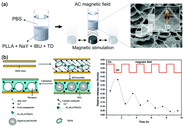 | ||
| Fig. 1 The magnetomechanical force for drug controlled release. (a) Illustration of magnetomechanical force-based controllable drug release enhancement. Reproduced with permission from ref. 45. Copyright 2016 Wiley-VCH. (b) Illustration of magnetomechanical force-based controllable drug release reduction. Reproduced with permission from ref. 47. Copyright 2015 Elsevier. | ||
Meanwhile, magnetomechanical force has tremendously fostered the development of drug release reduction and presented brand-new breakthroughs in therapeutic applications. For instance, Mao et al. combined silica-pillared clay (SPC) with a highly ordered interlayered mesopore structure with magnetic γ-Fe2O3 for controlled drug release and targeting.46 Such composites were featured by an unobstructed pore system, thus refraining from numerous application problems such as pore clogging, mass transfer restrictions, and limitations to further functionalization. More importantly, the drug release rate from these composites reduced dramatically due to the aggregation of γ-Fe2O3 triggered via non-contact magnetomechanical force. To be precise, the time for the drug release fraction of 50% was extended from 4 h to 5.5 h once exposed to a magnetic field of 150 mT. Nevertheless, γ-Fe2O3 on the outer surface influenced the number of Si–OH in the composites, resulting in a reduction in drug loading. Based on this, Mao et al. developed a novel cationic surfactant-aliphatic acid mixed route to obtain Fe3O4@SPC composites (Fig. 1b).47 In this route, oleic acid-formed organics could cooperate with cationic surfactants to generate interlayered mixed micelles, further ensuring that interlayered Fe3O4 formed with an outstanding dispersion and mesoporous structure and valid loading of IBU. Analogously, by reason of magnetomechanical force resultant particle aggregation, the release quantity of IBU was only 68.2–80.1% under a magnetic field of 150 mT for 10 h, while that without a magnetic field was 90%. Therefore, magnetomechanical force has a strong regulatory potential, implying the possibility of application in drug release reduction.
Impressively, the combination of magnetomechanical force and the anchoring technique has been a welcome addition to the drug controlled release currently. In this regard, Sun et al. developed a magnetic urchin-like microsystem on the basis of sunflower pollen grain to achieve transmembrane drug delivery (Fig. 2a–c).48 As a unique and renewable biomaterial, sunflower pollen grain was eligible to offer an enormous inner cavity structure for encapsulating drugs and nanospikes for drilling into cells. After the processing of acidolysis, sputtering, and vacuum loading, the prepared microsystem harboring the nickel coating was endowed with two magnetic response patterns (rolling and spinning). Thereinto, the rolling function was capable of delivering drugs to the required site rapidly, while the spinning function was beneficial for realising efficient membrane perforation for drug release after anchoring. In addition, Srivastava et al. gathered calcified porous acicular micromaterials with a length of 40–60 μm from dracaena and clad them with Fe–Ti shells for magnetic actuation drug release.49 Under an external rotating magnetic field, these calcified micromaterials were accountable for magnetic control to insert a single cell, which served as an anchoring mechanism for subsequent drug release. Owing to the cellular incision creation and site-directed drug release, this strategy based on acicular micromaterials significantly lowered lateral health damage induced by chemotherapy. Thus, the cooperation of magnetomechanical force and the anchoring technique demonstrates high intelligence and precision in cellular targeting, along with sufficient drug controlled release, presenting a promising avenue for future therapeutic applications.
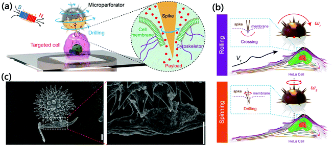 | ||
| Fig. 2 The combination of magnetomechanical force and the anchoring technique. (a) Illustration of drug controlled release based on the combination of magnetomechanical force and anchoring technique. (b) Illustration of the interaction with HeLa cells in rolling and spinning patterns. (c) Scanning electron microscopy images of a urchin-like microsystem anchoring single HeLa cells. Reproduced with permission from ref. 48. Copyright 2020 WILEY-VCH. | ||
Under an alternating magnetic field, magnetic materials are able to generate considerable magnetic hyperthermia,50 which enlightens the exploration of cooperative drug release controlled by magnetomechanical force and heat. For example, Yin et al. prepared a magnetic polymer composite micro-platform made of poly(D,L-lactide) (PLA) microspheres, γ-Fe2O3 nanoparticles (approximately 8 nm), and paclitaxel (an anti-cancer drug).51 Once injected directly into the body and guided by an externally applied magnetic field (750 G, 20 Hz) to the target location, two types of drug release mechanisms, natural degradation of PLA microspheres and release enhancement based on γ-Fe2O3 nanoparticles, could be activated simultaneously. Since the γ-Fe2O3 triggered magnetocaloric effect was extremely low and inadequate to influence the release behavior, as-generated magnetomechanical force leading to the physical deformation and ultimate breakage of the PLA matrix, was the main cause of elevated drug release. In addition, Liu et al. developed a novel nanomotor strategy for controllable drug delivery and release under an adjustable magnetic field (0.5 mT, 100 Hz).52 In this system, monodispersed CoFe2O4 nanoparticles were deposited on wormlike mesoporous silica nanotubes with ultrahigh surface area to form nanomotors, which integrated the functions of magnetic propulsion and drug loading. Subsequently, G-quadruplexes (highly ordered DNA structures) were engaged to modify as-formed nanomotors for improved stability and drug packaging. In this system, the drug release process could be simply controlled by tuning the magnetic field to initiate the thermal and mechanical effects of nanomotors. Interestingly, controlled drug release could still be realized by magnetomechanical force-triggered oscillation even removing the thermal effect. Taken together, the combined magnetomechanical force and other forms of energy are expected to be a highly potential drug controlled release strategy.
| Type | Saturation magnetization | Magnetic regulation system | Size | Magnetomechanical force | Application | Ref. |
|---|---|---|---|---|---|---|
| Ethylene-vinyl acetate@Fe–Cr@BSA composites | 1100 G, 60 Hz | r = 90–125 μm | Drug controlled release | 44 | ||
| Poly(L-lactic acid)@Faujasite@magnetostrictive Terfenol-D@Ibuprofen microporous membrane | AMF 100–200 mT, 0.3 Hz | r = 56 ± 13 μm | 45 | |||
| FexOy@silica-pillared clay@aspirin composites | 150 mT | r = 2.8–3.0 nm | 46 | |||
| Fe3O4@silica nanocomposites pillared clay@Ibuprofen composites | 150 mT | r = 2.5–2.8 nm | 47 | |||
| Ni/Ti-coated sunflower pollen grain@doxorubicin composites | 15 emu g−1 | RMF 5–15 mT, 10–100 Hz | r = 15 μm | 48 | ||
| Calcified porous microneedles coated with Fe–Ti@camptothecin | RMF 500–800 rpm | L = 40–60 μm | 49 | |||
| γ-Fe2O3@poly(D,L-lactide)@paclitaxel composites | 65.7 emu g−1 | AMF 750 G, 20 Hz | r = 972 ± 68 nm | 51 | ||
| Wormlike mesoporous silica nanotube@CoFe2O4 nanoparticles | AMF 0.5 mT, 100 Hz | L = 1–1.5 μm, r = 8 nm | 52 | |||
| Fe20Ni80 discs | 750 emu cm−3 | AMF 10 mT, 40 Hz | r = 1 μm, h = 60 nm | 26–53 pN | Cancer therapy | 58 |
| Permalloy magnetic discs | AMF 1 T, 20 Hz | r = 2 μm | 59 | |||
| Zn0.4Fe2.6O4@epidermal growth factor composites | 0.02 Am−2 g−1 | RMF 40 mT, 15 Hz | L = 4 μm | 20–50 pN | 60 | |
| Iron oxide magnetic nanoparticles@carboxymethyldextran@epidermal growth factor composites | 20 Am−2 kg−1 | AMF 42 kA m−1, 233 kHz | r = 61 ± 29 nm | 61 | ||
| Superparamagnetic iron oxide nanoparticles @lysosomal protein marker LAMP1 composites | DMF 30 mT, 5–15 Hz | r = 5.8 μm | 62 | |||
| Zn0.4Fe2.6O4@triphenylphosphonium composites | 44 emu g−1 | RMF 15 Hz, 40 mT | L = 1 μm | 4 pN | 63 | |
| Zn0.4Fe2.6O4@death receptor 4 composites | 161 emu g−1 | SMF 0.2 T | r = 15 nm | 30 fN | 64 | |
| Zn0.4Fe2.6O4@doxorubicin@death receptor 4 composites | 161 emu g−1 | SMF 0.5 T | r = 15 nm | 65 | ||
| Gold-coated magnetic microspheres | 65.65 emu g−1 | VMF 400 mT, 3 Hz | r = 1.56 ± 0.47 μm | 35.79 pN | 66 | |
| Doxorubicin@Zn0.2Fe2.8O4@poly(lactic-co-glycolic acid) composites | 22 emu g−1 | RMF 45 mT, 2000 rpm | r = 200 nm | 244.78 pN | 67 | |
| Hyaluronic acid@olaparib@polyethylenimine-Poly(D,L-lactide-co-glycolide)@Fe3O4 composites | 21.08 emu g−1 | RMF 7 T, 1000 rpm | r = 159.5 ± 2.3 nm | 68 | ||
| Fe3O4 magnetic nanoparticles coated with nanoscale graphene oxide | 50 emu g−1 | 0–20![[thin space (1/6-em)]](https://www.rsc.org/images/entities/char_2009.gif) 000 Oe 000 Oe |
r = 10 nm | Regenerative medicine | 80 | |
| Fe3O4@RGD peptide/Fe3O4@platelet-derived growth factor receptor α antibody | 60–120 mT | r = 250 nm | 81 | |||
| Fe3O4-Mesenchymal stem cell Sheets | 4000 G | 82 | ||||
| Nickel microtips | 57 μemu | 0.2 T, 1 Hz | H = 1 μm | 83 | ||
| Polycaprolactone@Fe3O4 nanofibre meshes | r = 50–100 nm, H = 100 μm, S = 16 cm × 7 cm | 86 | ||||
| CoFe2O4@poly(vinylidene fluoride) composites | 0–230 Oe, 0.3 Hz | r = 60, 80, 120 μm | 87 |
4. Magnetomechanical force for cancer therapy
Nowadays, cancer-relevant diseases remain a serious threat to human health globally owing to the abhorrent nature of cancer, namely diversity, complexity, and heterogeneity.53 Encouragingly, recent decades have witnessed the forward development of technologies against cancer invasion and morbidity including conventional clinical methods and novel therapeutic approaches.54–56 Among these therapeutic approaches, magnetomechanical force is a rising research hotspot with great potential for cancer therapy. In this therapeutic model, magnetic materials feature asymmetric geometric structures and efficient magnetic reversal play a vital role, since they exhibit a certain degree of advantages in generating magnetomechanical force when exposed to an alternating magnetic field.57As a precise therapeutic paradigm, magnetomechanical force is engaged to damage cellular membranes and organelles of targeted cancer cells, which can be classified as internalized and non-internalized forms. The former form is independent of internalized magnetic materials, whose sizes need not meet the wide requirements for cellular uptake. For example, Kim et al. reported for the first time a lithographically defined microdisc possessing a spin-vortex ground state for high magnetomechanical force induction (26–53 pN).58 In an alternating magnetic field (10 mT, 40 Hz), the microdisc vortices showed a shifting tendency to realize oscillation, thus transmitting magnetomechanical force to cancer cells. Importantly, the spin-vortex mediated mechanical stimulation not only resulted in compromised integrity of the cellular membranes, but initiated programmed cell death. Thanks to this, under a magnetic field with a magnitude as small as tens of oersteds, a frequency as low as tens of hertz, and an action time as short as 10 min, such a treatment model was sufficient to kill approximately 90% of the cancer cells in vitro. Correspondingly, the former form is taking advantage of internalized magnetic materials to mediate magnetomechanical force inside the cells. In this regard, the same team substantiated the antineoplastic effects (both in vitro and in vivo) of spin-vortex and disc-shaped permalloy nanomagnets (Fig. 3a–c).59 After co-incubation for 24 h, almost all U87 glioma cells efficiently ingested these nanomagnets without obvious alteration in cellular morphology. When the internalized nanomagnets were in the plane of the rotating magnetic field at 20 hertz, a powerful magnetomechanical force emerged, which damaged the cellular membrane integrity and caused the massive death of U87 glioma cells. Moreover, magnetomechanical force based on these nanomagnets was even capable of suppressing glioma growth significantly and increase the apoptotic area (26%), thus prolonging the survival rate of tumor-bearing mice. Therefore, magnetomechanical force-based physical damage is a novel approach to manipulating magnetic materials for tumor elimination.
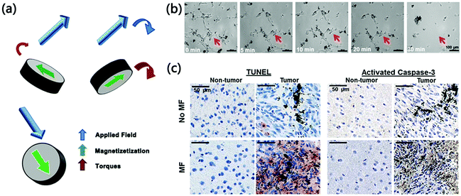 | ||
| Fig. 3 The magnetomechanical force-based physical damage for glioma elimination. (a) Magnetic response illustration of spin-vortex and disc-shaped nanomagnets. (b) Optical microscopy images of magnetomechanical force-based cellular physical disruption. (c) TUNEL staining and activated caspase-3 staining after magnetomechanical force-based physical treatment. Reproduced with permission from ref. 59. Copyright 2015 WILEY-VCH. | ||
Targeting strategies endow magnetic materials with the capacity of specifically reaching the surface or even the internal organelles of tumor cells, and have been a central topic regarding efficiency intensification of magnetomechanical force-based physical disruption. For instance, Shen et al. obtained a zinc-doped iron oxide nanoparticle possessing high magnetization, and further modified this nanoparticle with the epidermal growth factor (EGF) peptide to achieve specific targeting of tumor cells.60 Once internalized into tumor cells, such nanoparticles could assemble into slender aggregates under a rotating magnetic field at 15 hertz and 40 mT. On account of the aggregation effect, hundreds of pN could be produced inside tumor cells, which acutely devastated the plasma alongside lysosomal membranes, thus releasing hydrolases into the cytosol for programmed cell death and necrosis. Analogously, Domenech et al. harnessed iron oxide magnetic nanoparticles with EGF receptors to selectively mediate lysosomal membrane permeabilization (Fig. 4a and b).61 Since the capacity of lysosomal membrane permeabilization to promote cell death was independent of apoptosis defense mechanisms, this tactic based on magnetomechanical force efficiently activated the lysosomal death pathways and showed marked anti-tumor potential against a broad spectrum of tumors. In another work, Zhang et al. combined superparamagnetic iron oxide nanoparticles with antibodies targeting the lysosomal protein marker LAMP1 to induce apoptosis remotely.62 Owing to the binding of magnetic nanoparticles to endogenic LAMP1, approximately 71.2% of the prepared magnetic nanoparticles could accumulate along the lysosomal membranes preferentially. Subsequently, magnetic nanoparticles were rotationally activated via the dynamic magnetic field (30 mT, 5–15 Hz) to generate shear force and tear the lysosomal membranes, which therefore lowered the size and number of lysosomes to inhibit cell growth. In addition, Chen et al. synthesized a novel cubic-shape nanospinner, integrating zinc-doped iron oxide (Zn0.4Fe2.6O4) nanocubes for magnetomechanical force production and triphenylphosphonium (TPP) cations to target mitochondria (Fig. 4c and d).63 Impressively, the nanospinners had the characteristic to escape from lysosomes and head for mitochondria even in diverse cell lines. Due to the direct magnetomechanical force (approximate 4 pN) acting on mitochondria, the mechanical performance down-regulated the mitochondrial membrane potential to execute the cell apoptosis progress, hence effectively improving the apoptosis rate to 58%. As a consequence, magnetomechanical force-based physical disruption united with a well-established targeting ability is highly desirable in remote invasive cancer therapy.
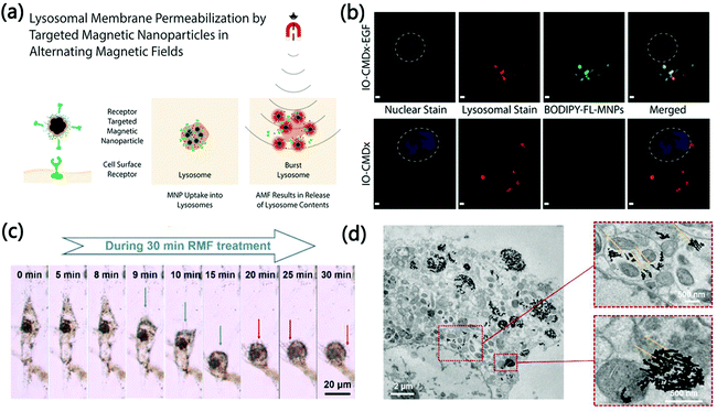 | ||
| Fig. 4 The combination of targeting strategies with magnetomechanical force-based physical disruption. (a) Illustration of lysosomal membrane permeabilization under an alternating magnetic field. (b) Confocal microscopy images of the intracellular distribution. Reproduced with permission from ref. 61. Copyright 2013 American Chemical Society. (c) Optical microscopy images of magnetomechanical force-based mitochondria-targeted physical disruption. (d) Transmission electron microscopy images of magnetomechanical force-based mitochondria-targeted physical disruption. Reproduced with permission from ref. 63. Copyright 2019 WILEY-VCH. | ||
In addition to the physical damage, magnetomechanical force has been used to stimulate mechanosensitive proteins on the cell membrane or organelle membrane, so as to initiate cell signaling pathways mechanically to induce cell death. In this aspect, Cho et al. designed a magnetic switch for apoptosis cell signaling, which was made of Zn0.4Fe2.6O4 nanoparticles and antibodies targeting the death receptor 4 (DR4).64 It is noteworthy that the attraction force between the nanoparticles was about 30 fN, inadequate to destroy the cell membranes but promote the nanoparticle clustering on the cell membranes. In a similar pattern to the biochemical ligand TRAIL, this magnetic switch achieved the clustering of the DR4s and the formation of a death-inducing signaling complex (DISC) in a remotely, non-invasively, and spatially and temporally regulated style. Based on this research, the same team developed a novel apoptosis trigger to induce complete death of multidrug-resistant cancer cells by virtue of DR4-targeting and magnetomechanical force (Fig. 5a and b).65 Importantly, such a trigger was conceived to launch apoptosis signals with single-cell level accuracy, simultaneously down-regulating the P-glycoprotein which was regarded as one of the main inducements of multidrug-resistance. In a word, magnetomechanical force-based signalling pathway activation is an emerging platform concept to cure tumors with high efficacy and specificity.
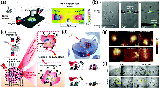 | ||
| Fig. 5 The combination of magnetomechanical force with other therapeutic modalities for oncotherapy. (a) Illustration of the magnetic setup for magnetomechanical force-based site-specific apoptosis activation. (b) Optical microscopy images showing the apoptotic activation process. Reproduced with permission from ref. 65. Copyright 2016 American Chemical Society. (c) Illustration of the combination therapy based on magnetomechanical force and photothermal therapy. Reproduced with permission from ref. 66. Copyright 2018 WILEY-VCH. (d) Illustration of the combination therapy based on magnetomechanical force and chemotherapy. (e) Atomic force microscopy images after magnetomechanical force/chemotherapy. (f) Transmission electron microscopy images after magnetomechanical force/chemotherapy. Reproduced with permission from ref. 67. Copyright 2020 Elsevier. | ||
Due to the aforementioned encouraging anti-tumor effects, combining magnetomechanical force with other therapeutic modalities to treat multitudinous cancers has aroused great attention. In a recent study, Chen et al. coated needle-like Fe3O4 nanoparticles with carbon and gold double layers to fabricate novel hedgehog-like microspheres for tumor ablation (Fig. 5c).66 Under a vibrating magnetic field with appropriate exposure time, frequency, and intension, sharp surfaces of the magnetic microspheres seriously dissevered cancer cells to restrain tumor growth via magnetomechanical force. By means of analysis and calculation, the maximum applied magnetomechanical force of a single microsphere was 35.79 pN. Integrated with laser-triggered photothermal ablation, solid tumors were completely eradicated after these two remote and noninvasive modes of synergistic local stimuli. In addition, to address the obstacle of drug-resistance, our team proposed a remotely controllable mechano-chemotherapeutic method relying on the rotating magnetic field (45 mT) and a therapeutic nanoagent composed of poly(lactic-co-glycolic acid) (PLGA), Zn0.2Fe2.8O4 nanoparticles and doxorubicin (DOX) (Fig. 5d–f).67 In this design, the generated magnetomechanical force (approximate 250 pN) was exploited for controlled release of DOX and physical disruption onto cell membranes. By right of different and complementary routes, such a mechano-chemotherapeutic method executed remarkable tumor cell death, regardless of the specifics of tumor cell drug resistance. In Addition, Zhang et al. prepared polyethylenimine (PEI)–PLGA carriers with the surface modification of hyaluronic acid (HA) to co-load Fe3O4 nanoparticles and Olaparib (an anti-cancer drug).68 Since the HA enabled the active targeting of the CD44 receptor overexpressing on the surface of triple negative breast cancer cells, this mechanical platform yielded magnetomechanical force through incomplete rotation, cooperating with Olaparib for cell synergistic destruction. Noticeably, the magnetomechanical force could cause two mechanical strikes including membrane structure destruction for cytolysis and lysosome-mitochondrial pathway activation for cell apoptosis. In brief, magnetomechanical force-based combination therapy is an attractive methodology to strengthen the antineoplastic effect; furthermore, this strategy may be adapted to remotely cure other types of diseases.
5. Magnetomechanical force for regenerative medicine
As an area attracting major attention, regenerative medicine employs live cells and tissues to gain renascent, healthy, and specially grown tissues for disease treatment (e.g. retinitis pigmentosa and macular degeneration).69–74 Although mechanical forces have been a robust manner of regenerative medicine to manipulate proteins on the cell surface, the timescales of the supplied mechanical forces easily fail to match that of the target signaling processes.75 Impressively, through the precise and remote control of cellular receptors, magnetomechanical force can initiate intended adhesion, proliferation, and differentiation signaling pathways for regenerative medicine.76,77Pluripotent stem cells occupy an important position in regenerative medicine strategies owing to their excellent differentiation characteristics.78,79 Regarding magnetomechanical force-based stem cell differentiation, Zhang et al. integrated Fe3O4 nanoparticles and graphene oxide into a nanoscale system for stem cell label and growth factor delivery (Fig. 6a–d).80 On the one hand, this nanoscale system was easily ingested via dental-pulp stem cells without causing any toxic side effects. Therefore, the stem cells labeled by the nanoscale system could constitute multilayered cell sheets under magnetomechanical force. On the other hand, with the introduction of Fe3O4 nanoparticles, bone-morphogenetic-protein-2 was successfully incorporated into multilayered cell sheets for bone formation, effectively realizing the establishment of an osteochondral complex. In a separate report, Hu et al. prepared magnetic bioreactors possessing the functions to apply magnetomechanical force to stem cells and target diverse cell surface receptors, which were used to explore the roles of PDGFRα and ανβ3 (two types of mechanical-sensitive cell membrane receptors).81 Compared with magnetic bioreactors targeting ανβ3, those targeting PDGFRαa induced a higher mineral-to-matrix ratio after magnetomechanical force stimulation, which enriched the cognition of mechanical force stimulation for bone regenerative engineering. In addition, Ishii et al. incubated mesenchymal stem cells with Fe3O4-liposome complexes to obtain magnetized stem cells for subsequent magnetomechanical force-guided formation of multilayered cell sheets.82 On account of the elevated expression of vascular endothelial growth factor and depressed apoptosis in ischemic tissues, this differentiation modality was distinguished with angiogenic and tissue preserving effects. Furthermore, Mary et al. succeeded in achieving cardiomyogenesis of embryonic stem cells using a novel magnetic pattern.83 After labeling with magnetic nanoparticles, stem cells could aggregate into embryoid bodies and readily be stimulated by means of magnetomechanical force. It is worth mentioning that cyclic magnetomechanical force stimulation was adequate to improve cardiomyogenesis, enabling more than 90% of the embryoid bodies to beat. In this way, gene expressions involved in mesoderm differentiation (e.g. Nkx2.5, Gata4, and Gata6) were greatly up-regulated (2-fold to 5-fold). In a word, the magnetomechanical force tissue-engineering strategy exhibits promising feasibility for future stem cell applications in regenerative medicine.
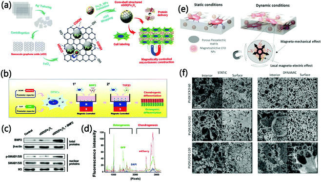 | ||
| Fig. 6 The magnetomechanical force for regenerative medicine. (a) Illustration of magnetomechanical force-based stem cell differentiation. (b) Illustration of the integrated osteochondral tissue constructive process. (c) Western blotting detection for bone-morphogenetic-protein-2 content and related signaling activation. (d) Fluorescence detection on frozen sections after one-week implantation. Reproduced with permission from ref. 77. Copyright 2017 WILEY-VCH. (e) Illustration of magneto-active 3D porous scaffolds for osteoblast proliferation. (f) Scanning electron microscopy images of 3D porous scaffolds after four-day incubation with preosteoblast cells. Reproduced with permission from ref. 84. Copyright 2019 American Chemical Society. | ||
Since natural tissue exists as a three-dimensional (3D) structure in which cells are spatially distributed, the desired scaffold for regenerative medicine had better be a 3D structure with a similar distribution of cells.84,85 To address this challenge, the emergence of magnetomechanical force demonstrates a novel strategy to establish such scaffolds. For instance, Zhu et al. developed magnetic meshes containing electrospun magnetic nanoparticles and polycaprolactone composite fibres for seeding osteoblast cells.86 Upon the action of magnetomechanical force, osteoblast cells were vertically arranged and tightly stacked, which could proliferate efficiently accompanied by the secretion of an extracellular matrix. As the main components of the extracellular matrix, type I collagen was able to fill the mesh interface and promote the construction of 3D scaffold-cell complexes. It follows that magnetomechanical force represents a valuable instrument for the construction and application of 3D scaffolds in the field of regenerative medicine. Impressively, the combination of magnetomechanical force and magnetoelectrical stimuli has been a full potential approach for skeletal muscle tissue engineering application. In this aspect, Fernandes et al. reported a magneto-active 3D porous scaffold to realize a significant proliferation of osteoblast cells (Fig. 6e and f).87 With the help of solvent casting, this 3D scaffold could be manufactured through the integration of piezoelectric polymer, poly(vinylidene fluoride), and magnetic CoFe2O4 particles. Importantly, local magnetomechanical and magnetoelectric performances of the scaffolds played a crucial role in expanding the preosteoblast proliferation, attributed to the imitation of the bone morphology and physical microenvironment. The same team reported a kind of magnetoelectric film which combined a piezoelectric polymer, poly(vinylidene fluoride-trifluoro-ethylene), and magnetostrictive particles (CoFe2O4). Upon cooperative magnetomechanical and magnetoelectrical stimulation by an applied magnetic field, the proliferation and differentiation of C2C12 myoblast cells were promoted.88 In another work, Ribeiro et al. proved that magnetoelectric Terfenol-D/poly(vinylidene fluoride-co-trifluoroethylene) composites were able to apply magnetomechanical and magnetoelectrical stimuli on MC3T3-E1 pre-osteoblast cells. Based on this, the cell proliferation was enhanced up to 25%, thereby offering a novel method for tissue engineering.89 Owing to their potential to promote cell proliferation, magnetoelectric biomaterials are promising to process a broad application in tissue engineering.
6. Perspectives
In this review, a series of therapeutic exploitations and applications based on magnetomechanical force including drug controlled release, cancer therapy, and regenerative medicine have been summarized, illustrating the innovation and uniqueness of magnetomechanical force in medical biology. All literature reports cited and discussed herein certify that magnetomechanical force paves a novel and potentially promising pathway for cutting-edge treatment paradigm. Nevertheless, magnetomechanical force is still in its infancy, and more surveys and investigations remain to be addressed for further development and innovation of this to-date therapeutic strategy.First, biosecurity is a critical focus in the design and fabrication of magnetic materials for magnetomechanical force generation. In addition to meeting the current standards of biomedical applications (e.g. biocompatibility and biodegradability), magnetic materials need to have significant therapeutic effects, thus bringing sufficient economic and social benefits.90 Accordingly, it is important to balance biosecurity, therapeutic effectiveness, and cost (e.g. raw material expenses, fabrication instruments, and preparation procedures), which remains a serious challenge nowadays.
Second, a single magnetic particle is generally incapable of imparting magnetomechanical force on demand in complex physical environments.91 Therefore, there is huge space to fully develop magnetic particle-based collective behavior, which efficiently executes assigned complex biological tasks due to the close cooperation between magnetic particles. In this aspect, future directions include the optimization of magnetic particle dose, the control of particle delivery, and the regulation of mechanical force stimulation in order to cure a wide range of diseases.
Third, accurate on-body and real-time maneuvering of the magnetomechanical force as well as its monitoring is extremely essential.92 Upon regulation of magnetic materials according to actual occasions including health status and physiology, magnetomechanical force is even capable of realizing individualized or personalized therapy. In this regard, the combination of imaging techniques and magnetomechanical force is promising to perform certain diagnostic and therapeutic missions in a visualization fashion.
Fourth, to improve the on-body fate of magnetomechanical force, physicochemical targeting (e.g. magnetic targeting) aims at targeting magnetic materials to specific treatment areas with remarkable temporal and spatial accuracy.93 In parallel, targeting ligands can endow magnetic materials with targeting performance for optimized tissue accumulation, blood circulation, and subsequent controllable magnetomechanical force modulation.
Fifth, with regard to future applications, magnetomechanical force promises to be a great candidate for targeted treatment and controllable drug release against microvascular thrombolysis on the strength of the locomotion and collective manners.94,95 In addition, magnetomechanical force has the potential to activate genetically encoded mechanosensitive ion channels (e.g. Piezo1 and TRPV-4),96,97 which can be utilized to explore sensory reception and tissue development.
In general, although in the initial stage, magnetomechanical force has secured its status in therapeutic applications on account of the concept of remote and precise stimulation. Herein, we strongly hope that this review will drive the future development and progress of magnetomechanical force and provide significant enlightenment and thoughts for novel therapeutic patterns.
Author contributions
Conceptualization: A. W. and F. Y. Writing – original draft preparation: J. Y. and C. Y. Writing – review and editing: R. Z. and X. X. Funding acquisition: A. W. and F. Y.Conflicts of interest
There are no conflicts to declare.Acknowledgements
The authors are grateful for financial support from the National Natural Science Foundation of China (51803228), the Youth Innovation Promotion Association, the Chinese Academy of Sciences (2022301), and the Ningbo 3315 Innovative Talent Project (2018-05-G).Notes and references
- G. G. Genchi, A. Marino, A. Grillone, I. Pezzini and G. Ciofani, Adv. Healthc. Mater., 2017, 6, 1700002 CrossRef PubMed.
- D. Atasoy and S. M. Sternson, Physiol. Rev., 2018, 98, 391–418 CrossRef CAS PubMed.
- J. Y. Lee, H. J. Park, J. H. Kim, B. P. Cho, S. R. Cho and S. H. Kim, Neurosci. Lett., 2015, 604, 167–172 CrossRef CAS PubMed.
- B. Hu, Y. Zhang, J. Zhou, J. Li, F. Deng, Z. Wang and J. Song, PLoS One, 2014, 9, 95168 CrossRef.
- E. Mortaz, F. A. Redegeld, M. W. van der Heijden, H. R. Wong, F. P. Nijkamp and F. Engels, Exp. Hematol., 2005, 33, 944–952 CrossRef CAS PubMed.
- T. Taghian, D. A. Narmoneva and A. B. Kogan, J. R. Soc., Interface, 2015, 12, 0153 CrossRef.
- A. Rettenmaier, T. Lenarz and G. Reuter, Biomed. Opt. Express, 2014, 5, 1014–1025 CrossRef PubMed.
- A. P. Liu, Biophys. J., 2016, 111, 1112–1118 CrossRef CAS PubMed.
- F. Hirth, The Monist, 1906, 16, 321–330 CrossRef.
- X. Qian, X. Han, L. Yu, T. Xu and Y. Chen, Adv. Funct. Mater., 2019, 30, 1907066 CrossRef.
- Y. Shen, Y. Cheng, T. Q.-P. Uyeda and G. R. Plaza, Ann. Biomed. Eng., 2017, 45, 2475–2486 CrossRef.
- A. Tay and D. Di Carlo, Curr. Med. Chem., 2017, 24, 537–548 CrossRef CAS PubMed.
- J. W. Kim, D. Seo, J. U. Lee, K. M. Southard, Y. Lim, D. Kim, Z. J. Gartner, Y. W. Jun and J. Cheon, Nat. Protoc., 2017, 12, 1871–1889 CrossRef CAS PubMed.
- S. Behrens and I. Appel, Curr. Opin. Biotechnol., 2016, 39, 89–96 CrossRef CAS.
- L. S. Schadler, Nanocompos. Sci. Technol., 2003, 2, 77–153 Search PubMed.
- P. M. Ajayan, L. S. Schadler and P. V. Braun, Nanocomposite science and technology, John Wiley & Sons, 2006 Search PubMed.
- J. Dobson, Nat. Nanotechnol., 2008, 3, 139–143 CrossRef CAS PubMed.
- D. J. Muller, J. Helenius, D. Alsteens and Y. F. Dufrene, Nat. Chem. Biol., 2009, 5, 383–390 CrossRef PubMed.
- C. Monzel, C. Vicario, J. Piehler, M. Coppey and M. Dahan, Chem. Sci., 2017, 8, 7330–7338 RSC.
- J. Howard and A. J. Hudspeth, Neuron, 1988, 1, 189–199 CrossRef CAS PubMed.
- Y. I. Golovin, S. L. Gribanovsky, D. Y. Golovin, N. L. Klyachko, A. G. Majouga, A. M. Master, M. Sokolsky and A. V. Kabanov, J. Controlled Release, 2015, 219, 43–60 CrossRef CAS PubMed.
- B. Hu, J. Dobson and A. J. El Haj, Nanomedicine, 2014, 10, 45–55 CrossRef CAS PubMed.
- B. Yigit, Y. Alapan and M. Sitti, Adv. Sci., 2019, 6, 1801837 CrossRef PubMed.
- H. W. Huang, M. S. Sakar, A. J. Petruska, S. Pane and B. J. Nelson, Nat. Commun., 2016, 7, 12263 CrossRef CAS PubMed.
- L. Ren, W. Wang and T. E. Mallouk, Acc. Chem. Res., 2018, 51, 1948–1956 CrossRef CAS PubMed.
- D. Jiles, Introduction to magnetism and magnetic materials, CRC Press, 2015 Search PubMed.
- N. A. Spaldin, Magnetic materials: fundamentals and applications, Cambridge University Press, 2010 Search PubMed.
- R. S.-M. Rikken, R. J.-M. Nolte, J. C. Maan, J. C.-M. van Hest, D. A. Wilson and P. C.-M. Christianen, Soft Matter, 2014, 10, 1295–1308 RSC.
- J. Yu, T. Xu, Z. Lu, C. I. Vong and L. Zhang, IEEE Trans. Robot., 2017, 33, 1213–1225 Search PubMed.
- B. Jang, E. Gutman, N. Stucki, B. F. Seitz, P. D. Wendel-Garcia, T. Newton, J. Pokki, O. Ergeneman, S. Pane, Y. Or and B. J. Nelson, Nano Lett., 2015, 15, 4829–4833 CrossRef CAS PubMed.
- K. Han, C. W. Shields, N. M. Diwakar, B. Bharti, G. P. Lopez and O. D. Velev, Sci. Adv., 2017, 3, 1701108 CrossRef PubMed.
- Z. Yang, L. Yang and L. Zhang, IEEE ASME Trans. Mechatron., 2021, 26, 3163–3174 Search PubMed.
- L. Yang, E. Yu, C.-I. Vong and L. Zhang, IEEE ASME Trans. Mechatron., 2019, 24, 1208–1219 Search PubMed.
- X. Du, M. Zhang, J. Yu, L. Yang, P. W.-Y. Chiu and L. Zhang, IEEE ASME Trans. Mechatron., 2021, 26, 1524–1535 Search PubMed.
- L. Yang, L. Zhang, IEEE Int. Confer. CASE, 2020, pp. 876–881.
- S. M. Khalil, A. Alfar, A. F. Tabak, A. Klingner, S. Stramigioli and M. Sitti, IEEE Int. Confer. AIM, 2017, pp. 1117–1122.
- A. W. Mahoney and J. J. Abbott, IEEE Trans. Robot., 2014, 30, 411–420 Search PubMed.
- C. Mavroidis and A. Ferreira, Nanorobotics: past, present, and future, Springer Science & Business Media, 2013, vol. 4, pp. 275–299 Search PubMed.
- S. Jeong, H. Choi, J. Choi, C. Yu, J.-O. Park and S. Park, Sens. Actuators, A, 2010, 157, 118–125 CrossRef CAS.
- H. Choi, K. Cha, J. Choi, S. Jeong, S. Jeon, G. Jang, J.-O. Park and S. Park, Sens. Actuators, A, 2010, 163, 410–417 CrossRef CAS.
- D. B. Pacardo, F. S. Ligler and Z. Gu, Nanoscale, 2015, 7, 3381–3391 RSC.
- R. A. Siegel, J. Controlled Release, 2014, 190, 337–351 CrossRef CAS PubMed.
- S. Bhatnagar and V. V. Venuganti, J. Nanosci. Nanotechnol., 2015, 15, 1925–1945 CrossRef CAS PubMed.
- E. R. Edelman, A. Fiorino, A. Grodzinsky and R. Langer, J. Biomed. Mater. Res., 1992, 26, 1619–1631 CrossRef CAS PubMed.
- J. Barbosa, D. M. Correia, R. Goncalves, C. Ribeiro, G. Botelho, P. Martins and S. Lanceros-Mendez, Adv. Healthcare Mater., 2016, 5, 3027–3034 CrossRef CAS.
- H. Mao, X. Liu, J. Yang, B. Li, C. Yao and Y. Kong, Mater. Sci. Eng., C, 2014, 40, 102–108 CrossRef CAS.
- H. Mao, K. Zhu, X. Liu, C. Yao and M. Kobayashi, Microporous Mesoporous Mater., 2016, 225, 216–223 CrossRef CAS.
- M. Sun, Q. Liu, X. Fan, Y. Wang, W. Chen, C. Tian, L. Sun and H. Xie, Small, 2020, 16, 1906701 CrossRef CAS.
- S. K. Srivastava, M. Medina-Sanchez, B. Koch and O. G. Schmidt, Adv. Mater., 2016, 28, 832–837 CrossRef CAS PubMed.
- A. K. Gupta and M. Gupta, Biomaterials, 2005, 26, 3995–4021 CrossRef CAS PubMed.
- H. Yin, S. Yu, P. S. Casey and G. M. Chow, Mater. Sci. Eng. C, 2010, 30, 618–623 CrossRef CAS.
- M. Liu, L. Pan, H. Piao, H. Sun, X. Huang, C. Peng and Y. Liu, ACS Appl. Mater. Interfaces, 2015, 7, 26017–26021 CrossRef CAS PubMed.
- R. L. Siegel, K. D. Miller and A. Jemal, Ca-Cancer J. Clin., 2020, 70, 7–30 CrossRef PubMed.
- Z. Xie, T. Fan, J. An, W. Choi, Y. Duo, Y. Ge, B. Zhang, G. Nie, N. Xie, T. Zheng, Y. Chen, H. Zhang and J. S. Kim, Chem. Soc. Rev., 2020, 49, 8065–8087 RSC.
- W. Fan, B. Yung, P. Huang and X. Chen, Chem. Rev., 2017, 117, 13566–13638 CrossRef CAS PubMed.
- C. Zhao, J. Chen, R. Zhong, D. S. Chen, J. Shi and J. Song, Angew. Chem., Int. Ed., 2021, 60, 9804–9827 CrossRef CAS PubMed.
- D. Cheng, X. Li, G. Zhang and H. Shi, Nanoscale Res. Lett., 2014, 9, 195 CrossRef PubMed.
- D. H. Kim, E. A. Rozhkova, I. V. Ulasov, S. D. Bader, T. Rajh, M. S. Lesniak and V. Novosad, Nat. Mater., 2010, 9, 165–171 CrossRef CAS PubMed.
- Y. Cheng, M. E. Muroski, D. Petit, R. Mansell, T. Vemulkar, R. A. Morshed, Y. Han, I. V. Balyasnikova, C. M. Horbinski, X. Huang, L. Zhang, R. P. Cowburn and M. S. Lesniak, J. Controlled Release, 2016, 223, 75–84 CrossRef CAS PubMed.
- Y. Shen, C. Wu, T. Q.-P. Uyeda, G. R. Plaza, B. Liu, Y. Han, M. S. Lesniak and Y. Cheng, Theranostics, 2017, 7, 1735–1748 CrossRef CAS PubMed.
- D. Maribella, M. B. Ileana, T. L. Madeline and R. Carlos, ACS Nano, 2013, 7, 5091–5101 CrossRef PubMed.
- E. Zhang, M. F. Kircher, M. Koch, L. Eliasson, S. N. Goldberg and E. Renstrom, ACS Nano, 2014, 8, 3192–3201 CrossRef CAS PubMed.
- M. Chen, J. Wu, P. Ning, J. Wang, Z. Ma, L. Huang, G. R. Plaza, Y. Shen, C. Xu, Y. Han, M. S. Lesniak, Z. Liu and Y. Cheng, Small, 2020, 16, 1905424 CrossRef CAS PubMed.
- M. H. Cho, E. J. Lee, M. Son, J. H. Lee, D. Yoo, J. W. Kim, S. W. Park, J. S. Shin and J. Cheon, Nat. Mater., 2012, 11, 1038–1043 CrossRef CAS PubMed.
- M. H. Cho, S. Kim, J. H. Lee, T. H. Shin, D. Yoo and J. Cheon, Nano Lett., 2016, 16, 7455–7460 CrossRef CAS PubMed.
- Y. Chen, P. Han, Y. Wu, Z. Zhang, Y. Yue, W. Li and M. Chu, Small, 2018, 14, 1802799 CrossRef PubMed.
- Y. Chenyang, Y. Fang, S. Li, M. Yuanyuan, S. G. Stanciu, L. Zihou, L. Chuang, O. U. Akakuru, X. Lipeng, H. Norbert, L. Huanming and W. Aiguo, Nano Today, 2020, 35, 100967 CrossRef.
- Y. Zhang, H. Hu, W. Tang, Q. Zhang, M. Li, H. Jin, Z. Huang, Z. Cui, J. Xu, K. Wang and C. Shi, J. Controlled Release, 2020, 322, 401–415 CrossRef CAS PubMed.
- C. Mason and P. Dunnill, Regener. Med., 2008, 3, 1–5 CrossRef PubMed.
- B. Nommiste, K. Fynes, V. E. Tovell, C. Ramsden, L. da Cruz and P. Coffey, Prog. Brain Res., 2017, 231, 225–244 Search PubMed.
- A. Webster, BMJ, 2017, 358, 4245 CrossRef PubMed.
- C. A. Cezar, E. T. Roche, H. H. Vandenburgh, G. N. Duda, C. J. Walsh and D. J. Mooney, Proc. Natl. Acad. Sci. U. S. A., 2016, 113, 1534–1539 CrossRef CAS PubMed.
- L. Chang, Y. H. Li, M. X. Li, S. B. Liu, J. Y. Han, G. X. Zhao, C. C. Ji, Y. Lyu, G. M. Genin, B. F. Bai and F. Xu, Chem. Eng. J., 2021, 420, 130398 CrossRef CAS.
- N. Y. Shi, Y. H. Li, L. Chang, G. X. Zhao, G. R. Jin, Y. Lyu, G. M. Genin, Y. F. Ma and F. Xu, Small Methods, 2021, 5, 2100276 CrossRef CAS PubMed.
- H. Kang, D. S.-H. Wong, X. Yan, H. J. Jung, S. Kim, S. Lin, K. Wei, G. Li, V. P. Dravid and L. Bian, ACS Nano, 2017, 11, 9636–9649 CrossRef CAS.
- M. Rotherham and A. J. El Haj, PLoS One, 2015, 10, 0121761 CrossRef PubMed.
- J. R. Henstock, M. Rotherham, H. Rashidi, K. M. Shakesheff and A. J. El Haj, Stem Cells Transl. Med., 2014, 3, 1363–1374 CrossRef CAS.
- E. Alsberg, E. Feinstein, M. P. Joy, M. Prentiss and D. E. Ingber, Tissue Eng., 2006, 12, 3247–3256 CrossRef CAS PubMed.
- D. Marot, M. Knezevic and G. V. Novakovic, Stem Cell Res. Ther., 2010, 1, 10 CrossRef.
- W. Zhang, G. Yang, X. Wang, L. Jiang, F. Jiang, G. Li, Z. Zhang and X. Jiang, Adv. Mater., 2017, 29, 1703795 CrossRef PubMed.
- B. Hu, A. J. El Haj and J. Dobson, Int. J. Mol. Sci., 2013, 14, 19276–19293 CrossRef.
- M. Ishii, R. Shibata, Y. Numaguchi, T. Kito, H. Suzuki, K. Shimizu, A. Ito, H. Honda and T. Murohara, Arterioscler., Thromb., Vasc. Biol., 2011, 31, 2210–2215 CrossRef CAS PubMed.
- G. Mary, A. Van de Walle, J. E. Perez, T. Ukai, T. Maekawa, N. Luciani and C. Wilhelm, Adv. Funct. Mater., 2020, 30, 2002541 CrossRef CAS.
- B. Sun, Y. Z. Long, H. D. Zhang, M. M. Li, J. L. Duvail, X. Y. Jiang and H. L. Yin, Prog. Polym. Sci., 2014, 39, 862–890 CrossRef CAS.
- R. Ng, R. Zang, K. K. Yang, N. Liu and S.-T. Yang, RSC Adv., 2012, 2, 10110–10124 RSC.
- G. Zhu, X. Shi and Y. Wang, Chem. Eng. J., 2021, 413, 127171 CrossRef CAS.
- M. M. Fernandes, D. M. Correia, C. Ribeiro, N. Castro and V. Correia, ACS Appl. Mater. Interfaces, 2019, 11, 45265–45275 CrossRef CAS.
- S. Ribeiro, C. Ribeiro, E. O. Carvalho, C. R. Tubio, N. Castro, N. Pereira, V. Correia, A. C. Gomes and S. Lanceros-Mendez, ACS Appl. Bio Mater., 2020, 3, 4239–4252 CrossRef CAS PubMed.
- X. Xue, Y. Hu, Y. Deng and J. Su, Adv. Funct. Mater., 2021, 31, 2009432 CrossRef CAS.
- C. Ribeiro, V. Correia, P. Martins, F. M. Gama and S. Lanceros-Mendez, Colloids Surf., B, 2016, 140, 430–436 CrossRef CAS PubMed.
- H. Xie, M. Sun, X. Fan, Z. Lin, W. Chen, L. Wang, L. Dong and Q. He, Sci. Robot., 2019, 4, 8006 CrossRef PubMed.
- A. Hillion, N. Hallali, P. Clerc, S. Lopez, Y. Lalatonne, C. Nous, L. Motte, V. Gigoux and J. Carrey, Nano Lett., 2022, 22, 1986–1991 CrossRef PubMed.
- Z. Zhou, Z. Shen and X. Chen, ACS Nano, 2020, 14, 7–11 CrossRef CAS PubMed.
- L. Wang, J. Wang, J. Hao, Z. Dong, J. Wu, G. Shen, T. Ying, L. Feng, X. Cai, Z. Liu and Y. Zheng, Adv. Mater., 2021, 33, 2105351 CrossRef CAS PubMed.
- Q. Wang, X. Du, D. Jin and L. Zhang, ACS Nano, 2022, 16, 604–616 CrossRef CAS PubMed.
- J.-u Lee, W. Shin, Y. Lim, J. Kim, W. R. Kim, H. Kim, J.-H. Lee and J. Cheon, Nat. Mater., 2021, 20, 1029–1036 CrossRef CAS PubMed.
- Y. Yu, C. Payne, N. Marina, A. Korsak, P. Southern, A. Garcia-Prieto, I. N. Christie, R. R. Baker, E. M.-C. Fisher, J. A. Wells, T. L. Kalber, Q. A. Pankhurst, A. V. Gourine and M. F. Lythgoe, Adv. Sci., 2022, 9, 2104194 CrossRef.
Footnote |
| † These authors contributed equally. |
| This journal is © The Royal Society of Chemistry 2022 |

