Fabrication of MnO2/graphene oxide composite nanosheets and their application in hydrazine detection†
Junyu
Lei
a,
Xiaofeng
Lu
*a,
Wei
Wang
a,
Xiujie
Bian
a,
Yanpeng
Xue
a,
Ce
Wang
*a and
Lijuan
Li
b
aAlan G. MacDiarmid Institute, Jilin University, Changchun, P. R. China 130012. E-mail: xflu@jlu.edu.cn; cwang@jlu.edu.cn; Tel: +86-431-85168292; Fax: +86-431-85168292
bDepartment of Chemistry and Biochemistry, California State University, Long Beach, CA 90840, USA
First published on 7th February 2012
Abstract
MnO2/graphene oxide (GO) composite nanosheets have been successfully fabricated by a simple wet-chemical synthetic route. The composition and structure of the resulting MnO2/GO composites were characterized by transmission electron microscopy (TEM), energy dispersive X-ray diffraction (EDX), Fourier-transform infrared (FT-IR) spectroscopy and X-ray diffraction (XRD) patterns. The MnO2/GO composite nanosheets exhibited good electrocatalytic behavior in detecting hydrazine at an applied potential of +0.6 V. The sensor showed a good linear dependence on hydrazine concentration in the range of 3 × 10⁻⁶ to 1.12 × 10⁻³ with a sensitivity of 1007 μA mM−1 cm−2. The detection limit is 0.16 μM at the signal-to-noise ratio of 3. Furthermore, the MnO2/GO modified electrode exhibits freedom from interference by other co-existing electroactive species. These results demonstrate that MnO2/GO composites have great potential in the application of hydrazine detection.
1. Introduction
Hydrazine, a colorless liquid compound and a human carcinogen, has received increased attention due to its various applications in many fields, such as fuel cells, photography chemicals, corrosive inhibitors, pharmacology, weapons, and so on.1–5Hydrazine is also a common reagent used as a reducing agent and catalyst in both laboratory and industry. However, hydrazine and its derivatives are toxic and have adverse health effects to the body through the digestive system, the respiratory system, as well as skin permeation.6 Therefore, it is highly desirable to afford a sensitive, innovative and reliable analytical tool for the effective detection of hydrazine. In recent years, electroanalytical techniques have emerged as a preferable method for detecting hydrazine, owing to their high sensitivity, high efficiency and low cost.9,10 Recently, numerous chemically modified electrodes have been prepared and used in detecting hydrazine for minimizing overvoltage effects and reducing the detection limit.To detect hydrazine with electrochemical methods, different materials have been used to modify the electrodes.10–13 Among those, manganese oxides (MnO2, Mn2O3) have been explored for the oxidation of hydrazine efficiently and selectively in electrocatalysis.7,8Manganese oxides are attractive inorganic materials owing to their low cost, high chemical stability and reasonably high specific capacitance and have been thoroughly investigated because of their important applications in catalysis and electrode materials in batteries.14–16 In recent years, several kinds of MnO2 nanoparticles and their composites were synthesized and used to construct biosensors.17 For example, Wang and co-workers synthesized MnO2/multi-walled carbon nanotubes (MWNTs) and nanocomposites by a sonochemical method for detecting hydrazine.8
Since graphene was experimentally discovered in 2004,18 it has captured the interest and imagination of researchers because of its unique properties, such as high surface area, excellent conductivity, ease of functionalization, robust mechanical properties, and low production cost.19 Thus, graphene nanosheets have received considerable interest for potential applications in many technological fields, including nanoelectronics, batteries, supercapacitors, field-effect transistors and sensors.20–24Graphene oxide (GO) could be regarded as graphene functionalized by carboxylic acid, hydroxyl, and epoxide groups.25 This unique structure provides a way to tune its electronic properties and may hold significant promise for novel sensors and optoelectronic devices.26,27 In addition, GO has advantages due to its easy preparation and potential low manufacturing cost as well.
In this study, the nanocomposites of GO/MnO2 were prepared and characterized using transmission electron microscopy (TEM), energy-dispersive X-ray spectroscopy (EDX), Fourier-transform infrared (FT-IR) spectroscopy and X-ray diffraction (XRD) measurements. Furthermore, the as-prepared samples were modified on the surface of a glassy carbon electrode (GCE), which was used in the detection of hydrazinevia an electrochemical method. The fabricated hydrazine sensor showed an ultrahigh sensitivity with 1007 μA mM−1 cm−2 and a low detection limit of 0.16 μM.
2. Experimental
2.1. Reagents and apparatus
Graphite powder was purchased from Sinopharm Chemical Reagent Co., Ltd.; and all other chemicals (hydrazine, H2SO4, KMnO4, KH2PO4, K2HPO4etc.) were obtained from Beijing Chemical Corporation. The chemicals were analytical grade and used without further purification.The morphology and composition of the products were characterized by TEM (JEM-1200EX), SEM, and EDX analyses (SSX-550, Shimadzu). FT-IR spectra were recorded on a BRUKER VECTOR22 Spectrometer using KBr pellets. XRD patterns were obtained with a Siemens D5005 diffractometer with Cu-Kα radiation. The electrochemical experiments were performed with a CHI 660C electrochemical workstation (Shanghai Chenhua). All experiments were carried out by a three-electrode system with a bare or modified glassy carbon electrode (GCE, 3 mm in diameter) as the working electrode, a saturated calomel electrode (SCE) as the reference electrode, and platinum wire as the auxiliary electrode.
2.2. Synthesis of MnO2/GO nanocomposites
GO was synthesized using a modified Hummers and Offeman's method.28,29 10 mg of GO was dispersed in 10 ml 2 M H2SO4 solution by ultrasonication for 2 h. Then KMnO4 particles were added into the solution under magnetic stirring at 90 °C. After 20 min, the mixture was poured into 20 ml distilled water. MnO2/GO nanocomposites were obtained by centrifuging and were washed with water several times.2.3. Electrochemical measurements
A glassy carbon electrode (GCE, 3 mm in diameter) was polished with 0.3 μm and 0.05 μm alumina slurries, followed by rinsing thoroughly with deionized water. The electrode was then washed with ethanol and deionized water under ultrasound conditions (each for 5 min), and allowed to dry at room temperature. In order to prepare the modified GCE, 1 mg ml−1 of GO/MnO2 nanocomposites in ethanol solution was obtained first by suspending 1 mg GO/MnO2 nanocomposites into 1 ml ethanol, and then, a 4.5 μl GO/MnO2 nanocomposites ethanol solution was mixed with 0.5 μl Nafion solution by supersonication for seconds. The suspension was dropped onto the surface of the GCE to obtain the MnO2/GO/GCE. The same method was used to prepare MnO2/GCE and Nafion coated GCE for control electrodes. All experiments were carried out at a temperature of 25 °C.3. Results and discussion
3.1. Characterization of MnO2/GO nanocomposites
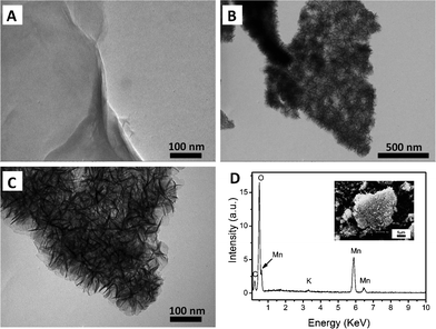
Fig. 1A shows a TEM image of graphene oxide nanosheets. It is found that the as-synthesized GO sheet has a smooth surface. After growing MnO2 on its surface, one can see that the GO nanosheet is covered with numerous clusters of MnO2 nanoparticles homogeneously as shown in Fig. 1B; when taking TEM image with a relatively high magnification (Fig. 1C), more details about the morphology of the nanocomposites are observed, in which GO nanosheets are composed of several layers and the length of each MnO2 nanoparticle are slice shaped with scales ranging from 30 to 50 nm. Furthermore, the as-prepared MnO2/GO nanocomposites can be subjected to ultrasonication in ethanol for 1 h with little loss of MnO2 from GO, indicating that the adsorption of MnO2 onto the surface of GO is stable. These aforementioned features of the synthetic MnO2/GO nanocomposites, along with their electrocatalytic activity toward the oxidation of hydrazine, virtually make them applicable as a new kind of electrochemical hydrazine sensor. | ||
| Fig. 1 TEM images of (A) GO nanosheet and (B) (C) GO/MnO2 nanocomposites at different magnification; (D) EDX of GO/MnO2 nanocomposites. Inset: SEM image of GO/MnO2 nanocomposites. | ||
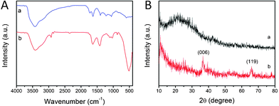
The chemical composition of the resulting MnO2/GO nanocomposites has been characterized by EDX, FT-IR spectra, and XRD measurements. Fig. 1D shows the EDX results of the MnO2/GO nanocomposites, which reveal the presence of C, Mn, and O in the sample. It also shows the presence of a small amount of K, reflecting what was doped in the MnO2 during the preparation process.31FT-IR spectra of the GO nanosheets and MnO2/GO nanocomposites are displayed in Fig. 2A. The results show several characteristic transmittance peaks of MnO2 and GO. The strong peaks at 513 cm−1 are assigned to the Mn–O vibration of MnO2 products. Other peaks in the spectra are the characteristic peaks of the GO and these characteristic peaks are consistent with the GO samples reported previously, which proves the formation of GO.30 Details are as follows: the wide peak around 3417 cm−1 can be attributed to the coupling O–H bond. The bands at 1631 and 1412 cm−1 are ascribed to the C![[double bond, length as m-dash]](https://www.rsc.org/images/entities/char_e001.gif) C vibrations and carboxyl O–H vibrations, respectively. The peak at 1106 and 1024 cm−1 are attributed to C–O–C vibrations. The composition of the as-synthesized MnO2/GO nanocomposites was also examined by XRD measurement (Fig. 2B). Compared to GO, the strong diffraction peaks at 2θ values of 36.6 and 65.9°, corresponding to the (006) and (119) main crystal planes of MnO2, were observed. These reflections could be indexed as the face-centered-cubic phase of MnO2 (JCPDS card No. 18-0802), indicating the formation of MnO2 nanoparticles. Furthermore, the diffraction peak at 10.9° is attributed to the (002) crystal plane of GO.
C vibrations and carboxyl O–H vibrations, respectively. The peak at 1106 and 1024 cm−1 are attributed to C–O–C vibrations. The composition of the as-synthesized MnO2/GO nanocomposites was also examined by XRD measurement (Fig. 2B). Compared to GO, the strong diffraction peaks at 2θ values of 36.6 and 65.9°, corresponding to the (006) and (119) main crystal planes of MnO2, were observed. These reflections could be indexed as the face-centered-cubic phase of MnO2 (JCPDS card No. 18-0802), indicating the formation of MnO2 nanoparticles. Furthermore, the diffraction peak at 10.9° is attributed to the (002) crystal plane of GO.
 | ||
| Fig. 2 (A) FT-IR spectra of (a) GO nanosheets and (b) GO/MnO2 nanocomposites; (B) XRD patterns for the (a) GO nanosheets and (b) GO/MnO2 nanocomposites. | ||
3.2. Electrochemical properties of MnO2/GO nanocomposites
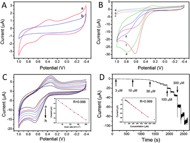
Direct electrochemical behaviors of both MnO2/GO modified GCE and bare GCE in pH 7.0 phosphate buffer solution were studied by cyclic voltammetry and the results are shown in Fig. 3A. At the MnO2/GO/GCE electrode, a pair of small oxidative and reductive peaks was observed (curve a), in which the anodic peak potential and the cathodic peak potential are located at +0.66 V and +0.36 V, respectively. These peaks are assignable to the oxidation and reduction of Mn species, which is ascribed to the overall redox process below.| MnO2(s) + 2e− + 4H3O+ = Mn2+(aq) + 6H2O |
 | ||
| Fig. 3 (A) CVs of (a) the GO/MnO2/GCE and (b) the bare GCE in 0.1 M pH = 7 PBS at a scan rate of 100 mV s−1; (B) CVs of (a, d) bare GCE, (b, e) MnO2/GCE, (c, f) GO/MnO2/GCE in the absence of hydrazine (a, b, c), and presence of 1 mM hydrazine (d, e, f) in pH = 7 PBS; (C) CVs of GO/MnO2/GCE in 0.1 M pH = 7 PBS and 0.3 mM hydrazine at various scan rates of 25, 50, 75, 100, 125, 150, 175, 200 mV s−1. Inset: plot of peak current vs. scan rate; (D) Amperometric response of the GO/MnO2/GCE with successive additions of hydrazine to 0.1 M pH = 7 PBS at an applied potential of 0.6 V. Inset: the corresponding calibration curve. | ||
At the bare GCE electrode, a comparative experiment, the current intensity was smaller than that of MnO2/GO/GCE, and no obvious oxidation and reduction peaks were obtained in the potential range of −0.4 to +1.0 V (curve b).
3.3. Electrooxidation of hydrazine at MnO2/GO modified GCE
In order to investigate the electrocatalytic activity of hydrazine on the MnO2/GO/GCE, MnO2/GCE and bare GCE were also introduced as controls in the electrocatalysis of hydrazine. The CV curves of bare GCE, MnO2/GCE, and MnO2/GO/GCE in a buffer solution (pH = 7) and in the presence of 1 mM hydrazine are presented in Fig. 3B. It shows clearly that all of these three electrodes have a small current intensity without hydrazine, and only very small oxidation peak currents are observed at about 0.7 V. In an environment containing 1 mM hydrazine, when using bare GCE, the current increases in the oxidation zone but without appearance of an oxidation peak (curve d). The CV obtained at MnO2/GCE (curve e) has a significantly higher current response compared to that of bare GCE, and there is an obvious oxidation peak at +0.78 V. A well-formed sharp catalytic oxidation peak at +0.68 V was obtained at MnO2/GO/GCE (curve f), which is lower potential than that of MnO2/GCE. And the oxidation peak current intensity is higher than that of MnO2/GCE, which is about 29 μA.The observations of current increasing and oxidation peak potential decreasing on MnO2/GO/GCE compared to that of the two other modified electrodes may be attributed as follows. Firstly, MnO2 nanoparticles have good catalytic properties for hydrazine oxidation on an electrode surface, which makes MnO2/GCE and MnO2/GO/GCE exhibit an extraordinary enhancement in the current intensity compared to that of the bare GCE. Secondly, GO can be regarded as graphene functionalized by carboxylic acid, hydroxyl, and epoxide groups, the properties of GO are sensitive to chemical doping, adsorbed or bound species, and structure distortion. As a result, a large number of MnO2 may be easily adsorbed on the rough surfaces of GO, which enlarge the surface area of the electrode and provide more electrocatalytic sites. Thirdly, the abundant specific functional groups on GO may enhance catalytic activity though the synergistic effect with the deposited MnO2 nanoparticles. Therefore, one can draw a conclusion from the above three statements that MnO2/GO/GCE electrodes have a great performance in electrocatalyzing hydrazine.
Fig. 3C shows the CV curves of MnO2/GO composite nanosheets modified GCE in pH = 7 phosphate buffer solution containing 0.3 mM hydrazine at different scan rates in the range of 25–200 mV s−1, and are used to evaluate the kinetics of the resulting MnO2/GO composite nanosheets modified electrodes. It is found that the peak current for the anodic oxidation of hydrazine is linearly proportional to the scan rates (Fig. 3C, inset), indicating a typical surface controlled electrochemical process.
Compared to CV curves, amperometric response under certain stirred conditions has a much higher current sensitivity and it can be used to ascertain the linear range and to calculate the limit of detection of hydrazine at the MnO2/GO/GCE. The amperometry experiments were carried out at 0.6 V in pH = 7 phosphate buffer solution under constant stirring. Fig. 3D displays a typical current–time response of the MnO2/GO/GCE on a step change of hydrazine concentration, and the corresponding calibration curve is shown in the inset of Fig. 3D. The steady-state current has a linear relationship with the concentration of hydrazine range up to 1.12 mM with a coefficient of 0.999. Furthermore, more data can be calculated: the detection limit (signal to noise ratio of 3) and sensitivity are 0.16 μM and 1007 μA mM−1 cm−2, respectively. It can be seen that the sensitivity and the detection limit of MnO2/GO/GCE are excellent, thus the MnO2/GO nanocomposites are a good candidate for a hydrazine sensor with a high performance.
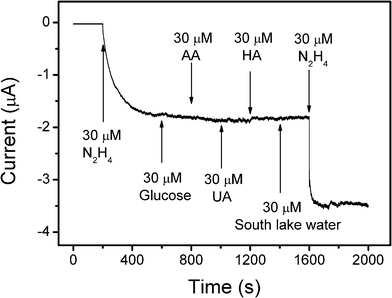
It is known that selectivity is a very important aspect of sensor performance. Some typical electroactive species may affect the response of the hydrazine during the electrochemical measurements, especially those compounds merged together with the hydrazine during the industrial process. The most common interfering species including ascorbic acid (AA), uric acid (UA), humic acid (HA), and glucose and they were chosen to evaluate the selectivity of the as-developed hydrazine sensor. In addition, naturally occurring compounds in the water samples from South Lake at Changchun city were also used to study the matrix effect without any pre-treatment. As shown in Fig. 4, it is found that the current responses almost did not change after the addition of the same concentration of glucose, AA, and UA to hydrazine. After adding the same dose of HA, the current increased a little, which can be neglected compared to the changes induced by hydrazine. Furthermore, no response occurred after injection of untreated South Lake water, indicating that the catalyst is not interfered with by the naturally occurring compounds in real water samples. After successive additions of different analytes, a further 30 μM hydrazine was injected into the solution, causing a current response equal to that at the first injection, indicating high selectivity of the MnO2/GO/GCE for detecting hydrazine. In addition to the good selectivity, the MnO2/GO/GCE electrode showed good stability as well. After one week from the initial measurements, the peak current changed little in response to 0.3 mM hydrazine at a scan rate of 100 mV s−1 (Fig. S2†).
 | ||
| Fig. 4 Amperometric response of GO/MnO2/GCE to 30 μM hydrazine and different interferences of 30 μM glucose, 30 μM ascorbic acid, 30 μM uric acid, 30 μM humic acid and 30 μM South Lake water in stirring 0.1 M pH = 7 PBS. The working potential was 0.6 V. | ||
4. Conclusions
In this work, MnO2/graphene oxide nanosheets have been successfully fabricated via a simple wet-chemical method. TEM studies show that MnO2 is well coated on the surface of GO nanosheets, while EDX and FT-IR were employed to confirm the elements and composition. XRD was also used to investigate the composition as well as the crystal structure. Additionally, the electrochemical study demonstrated that the MnO2/GO nanocomposites modified GCE showed excellent performance towards hydrazine oxidation in pH = 7 phosphate buffer solution. The as-prepared hydrazine sensor also has excellent properties such as great sensitivity, low detection limit, wide linear range, good selectivity and stability, indicating that MnO2/GO nanocomposites are a promising electrocatalyst for amperometric detection of hydrazine.Acknowledgements
This work is supported by the research grants from the National 973 Project (S2009061009), the National Natural Science Foundation of China (20904015, 50973038), Jilin Science and Technology Department project (20100101, 201115014) and State Key Laboratory for Modification of Chemical Fibers and Polymer Materials, Dong Hua University.References
- K. Yamada, K. Asazawa, K. Yasuda, T. Ioroi, H. Tanaka, Y. Miyazaki and T. Kobayashi, J. Power Sources, 2003, 115, 236 CrossRef CAS.
- U. Ragnarsson, Chem. Soc. Rev., 2001, 30, 205 RSC.
- B. A. Aigner, U. Darsow, M. Grosber, J. Ring and S. G. Plotz, Dermatology, 2010, 221, 300 CrossRef CAS.
- N. B. Patel, I. H. Khan and S. D. Rajani, Eur. J. Med. Chem., 2010, 45, 4293 CrossRef CAS.
- J. Waser, B. Gaspar, H. Nambu and E. M. Carreira, J. Am. Chem. Soc., 2006, 128, 11693 CrossRef CAS.
- S. Tafazoli, M. Mashregi and P. J. O'Brien, Toxicol. Appl. Pharmacol., 2008, 229, 94 CrossRef CAS.
- Y. Ding, C. Hou, B. Li and Y. Lei, Electroanalysis, 2011, 23, 1245 CrossRef CAS.
- M. Wang, C. Wang, G. Wang, W. Zhang and F. Bin, Electroanalysis, 2010, 22, 1123 CAS.
- M. S. Baymak and P. Zuman, Tetrahedron Lett., 2006, 47, 7991 CrossRef CAS.
- B. Sljukic, C. E. Banks, A. Crossley and R. G. Compton, Electroanalysis, 2006, 18, 1757 CrossRef CAS.
- C. Batchelor-McAuley, C. E. Banks, A. O. Simm, T. G. J. Jones and R. G. Compton, Analyst, 2006, 131, 106 RSC.
- B. K. Jena and C. R. Raj, J. Phys. Chem. C, 2007, 111, 6228 CAS.
- J. M. Pingarron, I. O. Hernandez, A. Gonzalez-Cortes and P. Yanez-Sedeno, Anal. Chim. Acta, 2001, 439, 281 CrossRef CAS.
- Y. S. Ding, X. F. Shen, S. Gomez, H. Luo, M. Aindow and S. L. Suib, Adv. Funct. Mater., 2006, 16, 549 CrossRef CAS.
- L. Espinal, S. L. Suib and J. F. Rusling, J. Am. Chem. Soc., 2004, 126, 7676 CrossRef CAS.
- M. Toupin, T. Brousse and D. Belanger, Chem. Mater., 2002, 14, 3946 CrossRef CAS.
- J. J. Xu, X. L. Luo, Y. Du and H. Y. Chen, Electrochem. Commun., 2004, 6, 1169 CrossRef CAS.
- K. S. Novoselov, A. K. Geim, S. V. Morozov, D. Jiang, Y. Zhang, S. V. Dubonos, I. V. Grigorieva and A. A. Firsov, Science, 2004, 306, 666 CrossRef CAS.
- A. K. Geim, Science, 2009, 324, 1530 CrossRef CAS.
- C. Berger, Z. M. Song, T. B. Li, X. B. Li, A. Y. Ogbazghi, R. Feng, Z. T. Dai, A. N. Marchenkov, E. H. Conrad, P. N. First and W. A. de Heer, J. Phys. Chem. B, 2004, 108, 19912 CrossRef CAS.
- Y. Wang, Z. Shi, Y. Huang, Y. Ma, C. Wang, M. Chen and Y. Chen, J. Phys. Chem. C, 2009, 113, 13103 CAS.
- X. Wang, Y. Ouyang, X. Li, H. Wang, J. Guo and H. Dai, Phys. Rev. Lett., 2008, 100, 6803 Search PubMed.
- E. Yoo, J. Kim, E. Hosono, H.-s. Zhou, T. Kudo and I. Honma, Nano Lett., 2008, 8, 2277 CrossRef CAS.
- F. Schedin, A. K. Geim, S. V. Morozov, E. W. Hill, P. Blake, M. I. Katsnelson and K. S. Novoselov, Nat. Mater., 2007, 6, 652 CrossRef CAS.
- X. Zhou, X. Huang, X. Qi, S. Wu, C. Xue, F. Y. C. Boey, Q. Yan, P. Chen and H. Zhang, J. Phys. Chem. C, 2009, 113, 10842 CAS.
- K. P. Loh, Q. Bao, G. Eda and M. Chhowalla, Nat. Chem., 2010, 2, 1015 CrossRef CAS.
- J. T. Robinson, F. K. Perkins, E. S. Snow, Z. Wei and P. E. Sheehan, Nano Lett., 2008, 8, 3137 CrossRef CAS.
- W. S. Hummers and R. E. Offeman, J. Am. Chem. Soc., 1958, 80, 1339 CrossRef CAS.
- L. J. Cote, F. Kim and J. X. Huang, J. Am. Chem. Soc., 2009, 131, 1043 CrossRef CAS.
- H.-L. Guo, X.-F. Wang, Q.-Y. Qian, F.-B. Wang and X.-H. Xia, ACS Nano, 2009, 3, 2653 CrossRef CAS.
- Y. Q. Dai, X. F. Lu, M. McKiernan, E. P. Lee, Y. M. Sun and Y. N. Xia, J. Mater. Chem., 2010, 20, 3157 RSC.
Footnote |
| † Electronic supplementary information (ESI) available. See DOI: 10.1039/c2ra01065h |
| This journal is © The Royal Society of Chemistry 2012 |
