Enzyme immobilization in biosensor constructions: self-assembled monolayers of calixarenes containing thiols
Dilek Odaci Demirkol*a,
Huseyin Bekir Yildiz*b,
Serkan Sayınc and
Mustafa Yilmazc
aEge University Faculty of Science Biochemistry Department, 35100 Bornova-Izmir, Turkey. E-mail: dilek.odaci@ege.edu.tr; Fax: +90 232 311 5485; Tel: +90 232 3115487
bKaramanoglu Mehmetbey University, Kamil Özdag Science Faculty, Chemistry Department, 70100, Karaman, Turkey. E-mail: yildizhb@kmu.edu.tr; Fax: +90 3382262116; Tel: +90 3382263840
cSelcuk University Chemistry Department, 42031 Konya, Turkey
First published on 23rd April 2014
Abstract
Herein, an amperometric glucose oxidase (GOx) biosensor is presented using calixarenes as an immobilization matrix of the biomolecule. Firstly, thiol-containing calixarenes (Calix-SH) were synthesized, then self-assembled monolayers (SAMs) of Calix-SH on a gold surface were formed and hydroxyl groups of Calix-SH were activated using 1,1′-carbonyldiimidazole (CDI) chemistry. To test the usability of Calix-SH modified surfaces as a biosensor, glucose oxidase was used as a model biological component. After optimization of preparation and working conditions, our results indicate that the Calix-SH/GOx biosensor has a linear range in the range 0.1–1.0 mM (LOD: 0.015 mM) for glucose with a 25 s response time. Finally, the application of the biosensor was examined to detect glucose in real samples. The glucose amounts were calculated as 19.460 ± 0.521 and 31.647 ± 2.125 mM in coke and fizzy drink (with orange), respectively. To confirm the reliability of the Calix-SH/GOx biosensor, the calculated glucose concentrations which were analyzed by the Calix-SH/GOx biosensor were compared to conventional spectrophotometric glucose kits. The glucose amounts in coke and fizzy drink were calculated as 18.509 ± 0.732 mM and 31.579 ± 4.466 mM, respectively.
Introduction
Immobilized enzymes are widely employed in various biomedical, environmental and industrial applications such as biosensors, fermentation technology, the food industry, biodiesel production and bioremediation, etc.1,2 To fabricate innovative biosensors and other biomolecule-based diagnostic techniques, immobilization of the biological component to solid surfaces is crucial. However, the relevant literature often cites that immobilization of enzymes generally causes a decline in enzyme activity or impaired catalytic characteristics. Therefore, the choice of efficient immobilization technique is of paramount significance to preserve catalytic properties of the enzyme. To date, different techniques in conjunction with various immobilization materials such as entrapment in natural and synthetic polymers,3–6 covalent attachment,7 adsorbtion,8 and cross-linking9 have been of choice to fabricate enzyme biosensors. Of them, self-assembled monolayers (SAMs) on the gold surfaces is one of the favorable strategies to prepare well-ordered structures onto the supports given that thiols can interact very strongly with metals such as gold, silver, platinum, palladium or copper, to name few. Because it allows controlling firmly dimension and properties of the surface as well as the nature of thiolic ends by various chemistries, SAM seems a meaningful approach to fabricate stable and reproducible surfaces for biosensor applications.10–12Calixarenes, cyclic oligomers of phenolic units linked through the ortho positions, are one of the remarkable macrocyclic host molecules.13 They are traditionally prepared via oligomerization of phenol and formaldehyde.14 Nowadays, they gain attention in various field of applications as they are effortlessly functionalized at either the upper and/or lower rim of the molecular structure and they form reversible complex with a large library of compounds including such as ions, amino acids, peptides and other biomolecules.15,16 By using the features of capturing cationic molecules, molecular recognition of aminoacids by calixarenes has opened a gain the application of them in protein microarrays.17 In another study, Chen et al. have reported the use of calixarenes for the first time as enzyme immobilization material. This group has reported the synthesis a novel derivative of calixarene, p-tert-butylthiacalix[4]arene tetra-amine (TC4TA), which was subsequently employed to adsorb GOx on the TC4TA modified electrode. In this design of TC4TA/GOD biosensor to detect glucose, the adsorption mechanism was purely based on ionic interactions between amino-groups of TC4TA and carboxyl groups of GOx.18 However, these ionic bonds are notoriously not of high stability as their strength strictly depends on pH. In this manuscript, we report the first synthesis of a thiol-containing calixarene named as 5,11,17,23-tetra-tert-butyl-25,27-bis(3-thiol-1-oxypropane)-26,28-dihydroxycalix[4]arene, (SH-Calix, for short hereafter) and its utilization to modify gold electrode surfaces via creation of self-assembled monolayers. Then, we have used GOx as a model enzyme to test application of calixarenes as an immobilization material in biosensor constructions. Glucose oxidase (GOx, glucose 1-oxidase, β-D-glucose: oxygen 1-oxidoreductase, EC 1.1.3.4) contains two identical subunits, which are flavine adenine dinucleotide (FAD) cofactor bound to the polypeptide chains and is a glycoprotein with 74% protein, 16.4% neutral sugar and 2.4% amino sugar.19 At neutral pH values, multipoint covalent attachment between multimeric enzymes such as GOx and immobilization material produce stabile enzymes without subunit dissociation. Because of reduction any conformational change involved in enzyme inactivation and increasing the enzyme stability, multipoint covalent attachment provides the rigidification of the enzyme structure.20 Generally, the reactive amines of aminoacids in the protein surface is useful to bind them to the surface of support.2
In this study, a two step-process was applied to immobilize Gox on the surface of gold electrode. In the first step, gold electrode surface was modified with Calix-SH to acquire a well-ordered surface to immobilize the enzyme. In here, we should note that interactions between gold and thiol groups of calixarenes direct free hydroxyl groups towards exterior side. Subsequently, these hydroxyl groups of calixarene are activated with CDI, upon which the activated support irreversibly reacts through free amine groups of GOx.21,22 The covalent bonds between hydroxyl groups of calixarenes well-ordered on the electrode surface and amine groups of enzyme were formed with 1,1′-carbonyldiimidazole (CDI) chemistry.23,24 Of course, the prime purpose of this technique is to bring the monolayer of GOx molecules on the surface of the calixarene-modified electrode, leaving all enzyme molecules equally accessible.25 Next, we have added glutaraldehyde to form the cross-links between BSA and GOx. However, glutaraldehyde did not solely cross-link BSA and GOx, but also cross-links BSA enzymes and BSA-to-GOx enzymes, leading to a fabrication of a biomembrane.26 Naturally, acquiring a biomembrane works in our favor as it offers several advantageous such as saturation of any non-specific binding sites,27 superior stability of Calix-SH/GOx biosensors and diminished noise of amperometric measurements and also inference effect of some molecules in real samples. Hence, glutaraldehyde as a crosslinking agent serves multiple purposes with the use of suitable reaction time, pH value and temperature as to crosslink enzyme molecules and enzyme to immobilization support through their amine groups.28 Besides, the choice of glutaraldehyde to form covalent bonds eliminates an additional reduction step to further stabilize the reaction product.26 In overall, our design of Calix-SH/GOx biosensor was schematized in Scheme 1. Upon the optimization of enzyme loading and pH on the biosensor response, analytical characteristics were studied and the application of Calix-SH/GOx biosensors was tested to determine the glucose amount in real samples.
 | ||
| Scheme 1 Schematic representation of Calix-SH/GOx biosensor. | ||
Materials and methods
Materials
Glucose oxidase [from Aspergillus niger, lyophilized powder, Type X-S, 147![[thin space (1/6-em)]](https://www.rsc.org/images/entities/char_2009.gif) 900 units per g solid; it includes catalase ≤5 units per mg protein (foreign activity) and may contain traces of amylase, maltase, glycogenase, invertase, and galactose oxidase. Its' protein composition is 65–85% and molecular weight: 160 kDa gel filtration], glucose, 1,1′-carbonyldiimidazole (CDI), dimethyl sulfoxide (DMSO), glutaraldehyde, bovine serum albumin (BSA) were purchased from Sigma.
900 units per g solid; it includes catalase ≤5 units per mg protein (foreign activity) and may contain traces of amylase, maltase, glycogenase, invertase, and galactose oxidase. Its' protein composition is 65–85% and molecular weight: 160 kDa gel filtration], glucose, 1,1′-carbonyldiimidazole (CDI), dimethyl sulfoxide (DMSO), glutaraldehyde, bovine serum albumin (BSA) were purchased from Sigma.
For the synthesis of calixarenes, TLC analyses were carried out on DC Alufolien Kieselgel 60 F254 (Merck). Generally, solvents were dried by storing them over molecular sieves (Aldrich; 4 Å, 8–12 mesh). All reactions, unless otherwise noted, were conducted under nitrogen atmosphere. All starting materials and reagents used were of standard analytical grade from Merck or Aldrich used without further purification. All aqueous solutions were prepared with deionized water that was passed through a Millipore milli-Q Plus water purification system.
Apparatus
PalmSens electrochemical measurement unit (Palm Instruments, Houten, Netherlands) was used to carry out chronoamperometric measurements. The experiments were performed in a reaction cell (10 mL) at room temperature using of three electrodes configuration, consisting of gold working electrode (BASI, USA), Ag/AgCl reference electrode (3 M KCl, Metrohm, Switzerland) and a platinum counter electrode (Metrohm, Switzerland). Pharmacia LKB Novaspec II spectrophotometer (LKB Biochrom, England) was used in colorimetric experiments as a reference method in sample applications.Melting points were determined on a Gallenkamp apparatus in a sealed capillary glass tube and are uncorrected. 1H NMR spectra were recorded on a Varian 400 MHz spectrometer. Elemental analyses were performed using a Leco CHNS-932 analyzer.
For the surface characterization of the prepared calixarene-based biosensors, JEOL5600-LU model scanning electron microscope (SEM) was used. To obtain samples for SEM analysis, gold slides were modified with same procedure which was used to prepare Calix-SH/GOx biosensors. The images were taken via using an acceleration voltage of 20 kV.
The spectrophotometric data to calculate enzyme activity and protein amount have been collected by means of a Lambda 35 UV/vis spectrometer purchased from Perkin-Elmer (USA).
Synthesis of SH-Calix, 5,11,17,23-tetra-tert-butyl-25,27-bis(3-thiol-1-oxypropane)-26,28-dihydroxycalix[4]arene
The compounds 1 and 2 were synthesized by procedures published in the literature.13,29 SH-Calix named as 5,11,17,23-tetra-tert-butyl-25,27-bis(3-thiol-1-oxypropane)-26,28-dihydroxycalix[4]arene (3) is herein reported for the first time (Scheme 2). For the synthesis of Calix-SH, a mixture of dialkyl bromide of p-tert-butylcalix[4]arene (2) (0.5 g, 0.561 mmol) and thiourea (0.14 g, 1.88 mmol) in CH3CN (40 mL) was refluxed. The reaction was monitored by using a TLC (CH2Cl2–hexane(1:1)). After 45 h, the solvent was removed under reduced pressure. The remaining was mixed with 0.19 g of KOH (3.423 mmol) and aliquot of 21 mL of deionized water and allowed to reflux for 2 h. The residue was extracted with 1 M HCl and CHCl3, dried over MgSO4 to afford pure compound 3 with 40% yield-m.p.: 203–205 °C. 1H NMR (400 MHz CDCl3): δ 1.19 (s, 18H, but), 1.22 (s, 18H, but), 1.57–1.69 (m, 2H, –SH), 2.42–2.48 (m, 4H, –CH2–), 3.24 (t, 4H, J = 8.0 Hz, –CH2–S), 3.38 (d, 4H, J = 12.8 Hz, Ar–CH2–Ar), 4.01 (t, 4H, J = 4.8 Hz, –CH2–O), 4.23 (d, 4H, J = 12.8 Hz, Ar–CH2–Ar), 7.01 (s, 4H, ArH), 7.07 (s, 4H, ArH), 9.13 (s, 2H, –OH). Anal. calcd. For C50H68O4S2: C, 75.33; H, 8.60. Found (%); C, 75.45; H, 8.79.
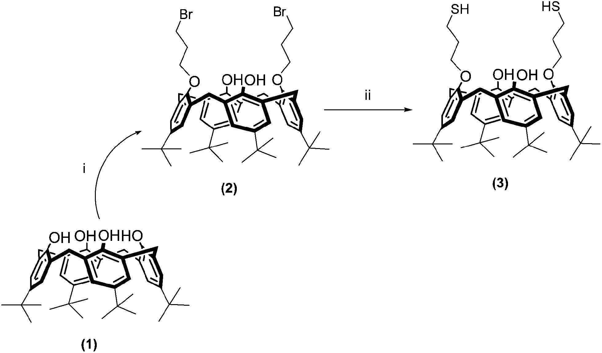 | ||
| Scheme 2 The synthetic route for synthesis of 5,11,17,23-tetra-tert-butyl-25,27-bis(3-thiol-1-oxypropane)-26,28-dihydroxycalix[4]arene (3). Reaction conditions: (i) K2CO3, CH3CN, NaI, 1,3-dibromopropane; (ii) thiourea, CH3CN, KOH. | ||
Fabrication of Calix-SH/GOx biosensors
Before each experiment, gold electrodes were polished with alumina powder (Gamma, 0.05; 0.1; 0.3; 1.0 μm). Then it etched in 0.5 M H2SO4 solution by cyclic-potential scanning between 0 and 1.5 V until a reproducible voltammetric response was obtained. Initially, 1 mg Calix-SH and 50 mg CDI were dissolved in 0.5 mL of DMSO and the gold electrode was immersed in the mixture of Calix-SH and CDI for 1.5 h at room temperature. Then 1 mg GOx was dissolved in 5 μL of pH 8.2; 0.1 M sodium phosphate buffer and spread over the Calix-SH modified gold electrodes. After that, the electrodes were allowed to stand at 4 °C overnight. Finally, the mixture of 2.5 μL of 1.0% of glutaraldehyde and 2.5 μL of BSA (0.5 mg mL−1) solutions prepared in pH 7.0; 50 mM phosphate buffer was spread over the surface of the modified electrodes and dried for 1 h at room temperature.Before and after immobilization steps, GOx activity of the prepared biosensors was checked according to ABTS method by enzymatically determining the concentration of the produced hydrogen peroxide by means of peroxidase (POD), according to the following procedure: 1 mL of ABTS (0.003 M), 0.3 mL of POD (6 U), 1 mL of glucose (0.3 M), and 0.7 mL of potassium phosphate buffer (50 mM; pH 6.0) were mixed and incubated for 5 min at 30 °C. Then electrodes were immerged into the reaction mixture and absorbance was followed for 3 min at 420 nm. The concentration of ABTS was in excess in respect to that of H2O2 so that the reaction was limited by the concentration of the latter product. H2O2 concentration was calculated by using the extinction coefficient of ABTS (ε420 = 43200 M−1 cm−1). One unit (U) of enzyme activity was defined as the amount of enzyme that catalyzed the production of 1 μmol H2O2 per minute under the experimental conditions.30,31 Protein contents of GOx solution before and after immobilization procedure were calculated according to the Bradford assay.32
Measurement procedure
All electrochemical measurements were performed at room temperature in an open vessel filled with the vigorously stirred 10 mL of 50 mM acetate buffer solution, pH 4.5. Increasing concentrations of glucose were added definite volumes of the stock solution of 1.0 M glucose (in pH 4.5, 50 mM acetate buffer). The response of the Calix-SH/GOx biosensor shows the decrease of dissolved O2 content upon exposure to glucose solutions. According to the reaction scheme, GOx uses β-D-glucose as substrate and converts it to D-glucono-1,5 lactone and hydrogen peroxide by oxidizing glucose at carbon-1:33| β-D-glucose + O2 → D-glucono-1,5-lactone + H2O2 |
In first generation amperometric biosensors, oxygen consumption and the formed hydrogen peroxide can be followed at −0.7 and +0.7 V, respectively. It is known that most common metabolites such as uric acid and ascorbic acid get oxidized and interfere with the electrochemical signal at higher potentials. It is therefore essential to apply the lowest possible electrode potential.10,33 According to this data, The decrease of dissolved O2 concentration was determined as the analytical signal of the biosensor at −0.7 V in this study.34 The steady-state current was typically achieved in 25 s. After each measurement, the working buffer was changed and the electrodes were washed with distilled water.
Sample application
The developed biosensors were tested with real samples (coke and fizzy with orange). Samples were degassed and diluted with working buffer and then injected into the reaction buffer instead of glucose. Calibration curves were used to determine the glucose contents in measured samples. The samples were also applied to a commercial enzyme assay kit based on spectrophotometric Trinder reaction (Cromatest, Glucose MR, Cat. no. 1129010) as the reference method and results were compared with those obtained with the constructed biosensors. In the Trinder reaction, the glucose is oxidized to D-gluconate by glucose oxidase (GOx) with the formation of hydrogen peroxide. In the presence of peroxidase (POD), a mixture of phenol and 4-aminoantipyrine (4-AAP) is oxidized by hydrogen peroxide to form a red quinoneimine dye proportional to the glucose concentration in the sample.35Results and discussion
Characterization of SH-Calix
p-tert-Butylcalix[4]arene (1) and its dialkyl bromide derivative (2) were synthesized according to the literature routes.29,36 The substitution of dibromide derivative of p-tert-butylcalix[4]arene (2) was conducted in the presence of CH3CN with thiourea and KOH solution to afford the cone conformer dithio substituted p-tert-butylcalix[4]arene (3) in 40% yield. The 1HNMR spectra of (3) has a typical AX pattern for the methylene bridge proton (ArCH2Ar) of calixarene by appearing two doublets at 3.38 and 4.23 ppm (12.8 Hz)37 (Fig. 1).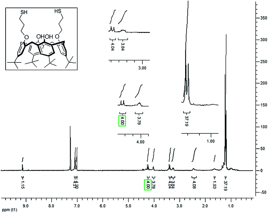 | ||
| Fig. 1 1H-NMR spectrum of 5,11,17,23-tetra-tert-butyl-25,27-bis(3-thiol-1-oxypropane)-26,28-dihydroxycalix[4]arene (3). | ||
Application of SH-Calix on the fabrication of enzyme biosensors
Calixarenes are synthesized by the oligomerisation of phenol and formaldehyde, offer several advantages in synthesis and applications as supramolecular platforms for molecular recognition, sensing and self-assembly, catalysis, nanotechnology, and drug discovery.14,15 In enzyme-based detection technologies, immobilization of proteins on the solid surface, without losing their activity is the critical point. Calixarenes are useful macromolecules to keep their structural motifs and thereby their functionality. In our study, thiol containing calixarenes (SH-Calix) are synthesized and gold electrode surfaces were modified by them via construction of SAMs. And free hydroxyl groups of SH-Calix were functional point to immobilize GOx in targeted position using CDI chemistry. To increase signal/noise ratios and to prevent the negative effects of interfering substances in samples, the surface was covered with BSA. After fabrication of Calix-SH/GOx biosensor, initially the obtained biomembrane was characterized. To evaluate of the conductivity of the biomolecule-modified gold electrode, Calix-SH/GOx biosensor was characterized by cyclic voltammetry (CV) using Fe(CN)63− as an electrochemical probe. CV obtained at the bare, SH-Calix and SH-Calix/GOx modified Au electrodes immersed in aqueous 0.1 M KCl containing 5 mM Fe(CN)63− is shown in Fig. 2A, respectively. A reversible electrochemical response for Fe(CN)63− was obtained. Well-defined oxidation and reduction peaks are observed at about 0.258 and 0.165 V for bare electrode (peak-to-peak separation of 93 mV), 0.244 and 0.151 V for SH-Calix modified electrode (peak-to-peak separation of 93 mV) and 0.338 and 0.139 V for Calix-SH/GOx-modified Au electrodes (peak-to-peak separation of 199 mV). According to obtained graphs, there is a decrease in the peak current when the Au electrode was coated step-by-step. Fig. 2B displays CVs of SH-Calix/GOx modified Au electrodes at the different scan rates (5; 10; 25; 50; 75; 100; 125; 150; 175; 200 mV s−1). The peak current increased linearly with the increasing square root of scan rate potential (v1/2). This suggests that the reactions on the SH-Calix/GOx modified Au electrodes were reversible and the mass transport phenomenon is mainly diffusion controlled. Fig. 2C depicts the change of the cyclic voltammograms of Calix-SH/GOx biosensor in the presence of glucose. | ||
| Fig. 2 (A) Cyclic voltammetric characterization of bare, Calix-SH and Calix-SH/GOx modified electrodes conducted in acetate buffer (pH 4.5) containing 5 mM Fe(CN)63−. (B) CV response of Calix-SH/GOx biosensor at different scan rates. (C) CV of Calix-SH/GOx biosensor in the absence and presence of glucose. | ||
The morphologies of SH-Calix and SH-Calix/GOx modified Au electrodes were characterized by SEM and the obtained images could be seen in Fig. 3. Data, was obtained from SEM images supported CV results. After immobilization steps and covering with BSA, the surface was smooth and the spaces between ridges were disappeared.
 | ||
| Fig. 3 SEM images of Calix-SH films (A) and Calix-SH/GOx films on the gold electrode surface. | ||
Effect of pH on the biosensor response
The working pH is an important factor that can significantly influence the performances of the biological catalysts. To obtain optimum pH of Calix-SH/GOx biosensor, the pHs were tested ranges from 3.5 to 5.5 (50 mM acetate buffers). The pH effect on the response of the Calix-SH/GOx biosensor is shown in Fig. 4. Maximum amperometric response was achieved at pH 4.5. Free glucose oxidase has a broad activity range of pH 4–7 (ref. 38) (as quoted by the manufacturer, Sigma-Aldrich) and obtained pH value as an optimum pH is agreement with the needed pH values reported for the free enzyme.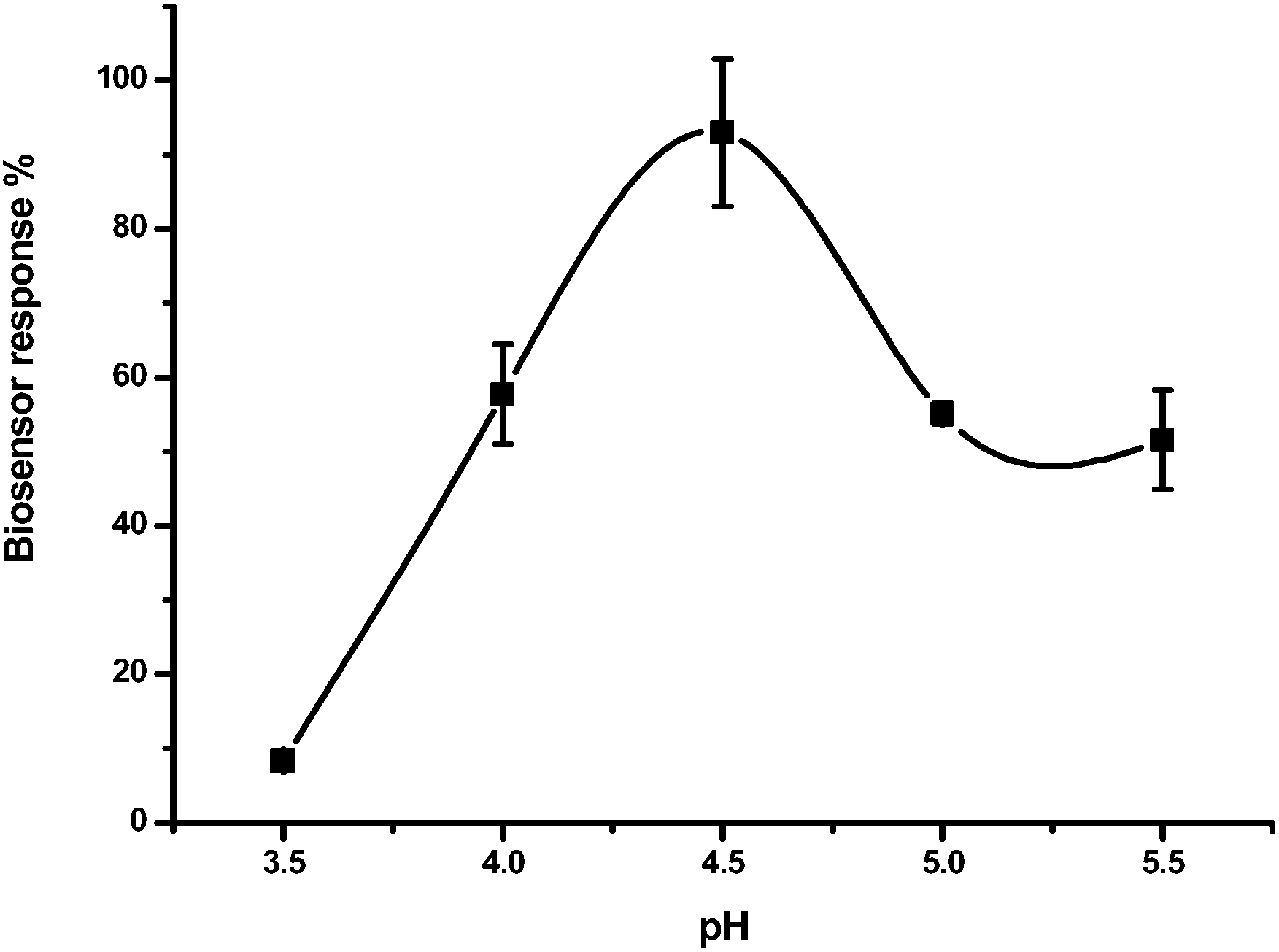 | ||
| Fig. 4 Effect of pH on the biosensor response of Calix-SH/GOx biosensor (in sodium acetate buffer, 50 mM, −0.7 V; error bars show S.D. of three measurements). | ||
Effect of enzyme amount on the biosensor response
To decide the enzyme content to obtain maximum sensitivity, the Calix-SH/GOx biosensors were fabricated by various GOx amounts (0.5 mg; 1.0 mg and 2.0 mg) and calibrated for glucose detection (Fig. 5). The highest signal appeared in the use of 1.0 mg of the enzyme. An enzyme loading of 0.5 mg was insufficient for detection of glucose and the efficiency of biosensor which was prepared using 2.0 mg GOx was lower than that for 1.0 mg. | ||
| Fig. 5 Effect of enzyme amount on the biosensor response (in sodium acetate buffer, 50 mM, pH 4.5, −0.7 V; error bars show S.D. of two or three measurements). | ||
As mentioned before, protein content and GOx activity of the prepared Calix-SH/GOx biosensor were checked using Bradford and ABTS method, respectively. Protein contents of Calix-SH/GOx biosensors which were fabricated using 0.5; 1.0 and 2.0 mg enzyme were calculated as 0.135; 0.30 and 0.64 mg according to the Bradford assay, respectively. Enzyme activities for Calix-SH/GOx biosensors which were fabricated using 0.5; 1.0 and 2.0 mg enzyme were found as 3.50; 24.56 and 11.72 units according to ABTS method, respectively. Because of the difficulties in the diffusion of glucose and oxygen in thick surface which contain biocomponents, higher amounts of GOx caused a decrease in the current. Linearity for substrates and the currents which were obtained using various electrodes modified with different enzyme amounts are depends on the electrode species, immobilization technique and also enzyme.3,4,33,39,40 Furthermore, the purity of the enzyme can bring about decrease in activity of enzyme when using an excess of protein.41 Here, GOx includes catalase, amylase, maltase, glycogenase, invertase, and galactose oxidase. During immobilization, contaminant proteins also was immobilized. When GOx was increased, the thickness of biocomponent layer was increased excessively because of impurities. Hence in all the following experiments, the enzyme amount of biosensor was kept at 1.0 mg. When 1.0 mg enzyme was used to prepare Calix-SH/GOx biosensor, activity yield was 87%. The yield was calculated considering the specific activity values for free and immobilized enzyme, which are expressed as U per mg protein.
Analytical characteristics
Different types of alkanethiols have been investigated as an immobilization matrix to fabricate biosensors via forming self assembled monolayers (SAMs). In this study, thiol-containing calixarene structures were tested as an alternative immobilization support or coatings to alkanethiols for the preparation of biosensors. Because of its effectiveness in bioconjugation, covalent bonds were used to combine hydroxyl groups of calixarene and amine groups of enzyme. The electrochemical signal of the Calix-SH/GOx biosensors toward glucose was investigated by chronoamperometrically. As shown in Fig. 6, current change was proportional to the glucose concentration in the range from 0.1 to 1.0 mM, and the linear regression equation was I (nA) = 159.732 + 31.624 (mM) (R2 = 0.995) with a detection limit of 0.015 mM (S/N = 3) and 25 s response time (inset of Fig. 6 shows the signal after glucose addition). When the concentration of glucose was more than 1.0 mM, substrate saturation was observed, showing a typical Michaelis–Menten kinetic mechanism.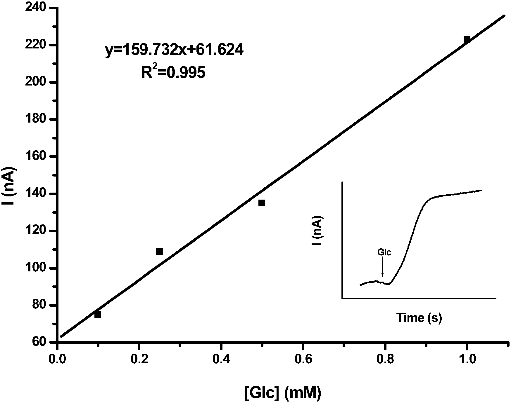 | ||
| Fig. 6 Calibration curve for glucose (in sodium acetate buffer, 50 mM, pH 4.5, −0.7 V; error bars show S.D. of three measurements. Inset: time dependent current response with the addition of 0.25 mM glucose). | ||
Some proteins such as gelatin, albumin have been added to architectures which has been used to prepare biosensor immobilization matrix.42,43 In the absence of these macromolecules, the repeated measurements are not carried out because of matrix decomposition containing biological material. To show importance of BSA in immobilization matrix, glucose biosensor was fabricated without BSA and glutaraldehyde. The obtained biosensor signal was noisy. Although the biosensor response time was faster than the biosensor which was prepared using BSA, the sensor response was decreased after five measurements for a standard glucose solution.
The repeatability of measurement was obtained by consecutive seven trials of 0.25 mM glucose using the same electrode. The relative standard deviation (R.S.D.) was calculated to be 4.7%. To evaluate the electrode-to-electrode reproducibility, three electrodes were prepared under the same conditions in different days. The result showed a R.S.D. of 2.6%, indicating a satisfactory reproducibility. To test operational stability, the biosensors response to 0.5 mM glucose standard solutions was followed. After measurements in 3 h, no decrease was observed in biosensor response. In order to compare the analytical performance of Calix-SH/GOx biosensors with the glucose biosensors based on calixarene in the literature, the characteristics of them were shown in Table 1. Pandya et al. developed non-enzymatic sensor combined with spectroscopic techniques for the detection of glucose based on calix[4]arene/phenyl boronic acid functionalized gold nanoparticles.44 Due to its ability to reversibly bind diol-containing compounds, its selectivity is low. But enzymatic sensors show the affinity and selectivity to their substrates. Coupling of enzymes and transducer are affiliated to success of immobilization techniques. Various types of materials such as synthetic/natural polymer membranes, composites and nanomaterials have been used to fabricate biological material based detection systems. Calixarenes can be used as an alternative to these materials for the immobilization of biological component of biosensors. For this aim, Chen et al. used the calixarenes for the first time as an enzyme immobilization matrix and after immobilization of glucose oxidase, amperometric glucose detection was carried out.18 Firstly, p-tert-butylthiacalix[4]arene tetra-amine (TC4TA) was synthesized and spread on the surface of the platinum disk electrode. Then, using EDC/NHS coupling reaction glucose oxidase was immobilized on the TC4TA modified electrode. Finally, amperometric response to glucose was followed as an amperometrically at +0.60 V vs. SCE. In our study, 5,11,17,23-tetra-tert-butyl-25,27-bis(3-thiol-1-oxypropane)-26,28-dihydroxycalix[4]arene (SH-Calix) was synthesized for the first time and used to modify gold electrode surfaces via creation of self-assembled monolayers (SAMs). Then glucose oxidase was immobilized the calixarene-modified surface. The success of the prepared biosensor depends on the immobilization technique. Among of other immobilization procedures, SAMs have some advantages such as easy preparation of well-designed functional surface, simplicity of preparation, versatility, stability, reproducibility and the possibility of introducing different chemical functionalities.
| Support | Immobilization technique | Principle of detection | Linearity | LOD | Ref. |
|---|---|---|---|---|---|
| — | Non-enzymatic | UV-visible spectrophotometry | 5–100 nM | 4.3 nM | 44 |
| Platinum disk electrode | GOX immobilization to adsorbed calixarene via EDC/NHS coupling reaction | Amperometric | 0.08–10 mM | — | 18 |
| Gold electrode | GOX immobilization to SAMs calixarene via CDI coupling reaction | Amperometric | 0.1–1.0 mM | 0.015 mM | This work |
Effect of some interferents on the biosensor response
The inference studies of Calix-SH/GOx biosensors have been investigated in the presence of some compounds such as galactose, mannose, cysteine, phenol and ethanol. Cysteine and phenol were chosen as a model compound of thiolic and phenolic electroactive species, respectively. Ethanol was used to test the interference effect of this compound to biosensor response when Calix-SH/GOx biosensor is applied to analyze glucose in alcoholic beverages. Galactose and mannose (0.1 mM) were selected to show substrate specificity of GOx and added to reaction medium instead of glucose, but no response were found for these monosaccharide. 0.1 mM of cysteine, phenol and ethanol were added to reaction buffer in the presence of 0.25 mM glucose and the signal was followed chronoamperometrically (Fig. 7). No significant change in the current response to glucose was obtained in the presence of these compounds. | ||
| Fig. 7 Interference by various compounds on the Calix-SH/GOx biosensor used in glucose determination (in sodium acetate buffer, 50 mM, pH 4.5, −0.7 V; error bars show S.D. of three measurements. [Glucose]: 0.25 mM; cysteine [Cys]: 0.1 mM; phenol [Ph]: 0.1 mM; ethanol [EtOH]: 0.1 mM). | ||
Sample application
The glucose concentrations of coke and fizzy with orange were quantitatively analyzed using Calix-SH/GOx biosensor under optimized conditions. Samples were added to reaction buffer as a substrate instead of glucose and the concentration of glucose in samples were calculated using calibration graph. The results were then compared to calculated glucose concentrations which were analyzed by spectrophotometric glucose kits as a conventional method in order to confirm the reliability of Calix-SH/GOx biosensor. The glucose amount of coke was calculated as 19.460 ± 0.521 and 18.509 ± 0.732 mM using Calix-SH/GOx biosensors and the reference method (recovery%: 105%), respectively (n = 3, data are given as the mean ± S.D. (standard deviation)). And glucose amount of fizzy (with orange) was calculated as 31.647 ± 2.125 and 31.579 ± 4.466 mM using Calix-SH/GOx biosensors and the reference method (n: 3; recovery%: 100%), respectively. The results proposed that Calix-SH/GOx biosensor can be successfully used for the determination of glucose in beverages.Conclusions
In summary, this study was defined the probability of developing a calixarene-based biosensor for monitoring glucose. Firstly, thiol containing calixarenes were synthesized. Then the electrochemical biosensing platform was established by SAMs of Calix-SH and GOx was immobilized the calixarene-modified gold surfaces via CDI chemistry. After that, the stability of immobilized enzyme was increased using BSA and glutaraldehyde and also addition of them prevents the interference of the other molecules in sample matrix. The current change was followed based on oxygen consumption during enzymatic reaction at −0.7 V. After optimization and characterization studies, the proposed Calix-SH/GOx biosensor was used successfully for glucose detection in real samples and it could provide as a valuable tool for other fields to determine glucose.Acknowledgements
Authors would like to thank the European Union through the COST Action CM1202 “Supramolecular photocatalytic water splitting (PERSPECT-H2O)” and the Scientific and Technological Research Council of Turkey (TUBITAK Grant Numbers 113T022) for the financial support of this research.Notes and references
- O. Barbosa, R. Torres, C. Ortiz, A. Berenguer-Murcia, R. C. Rodrigues and R. Fernandez-Lafuente, Biomacromolecules, 2013, 14(8), 2433 CrossRef CAS PubMed.
- R. C. Rodrigues, C. Ortiz, A. Berenguer-Murcia, R. Torres and R. Fernández-Lafuente, Chem. Soc. Rev., 2013, 42(15), 6290 RSC.
- I. Cakar, K. V. Ozdokur, B. Demir, E. Yavuz, D. Odaci Demirkol, S. Kocak, S. Timur and F. N. Ertas, Sens. Actuators, B, 2013, 185, 331 CrossRef CAS PubMed.
- O. Yilmaz, D. Odaci Demirkol, S. Gulcemal, A. Kilinç, S. Timur and B. Cetinkaya, Colloids Surf., B, 2012, 100, 62 CrossRef CAS PubMed.
- O. Habib, D. Odaci Demirkol and S. Timur, Food Anal. Methods, 2012, 5, 188 CrossRef.
- D. Odaci, M. U. Kahveci, E. L. Sahkulubey, C. Ozdemir, T. Uyar, S. Timur and Y. Yagci, Bioelectrochemistry, 2010, 79, 211 CrossRef CAS PubMed.
- B. Demir, M. Seleci, D. Ag, S. Cevik, E. E. Yalcinkaya, D. Odaci Demirkol, U. Anik and S. Timur, RSC Adv., 2013, 3, 7513 RSC.
- V. Stepankova, S. Bidmanova, T. Koudelakova, Z. Prokop, R. Chaloupkova and J. Damborsky, ACS Catal., 2013, 3(12), 2823 CrossRef CAS.
- M. Akin, A. Prediger, M. Yuksel, T. Höpfner, D. Odaci Demirkol, S. Beutel, S. Timur and T. Scheper, Biosens. Bioelectron., 2011, 26, 4532 CrossRef CAS PubMed.
- S. K. Aryaa, P. R. Solanki, M. Dattab and B. D. Malhotra, Biosens. Bioelectron., 2009, 24, 2810 CrossRef PubMed.
- N. K. Chaki and K. Vijayamohanan, Biosens. Bioelectron., 2002, 17, 1 CrossRef CAS.
- T. Wink, S. J. van Zuilen, A. Bult and W. P. van Bennekom, Analyst, 1997, 122, 43 RSC.
- S. Sayin, M. Yilmaz and M. Tavasli, Tetrahedron, 2011, 67, 3743 CrossRef CAS PubMed.
- L. Baldini, A. Casnati, F. Sansone and R. Ungaro, Chem. Soc. Rev., 2007, 36, 254 RSC.
- S. B. Nimse and T. Kim, Chem. Soc. Rev., 2013, 42, 366 RSC.
- R. Sharma, R. Margani, S. M. Mobin and R. Misra, RSC Adv., 2013, 3, 5785 RSC.
- S. W. Oh, J. D. Moon, H. J. Lim, S. Y. Park, T. Kim, J. Park, M. H. Han, M. Snyder and E. Y. Choi, FASEB J., 2005, 19, 1335 CAS.
- M. Chen, W. Zhang, R. Jiang and G. Diao, Anal. Chim. Acta, 2011, 687, 177 CrossRef CAS PubMed.
- H. Tsuge, O. Natsuaki and K. J. Ohashi, J. Biochem., 1975, 78(4), 835 CAS.
- C. Mateo, J. M. Palomo, G. Fernandez-Lorente, J. M. Guisan and R. Fernandez-Lafuente, Enzyme Microb. Technol., 2007, 40(6), 1451 CrossRef CAS PubMed.
- H. M. Lee, S. O. Song, S. H. Cha, S. B. Wee, K. Bischoff, S. W. Park, S. W. Son, H. G. Kang and M. H. Cho, J. Vet. Sci. Technol., 2013, 14(2), 143 CrossRef.
- F. Svec, Electrophoresis, 2006, 27(5–6), 947 CrossRef CAS PubMed.
- S. Akgöl, Y. Kaçar, A. Denizli and M. Y. Arca, Food Chem., 2001, 74(3), 281 CrossRef.
- G. T. Hermanson, Bioconjugate Techniques, 2008, p. 196 Search PubMed.
- M. Kjellander, A. M. A. Mazari, M. Boman, B. Mannervik and G. Johansson, Anal. Biochem., 2014, 446(1), 59 CrossRef CAS PubMed.
- O. Barbosa, C. Ortiz, A. Berenguer-Murcia, R. Torres, R. C. Rodrigues and R. Fernandez-Lafuente, RSC Adv., 2014, 4(4), 1583 RSC.
- E. Švábenská, D. Kovář, V. Krajíček, J. Přibyl and P. Skládal, Int. J. Electrochem. Sci., 2011, 6, 5968 Search PubMed.
- R. Fernandez-Lafuente, Enzyme Microb. Technol., 2009, 45(6–7), 405 CrossRef CAS PubMed.
- C. D. Gutsche and K. C. Nam, J. Am. Chem. Soc., 1988, 110, 6153 CrossRef CAS PubMed.
- D. Odaci, B. N. Gacal, B. Gacal, S. Timur and Y. Yagci, Biomacromolecules, 2009, 10, 2928 CrossRef CAS PubMed.
- D. Odaci Demirkol, K. Dornbusch, K.-H. Feller and S. Timur, Eng. Life Sci., 2011, 11, 182 CrossRef.
- M. M. Bradford, Anal. Biochem., 1976, 72, 248 CrossRef CAS.
- M. Yuksel, M. Akin, C. Geyik, D. Odaci Demirkol, C. Ozdemir, A. Bluma, T. Hopfner, S. Beutel, S. Timur and T. Scheper, Biotechnol. Prog., 2011, 27, 530 CrossRef CAS PubMed.
- H. Azak, E. Guler, U. Can, D. Odaci Demirkol, H. B. Yildiz, O. Talaz and S. Timur, RSC Adv., 2013, 3, 19582 RSC.
- P. Trinder, Annu. Rev. Clin. Biochem., 1969, 6, 24–27 CrossRef CAS PubMed.
- Z. T. Li, G. Z. Ji, C. X. Zhao, S. D. Yuan, H. Ding, C. Huang, A. L. Du and M. Wei, J. Org. Chem., 1999, 64, 3572 CrossRef CAS PubMed.
- C. Jaime, X. de Mendoza, P. Prados, P. M. Nieto and C. Sanchez, J. Org. Chem., 1991, 56, 3372 CrossRef CAS.
- As quoted by the manufacturer, Sigma-Aldrich; glucose oxidase from Aspergillus niger; sigma product information sheet.
- M. Karadag, C. Geyik, D. Odaci Demirkol, F. N. Ertas and S. Timur, Mater. Sci. Eng., C, 2013, 33, 634 CrossRef CAS PubMed.
- D. Odaci, A. Telefoncu and S. Timur, Sens. Actuators, B, 2008, 132, 159 CrossRef CAS PubMed.
- C. Garcia-Galan, A. Berenguer-Murcia, R. Fernandez-Lafuente and R. C. Rodrigues, Adv. Synth. Catal., 2011, 353(16), 2885 CrossRef CAS.
- C. Ozdemir, F. Yeni, D. Odaci and S. Timur, Food Chem., 2010, 119, 380 CrossRef CAS PubMed.
- M. Seleci, D. Ag, E. E. Yalcinkaya, D. Odaci Demirkol, C. Guler and S. Timur, RSC Adv., 2012, 2, 2112 RSC.
- A. Pandya, P. G. Sutariya and S. K. Menon, Analyst, 2013, 138(8), 2483 RSC.
| This journal is © The Royal Society of Chemistry 2014 |