Theoretical study of complexation of resveratrol with cyclodextrins and cucurbiturils: structure and antioxidative activity
Lilin Lu*ab,
Shufang Zhuc,
Haijun Zhang*a,
Faliang Lia and
Shaowei Zhanga
aThe State Key Laboratory of Refractories and Metallurgy, Wuhan University of Science and Technology, Wuhan 430081, China. E-mail: lulilin@wust.edu.cn; zhanghaijun@wust.edu.cn
bHubei Province Key Laboratory of Coal Conversion and New Carbon Materials, Wuhan University of Science and Technology, Wuhan 430081, China
cSchool of Resource and Environmental Engineering, Wuhan University of Science and Technology, Wuhan 430081, China
First published on 22nd January 2015
Abstract
Resveratrol is an outstanding natural antioxidant which is often found in a wide variety of plant species; its antioxidative activity has been recently reported as being influenced by complexation with macromolecules such as cyclodextrins (CDs). In this work, the complexation of resveratrol with CDs and cucurbiturils (CBs) has been studied by density functional theory calculations, the equilibrium geometries and the electronic structures of the complexes are investigated at the B3LYP/6-311G(d, p) level of theory, the antioxidative capabilities of the inclusion complexes have been elucidated based on H-atom transfer (HAT), sequential proton loss electron transfer (SPLET) and single electron transfer (SET) antioxidative mechanisms. The influence of inclusion complexation on the structure and antioxidative activity of resveratrol has been investigated. Our results show that resveratrol exhibits a non-planar geometry when it is included in CDs and CBs. Complexation of resveratrol with these two macromolecules results in negligible change in frontier orbital distribution, but distinct change in orbital energies. Different inclusion complexes and inclusion modes show different influences on 4′-OH bond dissociation enthalpy (4′-OH BDE), proton affinity (PA) and the electron transfer enthalpy (ETE) of the 4′-phenolate anion, and the ionization potential (IP) of resveratrol. Compared to cyclodextrins, cucurbiturils exhibit better performance in improving the antioxidative capacity of resveratrol.
1. Introduction
Natural polyphenolic compounds are considered to be health-promoting phytochemicals for their excellent antioxidant activity and beneficial antibacterial, antiglycemic, antiviral, anticarcinogenic, anti-inflammatory properties.1,2 Resveratrol (3,5,4′-trihydroxy-trans-stilbene, Fig. 1), a natural polyphenol that was found in white hellebore and many other plant species,3 has exhibited various bioactivities, such as antioxidant, anticancer, anti-aging and so on.4–8 There have been a number of reports suggesting that the mechanism by which resveratrol exerts its beneficial health effects is its intrinsic antioxidative capability.9–11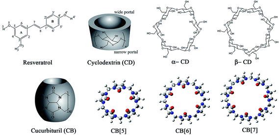 | ||
| Fig. 1 Chemical structures and molecular geometries of resveratrol, cyclodextrins and cucurbiturils. | ||
Over the past decades, considerable efforts have been devoted to study the antioxidative activity of resveratrol. The phenolic OH groups, especially 4′-OH, and the trans conformation, are believed to be responsible for the excellent antioxidative activity of resveratrol.12–16 Owing to its structural simplicity, resveratrol has aroused great interest in developing novel analogues with improved antioxidative activity. Various resveratrol-oriented analogues have been designed by introducing different substituent groups to aromatic rings of resveratrol,17–21 and electron-donating groups were found to be more effective than other substituent groups. Mikulski et al.22 found that the oligomers, glucosides of resveratrol, and trans-resveratrol-3-O-glucuronide exhibited more excellent antioxidative activity than resveratrol. Incorporation of chroman moiety of vitamin E into resveratrol scaffold, replacement of 4′-OH by mercapto group have also been reported to improve the antioxidative activity.23,24 In addition, several structural modifications in the stilbene scaffold have also been performed, such as elongation of conjugated chain,25,26 N-substituent of carbon–carbon double bond,27 and development of novel resveratrol analogues with cis-restricted conformation,28 to enhance the antioxidative capacity significantly.
Recently, the host–guest inclusion complexation between resveratrol and cyclodextrin (CD) has been reported by several research groups, the inclusion complexation has been characterized by various means,29–33 and the influence of host–guest inclusion on the antioxidative activity of resveratrol was studied. Sapino et al.34 and Lu et al.35 reported that the inclusion of resveratrol in cyclodextrin showed a negligible influence on DPPH radical scavenging capacity, but Lucas-Abellán et al.36,37 found that the radical scavenging capability of resveratrol increased significantly in the presence of cyclodextrin when ORAC and ABTS assays are employed. Since only a limited number of experimental studies with inconsistent results are presented about this, here we investigated the structures of complexes and the influence of complexation with cyclodextrin on antioxidative activity of resveratrol by employment of quantum chemical calculations to attempt to give a less biased evaluation. Generally, computational simulations can furnish information to clarify ambiguous experiment results, such as shown in many previous computational studies.38,39 Compared to CDs, cucurbiturils (CBs) is also an ideal molecular container due to its polar carbonyl portals and more complete hydrophobic cavity, which might make it more suitable for complexation with resveratrol. In this work, the complexation of resveratrol with CBs have also been studied, and the influence of complexation of resveratrol with CBs on the antioxidative activity of resveratrol have also been predicted. To our best knowledge, there are no experimental or theoretical studies about the complexation between resveratrol and CB so far. Fig. 1 displays the chemical structures of all investigated molecules, including resveratrol, cyclodextrins and cucurbiturils.
2. Computational methods
In view of the excellent performance in previous studies about antioxidative activity of resveratrol,14,16,26 Becke's three-parameter hybrid functional B3LYP40,41 was chosen to carried out the study of complexation of resveratrol with CDs and CBs. In this work, geometries of all investigated chemical systems were fully optimized without symmetry constraints by employing B3LYP using 6-31G basis set, unrestricted formulation was used for radical species. All optimized structures were confirmed to be real minima by frequency calculation at the same level of theory (no imaginary frequency), unscaled zero-point energies (ZPE) were abstracted from frequency analysis to make thermochemical correction to electronic energies and enthalpies of all studied species. Final energies were calculated by performing single-point calculations on the optimized geometries at 6-311G(d, p) standard basis set level for all the atoms in aqueous solution. The solvation effects were considered by means of the polarizable continuum model (radii = UFF), the dielectric constant (78.3553) of water was used thoroughly in this work.CDs are circular oligosaccharides with different portals (narrow portal and wide portal, shown in Fig. 1), so they have two complexation inclusion orientations, i.e. the A-ring of resveratrol orients towards narrow portal of CD and A-ring orients towards wide portal of CD, which were described as A/N and A/W inclusion mode in the following discussion. Whereas for pumpkin-shaped CBs which have two identical carbonyl-decorated portals, there is only one inclusion orientation.
Investigations of antioxidative activity of resveratrol in free form and complexed form have been performed based on three prevalent antioxidant mechanisms, i.e., H atom transfer (HAT) mechanism, sequential proton loss electron transfer (SPLET) mechanism and single electron transfer (SET) mechanism.26 Homolytic dissociation of phenolic O–H bond occurs in HAT mechanism, and bond dissociation enthalpies (BDEs) of O–H bond are used as the energetic parameter to evaluate the H atom transfer process. In SPLET mechanism heterolytic dissociation of O–H bond firstly occurs to lose a proton, and then electron transfers from phenolate anion to radical, proton affinity (PA) and electron transfer enthalpy (ETE) are used to evaluate the two subsequent process, respectively. In these two antioxidative mechanisms the active OH group plays a key role in exhibiting antioxidative activity. Based on our previous studies,26 only the most active 4′-OH was investigated to evaluate the influence of complexation between resveratrol and host macromolecules. In SET mechanism, single electron directly transfers from antioxidant to active radical, which are generally governed by ionization potential (IP) of antioxidant. O–H BDEs, PAs, ETEs and IPs, were calculated using the procedures as detailed in our previous work.26,42 The OH BDEs are calculated according to the formula BDE = Hr + Hh − Hp, where Hr is the enthalpy of the radical, Hh is the enthalpy of the H-atom (−0.49982 hartree at this level of theory), and Hp is the enthalpy of the parent molecule. PAs are calculated as the zero point energy (ZPE) corrected electronic energy difference between phenolate anions and neutral molecules. ETEs are calculated as the enthalpy difference between phenolate anions and the corresponding phenoxyl radicals. IPs are calculated as ZPE-corrected electronic energy difference between parent molecules and the corresponding cation radicals. In addition, the stability of radicals including phenoxyl radical and cation radical, and the unpaired electron delocalization in all radical species were investigated by spin density distribution analysis. All computations in this paper are performed using Gaussian 03 suite of programs.43
3. Results and discussion
3.1 Optimized geometries of complexes and host–guest inclusion model
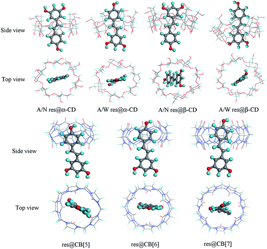
The stable complexes of resveratrol with CDs and CBs as optimized at B3LYP/6-31G level of theory are shown in Fig. 2. Vibrational frequency calculations at this level of theory were performed to confirm that the structures were true minima. To avoid possible computational artifacts coming from given initial complex structure, the full optimizations were re-performed to search stable conformation using different initial structure, that is A ring, B ring and double C![[double bond, length as m-dash]](https://www.rsc.org/images/entities/char_e001.gif) C bond of resveratrol situating in the cavity of host molecule (e.g. CDs and CBs), respectively. As displayed in Fig. 2, CDs and CBs are both capable of encapsulating resveratrol and form stable inclusion complexes, which are denoted as A/N res@α-CD, A/W res@α-CD, A/N res@β-CD, A/W res@β-CD, res@CB[5], res@CB[6] and res@CB[7] in the following discussion, respectively.
C bond of resveratrol situating in the cavity of host molecule (e.g. CDs and CBs), respectively. As displayed in Fig. 2, CDs and CBs are both capable of encapsulating resveratrol and form stable inclusion complexes, which are denoted as A/N res@α-CD, A/W res@α-CD, A/N res@β-CD, A/W res@β-CD, res@CB[5], res@CB[6] and res@CB[7] in the following discussion, respectively.
The inclusion model of resveratrol in CDs is significantly different from that of resveratrol in CBs. In res@CDs, resveratrol is included in hydrophobic cavity of CDs by vinyl moiety, with aromatic rings and OH groups locating in the portal or outside cavity. Whereas in res@CBs, resveratrol is included by B-ring moiety, with the 4′-OH closing to the carbonyl portal of CBs, and the A-ring and 3,5-OH groups locating outside cavity. In the complexes of resveratrol with CBs, the hydrogen atom of 4′-OH group orients towards the oxygen of carbonyl group in portal, thus forming hydrogen bond and contributing greatly to the stabilization of inclusion complexes. Similarly, in complexes of resveratrol with CDs, hydrogen bonds form between OH group of CD portal and OH group on A-ring of resveratrol.
Previous studies29,44 have discussed the inclusion mode between CDs and resveratrol based on force field computation, molecular dynamic simulation and experimental observations, but contradictory conclusions about the inclusion mode preference, i.e. A/N or A/W mode, have been drawn from them. In the present work, we found that CDs were able to include resveratrol forming stable inclusion complex in both inclusion modes. The calculated total energies of res@α-CD in A/N model is lower than that in A/W mode by 2.67 kcal mol−1, and the calculated total energies of res@β-CD in A/N mode is also lower than that in A/W mode by about 4.09 kcal mol−1, these indicate that for res@CD inclusion complex A/N mode is more stable than A/W mode. This is in good agreement with the conclusion of Troche-Pesqueira, et al.44
The equilibrium geometry of antioxidant molecule is critical factor that affects its antioxidative activity. Here, O–H bond lengths, neighboring C–O bond lengths, C![[double bond, length as m-dash]](https://www.rsc.org/images/entities/char_e001.gif) C bond length and the molecular planarity are selected as the structure parameters to characterize the influence of inclusion on molecular geometry of resveratrol. The results indicate that significant twist in the molecular geometry of resveratrol has been induced by inclusion in CDs and CBs, in comparison with strictly planar molecular configuration of free resveratrol. The twist between two aromatic rings and the phenolic O–H bonds twist out of the aromatic plane are characterized by torsion angles δ(C2–C1–C1′–C2′), ϕ(C2–C3–O3–H3), η(C6–C5–O5–H5) and φ(C3′–C4′–O4′–H4′). Detailed structure parameters have been listed in Table 1. As can be seen from the table, complexation of resveratrol with CDs and CBs shows negligible influence on the O–H bond length, the neighboring C–O bond length and C
C bond length and the molecular planarity are selected as the structure parameters to characterize the influence of inclusion on molecular geometry of resveratrol. The results indicate that significant twist in the molecular geometry of resveratrol has been induced by inclusion in CDs and CBs, in comparison with strictly planar molecular configuration of free resveratrol. The twist between two aromatic rings and the phenolic O–H bonds twist out of the aromatic plane are characterized by torsion angles δ(C2–C1–C1′–C2′), ϕ(C2–C3–O3–H3), η(C6–C5–O5–H5) and φ(C3′–C4′–O4′–H4′). Detailed structure parameters have been listed in Table 1. As can be seen from the table, complexation of resveratrol with CDs and CBs shows negligible influence on the O–H bond length, the neighboring C–O bond length and C![[double bond, length as m-dash]](https://www.rsc.org/images/entities/char_e001.gif) C bond length. All O–H bond lengths vary less than 0.006 Å. All C–O bond lengths vary less than 0.01 Å, and C
C bond length. All O–H bond lengths vary less than 0.006 Å. All C–O bond lengths vary less than 0.01 Å, and C![[double bond, length as m-dash]](https://www.rsc.org/images/entities/char_e001.gif) C bond length varies less than 0.002 Å. In most cases, OH group is found to be coplanar with the neighboring aromatic ring, with the exception of 3-OH in res@β-CD and 4′-OH in res@CB[7]. In res@β-CD complex, an intermolecular hydrogen bond between resveratrol and β-CD has formed to stabilize the inclusion system, which causes strong deviation of 3-OH from the aromatic planarity. Similarly, in res@CB[7] 4′-OH significantly deviates from the neighboring aromatic planarity and forms hydrogen bond with the carbonyl portal of CB[7] to stabilize the inclusion complex.
C bond length varies less than 0.002 Å. In most cases, OH group is found to be coplanar with the neighboring aromatic ring, with the exception of 3-OH in res@β-CD and 4′-OH in res@CB[7]. In res@β-CD complex, an intermolecular hydrogen bond between resveratrol and β-CD has formed to stabilize the inclusion system, which causes strong deviation of 3-OH from the aromatic planarity. Similarly, in res@CB[7] 4′-OH significantly deviates from the neighboring aromatic planarity and forms hydrogen bond with the carbonyl portal of CB[7] to stabilize the inclusion complex.
![[double bond, length as m-dash]](https://www.rsc.org/images/entities/char_e001.gif) C bond lengths (Å) and torsion angles (degree) of free resveratrol and complexed resveratrols
C bond lengths (Å) and torsion angles (degree) of free resveratrol and complexed resveratrols
| Bond lengths (Å) | Torsion angles (degree) | ||||||||||
|---|---|---|---|---|---|---|---|---|---|---|---|
| O3–H3 | C3–O3 | O5–H5 | C5–O5 | O4′–H4′ | C4′–O4′ | C7–C8 | δ(C2–C1–C1′–C2′) | ϕ(C2–C3–O3–H3) | η(C6–C5–O5–H5) | φ(C3′–C4′–O4′–H4′) | |
| Resveratrol | 0.976 | 1.393 | 0.976 | 1.394 | 0.976 | 1.391 | 1.352 | −179.997 | −179.996 | −179.969 | 180.000 |
| A/N res@α-CD | 0.976 | 1.394 | 0.976 | 1.397 | 0.977 | 1.389 | 1.354 | −163.735 | −179.165 | −176.931 | 178.686 |
| A/W res@α-CD | 0.976 | 1.390 | 0.975 | 1.404 | 0.977 | 1.389 | 1.353 | 147.466 | −179.658 | −179.851 | −179.460 |
| A/N res@β-CD | 0.995 | 1.386 | 0.976 | 1.403 | 0.976 | 1.391 | 1.352 | 174.639 | 147.372 | 176.458 | −179.778 |
| A/W res@β-CD | 1.019 | 1.404 | 0.975 | 1.405 | 0.977 | 1.390 | 1.353 | −168.547 | 56.551 | 177.003 | 179.993 |
| res@CB[5] | 0.976 | 1.398 | 0.976 | 1.398 | 0.983 | 1.386 | 1.350 | −156.259 | 179.797 | 179.841 | 174.637 |
| res@CB[6] | 0.976 | 1.402 | 0.976 | 1.403 | 0.980 | 1.374 | 1.352 | −173.854 | −179.893 | −179.893 | 178.299 |
| res@CB[7] | 0.976 | 1.403 | 0.976 | 1.402 | 0.982 | 1.379 | 1.352 | −135.078 | −179.600 | 178.940 | −165.002 |
3.2 Frontier molecular orbitals of free and complexed resveratrols and the influence of inclusion complexation
The distributions and energies of the highest occupied molecular orbital (HOMO) and the lowest unoccupied molecular orbital (LUMO) of antioxidant are important parameters of electronic structure, which indicate the active site of antioxidant molecules, electron donating and accepting capacity. The energy of HOMO determines the so-called SET antioxidative efficiency, higher HOMO energy is favorable for electron transfer process from antioxidant molecule to free radical.In the present work, the topologies and energies of HOMO and LUMO of free and complexed resveratrols are displayed in Fig. 3. As can be seen from it, the HOMO and LUMO of resveratrol are π type bonding and antibonding molecular orbitals, respectively, which spread over the whole molecular framework. In inclusion complexes of resveratrol with CDs and CBs, the HOMO and LUMO topologies are nearly the same as that in free resveratrol, indicating that the inclusion complexation exhibit negligible influence on the distribution of the frontier orbitals and the active site of resveratrol. But the inclusion complexation show remarkable influence on the energies of HOMO and LUMO. Compared with free resveratrol, inclusion in CDs induces slight decrease in HOMO and LUMO energies, and inclusion in CBs leads to significant increase in HOMO and LUMO energies. This indicates that the electron donating capacity of resveratrol is enhanced by inclusion in CBs, but inclusion of resveratrol in CDs is unfavorable for electron transfer process from antioxidant molecule to free radical. Moreover, the cavity size of host molecules and the inclusion mode show remarkable influence on HOMO energies. The HOMO energy of res@α-CD are lower than that of res@β-CD, A/W inclusion mode leads to even lower HOMO energy than the A/N inclusion mode. Among them the HOMO energy of A/N res@β-CD complex is very close to that of free resveratrol, the A/W res@α-CD is found to possess the lowest HOMO energy, which decreases by 0.11 eV as compared to that of free resveratrol. All res@CB complexes show higher HOMO energy relative to free resveratrol, the highest HOMO energy is presented in res@CB[5] case (−5.25 eV), and HOMO energy decreases with the increasing cavity size of CB molecules. By comparison with CD, resveratrol is predicted to be endowed with more antioxidative activity by inclusion in CB based on SET mechanism.
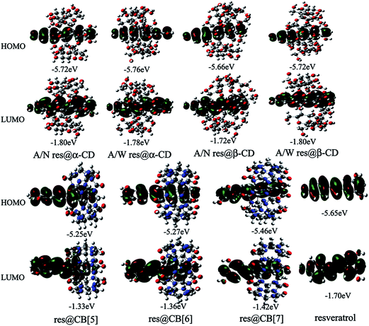 | ||
| Fig. 3 Topologies and energies of frontier orbitals (HOMO and LUMO) of free resveratrol and its inclusion complexes. | ||
3.3 Influence of inclusion complexation on the antioxidative activity of resveratrol
Generally, phenolic antioxidants exert their antioxidative capacities based on three antioxidative mechanisms, i.e. HAT, SPLET and SET. Lower O–H BDE, lower PA and ETE of phenolate anion and lower IP is favorable for antioxidative activity. In the present work, the influences of inclusions by CDs and CBs on the antioxidative activity of resveratrol is our chief concern. The 4′-OH BDEs, PAs and ETEs of 4′-phenolate anions, IPs of free resveratrol and complexed resveratrols are listed in Table 2.| 4′-OH BDE (kcal mol−1) | 4′-phenolate anion | IPs (kcal mol−1) | ||
|---|---|---|---|---|
| PAs (kcal mol−1) | ETEs (kcal mol−1) | |||
| Resveratrol | 76.92 | 295.59 | 95.14 | 124.55 |
| A/N res@α-CD | 78.57 | 289.02 | 104.69 | 129.83 |
| A/W res@α-CD | 77.57 | 294.25 | 97.31 | 128.63 |
| A/N res@β-CD | 77.29 | 294.45 | 97.03 | 124.58 |
| A/W res@β-CD | 79.25 | 298.12 | 94.92 | 121.50 |
| res@CB[5] | 76.39 | 306.74 | 83.65 | 115.87 |
| res@CB[6] | 84.55 | 316.92 | 80.85 | 116.25 |
| res@CB[7] | 78.77 | 315.44 | 77.22 | 117.89 |
The calculated 4′-OH BDE of free resveratrol is 76.92 kcal mol−1. When resveratrol is included in CDs or CBs, 4′-OH BDE of resveratrol increase remarkably. Especially for res@CB[6], 4′-OH BDE increases significantly by 7.63 kcal mol−1. These increase in 4′-OH BDE indicate that inclusion complexation of resveratrol in CDs or CBs is unfavorable for O–H homolytic dissociation. It should be noted the inclusion mode and the cavity size of host molecule both shows distinct influence on 4′-OH BDE in res@CD inclusion complexes. In A/N res@α-CD the 4′-OH BDE is 78.57 kcal mol−1, which is higher than 4′-OH BDE in A/W res@α-CD by 1.00 kcal mol−1. However, in res@β-CD complex 4′-OH BDE in A/N inclusion mode is lower than that in A/W mode by 1.96 kcal mol−1. Similarly, the cavity size of CB shows remarkable, but irregular influence on 4′-OH BDE in res@CBs.
PAs and ETEs of 4′-phenolate anions are also calculated to investigate the influence of inclusion complexation on the SPLET antioxidative mechanism. The calculated PA and ETE of 4′-phenolate anion of free resveratrol are 295.59 kcal mol−1 and 95.14 kcal mol−1, respectively. Inclusion of resveratrol in α-CD in A/N mode leads to a decrease of 6.57 kcal mol−1 in PA and an increase of 9.55 kcal mol−1 in ETE. While in A/W inclusion mode, a decrease of 1.34 kcal mol−1 in PA and an increase of 2.17 kcal mol−1 in ETE have been induced. In res@β-CD, the inclusion in A/N mode leads to a decrease of 1.14 kcal mol−1 in PA, an increase of 1.89 kcal mol−1 in ETE, and the inclusion of resveratrol in A/W mode induces an increase of 2.53 kcal mol−1 in PA and a decrease of 0.22 kcal mol−1 in ETE. As compare to ETE, PA is more important determinant for SPLET antioxidative mechanism, because the phenolate anion (ArO−) is considered to be much better electron donor which facilitates the following electron transfer process.26 Thus, based on our results, a conclusion can be drawn that the A/N inclusion mode and small cavity size of CD are favorable for enhancement of SPLET antioxidative activity of resveratrol. However, PA of 4′-phenolate anion in res@CBs is significantly higher than that in free resveratrol, and the larger cavity size leads to more increase in PA of 4′-phenolate anion. Although significant decreases in ETE of phenolate anion are induced, inclusion of resveratrol in CBs is considered to be unfavorable for improvement of SPLET antioxidative activity of resveratrol.
The influence of inclusion on IP of resveratrol has also been investigated. When resveratrol is included by α-CD in A/N mode and A/W mode, the corresponding IPs increase by 5.28 kcal mol−1 and 4.08 kcal mol−1, respectively. Inclusion of resveratrol by β-CD in A/N mode show negligible influence on IP, and the inclusion in A/W mode leads to a remarkable decrease of 3.05 kcal mol−1. In comparison with the A/W inclusion mode and β-CD, the A/N inclusion mode and the inclusion by α-CD result in more increase in IP. When resveratrol is included by CBs, IP decreases significantly as compared to free resveratrol. In res@CB[5], res@CB[6] and res@CB[7], the IP of complexed resveratrols decrease by 8.68 kcal mol−1, 8.30 kcal mol−1 and 6.66 kcal mol−1, respectively. Based on these results, we can conclude that inclusions of resveratrol by CDs are unfavorable but inclusions by CBs are favorable for SET antioxidative mechanism of resveratrol.
Lucas-Abellán et al.36 performed comparative study of different radical assays to measure the influence of presence of CDs on antioxidative activity of resveratrol, and they found that ORAC assay was the most sensitive to the increase in concentration of CDs, the ABTS assay also showed sensitive response, but the DPPH assay exhibited no effect. These experimental results can be well interpreted by the influence of inclusion complexation on the antioxidative activity of resveratrol presented in this study. Inclusion of resveratrol in CDs leads to significant increase in 4′-OH BDE and IP of resveratrol molecule, but the decrease in PA of phenolate anion. These indicate that the SPLET mechanism plays a major role in exhibiting its antioxidative activity when resveratrol is included by CDs. In ORAC assay the peroxyl anion radical (ROO˙−) is good proton accepter, which will facilitate the proton dissociation from 4′-OH in resveratrol to produce phenolate anion. In ABTS assay the produced ABTS˙+ can bond with the phenolate anion through electrostatic interaction, thus favoring the electron transfer from phenolate anion to ABTS˙+. Whereas in DPPH assay the neutral DPPH˙ radical can not facilitate the SPLET antioxidative mechanism. This should be the reason why the improvement in antioxidative activity of resveratrol included in CDs was observed when ORAC and ABTS assays are employed, but negligible effect on the scavenging capacity of DPPH radical was presented.
3.4 Structures of complexed radicals and anions of resveratrol and spin density distribution
For free resveratrol, 4′-phenoxyl radical, 4′-phenolate anion and cation radical are all in completely planar conformation.26 When it is included by CDs and CBs, phenoxyl radical, phenolate anion and cation radical undergo remarkable deformations, the molecular planarity of resveratrol is severely broken and the skeletons of resveratrol strongly bend to adapt the inclusion complexation with CDs and CBs.Spin density analysis is performed to investigate the unpaired electron delocalization, which has important influence on the stabilities of radical species. The detailed spin density distributions in 4′-phenoxyl radicals and cation radicals are displayed in Fig. 4 and 5, respectively. For all phenoxyl radicals, spin density is found to be evenly distributed over the remaining O-atom, B-ring and carbon–carbon double bond. In phenoxyl radical of res@CDs, spin densities are more confined to the remaining 4′-O-atom and B-ring, whereas in phenoxyl radical of res@CBs the spin densities on the remaining 4′-O-atom and B-ring are remarkably lower than that in phenoxyl radical of free resveratrol. This indicates that CD inclusion is unfavorable for unpaired electron delocalization and shows negative efforts in stabilization of phenoxyl radical, but CB inclusion is relatively favorable and contributes positively to stabilization of phenoxyl radical. For free cation radical of resveratrol, unpaired electron delocalizes over A-ring, carbon–carbon double bond and B-ring. The spin density distribution shows that A-ring and carbon–carbon double bond contribute more importantly to unpaired electron delocalization than B-ring and OH groups. In cation radicals of res@CD complexes, spin density distribution is even more unbalanced. Relatively, the A/W inclusion mode provides more balanced distribution than the A/N inclusion mode. Whereas for res@CB complexes, remarkable decrease in spin density of A-ring resulted by inclusion complexation leads to more delocalization of unpaired electron over A-ring and B-ring, and the influence increases with decreasing size of CB cavity, i.e., the smaller CB[5] results in more balanced distribution than the larger size CB molecules, CB[6] or CB[7]. These observations imply that inclusion in CDs shows negligible or very slight influence on unpaired electron delocalization of cation radical, while inclusion in CBs leads to more extent of unpaired electron delocalization. As compared to CDs, CBs are favorable for stabilization of cation radical, and help to enhance the antioxidative activity of resveratrol.
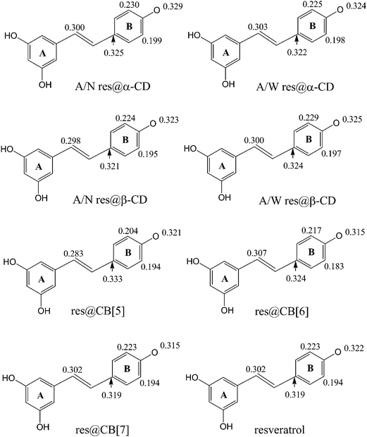 | ||
| Fig. 4 Spin density distribution in 4′-phenoxyl radicals of free resveratrol and complexed resveratrol in CDs and CBs. | ||
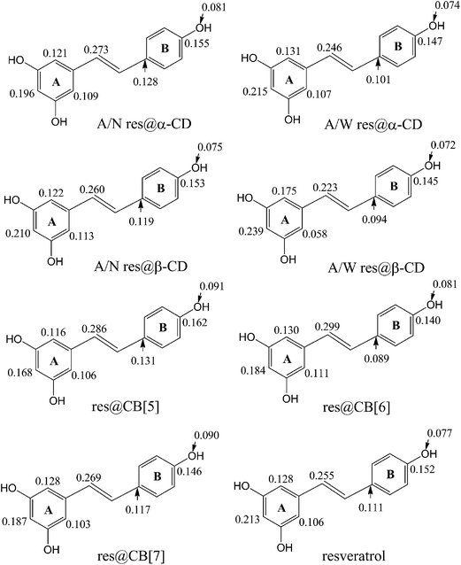 | ||
| Fig. 5 Spin density distribution in cation radicals of free resveratrol and complexed resveratrol in CDs and CBs. | ||
4. Conclusion
In the present work the inclusion complexations of resveratrol in CDs and CBs have been investigated by density functional theory calculation at B3LYP/6-311G(d, p) level of theory. The equilibrium geometries, electronic structures, antioxidative capabilities are investigated to determine the influence of inclusion by CDs and CBs on the properties of guest molecules, resveratrol.Our results manifest that different inclusion modes are presented between res@CD and res@CB complexes, and in the inclusion complexes the most remarkable change of resveratrol is the twist of molecular plane. Frontier orbital analysis shows that inclusion complexation influences the orbital distribution negligibly, but affects the energies of frontier orbitals significantly. Slight increase in 4′-OH BDE is caused by inclusion complexation by CDs and CBs, with the only exception of CB[5]. Inclusion by CD results in distinct decrease in PA, but increase in ETE of 4′-phenolate anion of resveratrol, and the influence of α-CD is more significant than that of β-CD. For res@CD complexes, the A/N inclusion mode is more favorable for heterolytic dissociation of 4′-OH, but more unfavorable for electron transfer process than the A/W inclusion mode. Contrary to the performance in CD inclusion, inclusion by CB leads to significant increase in PA and remarkable decrease in ETE of 4′-phenolate anion, and the cavity size of CB also shows important influence. In addition, a distinct increase in IP is induced by inclusion in α-CD, negligible change in IP presents in A/N res@β-CD but a decrease of 2.95 kcal mol−1 in IP presents in A/W res@β-CD. It is noteworthy that the IPs in res@CBs are significantly lower than free resveratrol, and the IP decreases with the increasing size of CB molecule. Spin density distribution analysis confirms that CD inclusion is unfavorable for unpaired electron delocalization in 4′-pheoxyl radical and cation radical, but CB inclusion favors the unpaired electron delocalization and stabilization of radical species. Compared to CDs, CBs is predicted to be more promising molecular container, which would improve the antioxidative capacity of resveratrol.
Acknowledgements
This work was financially supported by the fund of State Key Laboratory of Refractories and Metallurgy (G201405), National Natural Science Foundation of China (no. 51472184, 51472185), and State Basic Research Development Program of China (973 Program, 2014CB660802).References
- G. Duthie, S. Duthie and J. Kyle, Nutr. Res. Rev., 2000, 13, 79 CrossRef CAS PubMed.
- S. Quideau, D. Deffieux, C. Douat-Casassus and L. Pouysegu, Angew. Chem., Int. Ed., 2011, 50, 586 CrossRef CAS PubMed.
- J. A. Baur and D. A. Sinclair, Nat. Rev. Drug Discovery, 2006, 5, 493 CrossRef CAS PubMed.
- M. Jang, L. Cai, G. O. Udeani, K. V. Slowing, C. F. Thomas, C. W. W. Beecher, H. H. S. Fong, N. R. Farnsworth, A. D. Kinghorn, R. G. Mehta, R. C. Moon and J. M. Pezzuto, Science, 1997, 275, 218 CrossRef CAS.
- A. Bishayee, Cancer Prev. Res., 2009, 2, 409 CrossRef CAS PubMed.
- W. P. Chen, M. J. Su and L. M. Hung, Eur. J. Pharmacol., 2007, 554, 196 CrossRef CAS PubMed.
- D.-H. Yoon, O.-Y. Kwon, J. Y. Mang, M. J. Jung, D. Y. Kim, Y. K. Park, T.-H. Heo and S.-J. Kim, Biochem. Biophys. Res. Commun., 2011, 414, 49 CrossRef CAS PubMed.
- P. Saiko, A. Szakmary, W. Jaeger and T. Szekeres, Mutat. Res., 2008, 658, 68 CAS.
- C. C. Udenigwe, V. R. Ramprasath, R. E. Aluko and P. J. Jones, Nutr. Rev., 2008, 66, 445 CrossRef PubMed.
- H. Berrougui, G. Grenier, S. Loued, G. Drouin and A. Khalil, Atherosclerosis, 2009, 207, 420 CrossRef CAS PubMed.
- M. Ndiaye, C. Philippe, H. Mukhtar and N. Ahmad, Arch. Biochem. Biophys., 2011, 508, 164 CrossRef CAS PubMed.
- L. A. Stivala, M. Savio, F. Carafoli, P. Perucca, L. Bianchi, G. Maga, L. Forti, U. M. Pagnoni, A. Albini, E. Prosperi and V. Vannini, J. Biol. Chem., 2001, 276, 22586 CrossRef CAS PubMed.
- H. Cao, X. Pan, C. Li, C. Zhou, F. Deng and T. Li, Bioorg. Med. Chem. Lett., 2003, 13, 1869 CrossRef CAS.
- A. N. Queiroz, B. A. Q. Gomes, W. M. Moraes Jr and R. S. Borges, Eur. J. Med. Chem., 2009, 44, 1644 CrossRef CAS PubMed.
- F. Caruso, J. Tanski, A. Villegas-Estrada and M. J. Rossi, J. Agric. Food Chem., 2004, 52, 7279 CrossRef CAS PubMed.
- D. Mikulski, R. Gorniak and M. Molski, Eur. J. Med. Chem., 2010, 45, 1015 CrossRef CAS PubMed.
- J. G. Fang and B. Zhou, J. Agric. Food Chem., 2008, 56, 11458 CrossRef CAS PubMed.
- Y. J. Shang, Y. P. Qian, X. D. Liu, F. Dai, X. L. Shang, W. Q. Jia, Q. Liu, J. G. Fang and B. Zhou, J. Org. Chem., 2009, 74, 5025 CrossRef CAS PubMed.
- R. Amorati, M. Lucarini, V. Mugnaini, G. F. Pedulli, M. Roberti and D. Pizzirani, J. Org. Chem., 2004, 69, 7101 CrossRef CAS PubMed.
- K. Fukuhara, I. Nakanishi, A. Matsuoka, T. Matsumura, S. Honda, M. Hayashi, T. Ozawa, N. Miyata, S. Saito, N. Ikota and H. Okuda, Chem. Res. Toxicol., 2008, 21, 282 CrossRef CAS PubMed.
- M. Murias, W. Jäger, N. Handler, T. Erker, Z. Horvath, T. Szekeres, H. Nohl and L. Gille, Biochem. Pharmacol., 2005, 69, 903 CrossRef CAS PubMed.
- D. Mikulski and M. Molski, Eur. J. Med. Chem., 2010, 45, 2366 CrossRef CAS PubMed.
- J. Yang, G. Y. Liu, D. L. Lu, F. Dai, Y. P. Qian, X. L. Jin and B. Zhou, Chem.–Eur. J., 2010, 16, 12808 CrossRef CAS PubMed.
- X. Y. Cao, J. Yang, F. Dai, D. J. Ding, Y. F. Kang, F. Wang, X. Z. Li, G. Y. Liu, S. S. Yu, X. L. Jin and B. Zhou, Chem.–Eur. J., 2012, 18, 5898 CrossRef CAS PubMed.
- J. J. Tang, G. J. Fan, F. Dai, D. J. Ding, Q. Wang, D. L. Lu, R. R. Li, X. Z. Li, L. M. Hu, X. L. Jin and B. Zhou, Free Radical Biol. Med., 2011, 50, 1447 CrossRef CAS PubMed.
- L. Lu, S. Zhu, H. Zhang and S. Zhang, Comput. Theor. Chem., 2013, 1019, 39 CrossRef CAS PubMed.
- J. Lu, C. Li, Y. F. Chai, D. Y. Yang and C. R. Sun, Bioorg. Med. Chem. Lett., 2012, 22, 5744 CrossRef CAS PubMed.
- D. J. Ding, X. Y. Cao, F. Dai, X. Z. Li, G. Y. Liu, D. Lin, X. Fu, X. L. Jin and B. Zhou, Food Chem., 2012, 135, 1011 CrossRef CAS PubMed.
- Z. Lu, R. Chen, H. Liu, Y. Hu, B. Cheng and G. Zou, J. Inclusion Phenom. Macrocyclic Chem., 2009, 63, 295 CrossRef CAS.
- J. M. López-Nicolás, E. Núñez-Delicado, A. J. Pérez-López, Á. C. Barrachina and P. Cuadra-Crespo, J. Chromatogr. A, 2006, 1135, 158 CrossRef PubMed.
- C. Lucas-Abellán, I. Fortea, J. M. López-Nicolás and E. Núñez-Delicado, Food Chem., 2007, 104, 39 CrossRef PubMed.
- J. M. López-Nicolás and F. García-Carmona, Food Chem., 2008, 109, 868 CrossRef PubMed.
- C. Lucas-Abellán, M. I. Fortea, J. A. Gabaldón and E. Núñez-Delicado, Food Chem., 2008, 111, 262 CrossRef PubMed.
- S. Sapino, M. E. Carlotti, G. Caron, E. Ugazio and R. Cavalli, J. Inclusion Phenom. Macrocyclic Chem., 2009, 63, 171 CrossRef CAS.
- Z. Lu, B. Cheng, Y. Hu, Y. Zhang and G. Zou, Food Chem., 2009, 113, 17 CrossRef CAS PubMed.
- C. Lucas-Abellán, M. T. Mercader-Ros, M. P. Zafrilla, J. A. Gabaldón and E. Núñez-Delicado, Food Chem. Toxicol., 2011, 49, 1255 CrossRef PubMed.
- C. Lucas-Abellán, M. T. Mercader-Ros, M. P. Zafrilla, M. I. Fortea, J. A. Gabaldón and E. Núñez-Delicado, J. Agric. Food Chem., 2008, 56, 2254 CrossRef PubMed.
- Z. Ma and M. E. Tuckerman, Chem. Phys. Lett., 2011, 511, 177 CrossRef CAS PubMed.
- Z. Ma and D. F. Coker, J. Chem. Phys., 2008, 128, 244108 CrossRef CAS PubMed.
- A. D. Becke, J. Chem. Phys., 1993, 98, 5648 CrossRef CAS PubMed.
- C. Lee, W. Yang and R. G. Parr, Phys. Rev. B: Condens. Matter Mater. Phys., 1988, 37, 785 CrossRef CAS.
- L. Lu, M. Qiang, F. Li, H. Zhang and S. Zhang, Dyes Pigm., 2014, 103, 175 CrossRef CAS PubMed.
- M. J. Frisch, et al., Gaussian 03, Revision B.05, Gaussian, Inc., Pittsburgh PA 2003 Search PubMed.
- E. Troche-Pesqueira, I. Pérez-Juste, A. Navarro-Vázquez and M. M. Cid, RSC Adv., 2013, 3, 10242 RSC.
| This journal is © The Royal Society of Chemistry 2015 |

