Large area magnetic micropallet arrays for cell colony sorting
Wesley A.
Cox-Muranami
a,
Edward L.
Nelson
b,
G. P.
Li
a and
Mark
Bachman
*a
aBiomedical Engineering, University of California, Irvine, CA, USA. E-mail: mbachman@uci.edu
bSchool of Medicine, Department of Medicine, School of Biological Sciences, Department of Molecular Biology and Biochemistry, University of California, Irvine, CA, USA
First published on 19th November 2015
Abstract
A new micropallet array platform for adherent cell colony sorting has been developed. The platform consisted of thousands of square plastic pallets, 270 μm by 270 μm on each side, large enough to hold a single colony of cells. Each pallet included a magnetic core, allowing them to be collected with a magnet after being released using a microscope mounted laser system. The micropallets were patterned from 1002F epoxy resist and were fabricated on translucent, gold coated microscope slides. The gold layer was used as seed for electroplating the ferromagnetic cores within every individual pallet. The gold layer also facilitated the release of each micropallet during laser release. This array allows for individual observation, sorting and collection of isolated cell colonies for biological cell colony research. In addition to consistent release and recovery of individual colonies, we demonstrated stable biocompatibility and minimal loss in imaging quality compared to previously developed micropallet arrays.
Introduction
Isolating clonal cell colonies from heterogeneous sample populations is a process required for important biological research in which it is necessary to obtain clonal populations including stem cell and cancer research along with molecularly modified cells (over-expression or knock down) required for mechanistic studies of biology.1–4 Many cellular transformation assays such as viral transfection and drug effectiveness studies also require cell colony manipulation.5–7 These tasks are normally slow and labor intensive due to the necessity of extensive cell growth times and colony extractions followed by reseeding procedures.8 In the case of transfection assays, repetition of the growth and recovery cycle is often required in order to achieve adequate purity of the desired cells. Because the growth of recoverable colonies can take several weeks, studies involving cell transformation can become long-term projects. Furthermore, target cell colonies are typically identified based on their phenotypic features, often assessed on bulk populations maintained through in vitro cell culture. Some adherent cells, particularly primary cells in lieu of transformed cell lines, require some type of growth substrate (extracellular matrix) in order to retain their viability and functional capacity. The use of growth substrate is not selective for the cells of interest and when using heterogeneous cell populations can lead to a loss in recovered cell purity as a result of unwanted cells amassing around target colonies. Using standard methods, this requires multiple dissociation, dilution, and re-culturing steps to isolate clonal populations. Furthermore, some transfection procedures yield so few useful cell colonies that cells of interest can go unnoticed altogether or are outcompeted for space and nutrients by unmodified cells.Several new technologies have been developed to ease colony sorting procedures including advances in automated imaging and extraction systems. One group has developed a system of automated induced pluripotent stem cell selection with an integrated robotic arm and microscope.9 Another group paired an imaging system with a micropipette for colony extraction.10 While these methods solve issues associated with the time consuming imaging of cell cultures, they still rely on the utilization of shared cell culture plates as well as direct physical collection methods which limit extracted cell purity.
We report an approach to colony sorting that uses a new type of micropallet array. These arrays consist of small transparent micropallets, fabricated by photolithography on glass microscope slides that are designed to support biological samples for observation and subsequent isolation. This technology has demonstrated the possibility of single adherent cell sorting while maintaining cell adherence to a surface11 as well as for colonies.12,13 The technology has been demonstrated to be particularly useful for sorting adherent cells and colonies over conventional methods such as FACS. In micropallet assays, cells or colonies are seeded to the pellets. Once seeded, the cells are either observed for their differences in morphological features or fluorescently tagged to differentiate sample types. Once a target cell or colony is identified, the pallet that they are adhered to is released with a laser focused at the interface between the pallet and the glass slide. The released pallet can be recovered and moved to a new culture medium for further analysis. The technology was first utilized primarily for single cell sorting procedures; there have been recent efforts to expand the use of arrays to cell colony sorting.12–14 When the micropallets are made larger to accommodate cell colony growth, however, the laser energy required to eject the pallets becomes too high to preserve cell viability and structural integrity of the released pallet.15 This imposes a significant limitation to colony-sized micropallets, and various strategies have been explored to solve this problem. Efforts to address the laser energy issue have resulted in the designing of new types of micropallets with less stringent ejection requirements16 such as table-top style pallets which enabled lowered laser energy necessary to eject large pallets by minimizing surface area contact to the glass substrate. These schemes are clever, but require more complicated laser ejection methods. A newer ejection method designed specifically for large pallet release utilizing ultrasound to displace pallets for collection has been studied and shows great promise for pallet sizes much too great to eject with current methods.17 The standard pallet recovery method involves the inversion of the micropallet array over a culture well plate. To alleviate this time consuming, imprecise, somewhat random process that leads to limited downstream control of recovered cells, micropallet arrays infused with ferromagnetic iron nanoparticles were created to enable single structure collection with a magnet probe.18 This strategy greatly simplified the collection of individual pallets, but reduced the optical quality of the pallets.
In this paper, we report a newly developed micropallet array technology consisting of various sized patterned microstructures, which can be consistently released using a standard microscope mounted laser and collected using a magnetic probe. The array is fabricated on a thin gold layer, which allows for the electroplating of ferromagnetic cores in the pallets, and further acts as a light absorbing layer during laser release. We found that the gold layer heated up during laser irradiation, efficiently generating vapor microbubbles under the pallets, causing them to gently lift off the surface during laser release. The gold film was optically semi-transparent and did not result in a loss in phase contrast imaging quality. We successfully showed that individual cells sequestered to these magnetic micropallets could be developed into clonal colonies, analyzed, safely released, and magnetically transferred to separate growth substrates for further expansion or analysis.
Materials and methods
Gold thin film deposition
Thin films of gold were prepared on glass slides to form a seed layer for cell colony array fabrication. Standard microscope slides (VWR, Radnor, PA) were cleaned in piranha etchant solution (H2SO4 and NH4OH at a 3![[thin space (1/6-em)]](https://www.rsc.org/images/entities/char_2009.gif) :
:![[thin space (1/6-em)]](https://www.rsc.org/images/entities/char_2009.gif) 1 ratio) and rinsed thoroughly in deionized water (diH2O). Cleaned slides were dried with N2 gas and further dehydrated in a 135 °C oven for one hour. Titanium was evaporated using a Temescal CV-8 electron beam deposition tool (Vesco, Estero, FL) to the glass slides to a thickness of 38 Å to form an adhesion layer for gold deposition. The slides were coated once more with 200 Å of gold. Completed gold thin films were visually semi-transparent and passed standard tape-lift adherence tests. Electrical resistance of the films was required to be low enough to allow proper electroplating and was measured with a digital multimeter as a function of distance from a single corner of a slide. The light transmission characteristics of the gold thin films were analyzed with an Ocean Optics USB2000 Spectrometer (Ocean Optics, Dunedin, FL) paired with a tungsten halogen light source.
1 ratio) and rinsed thoroughly in deionized water (diH2O). Cleaned slides were dried with N2 gas and further dehydrated in a 135 °C oven for one hour. Titanium was evaporated using a Temescal CV-8 electron beam deposition tool (Vesco, Estero, FL) to the glass slides to a thickness of 38 Å to form an adhesion layer for gold deposition. The slides were coated once more with 200 Å of gold. Completed gold thin films were visually semi-transparent and passed standard tape-lift adherence tests. Electrical resistance of the films was required to be low enough to allow proper electroplating and was measured with a digital multimeter as a function of distance from a single corner of a slide. The light transmission characteristics of the gold thin films were analyzed with an Ocean Optics USB2000 Spectrometer (Ocean Optics, Dunedin, FL) paired with a tungsten halogen light source.
Fabrication of micropallet arrays
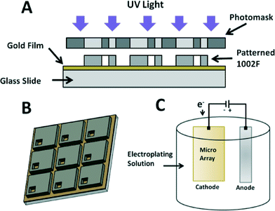
Large area micropallet arrays composed of squares with via holes were patterned onto gold-coated glass microscope slides. The gold-coated slides were cleaned with isopropyl alcohol, rinsed in diH2O and aspirated with N2 gas. The slides were then dehydrated in a 135 °C oven for one hour. 1002F photoresist was prepared as previously reported.19 EPON resin 1002F (Miller-Stephenson, Sylmar, CA) was dissolved in γ-butyrolactone (GBL) (Sigma-Aldrich) with triarylsulfonium hexafluoroantimonate salts (Dow Chemical, Torrance, CA) added to generate photosensitivity. Each component was prepared at a viscosity suitable for 50 μm thick film creation and stirred for 24 hours to break down all resin aggregates followed by degassing the solution using standard methods as previously described.19 Completed 1002F was spin coated onto the gold coated slides to a thickness of 50 μm and soft baked on a hotplate at 70 °C for 20 minutes followed by a second bake at 105 °C for 40 minutes to evaporate all solvents from the photoresist. Solidified 1002F films were patterned via exposure to a collimated UV light source (AB&M INC, Scotts Valley, CA) at 1200 mJ cm−2 through a photomask bearing a negative of the colony micropallet pattern. Four photomask designs were created for this study. One mask was composed of an array of squares at a width and height of 270 μm with borders of 50 μm separating adjacent pallets. This mask design was created to test the optical properties and biocompatibility of 1002F arrays fabricated on gold thin film coated glass slides. Three other masks were created using the same square size with the addition of square via holes of 50 μm by 50 μm (small), 75 μm by 75 μm (medium) and 100 μm by 100 μm (large). The via holes were intended to hold a magnetic core within the pallet. These were designed to be located in a single corner region of each pallet to preserve maximum unabridged growth area for cell colony development. All array designs included 2.5 mm borders with no photoresist around the edges of the glass slides in order to allow an electrical connection to a power source for electroplating. Fig. 1 is a schematic of the photolithography process for microarray fabrication and an example of a completed array. After UV exposure, the patterned 1002F film was baked at 65 °C for 7 minutes and then 95 °C for 18 minutes to finalize the curing process. The slides were developed in SU8 photoresist developer (MicroChem, Newton, MA) for four minutes and quenched with isopropyl alcohol. Finally, the slides were dehydrated with N2 gas and hard baked with a previously developed heating protocol for 1002F photoresist.Gold and ferromagnet electroplating
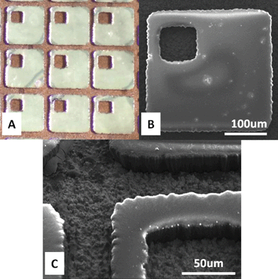
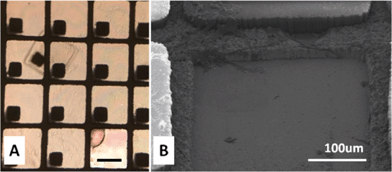
A multistep electroplating process was utilized to deposit gold-coated nickel structures onto all exposed gold thin film regions of the micropallet arrays. Before plating, the 1002F micropallet arrays were placed in a diH2O bath and sonicated at 47 kHz for 5 minutes at room temperature (27 °C) to displace any air trapped within the pallet via holes as well as the spaces between the structures. During sonication, additional surface perturbation was generated by pipetting diH2O over the surface of the arrays. It was found that neglecting sonication before electroplating resulted in some pallets without metal deposition within their via holes due to the inability for the electrolyte solution to displace trapped air. The first step of electroplating deposited a thin layer of gold to act as a biocompatible, oxidation resistant surface for the ferromagnetic nickel to follow. Arrays were immersed in a Technigold 25E electroplating gold solution (Technic Inc, Cranston, RI) and connected to a power source as a cathode at the exposed 2.5 mm border. A pure gold mesh anode was immersed in the same bath at an opposite end of the flask and grounded. Fig. 1 includes a representation of the setup that was utilized for all three electroplating steps.Gold electrodeposition was conducted for 2 minutes at an applied current of 0.5 A dm−2 at 60 °C to produce a 500 nm layer on all exposed conductive surfaces of the array. The arrays were dip washed three times in room temperature diH2O to remove excess plating solution and quench any ongoing reactions. A ferromagnetic nickel layer was then electroplated over the newly formed gold layer. Unlike the first plating step, sonication was no longer necessary due to maintained liquid within the via holes throughout the rest of the procedure. A Watts nickel bath solution (NiSO4·6H2O, NiCl2·6H2O, H3BO3), was prepared in a 50 °C hot bath. The glass slides were linked to a power source as cathode elements once more and immersed into the electroplating solution along with a nickel anode probe. A second dummy cathode was included in the solution to monitor plating rate as well as increase the overall active surface area of the system for better plating quality. Nickel coating was achieved with an applied current increased in three discreet steps. First, 0.5 A dm−2 was applied to the arrays for 3 minutes to form a first layer of nickel. The current was then raised to 1 A dm−2 and held for another 3 minutes. These first two steps were necessary to create an initial thin layer of nickel over the arrays to lower the sample resistance to homogenize plating rate over the entirety of the arrays. Plating with overly harsh conditions could lead to suboptimal brittle metal formation. The final current was set to 2 A dm−2 and held for 112 minutes. This procedure resulted in an approximately 30 μm thick layer of nickel plated over the previously deposited gold. Following another diH2O wash, the arrays were coated once more with a thin, 500 nm gold layer using the plating settings established in the first step of the process. Completed arrays were triple washed in diH2O and dehydrated with N2 gas. Images of the completed arrays are shown in Fig. 2.
Electroformed metal layer thickness was less than the 50 μm 1002F photoresist height of each pallet. This choice was made after it was empirically found that over plating of the magnetic elements lead to additional pallet anchoring forces, which were too great to overcome when ejecting the structures. Plating to a height lower than the pallet surfaces generated sufficient magnetic response from the ferromagnetic nickel while preserving the capability for laser ejection. As intended, no electrodeposition occurred directly onto the surfaces of the 1002F photoresist pallets due to the structures exhibiting no charge accumulation, leaving them clean and transparent.
Magnetic response analysis
The functional properties of the ferromagnetic nickel plated into each pallet of the arrays were tested via magnetic force response measurements in reaction to a magnetic probe. In order for the devices to work as intended, it was necessary for the nickel elements to express strong attraction to rare earth metal magnets small enough to be used for individual structure collection. To test magnetic attraction strength, individual pallets were physically removed from a plated array with an 18 gauge syringe needle and secured to a scale with double sided tape. A 1 mm diameter, cylindrical gold-coated neodymium rare earth metal magnet with a peak magnetic field strength of 225 Gauss was secured above the mass balance at a perpendicular orientation with respect to the secured pallet. Magnetic response measurements were characterized as the total mass forcibly attracted towards the probe as displayed as negative mass readings indicating vertical motion. Background force response to the magnet by the balance surface was analyzed before pallet placement and removed from all readings. Three different designs for ferromagnet square sizes were tested including 50 μm by 50 μm, 75 μm by 75 μm, and 100 μm by 100 μm square variations.Virtual airwall preparation
The electroplated micropallet arrays required additional surface property modifications in preparation for cell seeding. In order for successful cell retention and sequestering to take place, it was necessary to coat the arrays with a hydrophobic silane monolayer in order to enable the formation of virtual air walls between each individual pallet as well as within the via holes containing the magnetic elements of each structure. Cassie–Baxter wetting, which enabled virtual airwalls, was created as a function of the surface roughness of the system as well as its hydrophobicity. Silanization was conducted as previously described.20 In short, 200 μL of (heptadecafluoro-1,1,2,2-tetrahydrodecyl)trichlorosilane (Gelest, Morrisville, PA) was loaded on a weigh boat and placed into a dry-seal desiccator along with the microarrays. An oil-free vacuum pump was joined to the desiccator and used to lower the system pressure down to 7 Torr. The desiccator was then sealed and the arrays were held under vacuum for a minimum of 24 hours and kept stored in the desiccator until use.Protein surface coating for cell culture
Magnetic microarrays were coated in human fibronectin proteins (Millipore, Billerica, MA) to promote cellular adhesion to the surface of the pallets. Previous work has shown that extracellular matrix proteins and specific antibodies have a significant effect on the retention of cells to microstructures fabricated from 1002F photoresist.21,22 LabTek 8-well polystyrene cell culture chamber slides (Nunc, Naperville, IL) were mounted to the surface of the microarrays with poly(dimethylsiloxane) and placed in an oven at 65 °C for 15 minutes, solidifying the adhesive and creating a liquid seal. The arrays were sterilized in 70% ethanol and dehydrated in a sterile environment. 300 μL of a solution containing fibronectin at a concentration of 20 mg mL−1 in filtered double-distilled water (ddH2O) was pipetted into each chamber and incubated for one hour. Excess fibronectin not adhered to the arrays after an hour was removed with a series of half volume exchange washes with ddH2O. The fibronectin removal was followed by a second set of half volume exchanges with 70% ethanol to break down bridging polymerized fibronectin spanning the virtual walls as previously described.21 Ethanol, having a lower surface tension value than water, readily flowed into the gaps between micropallets and displaced air trapped in the spaces, removing any fibronectin, which may have settled over the air walls. Failure to break down virtual airwalls after fibronectin seeding has been shown to result in the formation of protein structures bridging adjacent micropallets that might allow unwanted cell travel from their original seeding location. Following a final sterilization with ethanol, the arrays were dried and stored in a sterile environment.Adherent cell culture and seeding procedure
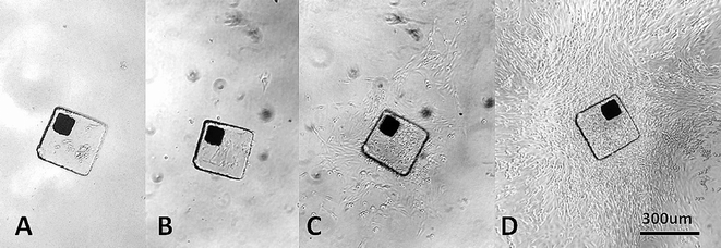
Three adherent cell lines were used to test cell growth on the magnetic microspallet arrays. HeLa, NIH/3T3 and rat208F cells were maintained in culture using vendor (ATCC) specified conditions. All cell types were maintained in polystyrene culture flasks incubated at 37 °C in 10% CO2. When ready for use, cells were removed from their culturing flasks with a trypsin–EDTA solution (0.25% trypsin; 1 mM EDTA) and vigorously pipetted after collection to generate single cell suspensions. The number of collected cells was determined manually using a standard hemocytometer (Reichert, Buffalo, NY). Recovered viable cells were resuspended at a concentration of 1000 cells per mL and one mL was pipetted into each well of an 8-well LabTek slide. Cell imaging studies were conducted on standard micropallets fabricated on the gold layer by seeding HeLa and 3T3 cells within a shared chamber at a 1![[thin space (1/6-em)]](https://www.rsc.org/images/entities/char_2009.gif) :
:![[thin space (1/6-em)]](https://www.rsc.org/images/entities/char_2009.gif) 1 ratio to test whether morphological differentiation was possible on the microarrays. To test cell growth potential on magnetic micropallets, rat208F cells were seeded to the arrays and grown for ten days to form colonies. Initial cell adhesion was confirmed at three hours from the seeding after which point the cultures were observed every 24 hours for viability and growth. Culture media was replaced every 72 hours. Fig. 3 displays the progression of cell seeding to confluence on the micropallets.
1 ratio to test whether morphological differentiation was possible on the microarrays. To test cell growth potential on magnetic micropallets, rat208F cells were seeded to the arrays and grown for ten days to form colonies. Initial cell adhesion was confirmed at three hours from the seeding after which point the cultures were observed every 24 hours for viability and growth. Culture media was replaced every 72 hours. Fig. 3 displays the progression of cell seeding to confluence on the micropallets.
Cell viability testing
The biocompatibility of magnetic arrays with gold coated nickel cores were directly compared to identical arrays lacking any gold electrodeposition, exposing the nickel coating to the cell culture media. Results were compared to a control slide with just 1002F micropallets fabricated on glass. The wells of an 8-well LabTek chamber adhered over the surface of the arrays were filled with 300 μL of rat208F culture media. Rat208F cells were seeded at a concentration of 1000 cells per mL in each of the eight wells. Cultures were grown on the arrays for seven days in an incubator at 37 °C with 10% CO2. To assess viability, a combination of 7-Aminoactinmycin (7AAD) and Annexin V conjugated to CF647 fluorescent dye (Annexin V) (EMD Millipore, Hayward, CA) was used. Annexin V targeted phosphatidylserine, a component of the phospholipid membrane, which is exposed on early apoptotic cells. 7AAD fluoresces upon intercalating with DNA, but requires compromised cell membranes to gain access to the genomic DNA, a later event in the cell death process. On day seven, for a positive control, one individual well from each slide was fixed with 2% paraformaldehyde. The media of the wells was replaced with four half volume exchanges of RPMI 1640 media (Life Technologies, Carlsbad, CA) and emptied to a final volume of 150 μL. 150 μL of 4% paraformaldehyde was added to the wells and left to fix the cells for 20 minutes. After fixation, the solution was replaced with RPMI 1640 once more and filled to 300 μL. The cell culture media in the remaining seven wells of each of the slides was replaced with the RPMI 1640 media with four half volume exchanges to a final volume of 300 μL. The combination of the two stains enabled the detection of varying levels of cellular health with Annexin V staining cells early in the apoptopic process and 7AAD staining late apoptotic and necrotic cells. Both stains were added to six non-fixed wells of each slide at 2.5% concentration and incubated for 20 minutes before visualization. A single well on each slide was left as a negative control with no exposure to the combination of Annexin V and 7AAD. The slides were imaged with an LSM 780 confocal microscope (Carl Zeiss, Oberkochen, Germany) with a two channel setup exciting at 633 nm and 561 nm for Annexin V and 7AAD respectively. Initial fluorescent peak emissions of 670 nm and 650 nm were confirmed using the wells containing the fixed cells as positive controls on each array.In order to compare the viability of cells before and after laser ejection, a Trypan blue exclusion test was performed at both points in time using HeLa and NIH/3T3 cell lines.26 Cells were seeded to the magnetic arrays in single wells of 4-well Labtek chambers at a concentration of 500 cells per mL in their respective culture medias in order to sequester single cells to individual pallets. After seven days of growth, two wells of each cell type were checked for viability by replacing the culture media with 1 mL of a solution of Trypan blue and phosphate buffered saline (PBS) at a 1![[thin space (1/6-em)]](https://www.rsc.org/images/entities/char_2009.gif) :
:![[thin space (1/6-em)]](https://www.rsc.org/images/entities/char_2009.gif) 1 ratio by volume. After five minutes, the cells were observed by phase contrast microscopy and those observed to have blue cytoplasm, indicating Trypan blue uptake, were considered non-viable. To test cell viability immediately following laser ejection, pallets bearing cell colonies were ejected via the methods described above and transferred to a 96-well culture dish with chambers filled with the Trypan blue/PBS solution. The cells were then observed by phase contrast microscopy after five minutes.
1 ratio by volume. After five minutes, the cells were observed by phase contrast microscopy and those observed to have blue cytoplasm, indicating Trypan blue uptake, were considered non-viable. To test cell viability immediately following laser ejection, pallets bearing cell colonies were ejected via the methods described above and transferred to a 96-well culture dish with chambers filled with the Trypan blue/PBS solution. The cells were then observed by phase contrast microscopy after five minutes.
Micropallet release and magnetic collection
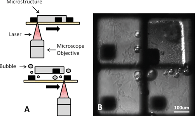
Cell colonies grown on the magnetic microarrays were visualized using an LSM 780 confocal microscope with a paired MaiTai Ti:Sapphire multiphoton laser system (Spectra-Physics, Santa Clara, CA). The MaiTai laser was utilized for micropallet ejection with its emission wavelength set to 790 nm, thus generating an effective irradiation wavelength of 395 nm at the laser focus point. Upon the discovery of a cell colony of interest to be recovered from an array, Zeiss' Zen microscope software was used to define a laser irradiation region drawn to match the perimeter of the structure holding the cell colony. The focus height of the microscope objective was set at the base of the micropallet to be ejected, targeting the gold thin film substrate below. The proper focus height could be repeatedly located by observing autofluorescent emissions from the glass slide in response to a 488 nm observation laser. Focus heights set at locations other than the film would not result in release. The interface between the micropallet and gold film was exposed to the MaiTai laser to an average energy of 88 mJ which equated to 8 passes of the laser over the defined region. The ejection of the micropallets followed a two-step event. First, initial delamination of the pallet at various points around the edges of the pallet was caused by the generation of localized plasma as previously shown.19 The delaminated regions allowed the flow of fluid under the micropallets. Laser energy absorption by the gold thin film resulted in the heating of the gold surface, which caused water vaporization leading to the generation of microbubbles between the surface of the gold film and the pallet structure. The bubbles expanded and lifted the remaining attached portions of the pallet away from the substrate. Displacement force generated by the bubbles effectively dislodged the micropallets while maintaining cellular adherence to their top surface. If laser irradiation was only targeted to the center of the structure, the pallet was not released, but instead resulted in localized charring of 1002F since fluid could not seep under the pallet. Metal film assisted laser absorption for biological material transfer has been previously described as a process deemed laser induced forward transfer (LIFT) which did not show significant detrimental effect on viability from the excitation of the metals.23–25 As with LIFT, micropallet ejection did not have any visible effect on adjacent structures or the viability of the colonies growing on them. Fig. 4 displays the micropallet ejection process.Cell colonies that were released from the array settled at a distance ranging from 30 μm to 500 μm from the ejection site. A 1.5 mm diameter polystyrene probe18 containing a removable neodymium magnet was sterilized with 70% ethanol and used to collect released pallets and transfer them to a separate culture plate. Cell colony collection involved moving the magnetic probe towards a released micropallet until the probe was at a distance close enough for the attraction force of the ferromagnet to overcome the gravitational force holding the pallet at its resting location. The collected pallet carrying the target cell colony was transferred to a 96-well Falcon polystyrene culture plate (Corning, Tewksbury, MA) wherein the pallet was released from the probe into fresh cell culture media, as the magnet was pulled back from the end of the probe eliminating the magnetic field effects on the captured pallet. The probe was rinsed in sterile diH2O before every collection to prevent culture contamination.
Results and discussion
Gold thin film characteristics
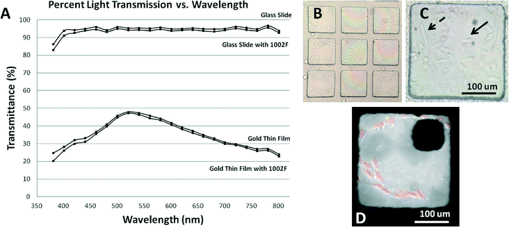
The average electrical resistance of 24 gold thin film coated glass slides followed a linear progression ranging from 12.4 Ω at a distance of 1 mm from a corner of the array to 23.8 Ω at 80 mm. These measured resistances were deemed acceptable for the required electroplating processes and allowed for consistent electrodeposition. Furthermore, the initial resistance of the films only had an impact on the first gold layer deposition step due to resistance values dropping considerably upon each successive metal plating. For example, the average measured resistance of the 30 μm nickel film was 0.5 Ω over the entirety of the array. The intended sorting capabilities of the microarrays required the gold thin films to have translucent imaging characteristics allowing for the visualization of cell colonies grown on them. Four types of slides were prepared for transmission analysis including an uncoated glass slide, a glass slide coated with a 50 μm layer of 1002F photoresist, a slide coated with the gold thin film, and finally a glass slide coated in gold and 50 μm of 1002F photoresist. The average measured light transmission properties of 5 of each type the films are shown in Fig. 5.Micropallet arrays fabricated on the gold thin films exhibited a loss in light transmission when compared to arrays fabricated on plain glass slides, but were still functional as cell colony imaging tools. Captured cells viewed with standard phase contrast microscopy were readily visible and distinguishable solely based on their morphological characteristics. The peak transmittance of the gold films was located within a range encompassing the most commonly utilized fluorescent probe wavelengths, allowing fluorescent signal transmission during standard cell staining and imaging procedures. The 7AAD and Annexin V stains, with peak fluorescent emissions of 650 nm and 670 nm respectively, were clearly visible on the arrays, suggesting that other commonly used fluorophores, such as FITC, would also be easily detected.
Magnetic property of micropallets
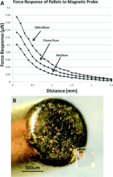
The magnetic retrieval of micropallets worked consistently for all ferromagnet component sizes. The attraction force generated by the magnetic probe was capable of collecting released structures and was not sufficiently strong to dislodge structures still adhered to the substrate, preventing any unintended cell collection. Fig. 6 shows the measured force response of three different ferromagnet sizes to the magnetic collection probe as a function of distance.The effective retrieval distance from the probe to the micropallet was directly related the measured force of attraction of each ferromagnet size. Minimum capture distances for the large, medium, and small micropallets were 0.6 mm, 0.5 mm, and 0.2 mm respectively. The micropallets preferentially adhered to the magnetic probe in an orientation, due to the placement of the ferromagnet core that kept the cell colony growth area separated from the probe surface, preventing unwanted compression on the cells. While transferring the pallets from the microarrays to separate culture wells, a droplet of culture media encompassed the probe end and kept the pallet wetted during transfer as previously shown.18
Magnetic micropallet release and recovery
The laser release of target cell colonies enabled direct capture of micropallets without an effect on the surrounding untargeted colonies. While the ferromagnets embedded within each pallet dislodged cleanly from the gold thin film, the metal plated around the proximity of the structures maintained its contact to the film. Due to the process requiring vapor bubbles for actuation, a liquid environment was required for proper function. Lack of liquid resulted in the ablation of the micropallets. Cell colonies remained adhered throughout the collection process and were viable upon reseeding post-recovery. The success rate of ejecting various sizes of micropallets was also tested to enable future research utilizing larger pallets than those typically used for cell colony sorting. Four square pallets with dimensions of 200 μm by 200 μm, 300 μm by 300 μm, 400 μm by 400 μm and 500 μm by 500 μm were able to be ejected from gold coated slides at a success rate greater than 90% for each pallet size (n = 30). The length and width of the largest pallets that were released for this study were comparable to those that were targeted for the recently described ultrasound release method.17 The total applied energy to release the pallets, however, was not optimized and thus varied from pallet to pallet requiring future work to design ideal ejection settings. Typical cell colony sorting pallets sized at 270 μm by 270 μm with embedded ferromagnets were investigated in detail showing a laser assisted ejection success rate of 95.1%. While the 88 mJ of energy required for ejection of the magnetic pallets using the vapor bubble method was much greater than that required for the similarly sized table top pallets,16 which only required around 36 μJ to release, the increased energy did not result in greater loss of viability in the captured cell colonies. It is likely that the Ni/Au mesh surrounding the ejected pallets acted as a heat sink, preventing significant damage to the captured cells on top of the pallets. Fig. 7 displays a released micropallet and its ejection site. These early results indicate that the gold absorption layer can be applied for micropallet arrays bearing pallets greater than previously utilized which could enable new types of adherent sample research. The conventional method for releasing micropallets using laser catapulting is primarily effective for pallets with dimensions less than 200 μm.15Cell viability and growth
Nickel-only plated micropallet arrays exhibited loss of cellular viability as visualized by 7AAD and Annexin V staining. Unhealthy cells were defined as those expressing either one of the stains or a combination of both, representing compromised viability. Of the cells grown on nickel-plated arrays (n = 1410), 9.9% were deemed unhealthy, 90.1% viability, after seven days. The proximity to the nickel core did not affect the viability of the cells. Of those that were compromised, 39% were located within a 150 μm by 150 μm square corner surrounding the nickel core and 61% were on the remaining surface of the pallets outside of the square (n = 140 compromised cells). These results indicate that cell viability was not associated with the proximity of the growing cells to the nickel cores, as the number of compromised cells counted was associated with the surface area allocated to them rather than their distance from the nickel core. Cells concurrently grown on arrays consisting of gold coated nickel cores maintained 99.9% percent viability, measured as above, (n = 1563) at 7 days, as did cells grown on uncoated glass slides with micropallets (n = 1521). Although the gold plating did not completely abrogate the decreased cell viability on the arrays, there was a substantial improvement in biocompatibility gained with the addition of the gold layer over the nickel.We evaluated the potential decrease in viability associated with the laser release process. Cells seeded to the magnetic arrays maintained significant viability immediately following laser ejection when assessed with Trypan blue exclusion. HeLa cells grown from single cells on the arrays for seven days exhibited 99.5% viability with an average of 35 cells per pallet before ejection. Once released from the array and collected, the cells had an average of 2.6 compromised cells per colony, which translated to a cell viability retention of 93% (n = 5 collected pallets). NIH/3T3 cells tested in the same manner as the HeLa cells grew to an average of 22 cells per pallet with a viability of 99.3% before ejection. Colonies ejected from the arrays had an average of 3.6 compromised cells per colony equating to 84% viability (n = 5). These results indicated that a significant portion of cells ejected using the laser absorptive gold layer were viable immediately following release.
While individual cell viability during cell expansion on the arrays is important, it was also important for the cells to maintain healthy growth after collection and reseeding in culture wells. Individual cell colonies were ejected, as above, from the gold coated magnetic arrays and transferred to a 96-well culture plate containing 300 μL of fresh cell media at one micropallet per well as described above. Twenty separate rat208F colony samples were collected and observed over a span of two weeks with culture media changes every 72 hours. All collected colonies exhibited further growth to the eventual confluence of the cultures within their wells. HeLa cell colonies collected in the same manner exhibited 100% growth after collection. Colonies derived from the less robust NIH/3T3 cell line had a 90% cell colony viability (n = 20 for both cell types). The cell colony viability maintained by this ejection method matched those previously displayed by previous methods designed for large pallet release.12–14 The initial resting position of the pallets did not have an effect on the cell growth. If the cells landed facing up on the pallet, the cells would simply grow down the side of the pallet to reach the culture well bottom and continue expansion. Cells captured facing down towards the bottom of the well were not crushed by the pallet but rather grew unhindered out from its sides. Fig. 8 displays the growth of a single captured rat208F cell colony seeded to a micropallet over a span of 7 days.
Conclusion
Large area magnetic micropallet arrays for cell colony sorting were fabricated on translucent gold thin films and filled with ferromagnetic cores. The electrical conductivity of the gold film enabled the electrodeposition of gold plated nickel cores to enable individual colony retrieval by a magnet probe. The arrays featured improved laser ejection mechanics and colony collection. The gold thin film substrate provided enhanced laser absorption beneath individual pallets that allowed use of lower powered laser systems such as those already paired to many common microscopes. This process was repeatable and did not have a negative impact on cell viability. A wide range of pallet sizes including those much larger than typically used for human cell sorting were able to be consistently ejected from the arrays using a two photon laser system typically included with modern, commercially available, confocal microscopes, negating the need for separate ejection equipment and methods. Magnetic collection of pallets provided quick and direct colony transfer without the need for time consuming and less precise gravitational transfer methods. The magnetic control of ejected micropallets can also enable further downstream cell colony manipulation within microsystems joined to standard arrays, a procedure which has yet to be explored, but could have a great impact on efforts to integrate micropallet arrays into lab-on-a-chip platforms.Imaging loss previously associated with other magnetic retrieval methods have been improved by maintaining the composition of the transparent photoresist structures and placing all ferromagnetic elements to one position in the pallet. These magnetic cores further acted as imaging fiducials. Although the semi-transparent nature of the gold layers led to an overall loss in light transmission, this work has laid the foundation for future work involving the use of more transparent conductive materials such as indium tin oxide as laser absorption layers. Another method being pursued to lower transmission loss caused by the gold layer is the plating of strategically patterned gold thin film layers. Cell morphology and fluorescent stains could clearly be observed on the micropallets, opening up the use of the arrays to multiple types of experimentation requiring cell phenotype differentiation. The creation of the arrays was straightforward and only required the use of commonly performed fabrication techniques such as electron beam vapor deposition, photolithography, and electroplating.
We believe that this technology has great implications towards enhancing adherent colony sorting protocols using micropallet arrays. The use of a semi-transparent gold layer allows for the inclusion of electroformed nickel cores as well as a bubble-assisted release mechanism for large area pallets using low power lasers. This enables broad practical application of micropallet array technology for use in cell colony assays.
Acknowledgements
This work was supported by the National Science Foundation's Integrative Graduate Education and Research Traineeship program (NSF-IGERT) “LifeChips” Award DGE-0549479. The work was made possible, in part, through access to the Optical Biology Core facility of the Developmental Biology Center, a Shared Resource supported in part by the National Cancer Institute of the National Institutes of Health under award number P30CA062203 to the Chao Family Comprehensive Cancer Center and the Center for Complex Biological Systems Support Grant (GM-076516) at the University of California, Irvine. This work was also funded in part by NSF I/UCRC: Center for Advanced Design and Manufacturing of Integrated Microfuidics (CADMIM) award #IIP-1362165. The content is solely the responsibility of the authors and does not necessarily represent the official views of the National Institutes of Health.References
- H. J. Unwalla and J. J. Rossi, Biotechnol. Genet. Eng. Rev., 2006, 23, 71–91 CrossRef CAS PubMed . Review.
- M. A. C. Huergo, M. A. Pasquale, P. H. Gonzalez, A. E. Bolzan and A. J. Arvia, Phys. Rev., 2012, 85, 011918 CAS.
- T. M. Yeung, S. C. Gandhi, J. L. Wilding, R. Muschel and W. F. Bodmer, Proc. Natl. Acad. Sci. U. S. A., 2010, 107(8), 3722–3727 CrossRef CAS PubMed.
- D. S. Kaufman, E. T. Hanson, R. L. Lewis, R. Auerbach and J. A. Thomson, Proc. Natl. Acad. Sci. U. S. A., 2001, 98(19), 10716–10721 CrossRef CAS PubMed.
- P. Vanparys, R. Corvi, M. J. Aardema, L. Gribaldo, M. Hayashi, S. Hoffmann and L. Schechtman, Mutat. Res., 2012, 744, 111–116 CAS.
- N. Maeda, M. Palmarini, C. Murgla and H. Fan, Proc. Natl. Acad. Sci. U. S. A., 2001, 98(8), 4449–4454 CrossRef CAS PubMed.
- K. Buch, T. Peters, T. Nawroth, M. Sanger, H. Schmidberger and P. Langguth, Radiat. Oncol., 2012, 7, 1 CrossRef PubMed.
- N. A. Franken, H. M. Rodermond, J. Stap, J. Haveman and C. Bree, Nat. Protoc., 2006, 1(5), 2315 CrossRef CAS PubMed.
- S. Haupt, J. Grutzner, B. H. Rath, H. Mohlig and O. Brustle, Nat. Methods, 2009, 6 Search PubMed.
- Z. Komyei, S. Beke, T. Mihalffy, M. Jelitai, K. J. Kovacs, Z. Szabo and B. Szabo, Sci. Rep., 2013, 3, 1088 Search PubMed.
- Y. Wang, G. Young, M. Bachman, C. E. Sims, G. Li and N. L. Albritton, Anal. Chem., 2007, 79, 2359–2366 CrossRef CAS PubMed.
- P. C. Gach, W. Xu, S. J. King, C. E. Sims, J. Bear and N. L. Albritton, Anal. Chem., 2012, 84, 10614–10620 CrossRef CAS PubMed.
- M. Charnley, M. Textor, A. Khademhosseini and M. P. Lutolf, Integr. Biol., 2009, 1, 625–634 RSC.
- J. Pai, K. Kluckman, D. O. Cowley, D. M. Bortner, C. E. Sims and N. L. Albritton, Analyst (Cambridge, U. K.), 2013, 138, 220 RSC.
- G. T. Salazar, Y. Wang, C. E. Sims, M. Bachman, G. Li and N. L. Albritton, J. Biomed. Opt., 2009, 13(3) Search PubMed.
- J. Pai, W. Xu, C. Sims and N. L. Albritton, Anal. Bioanal. Chem., 2010, 398(6), 2595–2604 CrossRef CAS PubMed.
- S. Guo, Y. Wang, N. Allbritton and X. Jiang, Appl. Phys. Lett., 2012, 101, 163703 CrossRef PubMed.
- N. M. Gunn, R. Chang, T. Westerhof, G. Li, M. Bachman and E. Nelson, Langmuir, 2010, 26(22), 17703–17711 CrossRef CAS PubMed.
- J. Pai, Y. Wang, G. T. Salazar, C. E. Sims, M. Bachman, G. P. Li and N. L. Albritton, Anal. Chem., 2007, 79(22), 8774–8780 CrossRef CAS PubMed.
- Y. Wang, M. Bachman, C. E. Sims, G. Li and N. L. Albritton, Anal. Chem., 2007, 79, 7104–7109 CrossRef CAS PubMed.
- N. M. Gunn, M. Bachman, G.-P. Li and E. L. Nelson, Fabrication and biological evaluation of uniform extracellular matrix coatings on discontinuous photolithography generated micropallet arrays, J. Biomed. Mater. Res., Part A, 2010, 95, 401–412 CrossRef PubMed.
- H. Shadpour, C. E. Sims and N. L. Albritton, Cytometry, Part A, 2009, 75A, 609–618 CrossRef CAS PubMed.
- J. M. Fernandez-Pradas, M. Colina, P. Serra, J. Dominguez and J. L. Morenza, Thin Solid Films, 2004, 453–454, 27–30 CrossRef CAS.
- B. Hopp, T. Smausz, Zs. Antal, N. Kresz, Zs. Bor and D. Chrisey, J. Appl. Phys., 2004, 96, 3478 CrossRef CAS.
- B. Hopp, T. Smausz, N. Kresz, N. Barna, Z. Bor, L. Kolozskari, D. B. Chrisey, A. Szabo and A. Nogradi, Tissue Eng., 2005, 11(11/12), 1817–1823 CrossRef CAS PubMed.
- W. Strober, Current Protocols in Immunology, 1997, A.3B.1–A.3B.2 Search PubMed.
| This journal is © The Royal Society of Chemistry 2016 |

