Solution assembly MoS2 nanopetals/GaAs n–n homotype heterojunction with ultrafast and low noise photoresponse using graphene as carrier collector†
Yan
Zhang
ab,
Yongqiang
Yu
b,
Xiaoyan
Wang
a,
Guoqing
Tong
c,
Longfei
Mi
a,
Zhifeng
Zhu
ab,
Xiangshun
Geng
b and
Yang
Jiang
*a
aSchool of Materials Science and Engineering, Hefei University of Technology, Hefei, Anhui 230009, P. R. China. E-mail: apjiang@hfut.edu.cn; apjiang2002@yahoo.com; Fax: +86-551-62904358; Tel: +86-551-62904358
bSchool of Electronic Science and Applied Physics, Hefei University of Technology, Hefei, Anhui 230009, P. R. China
cNational Laboratory of Solid State Microstructures and School of Electronics Science and Engineering/Collaborative Innovation Centre of Advanced Microstructures, Nanjing University Nanjing, Jiangsu 210093, P. R. China
First published on 25th November 2016
Abstract
MoS2, the classical representative of layered structure transition metal dichalcogenides (TMDCs), has been widely used as an ideal n-type semiconductor, offering an interesting opportunity to construct heterostructures with other 2D layered or 3D bulk materials for ultrafast optoelectronic applications. In this work, we report the synthesis of ultrathin MoS2 nanopetals via a solution-processable route, and the solution assembly of a 2D MoS2 nanopetal/GaAs n–n homotype heterojunction using graphene as the carrier collector. The fabricated devices have excellent photoresponse characteristics including a good detectivity of ∼2.28 × 10¹¹ Jones, a noise current approaching 0.015 pA Hz−1/2 at zero bias and notably a very fast response speed, up to ∼1.87/3.53 μs with a broad photoresponse range. More interestingly, the device could respond to fast pulsed illumination up to 1 MHz, far exceeding the performance of many current congeneric 2D nanostructured and solution-processable photodetectors reported. These results suggest that our devices, together with the solution assembly methodology of the device described herein, can be utilized to give large-scale integration of low-cost, high-performance photodetectors, thus opening up new possibilities for 2D layered material-based photovoltaic and optoelectronic applications in the future.
Introduction
Two-dimensional (2D) layered materials represented by graphene and layered transition metal dichalcogenides (TMDCs) have attracted tremendous attention because of their unique properties and the ease of construction of various structures compared with bulk materials. For graphene, a material that has been well studied for decades, its high optical transparency combined with excellent carrier mobility makes it highly applicable as a transparent electrode material in many devices, such as solar cells, sensors and optical–electrical devices.1–4 Meanwhile, TMDCs have also gradually become a new focus of fundamental research and technological applications due to their tunable bandgap, as well as excellent electronic and optical properties, just making up for the limitation of graphene which originates from its zero bandgap with semi-metal properties.5–7 The diversity of properties and the same 2D morphological features, make them very promising nano-platforms for many applications such as nano-electronics, photodetection and the assembly of ultrathin and flexible devices.Among various novel devices, photodetectors (PDs), a kind of very crucial device that can capture incident optical signals and instantly convert them to electrical signals, play an important role in many fields such as imaging, environment monitoring, remote control, optical communication, and biological sensing, etc.8,9 Researchers have exerted much effort to improve the performance of devices and develop novel photodetectors, including the choice and synthesis of different materials, as well as the design and optimization of device architecture, which have promoting effects on determining the detection ability.10 For example, much research has been done on 2D materials based PDs recently, and various types of high-performance 2D nanostructure-based PDs have been achieved.11–14 Compared with traditional PDs, these photodetectors based on 2D materials have great potential to give an improved performance, since some intriguing changes occur in their electronic and optical properties, which is probably because of the electron confinement and tight contact between the stacked layers. In particular, a heterojunction structure based on 2D layered materials offers the opportunity to further enhance the performance of devices by allowing more materials with different advantages to join in together, with the potential to fulfil many requirements for PDs such as ultra-sensitivity and fast response, benefiting from the combination of excellent electric characteristics of the different materials and the intrinsic advantages of the heterostructure.15–17
As the classical representative of TMDCs, MoS2 is widely used as an ideal photovoltaic semiconductor material, offering an interesting opportunity to construct heterostructures with other 2D layered or 3D bulk materials for ultrafast optoelectronic applications, and exhibits excellent optical and electrical properties.18–22 In the past few years, many MoS2 based PDs fabricated with different processes have been reported, suggesting its superior potential in optical–electrical fields.23–26 However, despite that, the photoresponsivity of most MoS2 based photodetectors is comparable to graphene-based devices, as well as some other nanostructured photodetectors,27–29 and devices’ response speed and operation frequency presented in most reports still needs improving to meet fully the requirements for applications in high-speed and high frequency optical communications nowadays. Up to now, the general preparation methods previously used are usually based on mechanical exfoliation or chemical vapor deposition (CVD) techniques.30–33 Nevertheless, of the high quality MoS2 crystals these methods can generate, the former only gives irregular and small sheets with low yield and low efficiency, while the latter often requires expensive facilities and complicated processing steps, which is bound to impede the practical utility and promotion of the corresponding devices. Additionally, as we know, MoS2 and other 2D materials tend to be obtained in the form of a dispersion of small nanosheets. Thus, a facile, efficient and scalable process suitable for various 2D materials is required urgently to realize the formation of thin films on diverse substrates. With this consideration, a solution-processed technique for the deposition of many kinds of films has attracted more attention and is being widely investigated as an alternative approach to construct nano-devices with improved performance.
By comparison, solution-process methods can prepare various materials in bulk quantities with different nanostructures or morphologies, which has many advantages such as being more efficient and feasible, together with being tunable, easy to control and substrate-free. More importantly, the product made by this route can be deposited easily on an arbitrary substrate and formed into uniform films through spin-coating or dip-coating assembly, which provides beneficial conditions for their application in various device constructions including heterojunction structures. To date, it has been proposed that various solution-process based 1D or 2D nanomaterials with different structures could be used as photodevices, and excellent optoelectrical performances have been achieved.34,35 However, to the best of our knowledge, solution-processed high performance photodetectors made from MoS2 nanosheets are limited, and more investigation still needs to be performed.
In light of the above investigation, in this work, we demonstrate the synthesis of MoS2 nanopetals via a facile solution method, and subsequently the solution assembly of a MoS2 nanopetal film/GaAs n–n homotype heterojunction based photodetector using graphene as a carrier collector. The heterojunction can exhibit a relatively wider response spectrum than a Schottky diode since the latter contains only one semiconductor material. Meanwhile, compared with a p–n junction, there are different band alignments in the n–n heterojunction such that depletion and accumulation regions were formed on opposite sides of the interface at thermal equilibrium, resulting in a majority carrier transport and improving the photoresponse speed of the device. Therefore, this method has the advantages of the kinetic reactivity of the solution process, excellent intrinsic optical and electronic properties of the two materials, efficient photo-carrier transport of the heterostructure, as well as effective collection and transfer of carriers using graphene as the transparent electrode, conceptually different from the current methodology based on mechanical exfoliation and chemical vapor deposition (CVD), emphasizing the exploration of large area, high-efficiency, and even flexible device species made from different nanostructures (1D or 2D nanomaterials) using a facile and universal process. In particular, our resultant devices exhibited an excellent photoelectric conversion performance, a high sensitivity to broad band light range, as well as stability and reproducibility, i.e. a good detectivity of ∼2.28 × 10¹¹ Jones, noise equivalent power (NEP) approaching 3.37 × 10⁻¹¹ W at zero bias, and notably an ultra-fast response speed up to 1.87/3.53 μs. More interestingly, the device could respond to fast pulse illumination up to 1 MHz, which is much better than most 2D nanostructured and solution-processable photodetectors reported so far,35–37 suggesting that this approach provides a new pathway for the design and fabrication of various solution-processable, large-area, high-speed photodetectors in the future.
Experimental section
Synthesis of MoS2 nanopetals
Hydrothermal synthesis was carried out in a 60 ml Teflon-lined stainless steel autoclave. All the reagents were of analytical grade purity (Shanghai Chemistry Co.) and used for synthesis as received without purification. In a typical procedure, 2 mmol of sodium molybdate (Na2MoO4·2H2O, 0.4888 g), 8 mmol of thiourea (CH4N2S, 0.624 g) and polyethylene glycol (PEG-10000, 0.0656 g) were dissolved in 40 ml deionized water, then hydrazine hydrate (N2H4·H2O, 0.18 g) was added into the solution under constant stirring. Distilled water was added to fill the autoclaves up to 80% of the total volume. Then the pH value of the mixture was adjusted to about 1.0 by adding 12 mol L−1 HCl solution dropwise. The resulting mixture was transferred into a 60 ml Teflon-lined stainless steel autoclave then sealed tightly and maintained at 200 °C for 24 h. After cooling down to room temperature naturally, the black precipitates were collected using centrifugation, then washed with plenty of deionized water and absolute ethanol several times to remove possible residual reactants and surfactant until the solution's pH ≈ 7, and dried under vacuum at 60 °C for 12 h in a vacuum oven. Finally, the products were annealed at 800 °C for 2 h in an Ar atmosphere. Subsequently, the molybdenum disulfide powder was dissolved in DMF solvent at the desired concentration and ultrasonicated for 12 h (the water in the sonicator was kept cold). The prepared MoS2 dispersions were centrifuged at 5000 rpm for 20 min to remove severely conglomerating MoS2 crystals and the yellow supernatant was collected and washed using ethanol and collected with a pipette. The sample dispersed in DMF had a concentration of ∼1 mg ml−1 for device fabrication.Materials characterization
The phases and crystal structures of the synthesized samples were identified using a powder X-ray diffraction (XRD) analysis system (D/MAX2500 V, Rigaku, Japan) equipped with Cu Kα radiation (λ = 0.15418 nm). Micro-Raman measurements were performed in a LabRam HR Evolution. AFM images were obtained using atomic force microscopy (AFM, BRUKER AXS Dimension) in the tapping mode after the samples were deposited on a SiO2/Si surface using spin coating. The morphological features of the samples and devices were obtained using field-emission scanning electron microscopy (FESEM; SU8020, Hitachi, Japan). The TEM images were taken on a high-resolution field-emission transmission electron microscope (HRTEM; JEM-2100F, JEOL, Japan).Device construction
To study the photoresponse properties, a vertically stacked MoS2 nanopetals/GaAs heterojunction based device was constructed using the following solution assembly route. A pre-cleaned Si-doped GaAs (100) wafer (1 × 1 cm2, n-type, 350 μm thick, resistivity of ≈(0.8–9) × 10−3 Ω cm) was chosen and first pretreated with a hydrophilic treatment using Ar-plasma for 10 min to ensure good adhesion of the MoS2 coatings. Ti/Au (5/20 nm) was used as the Ohmic contact electrode of the device and deposited on the back of a GaAs substrate using a high-vacuum electron-beam (EB) system with the assistance of a standard shadow mask, and the substrate subsequently underwent rapid thermal annealing (RTA) in vacuum for 5 min at 500 °C, thus alloying to improve the Ohmic contact. Then, the MoS2 nanopetals were re-dispersed in ethanol to make a homogeneous solution, and then drop and spin-coated onto the surface of the GaAs substrate, and dried in air naturally at room temperature, while the periphery of the substrate was protected with high-temperature resistant tape for insulation purposes. A thin MoS2 nanopetal film was formed and covered the bare substrate and tape surface. Afterwards, the semi-finished device was soaked in DI water with floating graphene films, and then slowly lifted to mount the graphene films on the substrate (more information is given in Fig. S1, ESI†).38 The assembled device was eventually transferred onto a copper foil, coated with silver conducting paint to form a good contact between the back side of the GaAs (Ti/Au back electrode) and the underlying copper. Meanwhile, high-purity silver conducting paint on the graphene side was selected as the top electrode leading wire of the device.Photoresponse measurements
The optoelectronic characterization of the device was performed using a semiconductor parameter analyzer system (Keithley 4200-SCS) at room temperature, and a laser diode (with wavelengths of 532, 650, and 808 nm) was used as the illumination source. The spectral response was studied using a built system composed of a xenon lamp (150 W), a monochromator (Omni-λ300) and a lock-in amplifier (SR830). The noise current was also measured with the same lock-in amplifier, and the device was kept in the dark operation cabinet during these measurements. The high-speed response investigation was performed using the above LD (650, 808 nm) modulated with a function generator (SIGLENT SDG5122), while the corresponding photocurrent under different frequencies of pulsed light illumination was recorded using a digital oscilloscope (Tektronix TDS2022B).Results and discussion
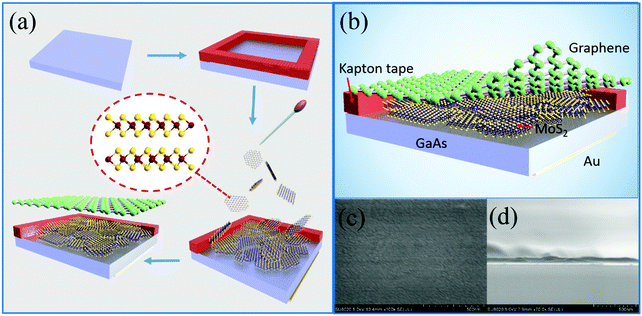
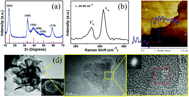
The fabrication process of the device is demonstrated in the schematic illustration in Fig. 1a, in which the inset shows a schematic drawing of the MoS2 crystal structure. Fig. 1b shows the structure of the obtained photodetector, in which the few-layer graphene film (FLG) was used as the top electrode and carrier collector by virtue of its excellent light transmission capability. Compared to common metal electrodes, it can make the light absorption of the device more effective and the transmission of carriers fluent, especially in the detection range of our device. Fig. 1c and d show SEM images of the top-view of the spin-coated MoS2 nanopetals film on the GaAs wafer from solution and its corresponding cross-section image of the obtained heterojunction.Fig. 2a depicts a typical powder X-ray diffraction (XRD) pattern of the MoS2 nanocrystals (upper profile) after annealing at 800 °C for 2 h and the corresponding standard Joint Committee on Powder Diffraction Standards (JCPDS) card no. 37-1492 (lower profile), which was consistent with previous reports for MoS2.39 The thickness of the synthesized MoS2 nanopetals was determined using Raman spectroscopy measurements via the energy difference between the E12g and A1g Raman modes (Fig. 2b). The E12g and A1g modes of the Raman band are located at 382.7 cm−1 and 407.5 cm−1, respectively, and the splitting is 24.8 cm−1, which is similar to that of MoS2 in a previous report,40 indicating that the sample is a layered MoS2 nanostructure. Moreover, a smooth surface morphology of the resulting MoS2 nanopetal film is observed using atomic force microscopy (AFM), as shown in Fig. 2c, with a thickness of ∼4 nm, also revealing that a layer structure of MoS2 is formed, and a uniform and smooth film can be made of MoS2 nanopetals. Fig. 2d shows a low-magnification transmission electron microscopy (TEM) image of the synthesized MoS2 crystals, showing a layer-like morphology of nanopetals. It can be seen clearly that many nanopetals aggregate together and assemble into each of the nanoflowers before ultrasonication and centrifugation, as shown in the inset of Fig. 2d, while independent MoS2 nanopetals can be found and are well dispersed in the collected supernatant (Fig. 2e), suggesting that the latter tends to give a large area, uniform film with good adhesion, easier for the heterojunction formation of our device. In order to further examine the quality of the MoS2 nanopetals, Fig. 2f shows a standard high-resolution TEM image of atoms arranged in a hexagonal pattern from the yellow square indicated in Fig. 2e, with the corresponding FFT (selected area electron diffraction pattern) of the red square inset, which not only gives an interplanar lattice spacing with intervals of 0.272 nm corresponding to the (010) facet of MoS2, but also clearly demonstrates the single crystal quality of the MoS2 nanopetals with the expected hexagonal crystal structure.
Based on the above high quality ultrathin MoS2 nanopetals, we constructed a 2D–3D heterostructure based device via a solution assembly method of spin-coating MoS2 nanopetals on a high-doped GaAs substrate, translating the optoelectronic properties into devices to investigate the photoresponse behaviour in detail.
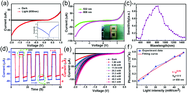
Fig. 3a plots the current–voltage (I–V) curves of our device based on vertical MoS2 nanopetals film/GaAs homotype heterojunction under dark and light illumination (650 nm, 20 mW cm−2). The inset of the figure shows the current under illumination on a logarithm scale. It is clearly seen that the black curve exhibits an obvious rectifying behavior associated with the heterojunction, with forward-to-reverse bias current ratios of 104 without illumination. Meanwhile the red curve is nonlinear and asymmetric with illumination, demonstrating the high sensitive photoresponse of our device. Notably, the curves also indicate that the heterojunction based on MoS2 nanopetals/GaAs shows photodiode-like behavior, where the reverse current increases but the forward current remains almost constant, similar to the device's function mode demonstrated in our previous work.26 The current–voltage (I–V) characteristics under incident light of wavelength 532 and 808 nm are also shown in Fig. 3b, and the inset shows a photograph of the device measured here. Fig. 3c presents the corresponding wavelength-dependent photocurrent response in the wavelength region of 300–1500 nm, used to determine the relative sensitivity to different wavelengths. The curve suggests clearly that the device exhibits a strong photocurrent response at 400–1300 nm under our testing conditions, which confirms that the combination of MoS2 and GaAs in our device broadens the spectral response range and provides a super capability of wide bandwidth photodetection. The spectral response of a pure GaAs/graphene based device (i.e. without MoS2) is given in Fig. S2b (ESI†), indicating that MoS2 makes a certain contribution to the wider spectral response of the device. Moreover, the I–V and time response characteristics of the GaAs/graphene device were also measured with 650 nm light illumination under the same conditions. As can be seen, the response speed of the GaAs/graphene based device is obviously slower than that in our work, especially to a high pulsed light signal, although it also exhibits a photoresponse and can act as a photodetector (see Fig. S2a, c and d, ESI†). Additionally, further I–V characteristics were measured for comparison, including the contact effect of the graphene/MoS2 nanopetal film and graphene/Ag electrode, to verify the origin of the heterojunction function in our photodetector (Fig. S3 and S4, ESI†). The resultant curves demonstrate that the graphene mainly functions as the collector and transport channel of the carriers in the device, and a reliable Ohmic contact can be formed between the graphene/Ag electrodes.
In order to further investigate the response characteristics of the heterojunction device, the time-dependent photoresponse was measured under periodic on/off laser illumination (650 nm, 20 mW cm−2) at different bias voltages, as illustrated in Fig. 3d. As can be seen, the device can be reversibly switched between a “low” current state in the dark and a “high” current state under light illumination quickly. The current of the heterojunction dramatically increases when the light irradiation is on (remaining almost the same for 6 cycles), giving a stable and repeatable current ON/OFF ratio (ION/IOFF) of 103 and 104 for biases of −1 V and −2 V, respectively. The above results indicate the promising potential of the device as a highly sensitive photodetector or photoswitch.
Next, the illuminated I–V characteristics of the photodetector with red light (650 nm) under different illumination intensities were measured (Fig. 3e) as the light intensity is another factor that determines the photocurrent in the heterojunction.41 Obviously, increasing the light intensity results in increasing the photocurrent without noticeable saturation, exhibiting a strong dependence of the photocurrent on light intensity, which proved that the photocurrent generation efficiency of the carriers is directly proportional to the luminous flux.42 The relationship between the photocurrent and light power can be expressed using the power law:
| Ip = cPb | (1) |
To further quantify the performance of our photodetector, the responsivity (R), one of the critical parameters that relates to the photodetector sensitivity, was also investigated. The responsivity (R) is defined as the photocurrent generated per unit power of the incident light on the effective area and it can be calculated using the following equation:
 | (2) |
Other figure-of-merits, the noise equivalent power (NEP) and specific detectivity (D*), were also evaluated to comprehensively analyze the optical–electrical performance of the photodetector. The noise equivalent power (NEP) is another important feature of a PD which represents the minimum optical input power that a detector can distinguish from the noise. A smaller NEP is desirable for a detector with high sensitivity. D* is commonly used as a material figure of merit to characterize the PD sensitivity to incident light (assuming shot noise from the dark current is the major contributor generally). Two parameters can be calculated with the following equation:
 | (3) |
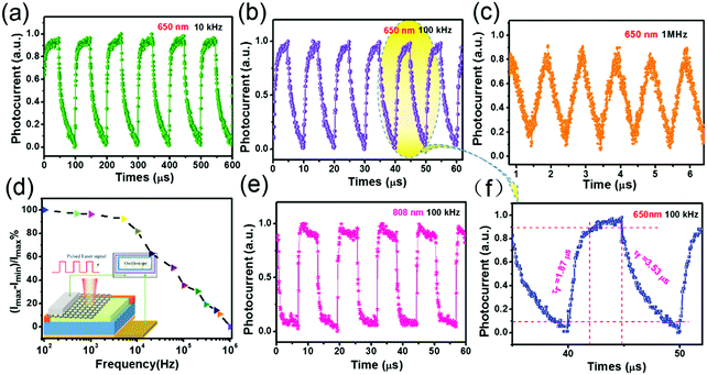
In order to ensure the PDs can significantly follow and record an optical signal faithfully, the response time, another critical parameter of a PD, should be shorter and change faster or equivalently, compared to the temporal variations in the signal. In our work, we illuminated our device with a pulsed incident light source with a varied pulsed frequency to evaluate further the potential of our PD for ultrafast photodetector application. In the schematic diagram of the measurement setup (shown in the inset of Fig. 4d), the photocurrent–time relationship of the PD was visualized on an oscilloscope as a high-frequency pulsing was applied to the system circuit to analyze the transient response to the excitation processes at zero bias voltage. Fig. 4a–c depict some representative results measured at zero bias as the red diode laser illumination was pulsed at several different frequencies varying from 100 Hz to 1 MHz using a function generator, and suggest that the device exhibits excellent stability and reproducibility whenever under low or high frequency (100 kHz), even up to a high frequency of ∼1 MHz. On magnifying the shape of a response pulse (one of the response periods) in Fig. 4d, the rise time τr and fall time τf under illumination of 650 nm laser were found to be 1.87 μs and 3.53 μs, respectively (Fig. 4f). A similar fast response speed was also observed under illumination of 808 nm laser (Fig. 4e), which is faster than that of most previously reported MoS2-based PDs and some other 2D-nanostructure photodetectors.11,52 Notably, even at a high frequency of 13![[thin space (1/6-em)]](https://www.rsc.org/images/entities/char_2009.gif) 000 Hz, the relative balance (Imax − Imin)/Imax of the photocurrent only decreases by less than 25%, as shown in Fig. 4d, suggesting that the device can be used to monitor ultrafast optical signals with very high frequency. The relative balance (Imax − Imin)/Imax of the photocurrent of the GaAs/graphene based device is also given for comparison (Fig. S7, ESI†), confirming that our PD is more suitable for high-frequency photodetection with high speed. All these results imply that our PDs may possess a potential to show an even shorter response time and work at much higher frequency if an appropriate input pulse is applied and the RC-time of the present circuit can be improved.53,54
000 Hz, the relative balance (Imax − Imin)/Imax of the photocurrent only decreases by less than 25%, as shown in Fig. 4d, suggesting that the device can be used to monitor ultrafast optical signals with very high frequency. The relative balance (Imax − Imin)/Imax of the photocurrent of the GaAs/graphene based device is also given for comparison (Fig. S7, ESI†), confirming that our PD is more suitable for high-frequency photodetection with high speed. All these results imply that our PDs may possess a potential to show an even shorter response time and work at much higher frequency if an appropriate input pulse is applied and the RC-time of the present circuit can be improved.53,54
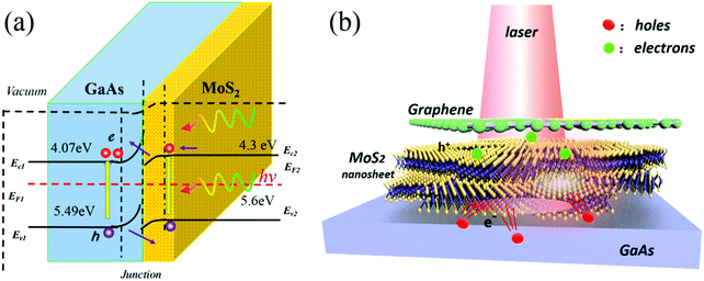
The excellent photoresponse of our device presented above could be ascribed to the following aspects. (i) Fig. 5a shows the energy band diagram of the MoS2 nanopetals/GaAs homotype heterojunction under light illumination at zero bias. As can be seen, MoS2 and GaAs form a diode where part of the photogenerated electrons diffuse from MoS2 towards the junction and are swept into GaAs, while holes move towards the MoS2. Compared to a p–n heterojunction, the n-MoS2/n-GaAs homotype heterojunction acts as a Schottky barrier to prevent leakage current in the dark, but easily overcomes the low barrier to enhance the photocurrent generation upon illumination. (ii) In this work, gallium arsenide (GaAs) was adopted to construct a heterojunction with MoS2 since it provides advantages over silicon for many applications,55,56 being more suitable for efficient optoelectronics devices, and improving high speed operation in high-frequency systems as expected on the basis of the high electron mobility in GaAs. (iii) The FLG here mainly functions as a transparent electrode and carrier collector since it can transport carriers fluently while it does not hinder the majority of the incident light to reach the junction interface because of its high optical transmittance. When the photo-generated electron–hole pairs under irradiation are quickly separated by the built-in electric field, the photo-induced carriers can be collected and transferred by the FLG film with its large area and uniform top electrode layer from the MoS2 nanopetals, as illustrated in Fig. 5b. Interestingly, the ultrathin and small 2D layered nanopetals, made using a solution-processable route, tend to make a strong adhesion easier with the flat, bulk GaAs substrate which provides a sufficient contact platform, thus constructing a 2D/3D heterostructure with a sharp, tight interface, as well as channels for carrier transport, all of which are beneficial to achieve enhanced detectivity and ultrafast response speed.
Conclusions
In summary, a novel photoelectrical device based on n-MoS2 nanopetals/n-GaAs was constructed via a facile solution assembly strategy using a plain homotype heterojunction architecture with graphene as the carrier collector. The prepared PDs showed excellent photoresponse characteristics including a good detectivity up to ∼2.28 × 10¹¹ Jones, a noise equivalent power (NEP) approaching 3.37 × 10⁻¹¹ W at zero bias, and notably a very fast response speed, i.e. ∼1.87 μs rise time and ∼3.53 μs fall time with a broad photoresponse range (400–1300 nm). Significantly, the device could respond to fast pulse illumination up to 1 MHz, far exceeding the performance of current congeneric MoS2-based photodetectors. Such high performance is actually attributed to the combination of excellent intrinsic optical and electronic properties of two materials, the simple but judicious homotype heterojunction structure, as well as the efficient carrier collector and transferring ability of graphene, offering a highway for the separation and transportation of photo-excited electron–hole pairs under illumination. The solution assembly methodology described herein, along with the high device performance of the MoS2 nanopetals/GaAs homotype heterojunction, may open up new possibilities for such 2D layered material-based photovoltaic and optoelectronic applications in the future.Acknowledgements
We would like to thank the National Natural Science Foundations of China (No. U1632151, 61076040) and the Specialized Research Fund for the Doctoral Program of Higher Education of China (No. 2012011111006) for financial support.References
- F. G. A. H. Castro Neto, N. M. R. Peres, K. S. Novoselov and A. K. Geim, Rev. Mod. Phys., 2009, 81, 109–155 CrossRef.
- M. Liu, X. Yin, E. Ulin-Avila, B. Geng, T. Zentgraf, L. Ju, F. Wang and X. Zhang, Nature, 2011, 474, 64–67 CrossRef CAS PubMed.
- X. Wang, A. Linjie Zhi and K. Müllen, Nano Lett., 2008, 8, 323–327 CrossRef CAS PubMed.
- P. Blake, P. D. Brimicombe, R. R. Nair, T. J. Booth, D. Jiang, F. Schedin, L. A. Ponomarenko, S. V. Morozov, H. F. Gleeson and E. W. Hill, Nano Lett., 2008, 8, 1704–1708 CrossRef PubMed.
- H. Xiao, Z. Zeng and Z. Hua, Chem. Soc. Rev., 2013, 42, 1934–1946 RSC.
- Q. H. Wang, K. Kalantar-Zadeh, A. Kis, J. N. Coleman and M. S. Strano, Nat. Nanotechnol., 2012, 7, 699–712 CrossRef CAS PubMed.
- I. Meric, M. Y. Han, A. F. Young, B. Ozyilmaz, P. Kim and K. L. Shepard, Nat. Nanotechnol., 2008, 3, 654–659 CrossRef CAS PubMed.
- K. J. Baeg, M. Binda, D. Natali, M. Caironi and Y. Y. Noh, Adv. Mater., 2013, 25, 4267–4295 CrossRef CAS PubMed.
- D. Silvano, Visualisation, 2001 Search PubMed.
- G. Konstantatos and E. H. Sargent, Nat. Nanotechnol., 2010, 5, 391–400 CrossRef CAS PubMed.
- S. Yang, C. Wang, C. Ataca, Y. Li, H. Chen, H. Cai, A. Suslu, J. C. Grossman, C. Jiang, Q. Liu and S. Tongay, ACS Appl. Mater. Interfaces, 2016, 8, 2533–2539 CAS.
- J. Song, J. Yuan, F. Xia, J. Liu, Y. Zhang, Y. L. Zhong, J. Zheng, Y. Liu, S. Li and M. Zhao, Adv. Electron. Mater., 2016, 2, 1600077 CrossRef.
- N. Huo, S. Yang, Z. Wei, S. S. Li, J. B. Xia and J. Li, Sci. Rep., 2014, 4, 5209 CAS.
- M. Buscema, D. J. Groenendijk, S. I. Blanter, G. A. Steele, V. D. Z. Hs and A. Castellanosgomez, Nano Lett., 2014, 14, 3347–3352 CrossRef CAS PubMed.
- H. P. Komsa and A. V. Krasheninnikov, Phys. Rev. B: Condens. Matter Mater. Phys., 2013, 88, 4049–4056 Search PubMed.
- J. Kang, S. Tongay, J. Zhou, J. Li and J. Wu, Appl. Phys. Lett., 2013, 102, 012111 CrossRef.
- X. Geng, Y. Yu, X. Zhou, C. Wang, K. Xu, Y. Zhang, C. Wu, L. Wang, Y. Jiang and Q. Yang, Nano Res., 2016, 1–11 Search PubMed.
- M. Ye, D. Winslow, D. Zhang, R. Pandey and Y. Yap, Photonics, 2015, 2, 288–307 CrossRef.
- Q. H. Wang, K. Kalantar-Zadeh, A. Kis, J. N. Coleman and M. S. Strano, Nat. Nanotechnol., 2012, 7, 699–712 CrossRef CAS PubMed.
- G. Cunningham, U. Khan, C. Backes, D. Hanlon, D. Mccloskey, J. F. Donegan and J. N. Coleman, J. Mater. Chem. C, 2013, 1, 6899–6904 RSC.
- A. Midya, A. Ghorai, S. Mukherjee, R. Maiti and S. K. Ray, J. Mater. Chem. A, 2016, 4, 4534–4543 CAS.
- N. Huo, Z. Wei, X. Meng, J. Kang, F. Wu, S. S. Li, S. H. Wei and J. Li, J. Mater. Chem. C, 2015, 3, 5467–5473 RSC.
- X. Zhong, W. Zhou, Y. Peng, Y. Zhou, F. Zhou, Y. Yin and D. Tang, RSC Adv., 2015, 5, 45239–45248 RSC.
- Y. Li, C. Y. Xu, J. Y. Wang and L. Zhen, Sci. Rep., 2014, 4, 7186 CrossRef CAS PubMed.
- O. Lopez-Sanchez, D. Lembke, M. Kayci, A. Radenovic and A. Kis, Nat. Nanotechnol., 2013, 8, 497–501 CrossRef CAS PubMed.
- Y. Zhang, Y. Yu, L. Mi, H. Wang, Z. Zhu, Q. Wu, Y. Zhang and Y. Jiang, Small, 2016, 12, 1062–1071 CrossRef CAS PubMed.
- B. Y. Zhang, T. Liu, B. Meng, X. Li, G. Liang, X. Hu and Q. J. Wang, Nat. Commun., 2013, 4, 1811 CrossRef PubMed.
- Z. Sun, Z. Liu, J. Li, G. A. Tai, S. P. Lau and F. Yan, Adv. Mater., 2012, 24, 5878–5883 CrossRef CAS PubMed.
- Y. Shen, X. Yan, Z. Bai, X. Zheng, Y. Sun, Y. Liu, P. Lin, X. Chen and Y. Zhang, RSC Adv., 2015, 5, 5976–5981 RSC.
- B. Radisavljevic and A. Kis, Nat. Mater., 2013, 12, 815–820 CrossRef CAS PubMed.
- J. Wu, H. Li, Z. Yin, H. Li, J. Liu, X. Cao, Q. Zhang and H. Zhang, Small, 2013, 9, 3314–3319 CAS.
- N. Perea-López, Z. Lin, N. R. Pradhan, A. Iñiguez-Rábago, A. Laura Elías, A. McCreary, J. Lou, P. M. Ajayan, H. Terrones, L. Balicas and M. Terrones, 2D Mater., 2014, 1, 011004 CrossRef.
- Z. Liu, M. Amani, S. Najmaei, Q. Xu, X. Zou, W. Zhou, T. Yu, C. Qiu, A. G. Birdwell, F. J. Crowne, R. Vajtai, B. I. Yakobson, Z. Xia, M. Dubey, P. M. Ajayan and J. Lou, Nat. Commun., 2014, 5, 5246 CrossRef PubMed.
- M. L. Geier, J. J. McMorrow, W. Xu, J. Zhu, C. H. Kim, T. J. Marks and M. C. Hersam, Nat. Nanotechnol., 2015, 10, 944–948 CrossRef CAS PubMed.
- L. Dou, Y. M. Yang, J. You, Z. Hong, W. H. Chang, G. Li and Y. Yang, Nat. Commun., 2014, 5, 5404 CrossRef CAS PubMed.
- C. Fan, Z. Wei, S. Yang and J. Li, RSC Adv., 2014, 4, 775–778 RSC.
- D. B. Velusamy, R. H. Kim, S. Cha, J. Huh, R. Khazaeinezhad, S. H. Kassani, G. Song, S. M. Cho, S. H. Cho, I. Hwang, J. Lee, K. Oh, H. Choi and C. Park, Nat. Commun., 2015, 8063, DOI:10.1038/ncomms9063.
- C. Xie, P. Lv, B. Nie, J. Jie, X. Zhang, Z. Wang, P. Jiang, Z. Hu, L. Luo and Z. Zhu, Appl. Phys. Lett., 2011, 99, 133113 CrossRef.
- S. Xu, D. Li and P. Wu, Adv. Funct. Mater., 2015, 25, 1127–1136 CrossRef CAS.
- H. Li, Q. Zhang, C. C. R. Yap, B. K. Tay, T. H. T. Edwin, A. Olivier and D. Baillargeat, Adv. Funct. Mater., 2012, 22, 1385–1390 CrossRef CAS.
- D. Wu, Y. Jiang, Y. Zhang, J. Li, Y. Yu, Y. Zhang, Z. Zhu, L. Wang, C. Wu, L. Luo and J. Jie, J. Mater. Chem., 2012, 22, 6206 RSC.
- Z. Fan, H. Razavi, J. W. Do, A. Moriwaki, O. Ergen, Y. L. Chueh, P. W. Leu, J. C. Ho, T. Takahashi and L. A. Reichertz, Nat. Mater., 2009, 8, 648–653 CrossRef CAS PubMed.
- S. Yang, S. Tongay, Y. Li, Q. Yue, J. B. Xia, S. S. Li, J. Li and S. H. Wei, Nanoscale, 2014, 6, 7226–7231 RSC.
- Z. Wang, K. Xu, Y. Li, X. Zhan, M. Safdar, Q. Wang, F. Wang and J. He, ACS Nano, 2014, 8, 4859–4865 CrossRef CAS PubMed.
- D. Jariwala, V. K. Sangwan, C.-C. Wu, P. L. Prabhumirashi, M. L. Geier, T. J. Marks, L. J. Lauhon and M. C. Hersam, Proc. Natl. Acad. Sci. U. S. A., 2013, 110, 18076–18080 CrossRef CAS PubMed.
- Q. Hong, Y. Cao, J. Xu, H. Lu, J. He and J. L. Sun, ACS Appl. Mater. Interfaces, 2014, 6, 20887–20894 CAS.
- C. Yim, M. O'Brien, N. McEvoy, S. Riazimehr, H. Schafer-Eberwein, A. Bablich, R. Pawar, G. Iannaccone, C. Downing, G. Fiori, M. C. Lemme and G. S. Duesberg, Sci. Rep., 2014, 4, 5458 CAS.
- L. Dou, Y. M. Yang, J. You, Z. Hong, W. H. Chang, G. Li and Y. Yang, Nat. Commun., 2014, 5, 5404 CrossRef CAS PubMed.
- D. S. Tsai, K. K. Liu, D. H. Lien, M. L. Tsai, C. F. Kang, C. A. Lin, L. J. Li and J. H. He, ACS Nano, 2013, 7, 3905–3911 CrossRef CAS PubMed.
- Q. Lin, A. Armin, D. M. Lyons, P. L. Burn and P. Meredith, Adv. Mater., 2015, 27, 2060–2064 CrossRef CAS PubMed.
- W. Choi, M. Y. Cho, A. Konar, J. H. Lee, G. B. Cha, S. C. Hong, S. Kim, J. Kim, D. Jena and J. Joo, Adv. Mater., 2012, 24, 5832–5836 CrossRef CAS PubMed.
- L. Wang, J. Jie, Z. Shao, Q. Zhang, X. Zhang, Y. Wang, Z. Sun and S. T. Lee, Adv. Funct. Mater., 2015, 25, 2910–2919 CrossRef CAS.
- C. Zhang, S. Wang, L. Yang, Y. Liu, T. Xu, Z. Ning, A. Zak, Z. Zhang, R. Tenne and Q. Chen, Appl. Phys. Lett., 2012, 100, 243101 CrossRef.
- T. Zhai, L. Li, X. Wang, X. Fang, Y. Bando and D. Golberg, Adv. Funct. Mater., 2010, 20, 4233–4248 CrossRef CAS.
- M. Bosi and C. Pelosi, Prog. Photovoltaics, 2007, 15, 51–68 CAS.
- O. Wada, Opt. Quantum Electron., 1988, 20, 441–474 CrossRef CAS.
Footnote |
| † Electronic supplementary information (ESI) available. See DOI: 10.1039/c6tc04414j |
| This journal is © The Royal Society of Chemistry 2017 |

