Improved cell viability for large-scale biofabrication with photo-crosslinkable hydrogel systems through a dual-photoinitiator approach†
Win Tun
Han‡
a,
Taesik
Jang‡
 b,
Shengyang
Chen
a,
Lydia Shi Hui
Chong
a,
Hyun-Do
Jung
b and
Juha
Song
 *a
*a
aSchool of Chemical and Biomedical Engineering, Nanyang Technological University, 70 Nanyang Drive, 637457, Singapore. E-mail: songjuha@ntu.edu.sg
bLiquid Processing & Casting Technology R&D Group, Korea Institute of Industrial Technology, Incheon 406-840, Republic of Korea
First published on 23rd October 2019
Abstract
Biofabrication with various hydrogel systems allows the production of tissue or organ constructs in vitro to address various challenges in healthcare and medicine. In particular, photocrosslinkable hydrogels have great advantages such as excellent spatial and temporal selectivity and low processing cost and energy requirements. However, inefficient polymerization kinetics of commercialized photoinitiators upon exposure to UV-A radiation or visible light increase processing time, often compromising cell viability. In this study, we developed a hydrogel crosslinking system which exhibited efficient crosslinking properties and desired mechanical properties with high cell viability, through a dual-photoinitiator approach. Through the co-existence of Irgacure 2959 and VA-086, the overall crosslinking process was completed with a minimal UV dosage during a significantly reduced crosslinking time, producing mechanically robust hydrogel constructs, while most encapsulated cells within the hydrogel constructs remained viable. Moreover, we fabricated a large PEGDA hydrogel construct with a single microchannel as a proof of concept for hydrogels with vasculature to demonstrate the versatility of the system. Our dual-photoinitiator approach allowed the production of this photocrosslinkable hydrogel system with microchannels, significantly improving cell viability and processing efficiency, yet maintaining good mechanical stability. Taken together, we envision the concurrent use of photoinitiators, Irgacure 2959 and VA-086, opening potential avenues for the utilization of various photocrosslinkable hydrogel systems in perfusable large artificial tissue for in vivo and ex vivo applications with improved processing efficiency and cell viability.
1. Introduction
Biofabrication is a multidisciplinary approach that encompasses various fields such as engineering, materials science and biology.1,2 Recent advances in biofabrication allows the production of tissue or organ constructs in vitro which can recapitulate the in vivo microenvironment of human native tissues to address the challenges in healthcare and medicine.3 This functional tissue construct can be used in many applications such as disease modelling,3,4 delivery systems for drug screening, biomolecules and genes,5,6 regenerative medicine7,8 and biohybrid robotics.9 Most commonly used biofabrication systems are based on extrusion,10–13 jetting14–17 and laser induced forward transfer (LIFT).18–20A crucial component in biofabrication is hydrogels which are three-dimensional (3D) polymer networks formed by physical entanglement of long polymer chains, intermolecular attractions or covalent bonds.21–23 Due to this ability of absorption and retention of water, hydrogels are suitable materials for applications such as in tissue engineering and regenerative medicine as they allow cell migration, attachment, growth and differentiation.24–26 Moreover, diffusibility and tunable mechanical properties of hydrogels can be achieved by varying the degree of crosslinking mimicking the in vivo microenvironment of tissues. Many hydrogels such as collagen, gelatin, chitosan, sodium alginate, hyaluronic acid, agarose and polyethylene glycol (PEG)-based hydrogels show desirable biocompatibility.27–29 Commonly, hydrogels are subjected to different methods of crosslinking to have intact and stable mechanical properties. In general, a crosslinking mechanism can be divided into three main categories: (1) chemical crosslinking, (2) photocrosslinking and (3) physical crosslinking.30,31 Among the crosslinking mechanisms, photocrosslinking or photopolymerization provides advantages such as low processing cost, ability to be performed at room temperature and low energy requirement.32
Formation of a hydrogel network by photocrosslinking is highly dependent on the initiation of free radicals with the use of cytocompatible photoinitiators which can either decompose (type I) or extract hydrogen from donor molecules (type II).33 The UV light with 365 nm wavelength has been used commonly for tissue engineering applications since this wavelength is biocompatible and non-susceptible to genetic damage in mammalian cells.34 To date, with the UV light of 365 nm, 1-[4-(2-hydroxyethoxy)-phenyl]-2-hydroxy-2-methyl-1-propanone (Irgacure 2959) is one of the most widely used photoinitiators due to its high availability and easy application in crosslinking under UV irradiation. Bryant et al. studied the NIH/3T3 cell viability with different photoinitiators and concentrations. In the study, Irgacure 2959 showed promising results due to its minimal cell toxicity in the hydrogel.35 Moreover, the study conducted by Williams et al. has also shown the cytocompatibility range of Irgacure 2959 concentration with several types of cells.36
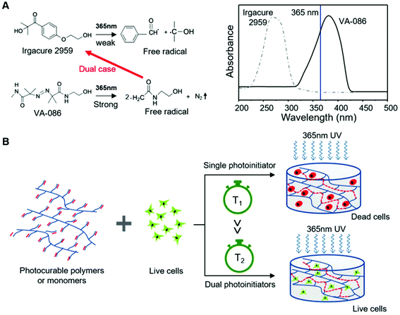
Despite the benefits of Irgacure 2959 such as high availability and ability to control the physical and mechanical properties of hydrogel fabrication, the free-radical species created during UV irradiation have detrimental effects on cell viability within the hydrogel. In addition, the absorbance of Irgacure 2959 at 365 nm is low (Fig. 1A). This low absorbance at 365 nm makes photoinitiated polymerization kinetics of Irgacure 2959 inefficient. Oftentimes, the intense and long exposure to UV light with wavelengths ranging from 360 nm to 400 nm is required during the photocrosslinking process with Irgacure 2959 and this process can potentially damage the cells. Moreover, its limited solubility in water at room temperature is one of the intrinsic shortcomings of Irgacure 2959.51 To circumvent the harmful effects of UV light exposure to the cells, the photocrosslinking of polymers using visible light initiating systems was developed. These visible light initiating systems include eosin Y, 1-vinyl-2-pyrrolidinone, and triethanolamine, excited with green light (530 nm)52,53 and camphorquinone, ethyl 4-N,N-dimethylaminobenzoate, triethanolamine, and the photosensitizer isopropyl thioxanthone, excited with blue light (470 nm).35 Although promising, the studies on these systems are still in a premature state and the studies on their compatibility with common photocrosslinkable hydrogels are still ongoing. On the other hand, many studies have shown the effectiveness and biocompatibility of using water-soluble azo photoinitiators such as 2,2′-azobis[2-methyl-N-(2-hydroxyethyl) propionamide] (VA-086) with UV light (365 nm).54,55 Occhetta et al. has proved the biocompatibility of VA-086 for the crosslinking of bone marrow stroma cell embedded methacrylate gelatin (GelMA) hydrogels. Besides biocompatibility, the mechanical stability of the hydrogel is an important factor in tissue engineering.56 Although VA-086 shows excellent biocompatibility, the formation of bubbles occurs within the hydrogel due to the generation of nitrogen gas as a by-product during crosslinking.43 This can greatly compromise the mechanical stability of the hydrogel. The pros and cons of various photoinitiators used in tissue engineering applications are listed in Table 1.
| Photoinitiator | Sources of irradiation and wavelength | Types of hydrogels | Pros | Cons | Ref. | |
|---|---|---|---|---|---|---|
| Name | Type | |||||
| Irgacure 2959 | Type I | UV (320–390 nm) | GelMA, MeHA, PEGDA | -Low toxicity up to 0.5 wt% Irgacure | -Low efficiency of initiation | 37–41 |
| -Good effectiveness and scalability of photocrosslinking | -Low extinction coefficient | |||||
| -Moderate water solubility | -Significant cell death due to longer UV exposure and higher UV dose | |||||
| -Good compatibility with common photocurable hydrogels | ||||||
| VA-086 | Type I | UV (365–385 nm) | GelMA, PEGDA | -Highly water soluble | -Bubble generation during reaction | 42–44 |
| -Biocompatible | ||||||
| -Good cell viability | ||||||
| Lithium phenyl-2,4,6-trimethylbenzoylphosphinate (LAP) | Type I | UV (320–390 nm) | PEGDA, GelMA | -Highly water soluble | -Reduced crosslinking efficiency due to slower curing rate when blue light is used | 45–47 |
| Blue light (405 nm) | -Can be used for both UV and visible light | |||||
| -Improved cell viability when blue light is used | -Low extinction coefficient | |||||
| Eosin Y | Type II | Visible light (400–800 nm) | PEGDA, GelMA | -Highly water soluble | -Require additional co-initiators and accelerators | 48–50 |
| -Good cell viability | ||||||
| -Broader range of activation wavelength | -Require further studies on compatibility with common photocurable hydrogels | |||||
In this research, our goal is to develop a hydrogel crosslinking system which possesses efficient crosslinking properties and desired mechanical properties with high cell viability. Polyethylene glycol diacrylate (PEGDA) is used as a representative photocurable hydrogel in this study due to its biocompatibility and tailorable mechanical properties. The efficient photocrosslinkable hydrogel system is achieved by photocrosslinking the PEGDA hydrogel with two commonly used photoinitiators, Irgacure 2959 and VA-086. As shown in Fig. 1A, different absorbance wavelengths are required to activate the two photoinitiators. The peak absorbance of Irgacure 2959 occurs at 265 nm. However, using this wavelength to activate Irgacure 2959 should damage the nucleic acid in the DNA of mammalian cells.57 The activation of Irgacure using 365 nm UV light is not very efficient. Contrarily, VA-086 can be effectively activated by UV light with wavelengths ranging from 300 nm to 420 nm. Thus, we hypothesize that free radicals produced by the activation of VA-086 will subsequently activate Irgacure 2959 to generate more free radicals which will improve the crosslinking efficiency of PEGDA as depicted in Fig. 1.
Through co-existence of those two photoinitiators, the overall photocrosslinking process should be completed with the minimal UV dosage during a shorter crosslinking time, enhancing the cell viability of encapsulated cells inside the hydrogels. To prove this, individual or dual photoinitiators with varying concentrations are first used for crosslinking PEGDA hydrogels with UV light of wavelength 365 nm and the optimal concentration of these photoinitiators is determined based on the mechanical performance of crosslinked hydrogels. Then, the cytocompatibility of those hydrogel systems is also assessed using mouse fibroblast cell line (L929) encapsulated in the PEGDA hydrogels. Finally, our dual-photoinitiator approach with Irgacure 2959 and VA-086 is implemented in a biofabrication process for the fabrication of a cell-laden large hydrogel construct with the vascular network. Taken together, we envisage that our dual-photoinitiator approach can extend the use of UV-curable hydrogel systems into cell-laden large constructs, with improved processing efficiency and cell viability.
2. Materials and methods
2.1. Materials
All chemicals were purchased from Sigma-Aldrich unless stated otherwise. The water-soluble photoinitiator 2,2-azobis[2-methyl-N-(2-hydroxyethyl) propionamide] (VA-086) was purchased from Wako Chemicals (Richmond, VA). PEGDA solution (Mn575) was crosslinked using two different photoinitiators, Irgacure 2959 and/or VA-86 to determine the crosslinking efficiency, rheological properties and cytocompatibility.2.2. Preparation of hydrogel solution with a single photoinitiator
PEGDA solution was diluted in DI water to obtain 5% (w/v) PEGDA solution. The solution was mixed using a vortex mixer. To prepare Irgacure 2959 (20% w/v) stock solution, 200 mg of Irgacure 2959 was dissolved in 1 ml of 70% (v/v) ethanol. The solution was mixed using a vortex mixer for 15 min until Irgacure 2959 was fully dissolved. For VA-086, the powder was added directly into PEGDA solution as VA-086 possesses higher solubility in water.Four different hydrogels with varying amounts of Irgacure 2959 solution were prepared: Irgacure solution (0.25%, 0.5%, 1% and 2% w/w) was mixed with 5% (w/v) PEGDA solution. Various amounts of VA-086 were added to 5% (w/v) PEGDA solution to acquire the VA-086 concentration of 0.05%, 0.1%, 0.3% and 0.5% (w/v).
2.3. Preparation of hydrogel solution with dual photoinitiators
5% (w/v) PEGDA solution with 0.5% (w/w) Irgacure 2959 was prepared as described previously. VA-086 powder (0.05% w/v) was mixed forming homogeneous suspension.2.4. Preparation of hydrogel solution with L929 fibroblasts
An L929 mouse fibroblast cell line (Bio-REV Pte Ltd, Singapore) was cultured at 37 °C and 5% CO2. The composition of culture medium consists of Dulbecco's modified Eagle's medium (DMEM) with 10% (v/v) fetal bovine serum and 1% (v/v) penicillin–streptomycin. All the cell culture reagents were purchased from Life Technologies (Grand Island, NY, USA). For the hydrogel with cells, L929 fibroblasts were expanded through passage 12 and encapsulated at 1![[thin space (1/6-em)]](https://www.rsc.org/images/entities/char_2009.gif) 000
000![[thin space (1/6-em)]](https://www.rsc.org/images/entities/char_2009.gif) 000 cells per ml in the hydrogel solution.
000 cells per ml in the hydrogel solution.
2.5. Photocrosslinking for hydrogel solution
To crosslink the hydrogel, 7 ml of each PEGDA-photoinitiator solution was pipetted into a Petri dish 35 mm in diameter. The hydrogel solution with Irgacure 2959 was crosslinked for 5 min, whereas the hydrogel solutions with VA-086 and a dual photoinitiator were crosslinked for 2 min using a UV lamp with a wavelength of 365 nm and an irradiation intensity of ∼19 mW cm−2 (Analytik Jena, Upland, CA, USA). The quantitative exposure of UV light with a 365 nm wavelength for 5 min and 2 min exposure are ∼95 mJ cm−2 and ∼38 mJ cm−2, respectively.2.6. Rheology
Rheological measurements of crosslinked hydrogels with various concentrations of photoinitiators (n = 3 per group) were carried out using a TA2000 rheometer (TA Instruments, Waters LLC New Castle, DE) with a 25 mm parallel-plate geometry at a gap of 7 mm at 25 °C. For a frequency sweep test, the crosslinked hydrogels were subjected to angular frequency scanning from 0.1 to 100 rad s−1 at 10% strain. Each hydrogel sample was used for only one test.2.7. Preparation of tissue constructs with a channel mimicking vasculature
To demonstrate a tissue construct with a channel mimicking a vessel, a top-down approach was adapted in our system. A negative mold was pre-printed using a commercially available fused deposition modelling printer (Dremel Digilab 3D20). In this study, a simple filament with a diameter of 500 μm was 3d printed and placed within the hydrogel to serve as a negative mold. The mold was placed in a premade container for perfusion. PEGDA hydrogel solutions with cells with different photoinitiators were placed in a mold. The hydrogel solution with a single photoinitiator was crosslinked for 5 min and the hydrogel solution with a dual photoinitiator was crosslinked for 2 min using a UV lamp with a wavelength of 365 nm (Analytik Jena, Upland, CA, USA). After curing, the negative mold was removed and the hollow channel was remained within the PEGDA hydrogel.2.8. Evaluation of cytocompatibility
Photocrosslinked cell laden PEGDA hydrogels were rinsed with 1× PBS for 3 times. Cell viability was assessed with the CCK-8 cell proliferation assay (Dojindo Molecular Technologies, Inc., USA). The photocrosslinked PEGDA hydrogel was immersed in fresh culture medium containing 10% of CCK-8 assay solution. The samples were then incubated for 6 hours at 37 °C and 5% CO2. 50 μl of supernatant was aliquoted in a 96 well plate for absorbance measurement (n = 5 per group). The absorbance values were obtained at 450 nm with a SpectraMax® M5 Multi-Mode Microplate Reader (California, United States).2.9. Optical and fluorescence microscopy
Cell viability of all the samples (n = 5 per group) was assessed with a LIVE/DEAD® Viability/Cytotoxicity assay kit from Molecular Probes (Life Technologies Ltd, Zug, Switzerland) after photocrosslinking. Samples were rinsed in 1× PBS for 3 times. Fluorescent marker Calcein AM and ethidium homodimer (EthD-1) were used for staining living cells and dead cells, respectively. A 2 mm thick slice was cut out and placed in a solution of 2 μM calcein AM- and 4 μM EthD-1 for 25 min at 37 °C. Living cells appeared in green using an argon laser (488 nm) and dead cells appeared in blue under a HeNe laser (543 nm). Imaging was done with a Zeiss LSM 800 confocal laser scanning microscope (Oberkochen, Germany). Bright field and fluorescence microscopic images were recorded using a CCD color digital camera (QImaging Retiga 4000R) connected to a system microscope (Olympus).2.10. Statistical analysis
All experimental results were demonstrated in the mean ± standard deviation (SD) for n ≥ 3. Samples between the groups at the same time point or within a single group at three different time points were compared using one-way ANOVA followed by Tukey's post-hoc analysis using the OriginPro 8 software. p < 0.05 was considered to be statistically significant.3. Results and discussion
3.1. Photocrosslinking of hydrogels with Irgacure 2959
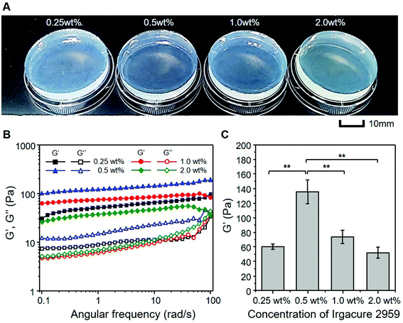
First, we assessed the photocrosslinking efficiency of Irgacure 2959 in terms of mechanical performance and biocompatibility of photocrosslinked PEGDA hydrogels. The concentration of Irgacure 2959 varied from 0.25 wt% to 2 wt% in PEGDA solution, while the photocrosslinking reaction for all PEGDA solutions was performed at 365 nm for 5 min (∼95 mJ cm−2). After photocrosslinking, all PEGDA hydrogels maintained their gel structures without support, thus they were sufficiently handleable, as shown in Fig. 2A. The result of frequency sweep test shown in Fig. 2B gave important information about the effect of Irgacure 2959 concentration on the viscoelastic behavior of PEG hydrogel. All PEGDA hydrogels cured with various concentrations of Irgacure 2959 behaved as gels as storage moduli, G′ were higher than loss moduli, G′′ over an entire range of angular frequency (tan![[thin space (1/6-em)]](https://www.rsc.org/images/entities/char_2009.gif) δ > 1). More importantly, G′ of PEGDA hydrogels was significantly influenced by the concentration of Irgacure 2959 (Fig. 2C). PEGDA hydrogels with 0.5 wt% Irgacure 2959 showed the highest G′, while G′ of PEGDA cured with 1% and 2% Irgacure 2959 was significantly lower than that with 0.5 wt% Irgacure 2959. The solubility of Irgacure 2959 in water is limited to not more than 2 wt% under ambient conditions. Even dissolving 0.5 wt% Irgacure in water required mechanical agitation and/or heating the solutions.51 To increase the solubility of Irgacure 2959, an organic solvent such as ethanol was used and it was reported that the solubility of Irgacure 2959 increased by tenfold in ethanol according to the data information from the manufacturer.58 Thus, all Irgacure 2959 solutions were prepared by dissolving Irgacure 2959 in 70% ethanol. Depending on the weight concentration of Irgacure 2959 in PEGDA solution, the designated volume of Irgacure 2959 solution was mixed with PEGDA solution. As a result, a higher concentration of Irgacure 2959 necessarily introduces larger ethanol impurity to the PEGDA solution with Irgacure 2959 and the existence of ethanol significantly reduces the crosslinking efficiency of Irgacure 2959. Therefore, increasing the concentration of Irgacure 2959 caused a significant decline in crosslinking efficiency due to the existence of higher volume of impurity. In addition, 0.5 wt% Irgacure 2959 has been commonly used for the preparation of photocrosslinked hydrogels.59
δ > 1). More importantly, G′ of PEGDA hydrogels was significantly influenced by the concentration of Irgacure 2959 (Fig. 2C). PEGDA hydrogels with 0.5 wt% Irgacure 2959 showed the highest G′, while G′ of PEGDA cured with 1% and 2% Irgacure 2959 was significantly lower than that with 0.5 wt% Irgacure 2959. The solubility of Irgacure 2959 in water is limited to not more than 2 wt% under ambient conditions. Even dissolving 0.5 wt% Irgacure in water required mechanical agitation and/or heating the solutions.51 To increase the solubility of Irgacure 2959, an organic solvent such as ethanol was used and it was reported that the solubility of Irgacure 2959 increased by tenfold in ethanol according to the data information from the manufacturer.58 Thus, all Irgacure 2959 solutions were prepared by dissolving Irgacure 2959 in 70% ethanol. Depending on the weight concentration of Irgacure 2959 in PEGDA solution, the designated volume of Irgacure 2959 solution was mixed with PEGDA solution. As a result, a higher concentration of Irgacure 2959 necessarily introduces larger ethanol impurity to the PEGDA solution with Irgacure 2959 and the existence of ethanol significantly reduces the crosslinking efficiency of Irgacure 2959. Therefore, increasing the concentration of Irgacure 2959 caused a significant decline in crosslinking efficiency due to the existence of higher volume of impurity. In addition, 0.5 wt% Irgacure 2959 has been commonly used for the preparation of photocrosslinked hydrogels.59
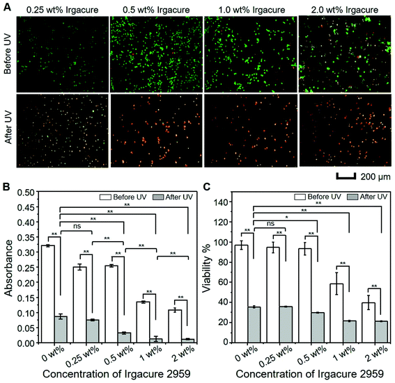
Moreover, cytocompatibility of both PEGDA solutions and hydrogels with varying concentrations of Irgacure 2959 was evaluated before and after photocrosslinking (Fig. 3). Fluorescence images obtained from live/dead staining indicate two important aspects of cytocompatibility before and after the crosslinking of PEGDA hydrogel, as shown in Fig. 3A. Firstly, it was observed that the number of dead cells (red stain) increased with increasing an amount of Irgacure 2959 before UV irradiation. This indicates that the cell viability within PEGDA solution decreases with increasing the concentration of Irgacure 2959 before UV irradiation. Secondly, most of the cell deaths occurred regardless of Irgacure 2959 concentration after 5 min UV irradiation to PEGDA solution. To assess quantitatively, CCK-8 assay and manual cell counting of live/dead cells were performed and compared with 2D cell culture in PEGDA solution (denoted as 0 wt% Irgacure 2959), as shown in Fig. 3C and D. From CCK-8 assay, the absorbance/optical density of cells exposed to 0.25 wt% and 0.5 wt% Irgacure 2959 was comparable to that of 0 wt% Irgacure 2959. However, the absorbance value decreased significantly when the cells were cultured with 1 wt% and 2 wt% of Irgacure 2959 (Fig. 3C). The cell viability measured from the direct counting of live/dead cells shows a similar tendency to that of the absorbance/optical density of cells by CCK-assay (Fig. 3D). These results indicated that higher Irgacure 2959 concentrations (>0.5 wt%) severely compromise the cell viability.
Based on the rheological analysis and cell viability of photocrosslinked PEDGA hydrogels, 0.5 wt% of Irgacure 2959 should be the most optimal conditions for photocrosslinking. Alternatively, decreasing UV irradiation time could be an option to increase cell viability. However, UV exposure significantly influences the mechanical stability of PEGDA hydrogels, particularly in the case of large-scale hydrogel constructs. In this study, we chose a cylindrical PEGDA hydrogel with a height of 1 cm and a diameter of 3 cm. To photocrosslink this volume of PEGDA hydrogels requires at least a UV irradiation time of 5 min for optimal mechanical stability (data not shown). Nevertheless, 5 min UV irradiation (∼95 mJ cm−2) significantly damages the cells encapsulated inside the PEGDA hydrogels. Thus, cell viability in our experiments appeared significantly lower compared to previously published studies by other groups.35,36,60 Long duration of the UV crosslinking process with Irgacure 2959 was not appropriate without any cell protection strategy for the fabrication of cell-laden large constructs.54,61
3.2. Photocrosslinking of hydrogels with VA-086
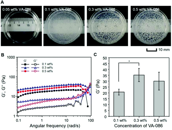
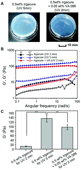
Besides Irgacure 2959, VA-086 has been known as one of the potential photoinitiators for photocrosslinkable hydrogels due to its excellent biocompatibility, high solubility in water and photocrosslinking efficiency for 365 nm UV irradiation. Despite those outstanding advantages, VA-086 has not been actively used for biofabrication due to its by-product, nitrogen gas, during the photocrosslinking reaction as indicated in Fig. 1A.43 We also confirmed the production of nitrogen gas which was trapped as gas bubbles inside the PEGDA hydrogels during UV crosslinking with VA-086 (Fig. 4A). The PEGDA solutions with VA-086 were crosslinked for 2 min. By increasing the amount of VA-086 in PEGDA solution, the size and total volume of entrapped bubbles seemed to be increased. The study done by Occhetta et al. also showed that UV crosslinking with VA-086 generated nitrogen gas during crosslinking and the gas created bubbles which made hydrogels cloudy.56The result of rheological analysis in Fig. 4B clearly substantiated that the storage moduli of G′ were higher than the loss moduli G′′ for all the samples (tan![[thin space (1/6-em)]](https://www.rsc.org/images/entities/char_2009.gif) δ > 1), indicating that gelation occurred in PEGDA with various concentrations (0.05–0.5 wt%) of VA-086 with a relatively shorter UV irradiation time of 2 min (∼38 mJ cm−2). However, the mechanical performance of PEGDA hydrogels crosslinked with VA-086 was considerably decreased due to the entrapped bubbles inside the hydrogels. PEGDA hydrogels crosslinked with Irgacure 2959 shows a storage modulus of ∼100 Pa, which is four times higher than that of PEGDA with VA-086 (∼25 Pa). Also, the storage modulus of PEGDA did not show any clear dependence on the amount of VA-086 due to the influence of those intrinsic defects inside PEGDA with VA-086 (Fig. 4C). We also found that increased UV irradiation time further decreased the rheological properties of PEGDA hydrogels along with the increased VA-086 concentrations since the nitrogen gas generation induced more bubble formation inside the hydrogel as reported by Billiet and his colleagues.61 Thus, considering the trade-off between the crosslinking density and trapped bubble amount of PEGDA hydrogels, 2 min UV irradiation (∼38 mJ cm−2) with 0.3–0.5 wt% VA-086 should be the optimal conditions for ensuring the mechanical stability of PEGDA hydrogels.
δ > 1), indicating that gelation occurred in PEGDA with various concentrations (0.05–0.5 wt%) of VA-086 with a relatively shorter UV irradiation time of 2 min (∼38 mJ cm−2). However, the mechanical performance of PEGDA hydrogels crosslinked with VA-086 was considerably decreased due to the entrapped bubbles inside the hydrogels. PEGDA hydrogels crosslinked with Irgacure 2959 shows a storage modulus of ∼100 Pa, which is four times higher than that of PEGDA with VA-086 (∼25 Pa). Also, the storage modulus of PEGDA did not show any clear dependence on the amount of VA-086 due to the influence of those intrinsic defects inside PEGDA with VA-086 (Fig. 4C). We also found that increased UV irradiation time further decreased the rheological properties of PEGDA hydrogels along with the increased VA-086 concentrations since the nitrogen gas generation induced more bubble formation inside the hydrogel as reported by Billiet and his colleagues.61 Thus, considering the trade-off between the crosslinking density and trapped bubble amount of PEGDA hydrogels, 2 min UV irradiation (∼38 mJ cm−2) with 0.3–0.5 wt% VA-086 should be the optimal conditions for ensuring the mechanical stability of PEGDA hydrogels.
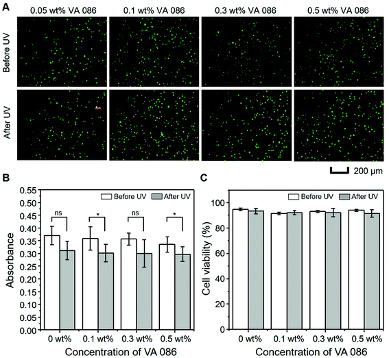
Qualitative and quantitative cytocompatibility tests of VA-086 with L929 cells were carried out as shown in Fig. 5. Cell viability of photocrosslinked hydrogels can be affected by both cytotoxicity of the photoinitiator or photocrosslinkable molecules and the total UV irradiation time. As compared to Irgacure 2959, VA-086 is known to be more biocompatible and does not require a longer UV irradiation time. Thus, cells were mostly viable regardless of VA-086 concentrations as shown in the fluorescence images of L929 cells of Fig. 5A. Similar to those cell viability tests with Irgacure 2959, both CCK-8 assay and direct counting of live/dead cells were performed and compared with the 2D cell culture in PEGDA solution (denoted as 0 wt% VA-086), as shown in Fig. 5B and C. Since the UV irradiation time of 2 min (∼38 mJ cm−2) was sufficiently short, the cell viability of 2D cell culture did not change after UV irradiation. In fact, cells for all hydrogel cases were highly viable, showing that their viability was approximately 95%. Despite excellent biocompatibility of VA-086, mechanical performance is still a critical issue as a photoinitiator of hydrogel scaffolds.
3.3. Photocrosslinking of hydrogels with Irgacure 2959 and VA-086
Irgacure 2959 and VA-086 clearly exhibit limitations in photocrosslinking of hydrogels due to either cell viability or mechanical stability. Since mechanical stability of hydrogel systems needs to be achieved without compromising cell viability, we chose Irgacure 2959 as a major photoinitiator and introduced a low concentration of VA-086 to the system minimizing nitrogen gas generation, yet producing sufficient free radicals in order to induce free radical generation of Irgacure 2959 via free radical chain reactions. Through this mechanism, dual photoinitiators of Irgacure 2959 and VA-086 can reduce the UV irradiation dosage over photocrosslinkable hydrogels and maintain cell viability.We confirmed our hypothesis by comparing three photocrosslinkable hydrogel systems: (1) PEGDA solution with 0.5 wt% Irgacure for a UV dosage of ∼38 mJ cm−2 (irradiation time of 2 min), (2) PEGDA solution with 0.5 wt% Irgacure for a UV dosage of ∼95 mJ cm−2 (irradiation time of 5 min) and (3) PEGDA solution with 0.5 wt% Irgacure + 0.05 wt% VA-086 for a UV dosage of ∼38 mJ cm−2 (irradiation time of 2 min). In the case of 0.5 wt% Irgacure for a UV dosage of ∼38 mJ cm−2, the PEGDA solution was not photocrosslinked properly, thus its hydrogel form was not maintained. However, for the other two cases, the crosslinked PEGDA hydrogels were in a good shape with sufficient mechanical stability, as shown in Fig. 6A. Although only 0.05 wt% VA-086 was contained in the PEGDA solution with 0.5 wt% Irgacure, a small amount of bubbles inside the hydrogel was still observed.
We further demonstrated the mechanical stability of PEGDA hydrogels using rheological tests in Fig. 6B and C. As predicted, the PEGDA hydrogel photocrosslinked with 0.5 wt% Irgacure for a UV dosage of ∼38 mJ cm−2 exhibits fluid-like behavior at high frequency (>80 rad s−1), implying that its mechanical stability was not sufficient for cell-laden scaffolds.
The other two hydrogel systems have almost similar rheological behavior even though the PEGDA solution with 0.5 wt% Irgacure for a UV dosage of ∼95 mJ cm−2 shows a significantly higher storage modulus than that with dual photoinitiators (Fig. 6C). The entrapped bubbles may reduce the mechanical properties of PEGDA hydrogels crosslinked with dual photoinitiators. From this mechanical assessment, we confirmed that VA-086 could effectively improve the photocrosslinking efficiency of Irgacure 2959 with minimal UV irradiation dosage.
The hypothesis about the sequential activation of Irgacure 2959 from activated VA-086 could also be proven by monitoring the weight fraction of PEGDA hydrogel during photocrosslinking as a function of UV irradiation time (Fig. S1†). PEGDA with either 0.5 wt% Irgacure 2959 or 0.05 wt% VA-086 was not crosslinked under 365 nm UV irradiation for 2 min (weight fraction of gel in PEGDA solution <5%). However, with the co-existence of 0.5 wt% Irgacure 2959 and 0.05 wt% VA-086, a rapid crosslinking reaction occurred during 2 min UV irradiation, almost completing 90% of crosslinking. Moreover, 405 nm visible light was used for the photocrosslinking of PEGDA where Irgacure cannot be activated (Fig. S2 and S3†). Thus, in the case of dual photoinitiators of Irgacure 2959 and VA-086, during irradiation with 405 nm blue light, the free radicals should be generated only from Irgacure due to free radical chain reactions initiated by activated VA-086. Compared to PEGDA with 0.05 wt% VA-086, PEGDA with dual photoinitiators was crosslinked faster compared to that with 0.05 wt% VA-086, implying the significant contribution of activated Irgacure 2959 for the crosslinking process.
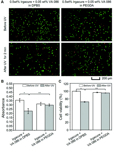
Since we already confirmed that the PEGDA hydrogel with 0.5 wt% Irgacure for a UV dosage of ∼95 mJ cm−2 showed low cell viability (∼40%) after crosslinking as shown in Fig. 3B, the cytocompatibility test of PEGDA hydrogels with a dual photoinitiator approach was further confirmed as shown in Fig. 7. As the control group, a 2D culture system in DPBS with dual photoinitiators was chosen to compare with other PEGDA photocrosslinking systems. Interestingly, the cells in DPBS were less viable than those in PEGDA after UV irradiation. In Fig. 7C, the cell viability in DPBS was decreased to 72% after UV irradiation, whereas for the hydrogel sample with a dual photoinitiator approach, almost 96% of the cell viability was observed. The activated photoinitiators produce free radicals in the solution under UV irradiation. Without photocrosslinkable molecules, these free radicals should directly interact with the cells, inducing cell death, whereas PEGDA molecules around the cells consume free radicals, blocking the direct contact of free radicals with the cells.62 Therefore, we concluded that the dual photoinitiators for photocrosslinkable hydrogels significantly improved cell viability, allowing photocrosslinkable molecules to be sufficiently crosslinked for good mechanical stability.
3.4. Fabrication of tissue constructs with channel mimicking vasculature
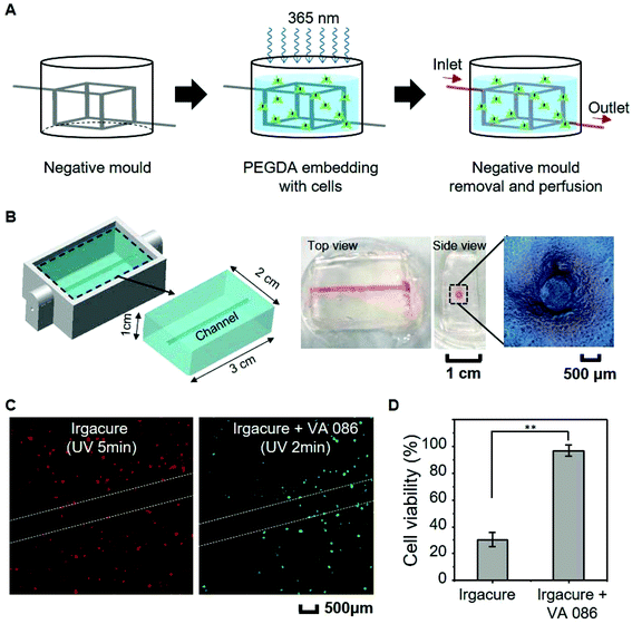
One of the most feasible applications for photocrosslinkable hydrogels should be the 3D supporting matrix of cells with or without vascular networks to mimic the microenvironment of native tissues. Low cell viability of hydrogels after photocrosslinking hinders the direct use of photocrosslinkable hydrogels. To overcome this issue, hydrogels such as fibrin or gelatin gels are usually enzymatically crosslinked for cell-laden large tissue constructs using molding procedures.27,30,62–64 Although this approach has been widely used due to high cell viability, enzymatic gelation is time consuming due to slower reaction kinetics compared to the photocrosslinking process.63,64 Therefore, the fabrication of hydrogels through photocrosslinking without compromising cellular activity is possible only by shortening the photocrosslinking time to reduce unnecessary UV irradiation to the cells or to use more cell-friendly visible light irradiation.62,65 In this context, we applied our approach to fabricate 3D tissue constructs with a simple vascular network using PEGDA hydrogels, as shown in Fig. 8. A typical approach to the fabrication of vascularized 3D scaffolds is illustrated in Fig. 8A. In brief, a negative mold was obtained by using a commercially available fused deposition modeling (FDM) 3D printer. Thereafter, the pre-fabricated mold was embedded within the hydrogel solution containing cells and two photoinitiators (0.5 wt% Irgacure + 0.05 wt% VA-086). The hydrogel solution was subjected to photocrosslinking under 365 nm UV irradiation at ∼38 mJ cm−2. The negative mold was removed after photocrosslinking the hydrogel. Using this approach, we constructed a thick vascularized artificial tissue with one simple channel using PEGDA hydrogels crosslinked by either Irgacure 2959 or dual photoinitiators (Fig. 8B). The crosslinked hydrogels with a volume of 2 cm × 3 cm × 1 cm had good mechanical stability, maintaining their shapes after removing the negative mold. The hollow structure has a diameter of about 500 μm. Cell viability of encapsulated cells within hydrogels was also assessed after UV irradiation (Fig. 8C and D). As expected, with dual photoinitiators, most cells remained viable (∼90%), whereas the cells in PEGDA hydrogels with Irgacure 2959 were almost dead, showing a low cell viability of less than 30%. The cell-laden PEGDA hydrogel constructs can be further cultured with cell culture medium or introduced to a perfusion system for long-term cell culturing.The photocurable hydrogels with dual photoinitiators could be fabricated within 2 min when the UV light source with 19.8 mW cm−2 was used, without further post-curing process. By contrast, a fibrinogen-incorporated gelatin matrix with cells required about 1 hour curing to induce the full gelation of the fibrinogen with the addition of thrombin and calcium ions.66 Moreover, this approach can be applied to other photocrosslinkable polymers such as GelMA which possessed an additional chemical crosslinking step.67 Thus, our dual photoinitiator approach enables the biofabrication process with photocurable hydrogels to be more effective and simpler without compromising the cell viability.
With a similar cell viability (>95%), the crosslinking efficiency of 365 nm UV irradiation was higher than that of 405 nm blue light irradiation, when dual photoinitiators were used for photocrosslinking (Fig. S3†). However, optimal conditions of our dual photoinitiator approach for visible light curing can be further investigated by tuning intensity and aperture of a visible light source or increasing VA-086 concentrations with ensuring minimal gas generation. More importantly, in this study, we used 365 nm UV light which still is a commonly used light source for crosslinking of photocurable hydrogels due to its excellent effectiveness and scalability of the photoinitiating system with well-established standardized equipment and a widely used photoinitiator, Irgacure 2959.37–40 Thus, we believe that our approach provides great benefits for researchers or industrial players who use the conventional UV crosslinking method with Irgacure 2959. In parallel, through dual photoinitiators which have different activation kinetics, adapting to the use of visible light for the photo-crosslinking of hydrogels should be accelerated, saving costs and time for developing and testing new photoinitiators.
4. Conclusions
We successfully developed a hydrogel crosslinking system which exhibited efficient crosslinking properties and desired mechanical properties with high cell viability, through a dual-photoinitiator approach. By using Irgacure 2959 and VA-086 concurrently, the UV dosage was minimized due to a shorter crosslinking time. PEGDA hydrogel fabricated using this approach demonstrated adequate mechanical stability without compromising the viability of L929 cells encapsulated within hydrogels compared to that using the conventional approach which only uses one type of photoinitiator, either Irgacure 2959 or VA-086. Moreover, we fabricated a large PEGDA hydrogel construct with a single microchannel as a proof of concept for hydrogels with vasculature to demonstrate the versatility of the system. Our dual-photoinitiator approach allows the production of this photocrosslinkable hydrogel system with microchannels, significantly improving cell viability and processing efficiency, yet maintaining good mechanical stability. Taken together, we envision the concurrent use of Irgacure 2959 and VA-086 as photoinitiators opening potential avenues for the utilization of various photocrosslinkable hydrogel systems in perfusable large artificial tissue for in vivo and ex vivo applications.Conflicts of interest
There are no conflicts to declare.Acknowledgements
This research was supported by the AcRF Tier 1 grant 2017-T1-001-246 (RG51/17) from the Ministry of Education of Singapore, and the A*STAR AME IRG grant A1983c0031 from A*STAR.References
- L. Moroni, J. A. Burdick, C. Highley, S. J. Lee, Y. Morimoto, S. Takeuchi and J. J. Yoo, Nat. Rev. Mater., 2018, 3, 21–37 CrossRef CAS PubMed.
- L. P. Silva, in 3D and 4D Printing in Biomedical Applications, ed. M. Maniruzzaman, pp. 373–421, 2019, DOI:10.1002/9783527813704.ch15.
- Z. Wu, X. Su, Y. Xu, B. Kong, W. Sun and S. Mi, Sci. Rep., 2016, 6, 24474 CrossRef CAS PubMed.
- J. Vanderburgh, J. A. Sterling and S. A. Guelcher, Ann. Biomed. Eng., 2017, 45, 164–179 CrossRef PubMed.
- C. J. Bishop, J. Kim and J. J. Green, Ann. Biomed. Eng., 2014, 42, 1557–1572 CrossRef PubMed.
- Y.-J. Choi, H.-G. Yi, S.-W. Kim and D.-W. Cho, Theranostics, 2017, 7, 3118–3137 CrossRef CAS PubMed.
- C. Khatiwala, R. Law, B. Shepherd, S. Dorfman and M. Csete, Gene Ther. Regul., 2012, 07, 1230004 CrossRef.
- V. K. Lee and G. Dai, Ann. Biomed. Eng., 2017, 45, 115–131 CrossRef PubMed.
- M. M. Stanton, C. Trichet-Paredes and S. Sanchez, Lab Chip, 2015, 15, 1634–1637 RSC.
- D. L. Cohen, E. Malone, H. Lipson and L. J. Bonassar, Tissue Eng., 2006, 12, 1325–1335 CrossRef CAS PubMed.
- L. Shor, S. Guceri, R. Chang, J. Gordon, Q. Kang, L. Hartsock, Y. An and W. Sun, Biofabrication, 2009, 1, 015003 CrossRef PubMed.
- K. Iwami, T. Noda, K. Ishida, K. Morishima, M. Nakamura and N. Umeda, Biofabrication, 2010, 2, 014108 CrossRef CAS PubMed.
- K. Holzl, S. Lin, L. Tytgat, S. Van Vlierberghe, L. Gu and A. Ovsianikov, Biofabrication, 2016, 8, 032002 CrossRef PubMed.
- R. J. Klebe, Exp. Cell Res., 1988, 179, 362–373 CrossRef CAS PubMed.
- T. Xu, J. Jin, C. Gregory, J. J. Hickman and T. Boland, Biomaterials, 2005, 26, 93–99 CrossRef CAS PubMed.
- X. Cui, T. Boland, D. D. D'Lima and M. K. Lotz, Recent Pat. Drug Delivery Formulation, 2012, 6, 149–155 CrossRef CAS PubMed.
- T. Xu, W. Zhao, J. M. Zhu, M. Z. Albanna, J. J. Yoo and A. Atala, Biomaterials, 2013, 34, 130–139 CrossRef CAS PubMed.
- J. A. Barron, P. Wu, H. D. Ladouceur and B. R. Ringeisen, Biomed. Microdevices, 2004, 6, 139–147 CrossRef CAS PubMed.
- F. Guillemot, A. Souquet, S. Catros, B. Guillotin, J. Lopez, M. Faucon, B. Pippenger, R. Bareille, M. Rémy, S. Bellance, P. Chabassier, J. C. Fricain and J. Amédée, Acta Biomater., 2010, 6, 2494–2500 CrossRef CAS PubMed.
- B. Guillotin, A. Souquet, S. Catros, M. Duocastella, B. Pippenger, S. Bellance, R. Bareille, M. Remy, L. Bordenave, J. Amedee and F. Guillemot, Biomaterials, 2010, 31, 7250–7256 CrossRef CAS PubMed.
- T. Billiet, M. Vandenhaute, J. Schelfhout, S. Van Vlierberghe and P. Dubruel, Biomaterials, 2012, 33, 6020–6041 CrossRef CAS PubMed.
- M. A. Daniele, A. A. Adams, J. Naciri, S. H. North and F. S. Ligler, Biomaterials, 2014, 35, 1845–1856 CrossRef CAS PubMed.
- E. M. Ahmed, J. Adv. Res., 2015, 6, 105–121 CrossRef CAS PubMed.
- L. Pan, G. Yu, D. Zhai, H. R. Lee, W. Zhao, N. Liu, H. Wang, B. C. K. Tee, Y. Shi, Y. Cui and Z. Bao, Proc. Natl. Acad. Sci. U. S. A., 2012, 109, 9287 CrossRef CAS PubMed.
- I. M. El-Sherbiny and M. H. Yacoub, Global Cardiol. Sci. Pract., 2013, 2013, 316–342 Search PubMed.
- D. Zhai, B. Liu, Y. Shi, L. Pan, Y. Wang, W. Li, R. Zhang and G. Yu, ACS Nano, 2013, 7, 3540–3546 CrossRef CAS PubMed.
- N. C. Hunt and L. M. Grover, Biotechnol. Lett., 2010, 32, 733–742 CrossRef CAS PubMed.
- Z. Li and M. Kawashita, J. Artif. Organs, 2011, 14, 163–170 CrossRef CAS PubMed.
- K. L. Spiller, S. A. Maher and A. M. Lowman, Tissue Eng., Part B, 2011, 17, 281–299 CrossRef CAS PubMed.
- J. Lee, M. J. Cuddihy and N. A. Kotov, Tissue Eng., Part B, 2008, 14, 61–86 CrossRef CAS PubMed.
- C. Chang, B. Duan, J. Cai and L. Zhang, Eur. Polym. J., 2010, 46, 92–100 CrossRef CAS.
- K. T. Nguyen and J. L. West, Biomaterials, 2002, 23, 4307–4314 CrossRef CAS PubMed.
- M. D. Purbrick, Polym. Int., 1996, 40, 315–315 CrossRef CAS.
- G. Wollensak, E. Spoerl and T. Seiler, Am. J. Ophthalmol., 2003, 135, 620–627 CrossRef CAS PubMed.
- S. J. Bryant, C. R. Nuttelman and K. S. Anseth, J. Biomater. Sci., Polym. Ed., 2000, 11, 439–457 CrossRef CAS PubMed.
- C. G. Williams, A. N. Malik, T. K. Kim, P. N. Manson and J. H. Elisseeff, Biomaterials, 2005, 26, 1211–1218 CrossRef CAS PubMed.
- G. Marchioli, L. Zellner, C. Oliveira, M. Engelse, E. D. Koning, J. Mano, Karperien, A. V. Apeldoorn and L. Moroni, J. Mater. Sci. Mater. Med., 2017, 28, 195–195 CrossRef PubMed.
- S. Liu, D. C. Yeo, C. Wiraja, H. L. Tey, M. Mrksich and C. Xu, Bioeng. Transl. Med., 2017, 2, 258–267 CrossRef CAS PubMed.
- A. Assmann, A. Vegh, M. Ghasemi-Rad, S. Bagherifard, G. Cheng, E. S. Sani, G. U. Ruiz-Esparza, I. Noshadi, A. D. Lassaletta, S. Gangadharan, A. Tamayol, A. Khademhosseini and N. Annabi, Biomaterials, 2017, 140, 115–127 CrossRef CAS PubMed.
- Y. S. Zhang, F. Davoudi, P. Walch, A. Manbachi, X. Luo, V. Dell'Erba, A. K. Miri, H. Albadawi, A. Arneri, X. Li, X. Wang, M. R. Dokmeci, A. Khademhosseini and R. Oklu, Lab Chip, 2016, 16, 4097–4105 RSC.
- S. Lin, W. Y. W. Lee, Q. Feng, L. Xu, B. Wang, G. C. W. Man, Y. Chen, X. Jiang, L. Bian, L. Cui, B. Wei and G. Li, Stem Cell Res. Ther., 2017, 8, 221 CrossRef PubMed.
- P. Occhetta, N. Sadr, F. Piraino, A. Redaelli, M. Moretti and M. Rasponi, Biofabrication, 2013, 5, 035002 CrossRef CAS PubMed.
- Z. Wang, X. Jin, R. Dai, J. F. Holzman and K. Kim, RSC Adv., 2016, 6, 21099–21104 RSC.
- M. Tromayer, A. Dobos, P. Gruber, A. Ajami, R. Dedic, A. Ovsianikov and R. Liska, Polym. Chem., 2018, 9, 3108–3117 RSC.
- M. Gou, X. Qu, W. Zhu, M. Xiang, J. Yang, K. Zhang, Y. Wei and S. Chen, Nat. Commun., 2014, 5, 3774 CrossRef CAS PubMed.
- A. Wenz, K. Borchers, G. E. M. Tovar and P. J. Kluger, Biofabrication, 2017, 9, 044103 CrossRef PubMed.
- G. A. Calderon, P. Thai, C. W. Hsu, B. Grigoryan, S. M. Gibson, M. E. Dickinson and J. S. Miller, Biomater. Sci., 2017, 5, 1652–1660 RSC.
- A. L. Y. Nachlas, S. Li, R. Jha, M. Singh, C. Xu and M. E. Davis, Acta Biomater., 2018, 71, 235–246 CrossRef CAS PubMed.
- P. Erkoc, F. Seker, T. Bagci-Onder and S. Kizilel, Macromol. Biosci., 2018, 18, 1700369 CrossRef PubMed.
- N. Annabi, D. Rana, E. Shirzaei Sani, R. Portillo-Lara, J. L. Gifford, M. M. Fares, S. M. Mithieux and A. S. Weiss, Biomaterials, 2017, 139, 229–243 CrossRef CAS PubMed.
- J.-P. Fouassier, D. Burr and F. Wieder, J. Polym. Sci., Part A: Polym. Chem., 1991, 29, 1319–1327 CrossRef CAS.
- S. L. Fenn and R. A. Oldinski, J. Biomed. Mater. Res., Part B, 2016, 104, 1229–1236 CrossRef CAS PubMed.
- D. Petta, A. R. Armiento, D. Grijpma, M. Alini, D. Eglin and M. D'Este, Biofabrication, 2018, 10, 044104 CrossRef CAS PubMed.
- A. D. Rouillard, C. M. Berglund, J. Y. Lee, W. J. Polacheck, Y. Tsui, L. J. Bonassar and B. J. Kirby, Tissue Eng., Part C, 2011, 17, 173–179 CrossRef CAS PubMed.
- S. Wang, O. Jeon, P. G. Shankles, Y. Liu, E. Alsberg, S. T. Retterer, B. P. Lee and C. K. Choi, Biomicrofluidics, 2016, 10, 011101–011101 CrossRef PubMed.
- P. Occhetta, R. Visone, L. Russo, L. Cipolla, M. Moretti and M. Rasponi, J. Biomed. Mater. Res., Part A, 2015, 103, 2109–2117 CrossRef CAS PubMed.
- S. E. Beck, R. A. Rodriguez, K. G. Linden, T. M. Hargy, T. C. Larason and H. B. Wright, Environ. Sci. Technol., 2014, 48, 591–598 CrossRef CAS PubMed.
- C. S. Chemicals, Ciba® IRGACURE®2959 Photoinitiator, 2011 Search PubMed.
- V. Chan, P. Zorlutuna, J. H. Jeong, H. Kong and R. Bashir, Lab Chip, 2010, 10, 2062–2070 RSC.
- T. U. Nguyen, K. E. Watkins and V. Kishore, J. Biomed. Mater. Res., Part A, 2019, 107, 1541–1550 CrossRef CAS PubMed.
- T. Billiet, E. Gevaert, T. De Schryver, M. Cornelissen and P. Dubruel, Biomaterials, 2014, 35, 49–62 CrossRef CAS PubMed.
- H. M. Pan, S. Chen, T. S. Jang, W. T. Han, H. D. Jung, Y. Li and J. Song, Biofabrication, 2019, 11, 025008 CrossRef CAS PubMed.
- A. L. Paguirigan and D. J. Beebe, Nat. Protoc., 2007, 2, 1782–1788 CrossRef CAS PubMed.
- D. B. Kolesky, K. A. Homan, M. A. Skylar-Scott and J. A. Lewis, Proc. Natl. Acad. Sci. U. S. A., 2016, 113, 3179–3184 CrossRef CAS PubMed.
- H. K. Park, M. Shin, B. Kim, J. W. Park and H. Lee, NPG Asia Mater., 2018, 10, 82–89 CrossRef CAS.
- L. Sando, S. Danon, A. G. Brownlee, R. J. McCulloch, J. A. M. Ramshaw, C. M. Elvin and J. A. Werkmeister, J. Tissue Eng. Regener. Med., 2011, 5, 337–346 CrossRef CAS PubMed.
- G. Basara, X. Yue and P. Zorlutuna, Gels, 2019, 5(3), 34 CrossRef PubMed.
Footnotes |
| † Electronic supplementary information (ESI) available. See DOI: 10.1039/c9bm01347d |
| ‡ These authors contributed equally to this work. |
| This journal is © The Royal Society of Chemistry 2020 |

