 Open Access Article
Heat and pressure-resistant room temperature irreversible sealing of hybrid PDMS–thermoplastic microfluidic devices via carbon–nitrogen covalent bonding and its application in a continuous-flow polymerase chain reaction†
Rajamanickam Sivakumar a,
Kieu The Loan Trinh
 a and
Nae Yoon Lee
 *b
*b
aDepartment of Industrial Environmental Engineering, College of Industrial Environmental Engineering, Gachon University, 1342 Seongnam-daero, Sujeong-gu, Seongnam-si, Gyeonggi-do 13120, Korea
bDepartment of BioNano Technology, Gachon University, 1342 Seongnam-daero, Sujeong-gu, Seongnam-si, Gyeonggi-do 13120, Korea. E-mail: nylee@gachon.ac.kr
First published on 25th April 2020
Abstract
In this study, we have introduced a facile room-temperature strategy for irreversibly sealing polydimethylsiloxane (PDMS) elastomers to various thermoplastics using (3-aminopropyl)triethoxysilane (APTES) and [2-(3,4-epoxycyclohexyl)ethyl]trimethoxysilane (ECTMS), which can resist heat and pressure after sealing due to the high chemical reactivity of the used chemicals. An irreversible chemical bond was realized at RT within 30 min through the initial activation of PDMS and thermoplastics using oxygen plasma, followed by surface modification using amino- and epoxy-based silane coupling reagents on either side of the substrates and then conformally contacting each other. Surface characterizations were performed using contact angle measurements, fluorescence measurements, X-ray photoelectron spectroscopy (XPS), and atomic force microscopy (AFM) to verify the successful surface modification of PDMS and thermoplastics. The tensile strengths of the bonded devices were 274.5 ± 27 (PDMS–PMMA), 591.7 ± 44 (PDMS–PS), 594.7 ± 25 (PDMS–PC), and 510 ± 47 kPa (PDMS–PET), suggesting the high stability of interfacial bonding. In addition, the results of the leakage test revealed that there was no leakage in the indigenously fabricated hybrid devices, even at high pressures, which is indicative of the robust bond strength between PDMS and thermoplastics obtained through the use of the chemical bonding method. Moreover, for the first time, the heat and pressure-resistant nature of the bonded PDMS–PC microfluidic device was assessed by performing a continuous-flow polymerase chain reaction (CF-PCR), which requires a high temperature and typically generates a high pressure inside the microchannel. The results demonstrated that the microfluidic device endured high heat and pressure during CF-PCR and successfully amplified the 210 bp gene fragment from the Shiga-toxin gene region of Escherichia coli (E. coli) O157:H7 within 30 min.
Introduction
A well-established technology is necessary for integrating complex microfluidic components, such as micro-pumps, micro-valves, and reservoirs, for the fabrication of a functional Lab-on-a-Chip (LOC) or microfluidic bioassay. To this end, the Micro Total Analysis System (μTAS) has been developed; however, the limitations of μTAS are that it is accessible only for silicon and related substrates and involves high fabrication cost due to the use of silicon. Hence, polymers were introduced to develop hybrid functional devices, but they were initially necessary only in the fabrication of a few parts of LOCs.1 However, in recent years, polymer micro-technology has become more versatile and reliable to facilitate the design and construction of all polymeric LOCs.PDMS is one of the preferred materials in hybrid microfluidic device fabrication and has many advantages over silicon and glass, such as flexibility, affordable price, and optical transparency. Additionally, PDMS can be modified into complex shapes within a short time and unlike silicon and glass, PDMS does not require an expensive cleanroom facility. Furthermore, PDMS can easily bond to other PDMS or silicon-like substrates by a simple surface activation method, and this property can be useful for the fabrication of disposable low-cost LOCs.2 Thermoplastics are also materials for hybrid frameworks, and these substrates have many attractive properties, such as high optical transparency, low thermal conductance, and good biocompatibility, which make them promising materials for bio-microelectromechanical system (bio-MEMS) calorimeters,3 electrochemical LOC,4 and other biosensor applications.5 Despite the many advantages of PDMS and thermoplastics, the strong sealing between the two substrates is one of the important aspects of the fabrication of LOCs. The strong sealing of the devices makes it possible to inject the fluids at high pressure, perform the experiments at high temperature, and avoid external contamination during device function.
To produce high-sealing devices, several bonding techniques have been developed. Overall, these techniques can be divided into two categories: (1) direct bonding and (2) indirect bonding. The former method, which includes thermal bonding, solvent bonding, and adhesive bonding, etc., requires high temperature and pressure to obtain mostly plastic–plastic devices and each method has its advantages and limitations. For instance, some bonding methods could change the channel dimensions by clogging, or alter the channel wall properties due to the high temperature and pressure. Moreover, the latter method (indirect bonding) has used several techniques to circumvent the channel clogging by using an intermediate substance to seal the two incompatible surfaces. The intermediate substances, such as glycidyl methacrylate,6,7 and 3-(trimethoxysilyl)propyl methacrylate8 have been cured by UV light or elevated temperature, which act as an adhesive for the bonding of PDMS with non-silicon-based materials. More significantly, the chemicals (3-aminopropyl)triethoxysilane (APTES) and (3-glycidyloxypropyl)trimethoxysilane (GPTMS) have been commonly used in many approaches for bonding PDMS to polyethylene terephthalate,9 polycarbonate,10–12 cyclic olefin copolymer,13 and poly(methyl methacrylate).14 Other chemicals, including 3-mercaptopropyltrimethoxysilane (MPTMS),15 bis[3-(triethoxysilyl)propyl]amine (BTESPA),16 and bis[3-(trimethoxysilyl)propyl]amine (BTMSPA),17 have also been introduced to bond thermoplastic or porous membranes to PDMS. However, most of the reported methods mentioned above have some limitations, such as longer times for the fabrication process and the requirement of high temperature and pressure for bond formation. In contrast, in the present study, we have fabricated high-strength hybrid microfluidic devices at RT with less fabrication time; these devices do not require additional pressure for bonding, which may be attributed to the usage of the highly reactive ring-strained cyclohexyl epoxysilane (ECTMS) that induces stronger bond formation between the PDMS and thermoplastics.
Recently, in the microfluidic chips for testing trans-epithelial electrical resistance (TEER) and anti-cancer drugs, 3-APTES and GPTMS have been used as silane reagents in device fabrication.11,12,18 The results of evaluating these microfluidic devices revealed that the silane reagents are compatible with cells without inhibiting cell growth in the microfluidic channels. Based on these findings, in the present study, we presume that ECTMS containing microfluidic devices can be used to perform various applications, such as polymerase chain reactions, cell culture experiments, and evaporation-based protein crystallization, since both the chemical and physical properties of ECTMS and GPTMS are almost analogous.
In recent years, the process of polymerase chain reaction (PCR) has been widely used to replicate DNA and, particularly, to produce copies of specific fragments of DNA by thermal cycling through three different temperatures. The quantity of DNA can be multiplied in each temperature cycle, which subsequently generates millions of DNA copies between 20 and 35 cycles. Nowadays, the development of miniaturized PCR devices has gained more attention;19–21 in the design of miniaturized PCR devices, continuous-flow-high-throughput PCR (CF-PCR) has many advantages. For instance, the temperature controlling system is much easier than chamber-based PCR, and the PCR mixture is pumped into a straight,22 serpentine,23,24 or circular25,26 microfluidic channel through the continuous flow with fixed temperature zones. The results obtained through the PCR process are highly accurate and reliable; therefore, it has been widely used in various fields including food sciences,27 agricultural sciences,28 and biomedical applications.29 Apart from PCR, the chemical and biological reactions were simultaneously monitored effectively with different concentrations of reagent using the microfluidics devices.30
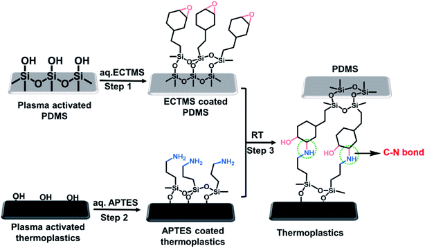
The present study aims to design stable and economic hybrid microfluidic devices that can tolerate high temperature and pressure using robust bonding strategies between PDMS and thermoplastic materials. This technique is based on the surface modification of the materials to be bonded. Briefly, both polymeric substrates are initially plasma-activated and then the terminal epoxy groups are introduced on the surface of the PDMS using 1% aqueous ECTMS, whereas terminal amine groups are introduced on the surface of the thermoplastics using 1% aqueous APTES. Finally, irreversible bonding occurs when the coated substrates contact each other at RT for 30 min as a result of the strong carbon–nitrogen (C–N) covalent bond formation between the two surfaces. Furthermore, the high pressure and thermal stability of the fabricated hybrid PDMS–PC microfluidic device was used to perform the CF-PCR to amplify a 210 bp gene fragment from the Shiga-toxin gene region of Escherichia coli (E. coli) O157:H7. To the best of our knowledge, there have been no previous reports on chemically surface-coated substrates to fabricate hybrid PDMS–PC microfluidic devices for CF-PCR applications that require such high bonding strength and temperature to amplify the DNA for foodborne pathogen detection.
Materials and methods
Chemicals and materials
(3-Aminopropyl)triethoxysilane (APTES; 99%) and [2-(3,4-epoxycyclohexyl)ethyl]trimethoxysilane (ECTMS; 98%) were purchased from Sigma-Aldrich. The PDMS prepolymer (Sylgard 184) and curing agent were obtained from Dow Corning. Poly(methyl methacrylate) (PMMA), polycarbonate (PC), polystyrene (PS), and polyethylene terephthalate (PET) were purchased from Goodfellow. Fluospheres amine (0.2 μm, red) and TE buffer (10 nM Tris–HCl, 0.1 nM EDTA, pH 8.0) were obtained from Thermo Fisher Scientific. Taq polymerase, PCR buffer solutions, and dNTPs were obtained from BioFact (Daejeon, Korea). Agarose powder was purchased from BioShop (Ontario, Canada). A 100 bp DNA size marker was purchased from Takara (Shiga, Japan). Ethidium bromide (EtBr)-based dye (Loading STAR) was purchased from Dynebio (Seongnam, Korea).Chemical bonding of PDMS to thermoplastics
The surface of the PDMS and thermoplastics were separately coated by using different chemicals such as ECTMS and APTES. The schematic process flow for bonding PDMS with thermoplastics is shown in Fig. 1. In this experiment, a flat elastomeric PDMS sheet was produced after the thermal curing of PDMS prepolymer and curing agent (10![[thin space (1/6-em)]](https://www.rsc.org/images/entities/char_2009.gif) :
:![[thin space (1/6-em)]](https://www.rsc.org/images/entities/char_2009.gif) 1 ratio) at 80 °C for 2 h. Before plasma treatment, the PDMS sheet and thermoplastics were washed with methanol, rinsed with deionized water, and then air-dried. Then, the PDMS sheet and thermoplastics were oxidized by oxygen plasma treatment for 1 min, which was followed by treatment with aqueous ECTMS (1 wt%) for PDMS and aqueous APTES (1 wt%) for thermoplastics at RT for 30 min. Next, both substrates were washed with water, methanol, dried with air, and immediately bonded with each other to form an irreversible bond via carbon–nitrogen covalent bonding. The time-lapse progress of the reaction is displayed as Fig. S1 in the ESI.† For better results, the 1 wt% ECTMS solution was stirred at RT for 90 min at the beginning, then plasma-activated PDMS was immersed in it. Generally, ECTMS is used as a coupling agent or adhesion promoter because terminal cyclic epoxide groups are highly reactive electrophiles that can react with common nucleophiles such as –NH2, –SH, and –OH. Hence, we made ECTMS-coated PDMS as an adhesive to covalently link with APTES-coated thermoplastics realized by the carbon–nitrogen bond (C–N bond) formation.
1 ratio) at 80 °C for 2 h. Before plasma treatment, the PDMS sheet and thermoplastics were washed with methanol, rinsed with deionized water, and then air-dried. Then, the PDMS sheet and thermoplastics were oxidized by oxygen plasma treatment for 1 min, which was followed by treatment with aqueous ECTMS (1 wt%) for PDMS and aqueous APTES (1 wt%) for thermoplastics at RT for 30 min. Next, both substrates were washed with water, methanol, dried with air, and immediately bonded with each other to form an irreversible bond via carbon–nitrogen covalent bonding. The time-lapse progress of the reaction is displayed as Fig. S1 in the ESI.† For better results, the 1 wt% ECTMS solution was stirred at RT for 90 min at the beginning, then plasma-activated PDMS was immersed in it. Generally, ECTMS is used as a coupling agent or adhesion promoter because terminal cyclic epoxide groups are highly reactive electrophiles that can react with common nucleophiles such as –NH2, –SH, and –OH. Hence, we made ECTMS-coated PDMS as an adhesive to covalently link with APTES-coated thermoplastics realized by the carbon–nitrogen bond (C–N bond) formation.
Surface characterizations
The contact angle measurements (Phoenix 300, Surface Electro-Optics, South Korea), fluorescence measurements (ProgRes CapturePro software), X-ray photoelectron spectroscopy, and atomic force microscopy (JPK NanoWizard II) were performed to characterize the functionalized surface. The XPS analyses were performed using an AxisHsi (Kratos Analytical, UK) equipped with an aluminum X-ray source (mono-gun, 1486.6 eV) with the pass energy of 40 eV. The pressure in the chamber was below 5 × 10⁻⁹ Torr before the data were recorded, and the voltage and current of the anode were 13 kV and 18 mA, respectively. The take-off angle was set at 45°. The binding energy of C1s (284.5 eV) was used as the reference. The resolution for the measurement of the binding energy was about 0.1 eV.Analysis of the bonding strength
![[thin space (1/6-em)]](https://www.rsc.org/images/entities/char_2009.gif) :
:![[thin space (1/6-em)]](https://www.rsc.org/images/entities/char_2009.gif) 1 (w/w) mixture of the PDMS prepolymer and a curing agent. For better visualization, the purple ink solution was injected into the microchannel and flow rates were systematically controlled at 0.1, 1.0, 10, 20, and 30 mL min−1 using a syringe pump. In addition, the burst test was conducted for the hybrid microfluidic devices by introducing compressed air into the microchannel through the silicon inlet and monitoring the maximum pressure when the bonded devices were disassembled.
1 (w/w) mixture of the PDMS prepolymer and a curing agent. For better visualization, the purple ink solution was injected into the microchannel and flow rates were systematically controlled at 0.1, 1.0, 10, 20, and 30 mL min−1 using a syringe pump. In addition, the burst test was conducted for the hybrid microfluidic devices by introducing compressed air into the microchannel through the silicon inlet and monitoring the maximum pressure when the bonded devices were disassembled.Temperature measurements for performing CF-PCR
The two-temperature measurement, including denaturation and annealing/extension, was applied for on-chip CF-PCR using a hybrid PDMS–PC microfluidic device. An infrared (IR) camera (FLIR Thermovision A320) was used to measure the surface temperature of the PC substrate. Also, the temperature controller was used for controlling the temperature during the PCR process as shown in Fig. S2 in the ESI.† In this procedure, the temperatures of two copper heaters were separately controlled for denaturation and annealing/extension as described in our previous studies.31–34 The temperature measured on the surface of the copper heaters was considered to be the temperature inside the microchannel since the PC where the microchannel was engraved was only 2 mm in thickness, and the relatively low thermal conductivity of the PC (0.22 W K−1 m−1) prevented heat dissipation and confined heat within the PC. The surface of the PC was then covered with black tape to reduce the heat reflection during temperature measurement using an IR camera. Ten spots were randomly selected for temperature measurement, and the average temperature was evaluated using an image analyzer (ThermaCAM researcher 2.8). The temperature for the annealing/extension zone was 56.91 ± 0.21 °C, and that for the denaturation zone was 94.63 ± 0.38 °C. Also, to ensure the stability of the device, the temperature was continuously observed over 2 h.Procedures for CF-PCR
For PCR application, a PDMS–PC microfluidic device (4.5 × 4.5 cm) with a serpentine microchannel with the width, depth, and total length of 300 μm, 200 μm, and 180 cm, respectively, was fabricated. The schematic illustration of the serpentine microchannel of a PDMS–PC microfluidic device is shown in Fig. S3 in the ESI.† For DNA templates used in the PCR processes, E. coli O157:H7 was collected from a liquid culture solution, and its DNA was directly isolated and added to the PCR reagents. The primer sequences for amplifying a 210 bp DNA fragment of the Shiga-toxin gene in E. coli O157:H7 were as follows: 5′-TGT AAC TGG AAA GGT GGA GTA TAC A-3′ (forward) and 5′-GCT ATT CTG AGT CAA CGA AAA ATA AC-3′ (reverse). The PCR mixture contained 5 μL buffer, 0.16 mM dNTPs mixture, 0.5 μM forward and reverse primers and 0.5 U μL−1 of Taq polymerase. For the on-chip amplification, the inner surface of the microchannel was passivated with a BSA solution (1.5 mg mL−1) to reduce the nonspecific adsorption of the PCR reagent on the walls of the microchannel. Next, the PCR reagent was introduced into the microchannel at 4 μL min−1, and amplifications were performed for 30 thermal cycles. PCR results were analyzed by using the gel electrophoresis method and were observed by trans-illuminated UV light using a Bio-Rad Molecular Imager Gel-Chem-Doc XR imaging system (Bio-Rad Laboratories, Hercules, CA, USA).Results and discussion
Surface analysis
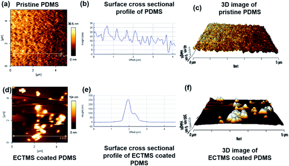
The oxygen plasma treatment is an excellent technique for increasing the hydrophilic properties of polymers since it can produce many hydroxyl groups (–OH) on the surface of the polymers, enabling the versatile chemical modification of the surface. Various analysis techniques, including contact angle measurements, fluorescence measurements, XPS measurements, and atomic force microscope (AFM) techniques, were performed to confirm the successful surface modification of PDMS and thermoplastics. The corresponding experimental results are discussed below. | ||
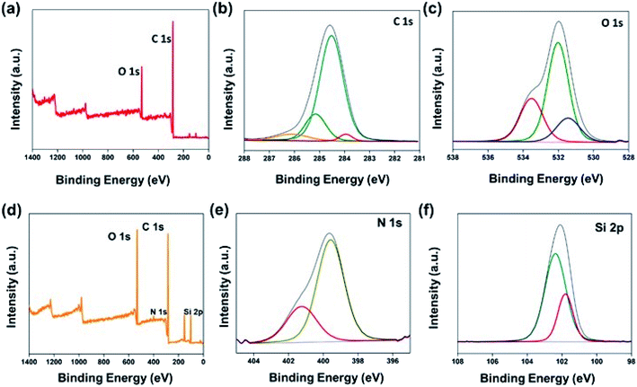
| Fig. 3 XPS for pristine PC showing (a) the survey spectrum, (b) C1s, (c) O1s, and for APTES-coated PC showing (d) the survey spectrum, (e) N1s, and (f) Si2p. | ||
| Substrate | Spectrum | Atomic ratio (%) |
|---|---|---|
| Pristine PDMS | O1s | 42.9 |
| C1s | 40.1 | |
| Si2p | 16.8 | |
| ECTMS-coated PDMS | O1s | 48.5 |
| C1s | 36.7 | |
| Si2p | 14.6 |
Bonding strength measurement
CF-PCR inside the microfluidic device
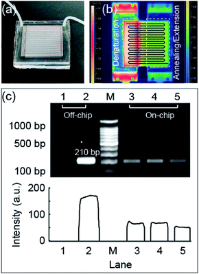
Fig. 6a shows a photo of the PDMS–PC microfluidic device fabricated using the bonding method introduced in the present study. The PDMS substrate with a serpentine microchannel was successfully sealed with a flat PC substrate to perform CF-PCR. Fig. 6b shows an IR camera image of the surface temperature of the PC substrate in two temperature zones (denaturation and annealing/extension). The positioning of the microfluidic device on heaters generally follows the schematic shown in Fig. S7 in the ESI† to ensure the on-chip PCR process. According to the results, the denaturation temperature was around 94.63 ± 0.38 °C (CV 0.41% (n = 10)), and the annealing/extension was approximately 56.91 ± 0.21 °C (CV 0.37% (n = 10)) for amplifying the 210 bp of E. coli O157:H7. We chose PC among the various polymers since PC has a relatively high glass transition temperature (145 °C), and the surface temperature of the PC substrate was constantly maintained over 120 min (Fig. S8 in the ESI†), indicating that our microfluidic device was suitable for performing on-chip CF-PCR. The 210 bp target gene from the Shiga-toxin gene region of E. coli O157:H7 was successfully amplified within 30 min (Fig. 6c).The negative (Lane 1) and positive (Lane 2) controls were performed using a thermocycler, and the results of CF-PCR are shown in Lanes 3–5 using our PDMS–PC microfluidic device. Using the hybrid PDMS–PC microfluidic device, the CF-PCR was successfully achieved to rapidly identify foodborne pathogens; the average intensity of the amplicons obtained using this method was approximately 35.53% as compared to the positive control. Also, to confirm the reproducibility of amplification using this microfluidic device, similar results were obtained after repeating the same experiment in triplicate for amplifying the 210 bp target gene. From those results, we concluded that this chemical-based bonding method for fabricating hybrid microfluidic devices has potential for CF-PCR, which always requires high temperature and pressure during the reaction.
Conclusions
In the present study, we successfully fabricated PDMS–thermoplastic microfluidic devices at RT capable of withstanding the elevated temperatures and high pressures realized by the strong covalent carbon–nitrogen bonding using silane reagents such as cyclohexyl epoxysilane and aminosilane. Cyclohexyl epoxysilane is highly reactive towards nucleophiles due to the ring strain of the epoxy group, which plays a crucial role in the strong PDMS–thermoplastic bond formation, the force of which was confirmed by tensile, leakage, and burst tests. Along with carbon–nitrogen bonding, the intermolecular hydrogen bond formed between hydroxyl (–OH) and secondary amine (–NH–) groups at the interface further strengthened the bonding between PDMS and thermoplastics. Moreover, the fabricated microfluidic devices can be used in cases when high-flow injection is mandatory, such as in micro-reactors and liquid chromatographic applications. According to our results, leakage did not occur even under high pressure, which was verified by the leakage test. The PDMS–PC microfluidic device was successfully adopted for performing CF-PCR owing to its excellent hydrolytic stability and has been applied to the amplification of the 210 bp gene fragment from the Shiga-toxin gene region of E. coli O157:H7. The introduced chemical-based bonding method is highly promising for fabricating PDMS–thermoplastic hybrid microfluidic devices for next-generation biomedical devices. It can also be anticipated that the proposed method can be used in various fields, including clinical applications, catalysis, and the food industry.Conflicts of interest
There are no conflicts to declare.Acknowledgements
This work was supported by the National Research Foundation of Korea (NRF) grants funded by the Korea government (MSIP) (No. NRF-2017R1A2B4008179) and the Korea government (MSIT) (No. NRF-2020R1A2B5B01001971).References
- C. Zhang and D. Xing, Chem. Rev., 2010, 110, 4910–4947 CrossRef PubMed.
- Y. Zheng, K. Kang, F. Xie, H. Li and M. Gao, BioChip J, 2019, 13, 217–225 CrossRef.
- S. Wang, S. Yu, M. S. Siedler, P. M. Ihnat, D. I. Filoti, M. Lu and L. Zuo, Rev. Sci. Instrum., 2016, 87, 105005 CrossRef PubMed.
- A. Baraket, M. Lee, N. Zine, N. Yaakoubi, J. Bausells and A. Errachid, Microchim. Acta, 2016, 183, 2155–2162 CrossRef.
- M. K. Yuzon, J. H. Kim and S. Kim, BioChip J, 2019, 13, 277–287 CrossRef.
- S. G. Im, K. W. Bong, C.-H. Lee, P. S. Doyle and K. K. Gleason, Lab Chip, 2009, 9, 411–416 RSC.
- J. Xu and K. K. Gleason, Chem. Mater., 2010, 22, 1732–1738 CrossRef CAS.
- P. Gu, K. Liu, H. Chen, T. Nishida and Z. H. Fan, Anal. Chem., 2011, 83, 446–452 CrossRef CAS.
- K. Aran, L. A. Sasso, N. Kamdar and J. D. Zahn, Lab Chip, 2010, 10, 548–552 RSC.
- L. Tang and N. Y. Lee, Lab Chip, 2010, 10, 1274–1280 RSC.
- B. M. Maoz, A. Herland, O. Y. F. Henry, W. D. Leineweber, M. Yadid, J. Doyle, R. Mannix, V. J. Kujala, E. A. FitzGerald, K. K. Parker and D. E. Ingber, Lab Chip, 2017, 17, 2294–2302 RSC.
- O. Y. F. Henry, R. Villenave, M. J. Cronce, W. D. Leineweber, M. A. Benz and D. E. Ingber, Lab Chip, 2017, 17, 2264–2271 RSC.
- B. Cortese, M. C. Mowlem and H. Morgan, Sens. Actuators, B, 2011, 160, 1473–1480 CrossRef CAS.
- I. R. G. Ogilvie, V. J. Sieben, B. Cortese, M. C. Mowlem and H. Morgan, Lab Chip, 2011, 11, 2455–2459 RSC.
- W. Wu, J. Wu, J.-H. Kim and N. Y. Lee, Lab Chip, 2015, 15, 2819–2825 RSC.
- K. S. Lee and R. J. Ram, Lab Chip, 2009, 9, 1618–1624 RSC.
- M. Tweedie, D. Sun, B. Ward and P. D. Maguire, Lab Chip, 2019, 19, 1287–1295 RSC.
- T. Nguyen, S. H. Jung, M. S. Lee, T.-E. Park, S.-k. Ahn and J. H. Kang, Lab Chip, 2019, 19, 3706–3713 RSC.
- L. Gorgannezhad, H. Stratton and N.-T. Nguyen, Micromachines, 2019, 10, 408 CrossRef PubMed.
- Y. Ding, J. Choo and A. J. deMello, Microfluid. Nanofluid., 2017, 21, 58 CrossRef.
- H. Yang, Z. Chen, X. Cao, Z. Li, S. Stavrakis, J. Choo, A. J. deMello, P. D. Howes and N. He, Anal. Bioanal. Chem., 2018, 410, 7019–7030 CrossRef CAS PubMed.
- O. Frey, S. Bonneick, A. Hierlemann and J. Lichtenberg, Biomed. Microdevices, 2007, 9, 711–718 CrossRef CAS.
- K. T. L. Trinh, H. Zhang, D.-J. Kang, S.-H. Kahng, B. D. Tall and N. Y. Lee, Int Neurourol J, 2016, 20, S38–S48 CrossRef.
- K. T. L. Trinh and N. Y. Lee, Talanta, 2018, 176, 544–550 CrossRef CAS.
- W. Wu, K. T. L. Trinh and N. Y. Lee, Analyst, 2015, 140, 1416–1420 RSC.
- A. C. Hatch, T. Ray, K. Lintecum and C. Youngbull, Lab Chip, 2014, 14, 562–568 RSC.
- D. Rodriguez-Lazaro, P. Gonzalez-García, E. Delibato, D. De Medici, R. M. García-Gimeno, A. Valero and M. Hernandez, Int. J. Food Microbiol., 2014, 184, 113–120 CrossRef CAS.
- A. Abd-Elmagid, P. A. Garrido, R. Hunger, J. L. Lyles, M. A. Mansfield, B. K. Gugino, D. L. Smith, H. A. Melouk and C. D. Garzon, J. Microbiol. Methods, 2013, 92, 293–300 CrossRef CAS PubMed.
- L. Cao, X. Cui, J. Hu, Z. Li, J. R. Choi, Q. Yang, M. Lin, L. Ying Hui and F. Xu, Biosens. Bioelectron., 2017, 90, 459–474 CrossRef CAS PubMed.
- J. Jeon, N. Choi, H. Chen, J. I. Moon, L. Chen and J. Choo, Lab Chip, 2019, 19, 674–681 RSC.
- Y. Zhang, K. T. L. Trinh, I.-S. Yoo and N. Y. Lee, Sens. Actuators, B, 2014, 202, 1281–1289 CrossRef CAS.
- W. Wu, K. T. L. Trinh, Y. Zhang and N. Y. Lee, RSC Adv., 2015, 5, 12071–12077 RSC.
- T. T. Nguyen, K. T. L. Trinh, W. J. Yoon, N. Y. Lee and H. Ju, Sens. Actuators, B, 2017, 242, 1–8 CrossRef CAS.
- K. T. L. Trinh, Q. N. Pham and N. Y. Lee, Sens. Actuators, B, 2019, 282, 1008–1017 CrossRef CAS.
- T. Trantidou, Y. Elani, E. Parsons and O. Ces, Microsyst. Nanoeng., 2017, 3, 16091 CrossRef CAS PubMed.
- V. Sunkara, D.-K. Park and Y.-K. Cho, RSC Adv., 2012, 2, 9066–9070 RSC.
- D. E. Packham, J. Adhes., 1992, 39, 137–144 CrossRef CAS.
- H. Zhang and N. Y. Lee, Appl. Surf. Sci., 2015, 327, 233–240 CrossRef CAS.
- V. Sunkara, D.-K. Park, H. Hwang, R. Chantiwas, S. A. Soper and Y.-K. Cho, Lab Chip, 2011, 11, 962–965 RSC.
- X. Wang, D. T. T. Phan, D. Zhao, S. C. George, C. C. W. Hughes and A. P. Lee, Lab Chip, 2016, 16, 868–876 RSC.
Footnote |
| † Electronic supplementary information (ESI) available. See DOI: 10.1039/d0ra02332a |
| This journal is © The Royal Society of Chemistry 2020 |


