 Open Access Article
Creative Commons Attribution 3.0 Unported Licence
Smart biomaterials for enhancing cancer therapy by overcoming tumor hypoxia: a review
Samar A. Salimab,
Taher A. Salaheldinc,
Mohamed M. Elmazard,
A. F. Abdel-Azizb and
Elbadawy A. Kamoun *ae
*ae
aNanotechnology Research Center (NTRC), The British University in Egypt (BUE), El-Sherouk City, Cairo 11837, Egypt. E-mail: e-b.kamoun@tu-bs.de; badawykamoun@yahoo.com; Tel: +20-1283320302
bBiochemistry Group, Dep. of Chemistry, Faculty of Science, Mansoura University, Egypt
cDepartment of Medicine, Case Western Reserve University School of Medicine, Cleveland, OH 44106, USA
dFaculty of Pharmacy, The British University in Egypt (BUE), El-Sherouk City, Cairo 11837, Egypt
ePolymeric Materials Research Dep., Advanced Technology and New Materials Research Institute (ATNMRI), The City of Scientific Research and Technological Applications (SRTA-City), New Borg Al-Arab City 21934, Alexandria, Egypt
First published on 25th November 2022
Abstract
Hypoxia is a distinctive feature of most solid tumors due to insufficient oxygen supply of the abnormal vasculature, which cannot work with the demands of the fast proliferation of cancer cells. One of the main obstacles to limiting the efficacy of cancer medicines is tumor hypoxia. Thus, oxygen is a vital parameter for controlling the efficacy of different types of cancer therapy, such as chemotherapy (CT), photodynamic therapy (PDT), photothermal therapy (PTT), immunotherapy (IT), and radiotherapy (RT). Numerous technologies have attracted much attention for enhancing oxygen distribution in humans and improving the efficacy of cancer treatment. Such technologies include treatment with hyperbaric oxygen therapy (HBO), delivering oxygen by polysaccharides (e.g., cellulose, gelatin, alginate, and silk) and other biocompatible synthetic polymers (e.g., PMMA, PLA, PVA, PVP and PCL), decreasing oxygen consumption, producing oxygen in situ in tumors, and using polymeric systems as oxygen carriers. Herein, this review provides an overview of the relationship between hypoxia in tumor cells and its role in the limitation of different cancer therapies alongside the numerous strategies for oxygen delivery using polysaccharides and other biomaterials as carriers and for oxygen generation.
1. Introduction
Cancer ranks as the chief cause of death and the main hurdle to significantly improving life expectancy worldwide. Regarding estimations from the World Health Organization (WHO) in 2019, in 112 of 183 countries, cancer is considered the principal cause of death before the age of 70 years and is ranked third in a further 23 countries.1 Female breast cancer and lung cancer are the most commonly diagnosed cancers. With a recorded 2.3 million new cases each year, breast cancer accounts for 11.7% of all cancers, followed by lung cancer (11.4%), colorectal (10.0%), prostate (7.3%), and stomach (5.6%) cancers. Lung cancer surpassed breast cancer as the primary reason for cancer mortality, with 1.8 million deaths (18%), followed by colorectal cancer (9.4%), liver cancer (8.3%), stomach cancer (7.7%), and female breast cancer (6.9%).1Tumor cells are abnormal cells that can regenerate rapidly. They are characterized by uncontrolled proliferation, transformation, and migration. Tumor cells resort to unusual metabolic pathways to obtain more energy to meet their needs for cell proliferation and migration, as their metabolism is more enthusiastic than that of normal cells.2 As a result of hypoxia, healthy cells are frequently tormented by insufficient oxygen supply, so they transform into cancer cells and the majority of solid tumors originate suddenly and sharply. According to recent studies, tumor hypoxia is a significant barrier to the actual treatment of cancer with immunotherapy and chemotherapy, but not radiotherapy.3 Both the intrinsic sensitivity of cancerous cells and the tumor microenvironment influence how cancer responds to chemotherapy. It is well established that tumor hypoxia promotes cancer cells' resistance to radiation and chemotherapy treatments. Moreover, cancer cell migration, growth dynamics, endoplasmic reticulum stress, angiogenesis, and aggressive characteristics are all impacted by hypoxia.4 Approximately 60% of advanced solid tumors include hypoxic regions, which is often related to poor survival. Hypoxia-inducible factor (HIF-1α) is a vital regulator of the molecular response to hypoxia. HIF-1α expression is reduced in normal conditions and enhanced by hypoxic conditions. Numerous gene products that influence the regulation of metabolism, cell cycle, angiogenesis, and apoptosis are altered by the activation of HIF-1 expression.4
This review explores new trends and strategies for overcoming tumor hypoxia toward enhancing cancer therapy protocols. Oxygen-generating polymeric and non-polymeric biomaterials from the last few decades are reviewed as promising materials for enhancing the efficacy of established cancer therapy protocols via reduction of tumor hypoxia.
1.1. Induction of solid tumors by hypoxia
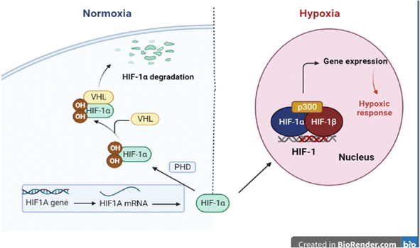
Oxygen-activated breakdown of HIF-1α results in locking genes are enhanced by the environment features with hypoxia properties. Hypoxia is a key factor for inspiring the transition of cobble-stone shaped cells (epithelial cells) to flat-spindle shaped cells (mesenchymal cells), with great potential for metastatic position formation, invasion, and motility proteins; this process is known as epithelial–mesenchymal transition (EMT). Therefore, there is a genuine connection between hypoxia and the proliferation of tumors (Fig. 1).5Under normoxia, the HIF1A gene is rapidly transcribed to HIF1A mRNA, which is then translated to HIF-1α protein. In normal oxygen conditions, the HIF-1α protein is constitutively hydroxylated by the prolyl hydroxylases (PHDs) enzymes, which allows binding of von Hippel–Lindau tumor suppressor protein (pVHL) and ubiquitin ligase, resulting in proteasomal degradation. Under hypoxia, hydroxylation no longer happens, permitting HIF-1α to penetrate the nucleus and form the active HIF transcription complex.
1.2. Oxygen-loaded sources in medical applications
Oxygen has a critical role as a signaling molecule and metabolic substrate.6 In hypoxic surroundings, human cells need to utilize lactic acid fermentation to yield ATP, which requires fifteen times more glucose to manufacture the same amount of ATP as oxidative phosphorylation. The principal cause of cell necrosis is the depletion of ATP stores,7 where hypoxia is the hallmark of ischemic tissue, which has also been shown to promote apoptosis in cells, further stressing the requirement to deliver oxygen. Approaches applied for local oxygen delivery can be mainly divided as mentioned below:As shown in eqn (1), calcium peroxide (CPO) is a solid inorganic peroxide with a high-energy covalent link that readily decomposes and produces oxygen in liquid media:
 | (1) |
The most popular inorganic peroxide used as an oxygen-releasing source is CPO due to its prolonged release time. Blending CPO into hydrophobic materials can limit the rate of oxygen release in a sustained manner due to the slow dispersion of water into hydrophobic materials and consequently the delayed decomposition of CPO. Additionally, a polymeric matrix's composition, shape, size, and surface chemistry can prevent early burst release and provide adjustable profile release in accordance with the carrier's specifications.12 Furthermore, cell micro-carriers have been demonstrated to be a potential strategy for repairing tissues with unconventional shapes, while injectable scaffolds can be used to quickly repair with simple surgical techniques.13
An oxygen-releasing antioxidant scaffold was prepared by incorporation of CPO into a polyurethane (PU) scaffold. This scaffold exhibited antioxidant behavior with the sustained release of oxygen are over a period of 10 days. The PU scaffold loaded with CPO reduced the effect of hypoxia in vitro and improved cell survival. In an in vivo skin flap model, the PU scaffold loaded with CPO (oxygen-releasing scaffold) prevented necrosis.14
Recently, a 3-polycaprolactone (PCL) and poly(glycerol sebacate) (PGS) mat loaded with a high concentration of CPO (up to 10%) was utilized as an oxygen source. This composite exhibited the continual discharge of oxygen for several days and meaningfully improved the rate of metabolism due to the decrease of hypoxia in the bone marrow-derived mesenchymal stem cells (BM-MSCs). The manufactured mats showed hints of potential antibacterial efficacy as well. However, for CPO to be used as an oxygen generator, it needs to be made into nanoparticles to ensure good dispersion plus homogeneity when loaded on the nanofibrous mat.15
Hydrogen peroxide (H2O2) is used with caution in biomedical applications due to its limited stability and potential for decomposition to produce reactive oxygen species (ROS) in the biological environment, which can lead to oxidative cell damage. The enzyme catalase, which is present in almost all living organisms, catalyzes the conversion of H2O2 into water and oxygen and can be utilized to stop H2O2 buildup.16
Due to its capacity to kill bacteria, fungi, and other pathogens, even at low concentrations, H2O2 is widely used in biomedical applications. It is also present in many oral care products.17 Hydrogen peroxide can be capsulated into polyvinyl pyrrolidone (PVP) to produce a PHP complex (PVP–H2O2 complex) as an oxygen-releasing nanoscaffold.18
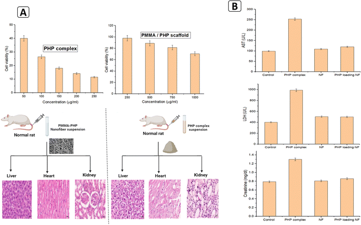
Our recent study explored the fabrication of poly(methyl methacrylate)-loaded PHP complex (PMMA–PHP complex) platforms as a new model for oxygen-releasing biomaterials with anticancer features. The PHP complex acts as an oxygen generator. The nanofibrous scaffold composed of PMMA + 10% PHP was found to have a decent distribution of PHP (PVP–H2O2) and exhibited an improvement of mechanical characteristics with uniform nanofibers (NF) as well as prolonged release of oxygen. Depending on the dosage manner, the nanofibers at a concentration of 1 mg ml−1 significantly reduced the viability of cells in several cancer cell lines. The viability of cancer cells was reduced to 30%, whereas the normal cells displayed exceptionally safe behavior at the same dose. Additionally, it was shown that the PHP complex, when in the form of a powder, was very hazardous to both healthy and malignant cells, even at low concentrations. However, by embedding the PHP complex in hydrophobic nanofibers, it was possible to avoid the burst release of H2O2.5
Mallepally et al. prolonged oxygen release from H2O2 to 24 hours by using PMMA as the encapsulating material.19 Furthermore, Li et al. demonstrated prolonged release of oxygen for up to 14 days by encapsulation of H2O2 into a high-molecular-weight polymer PLGA shell, through which oxygen gradually diffuses.20
Sodium percarbonate (SPC) has been extensively used as a source of anhydrous H2O2 in organic synthesis which then decomposed into oxygen and water.21
 | (2) |
McQuilling et al. used SPC to reduce the hypoxic problem on islets (found in clusters throughout the pancreas), commencing with islet separation and continuing 7 days after microencapsulation. They demonstrated that oxygen-producing substances, such as SPO, provide a potentially workable strategy in diabetic patients to replenish oxygen to transplanted islets that are either naked or encapsulated. Although these results revealed that SPO can improve islet viability and functionality, the authors noted that additional work is necessary to control oxygen generation.22
2. Strategies of using oxygen to overcome tumor hypoxia
There are four ways that oxygen can be used to reduce tumor hypoxia: hyperbaric oxygen therapy, delivering oxygen by carriers to tumors, decreasing tumor oxygen consumption, and generating oxygen in situ in the tumor.23 These strategies have some advantages and limitations, which will be discussed in the following sections.2.1. Hyperbaric oxygen therapy (HBO)
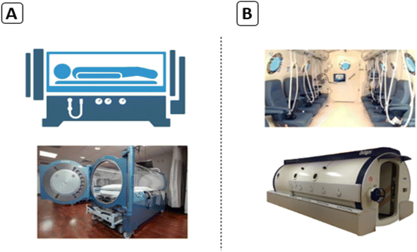
HBO therapy is termed an effective and safe treatment for different types of cancer.24 During HBO therapy, the patient breathes in 100% oxygen, typically at an absolute pressure of 2.5 atmospheres. HBO therapy can enhance oxygen levels in tissues, stimulate angiogenesis, decrease edema, and trigger collagen synthesis.25 Consequently, there are several means to implement HBO therapy; the most popular method is to use hyperbaric chambers, either mono-place or multi-place chambers (Fig. 2). Pure 100% oxygen is typically pressurized in mono-place chambers, while a mixture of air with oxygen is normally pressurized in multi-place chambers using an endotracheal tube or face-mask.26Hypoxia plays a critical role in developing tumor drug resistance and the failure of chemotherapy. Overall, there are four detectable mechanisms of drug resistance by hypoxia as follows: (1) hypoxia decreases the intracellular concentration of chemotherapy agents through the accumulation of the drug resistance protein P-glycoprotein, which might push therapeutic agents out. (2) Hypoxia alternates the signaling and metabolic pathways of tumor cells. (3) Hypoxia alters the redox condition of tumor cells. (4) Hypoxia induces mutations and gene instability in cancer cells.23
2.2. Delivery of oxygen by bio-carriers
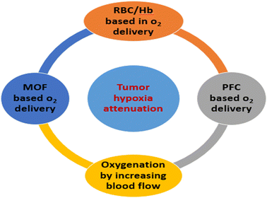
Studies have revealed that there is a critical role for O2 carriers in reducing hypoxia to enhance the efficiency of cancer therapies. Artificially carrying molecular O2 by innovative nanomaterials to the hypoxic site is one feasible method for increasing the concentration of O2. Thus, it reverses the hypoxia and consequently may improve the outcomes of traditional cancer treatments.27 There are different types of oxygen-based carriers, including red blood cell (RBC)/hemoglobin (Hb)-based O2 carriers, metal–organic framework (MOF)-based O2 carriers, and perfluorocarbon (PFC)-based O2 carriers, as well as oxygenation by increasing blood flow, as shown in Fig. 3.28Hemoglobin (Hb) is a metalloprotein that is responsible for the oxygen-carrying capacity of RBCs. Consequently, Hb is an interesting choice for scientists to create artificial oxygen transporters. Hb has a short blood circulation ability and poor stability in addition to its toxicity, so it needs to be conjugated with suitable carriers.30
Hb encapsulated in a liposome system revealed a higher affinity of direct oxygen delivery than RBC and was employed to improve the outcome of radiotherapy (RT) in vitro and in vivo. Murayama et al. proved that the tumor was significantly inhibited by radiation treatment accompanied with intravenous injection of Hb encapsulated in liposomes compared to treatment with radiation and unencapsulated Hb.31 Many efforts have been made to conjugate Hb with different types of polymers to create an oxygen delivery system to alleviate hypoxia and hence enhance cancer treatment. Wang et al. innovated an oxygen delivery nano-system by encapsulating polystyrene zinc phthalocyanine (PS-ZnPc) into Hb-conjugated polymeric micelles composed of poly(ethylene glycol)-block-poly (acrylic acid)-block-polystyrene.32
Hb-conjugated polymeric micelles produced more singlet oxygen (1O2) after light initiation and triggered photo-toxicity to cervical cancer cells in comparison with polymeric micelles without Hb conjugation.32 Correspondingly, Luo et al. enhanced the efficacy of PDT by developing a lipid–polymer hybrid nanoparticle system to mimic RBC seen in mammals.33 They complexed Hb with indocyanine green dye (ICG), and subsequently the complex was embedded into lipid–polymer nanoparticles with a DSPE–PEG shell and PLGA core. Tumor-bearing mice injected with Hb–ICG encapsulated nanoparticles showed broad inhibition of tumors due to oxidative damage by generating reactive oxygen species (ROS) from self-supply of oxygen.33 Hb-supported oxygen delivery is suitable for avoiding hypoxia accompanying drug resistance. Yang et al. encapsulated Hb and doxorubicin (DOX) into a liposome, which they called a DOX–Hb–liposome (DHL) system. In the tumor model, DHL overturned hypoxia and revealed potential antitumor properties compared to the DOX–liposome only without Hb.34
An oxygen-loaded pH-responsive multifunctional nano-drug carrier with improved chemo-photodynamic therapy effectiveness was successfully synthesized by Xie and coworkers. The rare earth-doped nanoparticles (NaYF4:Yb/Er@NaYbF4:Nd@NaGdF4) (UC) were engaged for dual-modal up-conversion and MR imaging. Moreover, the UC core–shell structural nanoparticles could efficiently stimulate photosensitizer Rose Bengal (RB) in a mesoporous silica shell (mSiO2) for PDT under laser irradiation (808 nm). The shell zeolitic imidazolate framework-90 (ZIF-90) breaks down under acidic conditions, which is the tumor environment, thus promoting the rapid release of DOX and oxygen to overcome tumor hypoxia. This resultant UCNPs-MOF multifunctional oxygen carrier demonstrated an intense antitumor effect both in vivo and in vitro.39
A number of anti-angiogenic therapeutic medications, specifically bevacizumab, sinomenine, combretastatin, and thalidomide, also successfully improved the therapeutic efficiency of numerous nanomedicines via fixing abnormal tumor vasculature.50,51 A high density of extracellular matrix (ECM) components, like collagen and hyaluronic acid, increases the pressure, which squeezed the tumor's arteries. Cancer-associated fibroblast (CAF) cells are principally responsible for producing these components. Angiotensin inhibitor losartan might decompress the intra-tumoral vessels by deactivating CAFs and reducing the manufacture of stromal collagen/HA. Therefore, losartan therapy improved vascular blood flow, decreased hypoxia in pancreatic and breast cancer simulations, and enhanced chemotherapy.52,53
2.3. Influence of decreasing oxygen consumption
The microenvironment of tumors is generally characterized by a limited amount of H2O2 and hypoxia, which result in restriction of the therapeutic efficiency of the combined treatment. Remarkably, modeling and simulation studies have demonstrated that reducing oxygen consumption is obviously more effective at enhancing tumor oxygenation than increasing oxygen delivery.54 Consequently, one of the novel alternative approaches for improving the accumulation of oxygen in tumors is reducing oxygen depletion. Recently, anticancer drug doxorubicin (DOX) was successfully loaded onto nanoparticles of copper–metformin (Dox@Cu–Met NPs) to stimulate chemotherapeutic effectiveness and starvation therapy by reducing O2 consumption and increasing H2O2 production, in addition to obstructing the production of ATP. The nano-sheet structure with an appropriate size of Dox@Cu–Met NPs degraded in response to high levels of GSH and the acidic environment of the tumor tissue. Dox@Cu–Met NPs can potentially enhance the concentration of O2 in the tumor environment. In vitro assay proved that Dox@Cu–Met NPs improves the sensitivity and selectivity of breast cancer cell model (MCF-7/ADR cells) to DOX and encourages the creation of ROS in tumor cells. Meng et al. also applied their DOX-loaded NPs in in vivo experiments, which confirmed their biosafety and ability to inhibit cancer cell progression. The combination of chemotherapy and nanoparticles can reduce oxygen consumption, resulting in the substantial reduction of tumor proliferation.552.4. Oxygen generation in situ in tumor
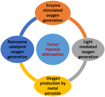
High levels of reactive oxygen species (ROS), specifically H2O2, are generated in tumor cells as the result of a defect of metabolic regulation and uncontrolled proliferation of tumor cells, which might be able to contribute to O2 creation to inhibit hypoxia.56 Large levels of ROS, primarily hydrogen peroxide (H2O2), are typically produced by the unchecked growth and malfunction of metabolic regulation in tumor cells. These ROS could be used for in situ O2 synthesis to combat hypoxia. As shown in Fig. 4, numerous methods have been developed so far to utilize endogenous H2O2 for cancer treatment and hypoxia relief.ROS are created as byproducts of cellular metabolism, such as hydroxyl radicals, singlet oxygen, superoxide, and peroxide,57 and affect cell homeostasis as well as signaling.58 Different human diseases can lead to extreme creation of ROS, which results in oxidative stress and many pathological features.59 Cancer cells possess high concentrations of ROS as a result of changes in their metabolic activities.60 The elevated intracellular ROS level in cancer cells, therefore, can be considered a cancer-specific stimulus for anticancer drug delivery. Recently, various ROS-responsive drug carriers have been employed to achieve effective anticancer therapy by regulating the release of anticancer drugs in response to the overproduction of intracellular ROS in cancer cells.
Zhou et al. developed a biological system based on Chlorella pyrenoidosa surrounded by calcium alginate, which they termed ALGAE (autotrophic light-triggered green oxygen-affording engine).68 Chlorella is a photosynthetic green algae and single autotrophic cell possessing photosynthetic pigments in its chloroplasts, which makes it a good candidate as a biocompatible oxygen resource.69 The ALGAE were fixed around the cancer cells in a slightly invasive way and could be reserved for an extended period for oxygen supplying because the Chlorella was protected via calcium alginate from scavenging by macrophages. A source of light energy was necessary for Chlorella to generate oxygen during PDT mediated by ALGAE treatment. The ALGAE stimulated oxygen generation through energy transformation and water splitting, and thus the process is ecofriendly and biosafe. ALGAE could surround cancer cells and produce abundant oxygen in vivo after irradiation.
During hypoxia-resistant PDT induced by ALGAE, light is not only one of the elements of PDT, but also the source of energy for Chlorella to generate oxygen. The oxygen is generated with light triggering by ALGAE through water splitting and energy transformation, and the overall process is economical and environmentally friendly. ALGAE can stay around tumor tissues and generate copious oxygen in vivo after receiving irradiation, and simultaneously the photosensitizer produces enough singlet oxygen, thus destroying the cancer cells and improving the efficiency of PDT.68
Bu et al. fabricated transferrin-modified MgO2 nanosheets (TMNSs), which effectively respond to the acidity of TME. MgO2 reacts rapidly with H+ to produce H2O2 and break down the structure of transferrin on the nanosheets.71 Subsequently, transferrin releases the ferric ions (Fe3+) and creates cytotoxic hydroxyl radicals (˙OH) by the Fenton reaction. The TMNSs generate a large concentration of H2O2 and release ˙OH, which has the capability to destroy tumor cells, while TMNSs in normal cells (alkaline condition) produce a small amount of H2O2, which is decomposed by catalase. The results confirmed the high selectivity of TMNSs for tumor cells.71
Lin et al. successfully developed PVP-modified ZnO2 NPs and fixed them with paramagnetic Mn2+ ions by the cation exchange method.72 In this nano-system, ZnO2 disintegrates into H2O2 and Zn2+ in the acidic environment of the tumor. One of their valuable observations is that Zn2+ enhanced the mitochondrial creation of ROS by preventing the electron transfer chain, which was proved also by other studies.73,74 The exogenous release of ROS combined with the endogenous generation led to a significant tumor-killing effect.72
Jiang et al. constructed nanoplatform mediated CDT by fabricating hybrid CaO2 and Fe3O4 NPs with hyaluronic acid (HA) as a stabilizer and a NIR fluorophore label in the form of CaO2–Fe3O4@HA NPs.75 The nanoplatform possessed a great capability for self-supplying H2O2 and generating ˙OH in the acidic conditions of the TME with delectable stability under physiological conditions. It also revealed great selectivity and sensitivity to tumor cells with an inhibiting rate of up to 70% by CDT, while being safe to normal cells.75
Feng et al. innovated novel TME-modulated nanozymes using tin ferrite SFO (SnFe2O4) for mediating phototherapy (PT), photothermal therapy (PTT) and chemotherapy (CT).78 The synthesized SFO nanozymes possessed both CAT- and GSH-like activities. The SFO nanozymes enhanced the PT efficiency by activating H2O2 to create oxygen to inhibit tumor hypoxia. Meanwhile, the SFO nanozyme in the TME efficiently effected CT by mediating H2O2 to generate (˙OH) in situ combined with the depletion of GSH to release the antioxidant ability of the tumors. Moreover, the SFO nanozyme irradiated with 808 nm laser exhibited an outstanding phototherapeutic effect on account of the improved PTT efficiency and excellent free radical generation performance.78
Recently, a multi-nanozyme design was constructed based on polymeric HA-mediated CuMnOx NPs (CMOH) overloaded with indocyanine green (ICG) to form HA–CuMnOx@ICG nanocomposite (CMOINC) with highly efficient ROS production, hyperthermia, oxygen self-evolving function, and GSH reduction capability for attaining hypoxic tumor therapy.79 The CMOH nanozyme system showed oxidase- and peroxidase-like activities, which could powerfully initiate H2O2 or O2 to produce superoxide radicals (˙O2−) or hydroxyl radicals (˙OH) in the TME, enriching the oxidative stress of the tumor. CMOINC was shown to be highly effective at generating 1O2 and in situ hyperthermia under light irradiation.79 Different nanozymes revealed high efficiency for integration with multiple treatment modalities. Thus, this strategy may provide a new dimension for the design of other TME-based anticancer strategies.28
2.5. Oxygen delivery-based polymeric biomaterials
Polymeric materials have become extensively widespread in numerous biomedical applications. Biomedical polymers can be classified into natural and synthetic polymers. Natural polymers like silk, alginate, cellulose, gelatin, agar, fibrin, collagen, pectin, and chitosan are of wide interest for medical applications, owing to their nontoxicity, biocompatibility with the human body, biodegradability, and ability to accelerate cell proliferation. However, they have the critical disadvantages of lack of mechanical strength and inappropriate degradation rate.80 Synthetic polymers (like polyacrylonitrile, polyurethanes, polyamides, polycarbonates, polyvinyl alcohol, poly caprolactone, polyvinyl chloride, and polyesters) are applied for regenerative medical applications owing to their desirable mechanical properties, good integration with the neighboring tissue, ability to enhance cell adhesion, differentiation, and good cell migration.81,82 Biomedical polymers can be divided into different dimensional classes, such as:83 (a) 0D, such as nano/micro spheres, shells or capsules, (b) 1D, such as fibers or suture materials, (c) 2D, such as coatings or films, (d) 3D solids, like porous or printed polymers, and (e) 3D soft, like gel or foam.Silk is composed of proteins, including the core protein of fibroin (70–80%) and adhesive proteins termed sericin (20–30%).84 It is produced from the cocoons of the larvae of silkworms85 or other insects like spiders.86 Silk possesses notable mechanical properties, biocompatibility, and flexibility, and remarkable degradation rates, both in vitro and in vivo.87 It has been permitted by the Food and Drug Administration (FDA) for utilization in the creation of sutures as a biomedical material in surgery.88 Arumugam et al. used an electrospun gold–silver nanoparticle-loaded-nanofiber scaffold of silk fibroin (SF) and cellulose acetate (CA/SF/Au–Ag) for anticancer applications. The silk in addition to cellulose acetate acted as the stabilizing agent for silver and gold ions with better biocompatibility. The fabricated scaffold had needle-shaped morphology with diameter of 86 nm and the Ag and Au nanoparticles were dispersed onto the fiber scaffold with an average size of 53 nm and 17 nm, respectively. Moreover, it powerfully activates the cytotoxic effects against MDA-MB-231 and MCF-7 human breast cancer cells with a potential IC50 value. They found that the SF/CA/Au–Ag composite nanofiber scaffold is a promising material for anticancer applications.89
In another study, Yang et al. developed a platform of SF loaded with MnO2 NPs and indocyanine green (ICG) as a photodynamic agent in addition to doxorubicin (DOX) as a chemotherapeutic agent that can be customized in the form of SF@MnO2/ICG/DOX (SMID) nano-system.90 This nano-system exhibited high reactivity with H2O2 in the TME, which was broken down into oxygen to improve PDT. They also proved that the SMID nano-system had a distinctive effect for PTT as the result of its strong photothermal response to NIR (near-infrared) radiation and stably conjugated ICG. Moreover, their animal studies confirmed that the SMID nano-system obviously enhanced tumor alleviation through the combination of PDT, PTT, and chemotherapy with low toxicity. SF-based bio-nanomaterial, therefore, represents a promising carrier for oxygen to reduce hypoxia of the TME and improve cancer therapy.
Alginate is a promising inert natural polymer that is easily extracted from algae, generally considered for its capability to facilitate drug delivery in vivo with unique features,91 like biodegradability, muco-adhesion, biocompatibility and biosafety.92 Moreover, alginate has been applied as a bio-carrier for liposomes to capture hydrophilic chemotherapeutic agents and assist their transport to the targeted cell.93 Alginate-based biomaterials are extensively applied in drug delivery, wound dressing, and tissue engineering.94 The combination of CaO2 and catalase encapsulated into an alginate solution was successfully fabricated as a source of oxygen release.95 Huang et al. established that implantation of alginate-encapsulated CaO2 close to the tumor area stimulated the decomposition of CaO2, when reacted with H2O, into H2O2 and calcium hydroxide, which then decayed to generate molecular oxygen. Subsequently, oxygen-generating depots in TME improve the response to chemotherapeutic administration of DOX.95
Cellulose and its derivatives have been utilized efficiently as a drug delivery system for numerous types of drugs.96–98 Both cellulose nanocrystals (CNCs) and cellulose nanofibrils are represented as important biocompatible and biodegradable nanomaterials with good biosafety.99,100 Targeting the TME has been the focus of various studies involving cancer therapeutics and epigenetics. Bhandari et al. developed oxygen nanobubbles (ONB) by encapsulating oxygen into nano-size carboxymethyl cellulosic nanobubbles for modifying the hypoxic area of tumors to obstruct tumor growth.101 ONB were able to significantly deplete tumor progression and accelerate survival rates in animal models. ONB reprogramed tumor suppressor- and hypoxia-associated genes like PDK-1 and MAT2A, preposition are aiding as an ultrasound contrast agent.101
Gelatin is a hydrophilic natural polymer extracted from the collagen in the bones and skin of animals, and it has been utilized as drug delivery carrier, a food additive and a scaffold for TE because of its elasticity and viscosity, as well as its specific temperature-controlled gelation behavior.102 In addition, above the specific gelation temperature, gelatin can be developed into an applicable ecofriendly film by solvent evaporation.103
Plasma-treated liquids were recently established to have discerning features for inhibiting cancer cells and have attracted interest for use toward plasma-based cancer therapies. Labay et al. showed that reactive oxygen and nitrogen species (RONS) can be generated by atmospheric pressure plasma jets in liquids and biological systems.104 They used gelatin solution to store RONS produced by atmospheric pressure plasma jets to design an innovative biomaterial for cancer treatment. The quantity of RONS created in gelatin is significantly upgraded with respect to water, with concentrations of H2O2 and NO2− between 2 to 12 times greater for the longest plasma treatments. Plasma-treated gelatin showed the discharge of RONS to a liquid media, which encouraged effective elimination of bone cancer cells with high selectivity. The results set the basis for the approach for developing hydrogels with a high capacity to deliver RONS to tumors.104
An alternative approach for oxygen supplementation was revealed by Mizukami et al., where they fabricated HepG2 spheroids containing GMS (gelatin microsphere) by first manufacturing 37 μm GMS by water–oil emulsification then freeze drying them; HepG2 hepatocyte cells were then incubated with GMS at several mixing ratios in agarose gel-based micro-wells.105 They combined GMS into the core of the HepG2 human hepatocyte spheroids to permit oxygen to reach the spheroid core. HepG2 cells in the GMS/HepG2 spheroids were more highly oxygenated than those in the GMS-free spheroids. The viability of HepG2 cells in the spheroids was increased by GMS incorporation and further the CYP1A1 of activity of the HepG2 cells was enhanced to metabolize 7-ethoxyresorufin. In addition, mRNA expression of the CYP1A1 gene was significantly affected by GMS integration. The results designated that combining GMS with HepG2 spheroids increased the bio-viability of the cells and the CYP1A1 metabolic activity.105
Liu et al. examined the effects of low oxygen conditions on cell survival and oxygen generation in a synthetic system. They established a 3D system using calcium peroxide (CaO2) and poly(lactic-co-glycolic acid) (PLGA) microspheres dispersed in a hydrogel. The synthetic oxygen-generating system was tested under hypoxic conditions using stem cells versus controls to examine its potential for oxygen generation in a period of up to 21 days. The hydrogel gave prolonged oxygen release, protected the microspheres, and enhanced cell adhesion and cell proliferation in a flexible manner. The system produced oxygen and supported cell growth, which is also predicted to stimulate stem cell growth and survival after implantation.107
3. Summary of our recent findings
The current research in regenerative medicine and tissue engineering for cancer treatment is focused on developing effective, low-cost, bioactive, and advanced biomaterials for targeting the therapeutic agents and curing tumor hypoxia. Our recent study investigated the fabrication of PMMA–PHP complex nanofibrous platforms and the generation of oxygen nanobubbles as a novel model of biomaterials possessing anticancer properties.3.1. Fabrication of oxygen-releasing nanofibrous scaffold
![[thin space (1/6-em)]](https://www.rsc.org/images/entities/b_char_2009.gif) :
:![[thin space (1/6-em)]](https://www.rsc.org/images/entities/b_char_2009.gif) H2O2) complex and PMMA/PHP scaffold as an oxygen source. The utilization of the PHP (PVP
H2O2) complex and PMMA/PHP scaffold as an oxygen source. The utilization of the PHP (PVP![[thin space (1/6-em)]](https://www.rsc.org/images/entities/char_2009.gif) :
:![[thin space (1/6-em)]](https://www.rsc.org/images/entities/char_2009.gif) H2O2) complex as an oxygen source previously proved the importance of oxygen in the treatment of cancer. The different ratios of PVP
H2O2) complex as an oxygen source previously proved the importance of oxygen in the treatment of cancer. The different ratios of PVP![[thin space (1/6-em)]](https://www.rsc.org/images/entities/char_2009.gif) :
:![[thin space (1/6-em)]](https://www.rsc.org/images/entities/char_2009.gif) H2O2 used to gain the complex using procedures were clearly described previously.5 Notably, the yield of the developed complex climbed with increasing H2O2 ratio up to 1
H2O2 used to gain the complex using procedures were clearly described previously.5 Notably, the yield of the developed complex climbed with increasing H2O2 ratio up to 1![[thin space (1/6-em)]](https://www.rsc.org/images/entities/char_2009.gif) :
:![[thin space (1/6-em)]](https://www.rsc.org/images/entities/char_2009.gif) 1.5, at which point it started to decrease. In contrast to the yields for the other investigated ratios, a 1
1.5, at which point it started to decrease. In contrast to the yields for the other investigated ratios, a 1![[thin space (1/6-em)]](https://www.rsc.org/images/entities/char_2009.gif) :
:![[thin space (1/6-em)]](https://www.rsc.org/images/entities/char_2009.gif) 1.5 ratio of PVP
1.5 ratio of PVP![[thin space (1/6-em)]](https://www.rsc.org/images/entities/char_2009.gif) :
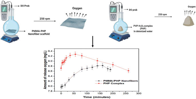
:![[thin space (1/6-em)]](https://www.rsc.org/images/entities/char_2009.gif) H2O2 provided the optimal yield of the complex of 66%. The PMMA + 10% PHP scaffold was reported to have the highest mechanical performance with smooth nanofibers, as shown before by examining its swelling and degrading properties. It also had excellent dispersion of the PHP complex. The concentration of oxygen released from both the PMMA scaffold and the PHP complex is presented in Fig. 5. As shown in the graph, the PHP complex in powder form released oxygen in a faster manner compared to the oxygen released from the PMMA scaffold loaded with the PHP complex. These results matched with those reported by Ahmed and his coworkers for their research based on measuring dissolved oxygen (DO). The DO increased over time (for 120 minutes) in the case of oxygen nanobubbles (ONB), while it decreased when air nanobubbles (ANB) were dispersed into deionized water.108
H2O2 provided the optimal yield of the complex of 66%. The PMMA + 10% PHP scaffold was reported to have the highest mechanical performance with smooth nanofibers, as shown before by examining its swelling and degrading properties. It also had excellent dispersion of the PHP complex. The concentration of oxygen released from both the PMMA scaffold and the PHP complex is presented in Fig. 5. As shown in the graph, the PHP complex in powder form released oxygen in a faster manner compared to the oxygen released from the PMMA scaffold loaded with the PHP complex. These results matched with those reported by Ahmed and his coworkers for their research based on measuring dissolved oxygen (DO). The DO increased over time (for 120 minutes) in the case of oxygen nanobubbles (ONB), while it decreased when air nanobubbles (ANB) were dispersed into deionized water.108
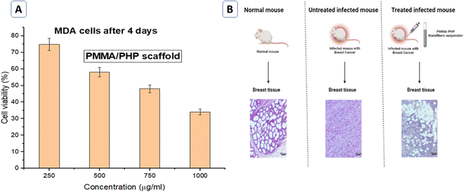 | ||
| Fig. 7 (A) Anticancer activity evaluation in vitro (MDA cells). (B) Photomicrographs of H&E stained breast tissue for −ve and +ve control mice and mice treated with and PHP-loaded NF. | ||
Clear morphological alterations are often present in the tumor's core, which is significantly observed in a positive control mouse, which also showed multinuclear tissue in addition to increased vessel density in the tumor with dense leukocyte infiltration. The tumor's malignant cells differ noticeably in terms of their cellular and nuclear structure, with vesicular nuclei and obvious nucleoli; this result is in agreement with those reported by Zheng et al.110 Moreover, the mice treated with PHP-loaded NF showed a decrease in the percentage of necrosis and mitosis compared to the positive control mice, as shown in Fig. 7(B). The PMMA–PHP complex nanofibrous platform was suggested as a potential biomaterial for cancer treatment based on our findings.
3.2. Creation and characterization of oxygen nanobubbles (ONBs) as an oxygen source
For many years, medical professionals have used nanobubbles (NBs), vesicles with a spherical shell and a core, as ultrasonic (US) contrast agents.111 Due to their miniscule size, US contrast agents are commonly referred to as “nanobubbles”. A non-invasive real-time molecular imaging technique based on the optical absorption of tissues, photoacoustic imaging, also uses NBs as contrast agents.112 Due to their nanoscale size, which increases their penetration through cells, nanobubbles have been studied for diagnostic and therapeutic applications. When high US frequencies are used, nanobubbles have similar echogenic characteristics to microbubbles and are retained in tumors for longer periods of time than microbubbles, according to Yin et al.113 NBs can be created using a variety of techniques, including sonication, agitation, and the use of microfluidic devices to deliver oxygen to precise locations. Although flexible and easy to construct and position for applications, phospholipid shells are hindered by their short half-life. Incorporating surfactants like PEG has been done in a variety of ways to stabilize phospholipid bubbles at the nanoscale. A previous study used oral administration of lipid-based oxygen NBs for enhancing the tumor response to sonodynamic therapy (SDT), where the results showed a significant drop in the rate of tumor growth in the groups treated with oxygen NBs for either 5 or 20 minutes before SDT.114In our recent study, NBs were prepared from a mixture of 25.2 mg of DPPC (1,2-dipalmitoyl-sn-glycero-3-phosphocholine) and 8.4 mg of DPSE–PEG2000 (1,2-distearoyl-sn-glycero-3-phosphoethanolamine-N-[methoxy(polyethylene glycol)-2000]) in a 75![[thin space (1/6-em)]](https://www.rsc.org/images/entities/char_2009.gif) :
:![[thin space (1/6-em)]](https://www.rsc.org/images/entities/char_2009.gif) 25 molar ratio were dissolved in a 1
25 molar ratio were dissolved in a 1![[thin space (1/6-em)]](https://www.rsc.org/images/entities/char_2009.gif) :
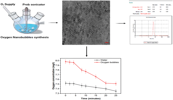
:![[thin space (1/6-em)]](https://www.rsc.org/images/entities/char_2009.gif) 1 mixture of methanol and chloroform inside a three-neck conical flask, as shown in Fig. 8. A thin-layered lipid film was obtained by drying in a hot air vacuum. The lipid layer was then re-suspended into 10 ml of PBS with 10% glycerol to get the final concentration of 3.36 mg ml−1 of lipids, which was then sonicated at 50 °C using a water bath until a milky suspension was formed. Furthermore, the suspension was sonicated using probe-sonication with pulsed mode at 190 W for 5 min in the presence of an oxygen supply to fabricate nanobubbles.111 The size distribution and zeta potential of the oxygen nanobubbles were determined using a zeta-sizer (Malvern, USA).
1 mixture of methanol and chloroform inside a three-neck conical flask, as shown in Fig. 8. A thin-layered lipid film was obtained by drying in a hot air vacuum. The lipid layer was then re-suspended into 10 ml of PBS with 10% glycerol to get the final concentration of 3.36 mg ml−1 of lipids, which was then sonicated at 50 °C using a water bath until a milky suspension was formed. Furthermore, the suspension was sonicated using probe-sonication with pulsed mode at 190 W for 5 min in the presence of an oxygen supply to fabricate nanobubbles.111 The size distribution and zeta potential of the oxygen nanobubbles were determined using a zeta-sizer (Malvern, USA).
The obtained results showed that the spherical NBs were created clearly with moderate stability and negative charge due to use of a mixture of phospholipids as the shell. Moreover, the stability was enhanced by adding glycerol, which establishes an equilibrium between the shell and the gas core. The oxygen was released in a continuous manner for up to 20 minutes compared with the same volume of water, which was 30 ml. This estimation ensured the successful fabrication of oxygen nanobubbles with moderate stability and further experiments are required to improve their stability.
4. Conclusions
The limitation of different cancer therapies is strongly dependent upon tumor hypoxia. There have been numerous attempts through different approaches to reverse hypoxia of cancer cells to date; however, there is no treatment approved by the FDA to reverse tumor hypoxia. Recently, two novel products have been reported to get rid of tumor hypoxia, however two products are currently still undergoing further clinical trials. The first treatment is designed to increase the diffusion of oxygen. The second treatment is fluorocarbons with low boiling points, which have the capability to act as an oxygen delivery system with high efficiency. Prospective, randomized, and placebo-controlled clinical trials will be essential to reveal the efficacy of oxygen as a novel supplement for overcoming tumor hypoxia. Moreover, to establish oxygen as an alternative method for enhancing cancer treatment, there are further issues that urgently need to be explored, such as focusing on all the factors that lead to hypoxia in normal cells, converting them to tumor cells, studying if the hypoxia in tumor cells is reversible or irreversible, understanding the mechanism of oxygen inside the tumor cells and the catalase function for decomposing the H2O2 into water and oxygen, and optimizing the conditions for enhancing cancer treatment using oxygen.Conflicts of interest
The authors report no conflicts of interest in this work.References
- H. Sung, J. Ferlay, R. L. Siegel, M. Laversanne, I. Soerjomataram, A. Jemal and F. Bray, Global Cancer Statistics 2020: GLOBOCAN Estimates of Incidence and Mortality Worldwide for 36 Cancers in 185 Countries, Ca-Cancer J. Clin., 2021, 71(3), 209–249, DOI:10.3322/caac.21660.
- Z. Tang, Z. Xu, X. Zhu and J. Zhang, New Insights into Molecules and Pathways of Cancer Metabolism and Therapeutic Implications, Cancer Commun., 2021, 41(1), 16–36, DOI:10.1002/cac2.12112.
- K. Graham and E. Unger, Overcoming Tumor Hypoxia as a Barrier to Radiotherapy, Chemotherapy and Immunotherapy in Cancer Treatment, Int. J. Nanomed., 2018, 13, 6049–6058, DOI:10.2147/IJN.S140462.
- F. Y. Cheng, C. H. Chan, B. J. Wang, Y. L. Yeh, Y. J. Wang and H. W. Chiu, The Oxygen-Generating Calcium Peroxide-Modified Magnetic Nanoparticles Attenuate Hypoxia-Induced Chemoresistance in Triple-Negative Breast Cancer, Cancers, 2021, 13(4), 1–16, DOI:10.3390/cancers13040606.
- S. A. Salim, E. A. Kamoun, S. Evans, T. H. Taha, E. M. El-Fakharany, M. M. Elmazar, A. F. Abdel-Aziz, R. H. Abou-Saleh and T. A. Salaheldin, Novel Oxygen-Generation from Electrospun Nanofibrous Scaffolds with Anticancer Properties: Synthesis of PMMA-Conjugate PVP–H2O2 Nanofibers, Characterization, and in Vitro Bio-Evaluation Tests, RSC Adv., 2021, 11(33), 19978–19991 RSC.
- A. L. Farris, A. N. Rindone and W. L. Grayson, Oxygen Delivering Biomaterials for Tissue Engineering, J. Mater. Chem. B, 2016, 4(20), 3422–3432, 10.1039/c5tb02635k.
- M.-Y. Wu, G.-T. Yiang, W.-T. Liao, A. P.-Y. Tsai, Y.-L. Cheng, P.-W. Cheng, C.-Y. Li and C.-J. Li, Current Mechanistic Concepts in Ischemia and Reperfusion Injury, Cell. Physiol. Biochem., 2018, 46(4), 1650–1667 CrossRef CAS PubMed.
- Y. Evron, C. K. Colton, B. Ludwig, G. C. Weir, B. Zimermann, S. Maimon, T. Neufeld, N. Shalev, T. Goldman and A. Leon, Long-Term Viability and Function of Transplanted Islets Macroencapsulated at High Density Are Achieved by Enhanced Oxygen Supply, Sci. Rep., 2018, 8(1), 1–13 CAS.
- M. M. Coronel, J. P. Liang, Y. Li and C. L. Stabler, Oxygen Generating Biomaterial Improves the Function and Efficacy of Beta Cells within a Macroencapsulation Device, Biomaterials, 2019, 210, 1–11, DOI:10.1016/j.biomaterials.2019.04.017.
- G. Camci-Unal, N. Alemdar, N. Annabi and A. Khademhosseini, Oxygen-releasing Biomaterials for Tissue Engineering, Polym. Int., 2013, 62(6), 843–848 CrossRef CAS PubMed.
- E. Mohseni-Vadeghani, R. Karimi-Soflou, S. Khorshidi and A. Karkhaneh, Fabrication of Oxygen and Calcium Releasing Microcarriers with Different Internal Structures for Bone Tissue Engineering: Solid Filled versus Hollow Microparticles, Colloids Surf., B, 2021, 197, 111376 CrossRef CAS PubMed.
- H. Y. Kim, S. Y. Kim, H.-Y. Lee, J. H. Lee, G.-J. Rho, H.-J. Lee, H.-C. Lee, J.-H. Byun and S. H. Oh, Oxygen-Releasing Microparticles for Cell Survival and Differentiation Ability under Hypoxia for Effective Bone Regeneration, Biomacromolecules, 2019, 20(2), 1087–1097 CrossRef CAS PubMed.
- L. Gao, Z. Huang, S. Yan, K. Zhang, S. Xu, G. Li, L. Cui and J. Yin, Sr-HA-Graft-Poly (γ-Benzyl-L-Glutamate) Nanocomposite Microcarriers: Controllable Sr2+ Release for Accelerating Osteogenenisis and Bony Nonunion Repair, Biomacromolecules, 2017, 18(11), 3742–3752 CrossRef CAS PubMed.
- P. A. Shiekh, A. Singh and A. Kumar, Oxygen-Releasing Antioxidant Cryogel Scaffolds with Sustained Oxygen Delivery for Tissue Engineering Applications, ACS Appl. Mater. Interfaces, 2018, 10(22), 18458–18469 CrossRef CAS PubMed.
- T. Abudula, K. Gauthaman, A. H. Hammad, K. Joshi Navare, A. A. Alshahrie, S. A. Bencherif, A. Tamayol and A. Memic, Oxygen-Releasing Antibacterial Nanofibrous Scaffolds for Tissue Engineering Applications, Polymers, 2020, 12(6), 1233 CrossRef CAS PubMed.
- M. Gholipourmalekabadi, S. Zhao, B. S. Harrison, M. Mozafari and A. M. Seifalian, Oxygen-Generating Biomaterials: A New, Viable Paradigm for Tissue Engineering?, Trends Biotechnol., 2016, 34(12), 1010–1021 CrossRef CAS PubMed.
- J. Szymańska, Antifungal Efficacy of Hydrogen Peroxide in Dental Unit Waterline Disinfection, Ann. Agric. Environ. Med., 2006, 13(2), 313–317 Search PubMed.
- D. Modhave, B. Barrios and A. Paudel, PVP-H2O2 Complex as a New Stressor for the Accelerated Oxidation Study of Pharmaceutical Solids, Pharmaceutics, 2019, 11(9), 457 CrossRef CAS PubMed.
- R. R. Mallepally, C. C. Parrish, M. A. M. Mc Hugh and K. R. Ward, Hydrogen Peroxide Filled Poly (Methyl Methacrylate) Microcapsules: Potential Oxygen Delivery Materials, Int. J. Pharm., 2014, 475(1–2), 130–137 CrossRef CAS PubMed.
- Z. Li, X. Guo and J. Guan, An Oxygen Release System to Augment Cardiac Progenitor Cell Survival and Differentiation under Hypoxic Condition, Biomaterials, 2012, 33(25), 5914–5923 CrossRef CAS PubMed.
- J. Gao, J. Song, J. Ye, X. Duan, D. D. Dionysiou, J. S. Yadav, M. N. Nadagouda, L. Yang and S. Luo, Comparative Toxicity Reduction Potential of UV/Sodium Percarbonate and UV/Hydrogen Peroxide Treatments for Bisphenol A in Water: An Integrated Analysis Using Chemical, Computational, Biological, and Metabolomic Approaches, Water Res., 2021, 190, 116755 CrossRef CAS PubMed.
- J. P. McQuilling, S. Sittadjody, S. Pendergraft, A. C. Farney and E. C. Opara, Applications of Particulate Oxygen-Generating Substances (POGS) in the Bioartificial Pancreas, Biomater. Sci., 2017, 5(12), 2437–2447 RSC.
- X. Wang, S. Li, X. Liu, X. Wu, N. Ye, X. Yang and Z. Li, Boosting Nanomedicine Efficacy with Hyperbaric Oxygen Therapy, in Bio-Nanomedicine for Cancer Therapy, Springer, 2021, pp. 77–95 Search PubMed.
- M. C. T. Batenburg, W. Maarse, F. van der Leij, I. O. Baas, O. Boonstra, N. Lansdorp, A. Doeksen, D. H. J. G. van den Bongard and H. M. Verkooijen, The Impact of Hyperbaric Oxygen Therapy on Late Radiation Toxicity and Quality of Life in Breast Cancer Patients, Breast Cancer Res. Treat., 2021, 1–9 Search PubMed.
- I. Moen and L. E. B. Stuhr, Hyperbaric Oxygen Therapy and Cancer—A Review, Target. Oncol., 2012, 7(4), 233–242 CrossRef PubMed.
- K. D. Torp and H. M. Murphy-Lavoie, Acute Traumatic Ischemia Hyperbaric Evaluation and Treatment, StatPearls, 2021 Search PubMed.
- M. P. Krafft, Alleviating Tumor Hypoxia with Perfluorocarbon-Based Oxygen Carriers, Curr. Opin. Pharmacol., 2020, 53, 117–125 CrossRef CAS PubMed.
- A. Sahu, I. Kwon and G. Tae, Improving Cancer Therapy through the Nanomaterials-Assisted Alleviation of Hypoxia, Biomaterials, 2020, 228, 119578 CrossRef CAS PubMed.
- W. Tang, Z. Zhen, M. Wang, H. Wang, Y. Chuang, W. Zhang, G. D. Wang, T. Todd, T. Cowger and H. Chen, Red Blood Cell-facilitated Photodynamic Therapy for Cancer Treatment, Adv. Funct. Mater., 2016, 26(11), 1757–1768 CrossRef CAS PubMed.
- A. M. Piras, A. Dessy, F. Chiellini, E. Chiellini, C. Farina, M. Ramelli and E. Della Valle, Polymeric Nanoparticles for Hemoglobin-Based Oxygen Carriers, Biochim. Biophys. Acta, Proteins Proteomics, 2008, 1784(10), 1454–1461 CrossRef CAS PubMed.
- C. Murayama, A. T. Kawaguchi, K. Ishikawa, A. Kamijo, N. Kato, Y. Ohizumi, S. Sadahiro and M. Haida, Liposome-encapsulated Hemoglobin Ameliorates Tumor Hypoxia and Enhances Radiation Therapy to Suppress Tumor Growth in Mice, Artif. Organs, 2012, 36(2), 170–177 CrossRef CAS PubMed.
- S. Wang, F. Yuan, K. Chen, G. Chen, K. Tu, H. Wang and L.-Q. Wang, Synthesis of Hemoglobin Conjugated Polymeric Micelle: A ZnPc Carrier with Oxygen Self-Compensating Ability for Photodynamic Therapy, Biomacromolecules, 2015, 16(9), 2693–2700 CrossRef CAS PubMed.
- Z. Luo, M. Zheng, P. Zhao, Z. Chen, F. Siu, P. Gong, G. Gao, Z. Sheng, C. Zheng and Y. Ma, Self-Monitoring Artificial Red Cells with Sufficient Oxygen Supply for Enhanced Photodynamic Therapy, Sci. Rep., 2016, 6(1), 1–11 CrossRef CAS PubMed.
- J. Yang, W. Li, L. Luo, M. Jiang, C. Zhu, B. Qin, H. Yin, X. Yuan, X. Yin and J. Zhang, Hypoxic Tumor Therapy by Hemoglobin-Mediated Drug Delivery and Reversal of Hypoxia-Induced Chemoresistance, Biomaterials, 2018, 182, 145–156 CrossRef CAS PubMed.
- H. Furukawa, K. E. Cordova, M. O’Keeffe and O. M. Yaghi, The Chemistry and Applications of Metal-Organic Frameworks, Science, 2013, 341(6149), 1230444 CrossRef PubMed.
- H.-C. Zhou, J. R. Long and O. M. Yaghi, Introduction to Metal–Organic Frameworks, Chem. Rev., 2012, 112(2), 673–674 CrossRef CAS PubMed.
- J. B. DeCoste, M. H. Weston, P. E. Fuller, T. M. Tovar, G. W. Peterson, M. D. LeVan and O. K. Farha, Metal–Organic Frameworks for Oxygen Storage, Angew. Chem., 2014, 126(51), 14316–14319 CrossRef.
- S. Gao, P. Zheng, Z. Li, X. Feng, W. Yan, S. Chen, W. Guo, D. Liu, X. Yang and S. Wang, Biomimetic O2-Evolving Metal-Organic Framework Nanoplatform for Highly Efficient Photodynamic Therapy against Hypoxic Tumor, Biomaterials, 2018, 178, 83–94 CrossRef CAS PubMed.
- Z. Xie, X. Cai, C. Sun, S. Liang, S. Shao, S. Huang, Z. Cheng, M. Pang, B. Xing and A. A. Al Kheraif, O2-Loaded PH-Responsive Multifunctional Nanodrug Carrier for Overcoming Hypoxia and Highly Efficient Chemo-Photodynamic Cancer Therapy, Chem. Mater., 2018, 31(2), 483–490 CrossRef.
- J. Jägers, A. Wrobeln and K. B. Ferenz, Perfluorocarbon-Based Oxygen Carriers: From Physics to Physiology, Pfluegers Arch., 2021, 473(2), 139–150 CrossRef PubMed.
- N. Moasefi, M. Fouladi, A. H. Norooznezhad, R. Yarani, A. Rahmani and K. Mansouri, How Could Perfluorocarbon Affect Cytokine Storm and Angiogenesis in Coronavirus Disease 2019 (COVID-19): Role of Hypoxia-Inducible Factor 1α, Inflammation Res., 2021, 1–4 Search PubMed.
- Y. Que, Y. Liu, W. Tan, C. Feng, P. Shi, Y. Li and H. Xiaoyu, Enhancing Photodynamic Therapy Efficacy by Using Fluorinated Nanoplatform, ACS Macro Lett., 2016, 5(2), 168–173 CrossRef CAS PubMed.
- H. Ren, J. Liu, F. Su, S. Ge, A. Yuan, W. Dai, J. Wu and Y. Hu, Relighting Photosensitizers by Synergistic Integration of Albumin and Perfluorocarbon for Enhanced Photodynamic Therapy, ACS Appl. Mater. Interfaces, 2017, 9(4), 3463–3473 CrossRef CAS PubMed.
- F. Mao, L. Wen, C. Sun, S. Zhang, G. Wang, J. Zeng, Y. Wang, J. Ma, M. Gao and Z. Li, Ultrasmall Biocompatible Bi2Se3 Nanodots for Multimodal Imaging-Guided Synergistic Radiophotothermal Therapy against Cancer, ACS Nano, 2016, 10(12), 11145–11155 CrossRef CAS PubMed.
- M. Gao, C. Liang, X. Song, Q. Chen, Q. Jin, C. Wang and Z. Liu, Erythrocyte-membrane-enveloped Perfluorocarbon as Nanoscale Artificial Red Blood Cells to Relieve Tumor Hypoxia and Enhance Cancer Radiotherapy, Adv. Mater., 2017, 29(35), 1701429 CrossRef PubMed.
- W.-C. Huang, W.-H. Chiang, Y.-H. Cheng, W.-C. Lin, C.-F. Yu, C.-Y. Yen, C.-K. Yeh, C.-S. Chern, C.-S. Chiang and H.-C. Chiu, Tumortropic Monocyte-Mediated Delivery of Echogenic Polymer Bubbles and Therapeutic Vesicles for Chemotherapy of Tumor Hypoxia, Biomaterials, 2015, 71, 71–83 CrossRef CAS PubMed.
- J. D. Martin, G. Seano and R. K. Jain, Normalizing Function of Tumor Vessels: Progress, Opportunities, and Challenges, Annu. Rev. Physiol., 2019, 81, 505–534 CrossRef CAS PubMed.
- W. Jiang, Y. Huang, Y. An and B. Y. S. Kim, Remodeling Tumor Vasculature to Enhance Delivery of Intermediate-Sized Nanoparticles, ACS Nano, 2015, 9(9), 8689–8696 CrossRef CAS PubMed.
- S. Guo, C. M. Lin, Z. Xu, L. Miao, Y. Wang and L. Huang, Co-Delivery of Cisplatin and Rapamycin for Enhanced Anticancer Therapy through Synergistic Effects and Microenvironment Modulation, ACS Nano, 2014, 8(5), 4996–5009 CrossRef CAS PubMed.
- D. Wang, J. Fu, Y. Shi, D. Peng, L. Yuan, B. He, W. Dai, H. Zhang, X. Wang and J. Tian, The Modulation of Tumor Vessel Permeability by Thalidomide and Its Impacts on Different Types of Targeted Drug Delivery Systems in a Sarcoma Mouse Model, J. Controlled Release, 2016, 238, 186–196 CrossRef CAS PubMed.
- A. B. Satterlee, J. D. Rojas, P. A. Dayton and L. Huang, Enhancing Nanoparticle Accumulation and Retention in Desmoplastic Tumors via Vascular Disruption for Internal Radiation Therapy, Theranostics, 2017, 7(2), 253 CrossRef CAS PubMed.
- V. P. Chauhan, J. D. Martin, H. Liu, D. A. Lacorre, S. R. Jain, S. V. Kozin, T. Stylianopoulos, A. S. Mousa, X. Han and P. Adstamongkonkul, Angiotensin Inhibition Enhances Drug Delivery and Potentiates Chemotherapy by Decompressing Tumour Blood Vessels, Nat. Commun., 2013, 4(1), 1–11 Search PubMed.
- P. Papageorgis, C. Polydorou, F. Mpekris, C. Voutouri, E. Agathokleous, C. P. Kapnissi-Christodoulou and T. Stylianopoulos, Tranilast-Induced Stress Alleviation in Solid Tumors Improves the Efficacy of Chemo-and Nanotherapeutics in a Size-Independent Manner, Sci. Rep., 2017, 7(1), 1–12 CrossRef PubMed.
- W. Yu, T. Liu, M. Zhang, Z. Wang, J. Ye, C.-X. Li, W. Liu, R. Li, J. Feng and X.-Z. Zhang, O2 Economizer for Inhibiting Cell Respiration to Combat the Hypoxia Obstacle in Tumor Treatments, ACS Nano, 2019, 13(2), 1784–1794 CAS.
- X. Meng, X. Zhang, Y. Lei, D. Cao and Z. Wang, Biodegradable Copper–Metformin Nanoscale Coordination Polymers for Enhanced Chemo/Chemodynamic Synergistic Therapy by Reducing Oxygen Consumption to Promote H2O2 Accumulation, J. Mater. Chem. B, 2021, 9(8), 1988–2000 RSC.
- L. Meng, Y. Cheng, X. Tong, S. Gan, Y. Ding, Y. Zhang, C. Wang, L. Xu, Y. Zhu, J. Wu, Y. Hu and A. Yuan, Tumor Oxygenation and Hypoxia Inducible Factor-1 Functional Inhibition via a Reactive Oxygen Species Responsive Nanoplatform for Enhancing Radiation Therapy and Abscopal Effects, ACS Nano, 2018, 12(8), 8308–8322, DOI:10.1021/acsnano.8b03590.
- S. J. Forrester, D. S. Kikuchi, M. S. Hernandes, Q. Xu and K. K. Griendling, Reactive Oxygen Species in Metabolic and Inflammatory Signaling, Circ. Res., 2018, 122(6), 877–902 CrossRef CAS PubMed.
- P. D. Ray, B.-W. Huang and Y. Tsuji, Reactive Oxygen Species (ROS) Homeostasis and Redox Regulation in Cellular Signaling, Cell. Signalling, 2012, 24(5), 981–990 CrossRef CAS PubMed.
- G. H. Kim, J. E. Kim, S. J. Rhie and S. Yoon, The Role of Oxidative Stress in Neurodegenerative Diseases, Exp. Neurobiol., 2015, 24(4), 325 CrossRef PubMed.
- A. Costa, A. Scholer-Dahirel and F. Mechta-Grigoriou, The Role of Reactive Oxygen Species and Metabolism on Cancer Cells and Their Microenvironment, in Seminars in cancer biology, Elsevier, 2014, vol. 25, pp. 23–32 Search PubMed.
- S. Sadeghi Mohammadi, Z. Vaezi, B. Shojaedin-Givi and H. Naderi-Manesh, Chemiluminescent Liposomes as a Theranostic Carrier for Detection of Tumor Cells under Oxidative Stress, Anal. Chim. Acta, 2019, 1059, 113–123, DOI:10.1016/J.ACA.2019.01.045.
- H. Chen, W. He and Z. Guo, An H2O2-Responsive Nanocarrier for Dual-Release of Platinum Anticancer Drugs and O2: Controlled Release and Enhanced Cytotoxicity against Cisplatin Resistant Cancer Cells, Chem. Commun., 2014, 50(68), 9714–9717, 10.1039/c4cc03385j.
- T. Liu, N. Zhang, Z. Wang, M. Wu, Y. Chen, M. Ma, H. Chen and J. Shi, Endogenous Catalytic Generation of O2 Bubbles for In Situ Ultrasound-Guided High Intensity Focused Ultrasound Ablation, ACS Nano, 2017, 11(9), 9093–9102, DOI:10.1021/acsnano.7b03772.
- S. Z. F. Phua, G. Yang, W. Q. Lim, A. Verma, H. Chen, T. Thanabalu and Y. Zhao, Catalase-Integrated Hyaluronic Acid as Nanocarriers for Enhanced Photodynamic Therapy in Solid Tumor, ACS Nano, 2019, 13(4), 4742–4751, DOI:10.1021/acsnano.9b01087.
- C. Lee, K. Lim, S. S. Kim, L. X. Thien, E. S. Lee, K. T. Oh, H. G. Choi and Y. S. Youn, Chlorella-Gold Nanorods Hydrogels Generating Photosynthesis-Derived Oxygen and Mild Heat for the Treatment of Hypoxic Breast Cancer, J. Controlled Release, 2019, 294, 77–90, DOI:10.1016/J.JCONREL.2018.12.011.
- W.-L. Liu, T. Liu, M.-Z. Zou, W.-Y. Yu, C.-X. Li, Z.-Y. He, M.-K. Zhang, M.-D. Liu, Z.-H. Li, J. Feng and X.-Z. Zhang, Aggressive Man-Made Red Blood Cells for Hypoxia-Resistant Photodynamic Therapy, Adv. Mater., 2018, 30(35), 1802006, DOI:10.1002/adma.201802006.
- R. Q. Li, C. Zhang, B. R. Xie, W. Y. Yu, W. X. Qiu, H. Cheng and X. Z. Zhang, A Two-Photon Excited O2-Evolving Nanocomposite for Efficient Photodynamic Therapy against Hypoxic Tumor, Biomaterials, 2019, 194, 84–93, DOI:10.1016/J.BIOMATERIALS.2018.12.017.
- T.-J. Zhou, L. Xing, Y.-T. Fan, P.-F. Cui and H.-L. Jiang, Light Triggered Oxygen-Affording Engines for Repeated Hypoxia-Resistant Photodynamic Therapy, J. Controlled Release, 2019, 307, 44–54, DOI:10.1016/j.jconrel.2019.06.016.
- S. M. Nabavi and A. S. S. Silva, Nonvitamin and Nonmineral Nutritional Supplements, Elsevier Science, 2018 Search PubMed.
- J. He, L. H. Fu, C. Qi, J. Lin and P. Huang, Metal Peroxides for Cancer Treatment, Bioact. Mater., 2021, 6(9), 2698–2710, DOI:10.1016/J.BIOACTMAT.2021.01.026.
- Z. Tang, Y. Liu, D. Ni, J. Zhou, M. Zhang, P. Zhao, B. Lv, H. Wang, D. Jin and W. Bu, Biodegradable Nanoprodrugs: “Delivering” ROS to Cancer Cells for Molecular Dynamic Therapy, Adv. Mater., 2020, 32(4), 1904011, DOI:10.1002/adma.201904011.
- L.-S. Lin, J.-F. Wang, J. Song, Y. Liu, G. Zhu, Y. Dai, Z. Shen, R. Tian, J. Song, Z. Wang, W. Tang, G. Yu, Z. Zhou, Z. Yang, T. Huang, G. Niu, H.-H. Yang, Z.-Y. Chen and X. Chen, Cooperation of Endogenous and Exogenous Reactive Oxygen Species Induced by Zinc Peroxide Nanoparticles to Enhance Oxidative Stress-Based Cancer Therapy, Theranostics, 2019, 9(24), 7200–7209, DOI:10.7150/thno.39831.
- V. Kolenko, E. Teper, A. Kutikov and R. Uzzo, Zinc and Zinc Transporters in Prostate Carcinogenesis, Nat. Rev. Urol., 2013, 10(4), 219–226, DOI:10.1038/nrurol.2013.43.
- R. B. Franklin and L. C. Costello, Zinc as an Anti-Tumor Agent in Prostate Cancer and in Other Cancers, Arch. Biochem. Biophys., 2007, 463(2), 211–217, DOI:10.1016/j.abb.2007.02.033.
- Y. Han, J. Ouyang, Y. Li, F. Wang and J.-H. Jiang, Engineering H2O2 Self-Supplying Nanotheranostic Platform for Targeted and Imaging-Guided Chemodynamic Therapy, ACS Appl. Mater. Interfaces, 2020, 12(1), 288–297, DOI:10.1021/acsami.9b18676.
- X. Wang, Y. Hu and H. Wei, Nanozymes in Bionanotechnology: From Sensing to Therapeutics and Beyond, Inorg. Chem. Front., 2016, 3(1), 41–60, 10.1039/C5QI00240K.
- D. P. Cormode, L. Gao and H. Koo, Emerging Biomedical Applications of Enzyme-Like Catalytic Nanomaterials, Trends Biotechnol., 2018, 36(1), 15–29, DOI:10.1016/j.tibtech.2017.09.006.
- L. Feng, B. Liu, R. Xie, D. Wang, C. Qian, W. Zhou, J. Liu, D. Jana, P. Yang and Y. Zhao, An Ultrasmall SnFe2O4 Nanozyme with Endogenous Oxygen Generation and Glutathione Depletion for Synergistic Cancer Therapy, Adv. Funct. Mater., 2021, 31(5), 2006216, DOI:10.1002/adfm.202006216.
- W. Lv, M. Cao, J. Liu, Y. Hei and J. Bai, Tumor Microenvironment-Responsive Nanozymes Achieve Photothermal-Enhanced Multiple Catalysis against Tumor Hypoxia, Acta Biomater., 2021, 135, 617–627, DOI:10.1016/j.actbio.2021.08.015.
- D. P. Bhattarai, L. E. Aguilar, C. H. Park and C. S. Kim, A Review on Properties of Natural and Synthetic Based Electrospun Fibrous Materials for Bone Tissue Engineering, Membranes, 2018, 8(3), 62 CrossRef PubMed.
- M. C. Hacker, J. Krieghoff and A. G. Mikos, Synthetic Polymers, in Principles of regenerative medicine, Elsevier, 2019, pp. 559–590 Search PubMed.
- C. Zhang, J. Chen, Y. Song, J. Luo, P. Jin, X. Wang, L. Xin, F. Qiu, J. Yao, G. Wang and P. Huang, Ultrasound-Enhanced Reactive Oxygen Species Responsive Charge-Reversal Polymeric Nanocarriers for Efficient Pancreatic Cancer Gene Delivery, ACS Appl. Mater. Interfaces, 2022, 14(2), 2587–2596, DOI:10.1021/acsami.1c20030.
- A. B. Shcherbakov, V. V. Reukov, A. V. Yakimansky, E. L. Krasnopeeva, O. S. Ivanova, A. L. Popov and V. K. Ivanov, Polymers CeO2 Nanoparticle-Containing Polymers for Biomedical Applications: A Review, Polymers, 2021, 13(6), 924, DOI:10.3390/polym13060924.
- L. S. Wray, X. Hu, J. Gallego, I. Georgakoudi, F. G. Omenetto, D. Schmidt and D. L. Kaplan, Effect of Processing on Silk-based Biomaterials: Reproducibility and Biocompatibility, J. Biomed. Mater. Res., Part B, 2011, 99(1), 89–101 CrossRef PubMed.
- D. N. Rockwood, R. C. Preda, T. Yücel, X. Wang, M. L. Lovett and D. L. Kaplan, Materials Fabrication from Bombyx mori Silk Fibroin, Nat. Protoc., 2011, 6(10), 1612–1631 CrossRef CAS PubMed.
- J. Melke, S. Midha, S. Ghosh, K. Ito and S. Hofmann, Silk Fibroin as Biomaterial for Bone Tissue Engineering, Acta Biomater., 2016, 31, 1–16 CrossRef CAS PubMed.
- W. Huang, S. Ling, C. Li, F. G. Omenetto and D. L. Kaplan, Silkworm Silk-Based Materials and Devices Generated Using Bio-Nanotechnology, Chem. Soc. Rev., 2018, 47(17), 6486–6504 RSC.
- F. Chen, D. Porter and F. Vollrath, Morphology and Structure of Silkworm Cocoons, Mater. Sci. Eng., C, 2012, 32(4), 772–778 CrossRef CAS.
- M. Arumugam, B. Murugesan, N. Pandiyan, D. K. Chinnalagu, G. Rangasamy and S. Mahalingam, Electrospinning Cellulose Acetate/Silk Fibroin/Au-Ag Hybrid Composite Nanofiber for Enhanced Biocidal Activity against MCF-7 Breast Cancer Cell, Mater. Sci. Eng., C, 2021, 123, 112019 CrossRef CAS PubMed.
- R. Yang, M. Hou, Y. Gao, S. Lu, L. Zhang, Z. Xu, C. M. Li, Y. Kang and P. Xue, Biomineralization-Inspired Crystallization of Manganese Oxide on Silk Fibroin Nanoparticles for in Vivo MR/Fluorescence Imaging-Assisted Tri-Modal Therapy of Cancer, Theranostics, 2019, 9(21), 6314–6333, DOI:10.7150/thno.36252.
- A. Abruzzo, Chitosan Based Hydrogels for Transmucosal Drug Delivery, 2013 Search PubMed.
- I. Akbarzadeh, M. Shayan, M. Bourbour, M. Moghtaderi, H. Noorbazargan, F. Eshrati Yeganeh, S. Saffar and M. Tahriri, Preparation, Optimization and In-Vitro Evaluation of Curcumin-Loaded Niosome@Calcium Alginate Nanocarrier as a New Approach for Breast Cancer Treatment, Biology, 2021, 10(3), 173, DOI:10.3390/biology10030173.
- J. B. Buse, R. A. DeFronzo, J. Rosenstock, T. Kim, C. Burns, S. Skare, A. Baron and M. Fineman, The Primary Glucose-Lowering Effect of Metformin Resides in the Gut, Not the Circulation: Results from Short-Term Pharmacokinetic and 12-Week Dose-Ranging Studies, Diabetes Care, 2016, 39(2), 198–205 CrossRef CAS PubMed.
- K. Y. Lee and D. J. Mooney, Alginate: Properties and Biomedical Applications, Prog. Polym. Sci., 2012, 37(1), 106–126 CrossRef CAS PubMed.
- C.-C. Huang, W.-T. Chia, M.-F. Chung, K.-J. Lin, C.-W. Hsiao, C. Jin, W.-H. Lim, C.-C. Chen and H.-W. Sung, An Implantable Depot That Can Generate Oxygen in Situ for Overcoming Hypoxia-Induced Resistance to Anticancer Drugs in Chemotherapy, J. Am. Chem. Soc., 2016, 138(16), 5222–5225, DOI:10.1021/jacs.6b01784.
- M. M. Merza, F. M. Hussein and R. R. AL-ani, Physical Properties and Biological Activity of Methyldopa Drug Carrier Cellulose Derivatives. Theoretical Study, Egypt. J. Chem., 2021, 64(8), 4081–4090 Search PubMed.
- C. J. Wijaya, S. Ismadji and S. Gunawan, A Review of Lignocellulosic-Derived Nanoparticles for Drug Delivery Applications: Lignin Nanoparticles, Xylan Nanoparticles, and Cellulose Nanocrystals, Molecules, 2021, 26(3), 676 CrossRef CAS PubMed.
- Z. Liu, S. Zhang, B. He, S. Wang and F. Kong, Synthesis of Cellulose Aerogels as Promising Carriers for Drug Delivery: A Review, Cellulose, 2021, 1–18 Search PubMed.
- W. Liu, H. Du, H. Liu, H. Xie, T. Xu, X. Zhao, Y. Liu, X. Zhang and C. Si, Highly Efficient and Sustainable Preparation of Carboxylic and Thermostable Cellulose Nanocrystals via FeCl3-Catalyzed Innocuous Citric Acid Hydrolysis, ACS Sustainable Chem. Eng., 2020, 8(44), 16691–16700 CrossRef CAS.
- H. Du, W. Liu, M. Zhang, C. Si, X. Zhang and B. Li, Cellulose Nanocrystals and Cellulose Nanofibrils Based Hydrogels for Biomedical Applications, Carbohydr. Polym., 2019, 209, 130–144 CrossRef CAS PubMed.
- P. N. Bhandari, Y. Cui, B. D. Elzey, C. J. Goergen, C. M. Long and J. Irudayaraj, Oxygen Nanobubbles Revert Hypoxia by Methylation Programming, Sci. Rep., 2017, 7(1), 1–14 CrossRef CAS PubMed.
- J. Park, J. Nam, H. Yun, H.-J. Jin and H. W. Kwak, Aquatic Polymer-Based Edible Films of Fish Gelatin Crosslinked with Alginate Dialdehyde Having Enhanced Physicochemical Properties, Carbohydr. Polym., 2021, 254, 117317 CrossRef CAS PubMed.
- S. Yadav, G. K. Mehrotra, P. Bhartiya, A. Singh and P. K. Dutta, Preparation, Physicochemical and Biological Evaluation of Quercetin Based Chitosan-Gelatin Film for Food Packaging, Carbohydr. Polym., 2020, 227, 115348 CrossRef CAS PubMed.
- C. Labay, M. Roldán, F. Tampieri, A. Stancampiano, P. E. Bocanegra, M.-P. Ginebra and C. Canal, Enhanced Generation of Reactive Species by Cold Plasma in Gelatin Solutions for Selective Cancer Cell Death, ACS Appl. Mater. Interfaces, 2020, 12(42), 47256–47269, DOI:10.1021/acsami.0c12930.
- Y. Mizukami, A. Moriya, Y. Takahashi, K. Shimizu, S. Konishi, Y. Takakura and M. Nishikawa, Incorporation of Gelatin Microspheres into HepG2 Human Hepatocyte Spheroids for Functional Improvement through Improved Oxygen Supply to Spheroid Core, Biol. Pharm. Bull., 2020, 43(8), 1220–1225 CrossRef CAS PubMed.
- W. Lv, H. Xia, K. Y. Zhang, Z. Chen, S. Liu, W. Huang and Q. Zhao, Photothermal-Triggered Release of Singlet Oxygen from an Endoperoxide-Containing Polymeric Carrier for Killing Cancer Cells, Mater. Horiz., 2017, 4(6), 1185–1189 RSC.
- L. Liu, Z. Peng, C. Wang, C. Wang, C. Liu, L. Zhu and C. Tang, Effect of Synthetic Oxygen-Generating System on Cell Survival under Hypoxic Condition in Vitro, J. Biomater. Sci., Polym. Ed., 2021, 32(8), 967–979 CrossRef CAS PubMed.
- A. K. A. Ahmed, C. Sun, L. Hua, Z. Zhang, Y. Zhang, W. Zhang and T. Marhaba, Generation of Nanobubbles by Ceramic Membrane Filters: The Dependence of Bubble Size and Zeta Potential on Surface Coating, Pore Size and Injected Gas Pressure, Chemosphere, 2018, 203, 327–335, DOI:10.1016/J.CHEMOSPHERE.2018.03.157.
- K. Y. Sletta, M. K. Tveitarås, N. Lu, A. S. T. Engelsen, R. K. Reed, A. Garmann-Johnsen and L. Stuhr, Oxygen-Dependent Regulation of Tumor Growth and Metastasis in Human Breast Cancer Xenografts, PLoS One, 2017, 12(8), e0183254, DOI:10.1371/journal.pone.0183254.
- L. Zheng, B. Zhou, X. Meng, W. Zhu, A. Zuo, X. Wang, R. Jiang and S. Yu, A Model of Spontaneous Mouse Mammary Tumor for Human Estrogen Receptor- and Progesterone Receptor-Negative Breast Cancer, Int. J. Oncol., 2014, 45(6), 2241–2249, DOI:10.3892/ijo.2014.2657.
- M. S. Khan, J. Hwang, Y. Seo, K. Shin, K. Lee, C. Park, Y. Choi, J. W. Hong and J. Choi, Engineering Oxygen Nanobubbles for the Effective Reversal of Hypoxia, Artif. Cells, Nanomed., Biotechnol., 2018, 46(suppl. 3), S318–S327, DOI:10.1080/21691401.2018.1492420.
- R. Cavalli, M. Soster and M. Argenziano, Nanobubbles: A Promising Efficient Tool for Therapeutic Delivery, Ther. Delivery, 2016, 1, 117–138, DOI:10.4155/tde.15.92.
- T. Yin, P. Wang, R. Zheng, B. Zheng, D. Cheng, X. Zhang and X. Shuai, Nanobubbles for Enhanced Ultrasound Imaging of Tumors, Int. J. Nanomed., 2012, 7, 895–904, DOI:10.2147/IJN.S28830.
- J. Owen, K. Logan, H. Nesbitt, S. Able, A. Vasilyeva, E. Bluemke, V. Kersemans, S. Smart, K. A. Vallis and A. P. McHale, Orally Administered Oxygen Nanobubbles Enhance Tumor Response to Sonodynamic Therapy, Nano Sel., 2022, 3(2), 394–401 CrossRef CAS.
| This journal is © The Royal Society of Chemistry 2022 |

