 Open Access Article
Creative Commons Attribution 3.0 Unported Licence
Correction: Chrysomycins A–C, antileukemic naphthocoumarins from Streptomyces sporoverrucosus
Shreyans K. Jainab,
Anup S. Pathaniabc,
Rajinder Parshadd,
Chandji Rainad,
Asif Aliab,
Ajai P. Guptae,
Manoj Kushwahae,
Subrayashastry Aravindaf,
Shashi Bhushanbc,
Sandip B. Bharate*bf and
Ram A. Vishwakarma*abf
aNatural Products Chemistry Division, Indian Institute of Integrative Medicine (CSIR), Canal Road, Jammu, 180001, India. E-mail: ram@iiim.ac.in; Fax: +91-191-2569333; Tel: +91-191-2569111
bAcademy of Scientific & Innovative Research (AcSIR), Anusandhan Bhawan, 2 Rafi Marg, New Delhi, 110001, India
cCancer Pharmacology Division, Indian Institute of Integrative Medicine (CSIR), Canal Road, Jammu, 180001, India
dFermentation Division, Indian Institute of Integrative Medicine (CSIR), Canal Road, Jammu, 180001, India
eQuality Control and Quality Assurance Division, Indian Institute of Integrative Medicine (CSIR), Canal Road, Jammu, 180001, India
fMedicinal Chemistry Division, Indian Institute of Integrative Medicine (CSIR), Canal Road, Jammu, 180001, India. E-mail: sbharate@iiim.ac.in
First published on 19th August 2022
Abstract
Correction for ‘Chrysomycins A–C, antileukemic naphthocoumarins from Streptomyces sporoverrucosus’ by Shreyans K. Jain et al., RSC Adv., 2013, 3, 21046–21053, https://doi.org/10.1039/c3ra42884b.
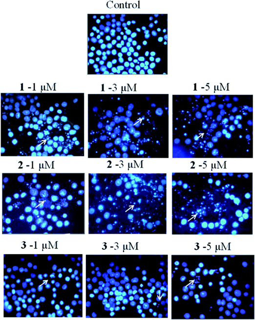
The authors regret that incorrect versions of Fig. 6 and Fig. 7 were included in the original article. The correct versions of Fig. 6 and 7 are presented below.
The Royal Society of Chemistry apologises for these errors and any consequent inconvenience to authors and readers.
| This journal is © The Royal Society of Chemistry 2022 |


