 Open Access Article
Creative Commons Attribution 3.0 Unported Licence
Induced-volatolomics for the design of tumour activated therapy†
Rémi
Châtre‡
a,
Estelle
Blochouse‡
a,
Rony
Eid
a,
Fabiola
Djago
a,
Justin
Lange
a,
Mehrad
Tarighi
a,
Brigitte
Renoux
a,
Julien
Sobilo
 b,
Alain
Le Pape
b,
Jonathan
Clarhaut
ac,
Claude
Geffroy
a,
Isabelle
Opalinski
a,
Wei
Tuo
a,
Sébastien
Papot
b,
Alain
Le Pape
b,
Jonathan
Clarhaut
ac,
Claude
Geffroy
a,
Isabelle
Opalinski
a,
Wei
Tuo
a,
Sébastien
Papot
 *ad and
Pauline
Poinot
 *a
*a
aUniversity of Poitiers, UMR CNRS 7285, Institut de Chimie des Milieux et Matériaux de Poitiers (IC2MP), Equipe Labellisée Ligue Contre le Cancer, 4 Rue Michel-Brunet, TSA 51106, 86073 Poitiers Cedex 9, France. E-mail: pauline.poinot@univ-poitiers.fr; sebastien.papot@univ-poitiers.fr
bUAR No. 44 PHENOMIN TAAM-Imagerie In Vivo, CNRS, 3B Rue de la Férollerie, F-45071 Orléans, France
cCHU de Poitiers, 2 Rue de la Miléterie, CS 90577, F-86021 Poitiers, France
dSeekyo SA, 2 Avenue Galilée, BP 30153, 86961 Futuroscope, France
First published on 11th April 2023
Abstract
The discovery of tumour-associated markers is of major interest for the development of selective cancer chemotherapy. Within this framework, we introduced the concept of induced-volatolomics enabling to monitor simultaneously the dysregulation of several tumour-associated enzymes in living mice or biopsies. This approach relies on the use of a cocktail of volatile organic compound (VOC)-based probes that are activated enzymatically for releasing the corresponding VOCs. Exogenous VOCs can then be detected in the breath of mice or in the headspace above solid biopsies as specific tracers of enzyme activities. Our induced-volatolomics modality highlighted that the up-regulation of N-acetylglucosaminidase was a hallmark of several solid tumours. Having identified this glycosidase as a potential target for cancer therapy, we designed an enzyme-responsive albumin-binding prodrug of the potent monomethyl auristatin E programmed for the selective release of the drug in the tumour microenvironment. This tumour activated therapy produced a remarkable therapeutic efficacy on orthotopic triple-negative mammary xenografts in mice, leading to the disappearance of tumours in 66% of treated animals. Thus, this study shows the potential of induced-volatolomics for the exploration of biological processes as well as the discovery of novel therapeutic strategies.
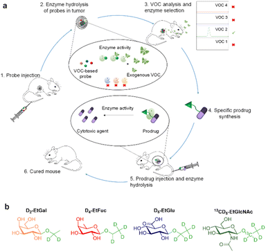
Enzymes are major targets for drug discovery in oncology.1,2 The current therapeutic arsenal includes numerous enzyme inhibitors3 and enzyme-responsive antibody–drug conjugates4 for the treatment of a wide range of malignancies. Thus, the design of novel strategies dedicated to the exploration of tumour-associated enzymes is of prime importance for cancer therapy and diagnosis.5,6 Within this framework, activable imaging probes have been developed to detect the dysregulation of some enzyme activities in tumours.7–17 However, most of these turn-on probes suffer from low signal penetration depth and background noise.18,19 Recently, we introduced the concept of induced-volatolomics for cancer diagnosis.20,21 We demonstrated that a volatile organic compound (VOC)-based probe can be activated in vivo by an enzyme overexpressed in solid tumours to trigger the release of an exogenous volatile tracer in the breath of individuals, thereby enabling cancer detection. Herein, we present an original approach allowing real time and simultaneous monitoring of the activity of several tumour-associated enzymes by means of a VOC-based probe cocktail (Fig. 1). This induced-volatolomics modality allowed the identification of upregulated enzymes within the tumour, leading to the development of a new enzyme-targeted drug candidate.
In this proof-of-concept, we chose to target glycolytic activity of tumours since glycosidase dysregulation constitutes a hallmark of many cancers.22–26 β-Galactosidase (β-Gal), α-L-fucosidase (α-Fuc), β-glucuronidase (β-Glu) and N-acetyl-β-D-glucosaminidase (β-GlcNAc) were selected as the target enzymes since their overexpression has been reported in several solid tumours.27–34 Thus, the probe cocktail was composed of four ethyl glycosides i.e. D2-ethyl-β-D-galactopyranoside (D2-EtGal), D4-ethyl-α-L-fucopyranoside, (D4-EtFuc), D5-ethyl-β-D-glucuronide (D5-EtGlu) and 13CD5-ethyl-N-acetyl-β-D-glucosamine (13CD5-EtGlcNAc), designed to release D2-, D4-, D5- and 13CD5-ethanol isotopes, respectively, in the presence of the corresponding glycosidase.
After cocktail administration (Fig. 1, step 1), VOC-based probes can be converted into labelled ethanol exclusively if the corresponding glycosidase is overexpressed in the tumour mass (step 2). The volatile tracers can diffuse in the blood stream and be monitored in an animal's breath as reporters of the glycolytic activity prevailing in the tumour (step 3). This screening phase permitted to identify β-GlcNAc as a target for the development of a tumour activated therapy. On this basis, we designed and evaluated (steps 4 and 5) the first β-GlcNAc prodrug of the potent monomethyl auristatin E (MMAE) that demonstrated a remarkable therapeutic efficacy on orthotopic triple-negative mammary tumour in mice (step 6).
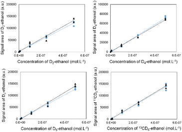
To ensure the selective detection of D2-, D4-, D5- and 13CD5-ethanol molecules, we first developed an analytical strategy involving the GC-MRM-MS methodology (see the ESI† for more details).
We validated its performance by drawing calibration curves for each labelled ethanol, in the absence and presence of the three other isotopes (Fig. 2). The superposition of both curves indicates that the isotope signals did not interfere with each other.
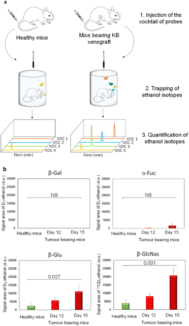
Since β-Gal, α-Fuc, β-Glu and β-GlcNAc are lysosomal enzymes widely distributed in healthy tissues, we next evaluated the dose of probes that has to be injected in mice for avoiding unselective activation. Indeed, at too high doses, probes can penetrate across cell membranes and then be hydrolysed by intracellular glycosidases in non-target tissues. Thus, we conducted preliminary experiments in healthy mice to determine the lowest detectable signal (LDS) for each VOC-based probe. For this purpose, we administered increasing probe concentrations to the animals that were then placed for over 1 h 30 in airtight cages equipped with an SPME fibre (Fig. 3a).
During the last 30 minutes, exhaled D2-, D4-, D5- and 13CD5-ethanol isotopes were trapped and subsequently analysed by GC-MRM-MS. Under these conditions, injection of 1, 100, 10 and 10 μg kg−1 doses of D2-EtGal, D4-EtFuc, D5-EtGlu and 13CD5-EtGlcNAc respectively, produced the LDS for the corresponding volatile tracers (see the ESI†). Thus, these probe concentrations were selected for the selective detection of glycosidases overexpressed in the tumour extracellular matrix.
Thereafter, our experimental protocol was applied to highlight the potential upregulation of glycosidases in malignant tissues. The cocktail of probes was administered to athymic nude mice bearing subcutaneous human KB xenografts (cervical cancer cell line) on days 12 and 15 post-tumour implantation. A control experiment was also carried out with healthy animals. On days 12 and 15, the amounts of D5- and 13CD5-ethanol were significantly higher in the breath of cancer animals compared to those emitted by their healthy counterparts (Fig. 3b). In contrast, the concentrations of D2- and D4-ethanol exhaled by tumour-bearing mice remained in the background noise. These results indicate that both β-Glu and β-GlcNAc exhibit increased catalytic activities in tumours while β-Gal and α-Fuc are not upregulated. Although the overexpression of β-Glu in solid tumours has been widely reported in the literature,35,36 the presence of elevated concentrations of β-GlcNAc remains weakly documented so far.37,38 These outcomes emphasize the potential of induced-volatolomics modalities to identify novel enzyme targets for drug discovery. Interestingly, between days 12 and 15, enzymatic activities of β-Glu and β-GlcNAc increased concomitantly with tumour growth (1.9- and 2.5-fold, respectively). Under such circumstances, the VOC-based probe cocktail seems to be a useful tool for monitoring disease progression in real time.21
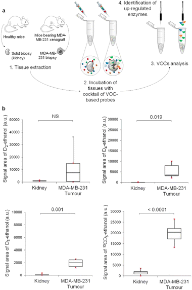
We next investigated our probe cocktail ex vivo on tumour biopsies. The aim of this trial was to compare simultaneously the catalytic activity of the four glycosidases in tumours and healthy tissues such as kidneys. In these experiments, we used D2-EtGal, D4-EtFuc, D5-EtGlu and 13CD5-EtGlcNAc probes at 10−6, at 10−4, 10−6, and 10−7 mol L−1, respectively, since these concentrations produced the LDS for ethanol isotopes when used on healthy tissues (see the ESI†).
Thus, MDA-MB-231 human mammary tumours harvested from nude mice with orthotopic xenografts were incubated in acetate buffer (pH 5) in the presence of the cocktail of VOC-based probes (Fig. 4a). Volatile tracers released in the sample headspace were monitored by GC-MRM-MS (Fig. 4b). The same experiment was also conducted with mice kidneys. Excepted for D2-ethanol, concentrations of the volatile tracers released in the headspace were higher for tumour than for kidney samples. These results indicated the presence of an elevated level of α-Fuc, β-Glu and β-GlcNAc in MDA-MB-231 xenografts compared to in healthy tissues. In contrast, β-Gal activity in tumours was highly variable and not significantly different than that of the control group. As was the case for KB tumours, β-GlcNAc was identified once again as the most active glycosidase. Indeed, 13CD5-ethanol was the volatile tracer exhibiting the highest GC-RMM-MS signal, although 13CD5-EtGlcNAc was the probe incubated at the lowest concentration (10−7 mol L−1).
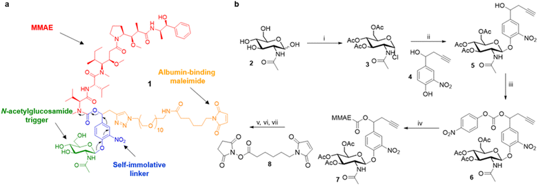
Overall, these results suggest that the overexpression of β-GlcNAc could be a hallmark of solid tumours. Therefore, this glycosidase appeared to be an attractive target for the development of a novel enzyme-activated cancer therapy. With the aim to explore this therapeutic strategy, we designed the β-GlcNAc-responsive albumin-binding prodrug 1 programmed for the selective delivery of MMAE in the tumour microenvironment39–41 (Fig. 5a). Once in the blood stream, 1 is expected to bind selectively to the free thiol of circulating albumin through Michael addition. The resulting bioconjugate will then accumulate in the tumour microenvironment where the glycosidic bond will be cleaved by extracellular β-GlcNAc, hence triggering the release of MMAE via the self-immolative mechanism42–44 described in Fig. 5a. It is worth mentioning here that glycosidase-responsive albumin-binding prodrugs are more efficient than their non-binding counterparts since they lead to increased drug deposition in the tumor microenvironment combined with a prolonged plasma half-life.39,45
The tumour-activated prodrug 1 was accessible from the commercially available β-N-acetylglucosamide 2 (Fig. 5b). First, this latter one was converted into the glycoside donor 3 (61%) that was coupled subsequently with the nitrophenol 4 (ref. 46 and 47) to afford compound 5 in 68% yield. Treatment of the benzyl alcohol 5 with 4-nitrophenyl chloroformate led to the activated carbonate 6 (72%). MMAE was then introduced via nucleophilic substitution in the presence of hydroxybenzo-triazole to give the carbamate 7 (93%). The three last steps of the synthesis were undertaken without intermediate purification. Thus, alkyne 7 reacted with O-(2-aminoethyl)-O-(2-azidoethyl)-nonaethylene glycol and Cu(CH3CN)4PF6 to form the corresponding triazole through copper(I)-catalysed azide–alkyne 1,3-cycloaddition. Deprotection of the glycoside followed by the introduction of the maleimide moiety using the N-hydroxysuccinimide ester 8 finally led to the drug delivery system 1 (24% over 3 steps).
As enzymatic hydrolysis of the β-N-acetylglucosamide trigger is the key step in the process of drug release, we first verified that the carbohydrate moiety was still accessible to β-GlcNAc once bound to albumin. For this purpose, prodrug 1 was incubated with human serum albumin (HSA) at 37 °C in order to form the corresponding bioconjugate through Michael addition. Under these conditions, nearly 90% of 1 was converted in two hours as a result of its rapid binding with the protein (Fig. 6a). The formation of the albumin conjugate was confirmed by trypsin digestion followed by HPLC/HRMS analysis (see the ESI†). When β-GlcNAc was incubated in the medium, the full release of MMAE was achieved in three hours indicating that the β-N-acetylglucosamide was a substrate of the enzyme for the activating enzyme even with the proximity of the bulky albumin (Fig. 6b). In contrast, the release of MMAE was not observed after 22 hours of incubation in the absence of β-GlcNAc (see the ESI†). Thus, these results highlighted that the prodrug 1 possessed the capacity both to bind covalently to HSA and release MMAE following enzymatic activation.
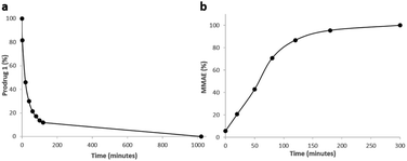 | ||
| Fig. 6 (a) Disappearance of 1 over time when placed in the presence of HSA at 37 °C. (b) Kinetics of MMAE release from 1 linked to HSA in the presence of β-GlcNAc (0.227 U mL−1). | ||
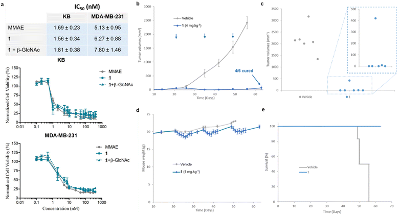
We then examined the antiproliferative activity of N-acetylglucosamide 1 against human KB and MDA-MB-231 tumour cell lines. As shown in Fig. 7a, the prodrug 1 exhibited a similar cytotoxicity to free MMAE, with IC50 values in the low nanomolar range. This result indicated that 1 was activated by β-GlcNAc produced by tumour cells,38 leading to the release of the drug. Since the co-incubation of the prodrug with β-GlcNAc did not induce a supplementary cytotoxicity, it appeared that the level of the glycosidase generated by tumour cells in the culture medium was sufficiently elevated for the full activation of 1. In order to confirm that the cytotoxicity of the β-N-acetylglucosamide 1 was due to its enzymatic activation in the culture medium, we investigated the antiproliferative activities of the galactoside48 and glucuronide39 analogues against MDA-MB-231 tumor cells (see the ESI†). In contrast to 1, when these prodrugs were incubated in the absence of the corresponding enzyme (β-Gal or β-Glu), they appeared far less toxic than MMAE. On the other hand, addition of the activating enzyme in the culture medium triggered a strong antiproliferative activity due to the enzyme-catalysed release of the drug. As β-galactoside, β-glucuronide and β-N-acetylglucosamide prodrugs differ exclusively by the nature of their glycoside triggers, these results showed that the cytotoxicity observed on MDA-MB-231 tumor cells is due to the β-GlcNAc-mediated activation of 1.
We next pursued our study by assessing the antitumour activity of the β-GlcNAc-sensitive prodrug 1 in Balb/c athymic mice bearing orthotopic MDA-MB-231 triple-negative mammary tumours (Fig. 7). The animals received i.v. injections of 1 at 4 mg kg−1 once every two weeks. Following this protocol, a remarkable reduction of tumour volume was observed in all treated animals (Fig. 7b). At the end of the experiment (day 63), 66% of the mice treated with 1 exhibited complete remission (Fig. 7c). In contrast, a previous study showed that the free MMAE produced only a poor anticancer activity, even when administered at its maximal tolerated dose.39 The successive i.v. administrations of 1 (1.54 mg per kg per injection MMAE equivalents) were well tolerated without major body weight loss (Fig. 6d) or detectable side effects. These outcomes demonstrated that the derivatization of MMAE in the form of the β-N-acetylglucosamide 1 significantly reduced its toxicity in vivo, allowing the administration of at least 3-fold the maximal tolerate dose (MTD) of the drug.39 Furthermore, the therapeutic efficacy obtained with 1 confirmed the selective and efficient enzyme-mediated release of MMAE in the tumour mass. Thus, this study showed that the targeting of tumour-associated β-GlcNAc by means of the enzyme-responsive albumin-binding prodrug is a promising therapeutic strategy for the treatment of triple-negative breast cancer (TNBC), a clinically aggressive disease for which current drugs are ineffective.
Conclusions
In this study, we demonstrated that a cocktail of VOC-based probes allowed the screening of several tumour-associated enzyme activities in both living mice and tumour biopsies. This induced-volatolomics modality led to the identification of an enzyme target, β-GlcNAc, which was exploited for the selective delivery of an anticancer drug that produced an impressive anticancer activity in mice bearing TNBC xenografts. Hence, our work highlights the potential of induced-volatolomics for the discovery of tumour-associated enzyme targets and the development of novel therapeutic strategies. Numerous VOC-based probe cocktails can be designed for the multimodal exploration of enzymatic dysregulation related to various diseases. Thus, induced-volatolomics modalities could become powerful tools to accelerate the process of drug discovery in the near future.Ethical statement
Experimental procedures involving animals were conducted according to protocol no. 2017053011069010 approved by the national ethical committee and carried out in accordance with the guidelines of the French Agriculture and Forestry Ministry (decree 2013-118) and the European Communities Council Directive (2010/63/UE).Data availability
All relevant data supporting this article have been included in the main text and the ESI.†Author contributions
P. P. and S. P. supervised the whole study. P. P., C. G. and M. T. supervised the induced-volatolomics approach and the development of analytical methods. P. P. and J. C. designed in vivo studies to test the efficiency of the cocktail of probes for highlighting highly active enzymes in tumours. J. L., J. C., F. D. and E. B. performed the in vitro and in vivo experiments using VOC-based probes. R. E. completed the VOC-based probe sensitivity study. J. C. and E. B. performed cell viability assays. S. P., I. O., B. R., R. C. and W. T. designed and synthesized the anticancer agent and VOC-based probes. R. C. and R. E. performed biochemical assays to certify the formation of the albumin–prodrug conjugate. J. S. and A. L. P. designed and supervised the experiments in tumour bearing mice treated with the enzyme-responsive prodrug. J. L., E. B. and R. C. collected all the data and reconstructed the figures. J. L., E. B. and R. C. wrote the methods section. P. P. and S. P. wrote the paper with the contribution of all authors.Conflicts of interest
There are no conflicts to declare.Acknowledgements
The authors thank La Ligue contre le Cancer (Ligue Nationale, Comités Vienne and Deux-Sèvres), and the “Région Nouvelle Aquitaine” for financial support. This work has also been funded by the ANR through the Young Researcher grant “Volatolomix” (ANR-21-CE18-0009). We also thank the CNRS which supplied the project through the “AAP Emergence”.Notes and references
- G. A. Holdgate, T. D. Meek and R. L. Grimley, Nat. Rev. Drug Discovery, 2018, 17, 115–132 CrossRef CAS PubMed.
- A. C. Rufer, Drug Discov. Today, 2021, 26, 875–886 CrossRef CAS PubMed.
- E. Rajakumara, S. Abhishek, K. Nitin, D. Saniya, P. Bajaj, U. Schwaneberg and M. D. Davari, ACS Chem. Biol., 2022, 17, 266–280 CrossRef CAS PubMed.
- A. Beck, L. Goetsch, C. Dumontet and N. Corvaïa, Nat. Rev. Drug Discovery, 2017, 16, 315–337 CrossRef CAS PubMed.
- N. Khiar-Fernández, J. Macicior, B. Marcos-Ramiro and S. Ortega-Gutiérrez, Eur. J. Org. Chem., 2021, 2021, 1307–1320 CrossRef.
- M.-J. Blanco and K. M. Gardinier, ACS Med. Chem. Lett., 2020, 11, 228–231 CrossRef CAS PubMed.
- W. Chyan and R. T. Raines, ACS Chem. Biol., 2018, 13, 1810–1823 CrossRef CAS PubMed.
- L. Wu, Z. Armstrong, S. P. Schröder, C. de Boer, M. Artola, J. M. Aerts, H. S. Overkleeft and G. J. Davies, Curr. Opin. Chem. Biol., 2019, 53, 25–36 CrossRef CAS PubMed.
- H. Fang, B. Peng, S. Y. Ong, Q. Wu, L. Li and S. Q. Yao, Chem. Sci., 2021, 12, 8288–8310 RSC.
- Y. Kuriki, T. Yoshioka, M. Kamiya, T. Komatsu, H. Takamaru, K. Fujita, H. Iwaki, A. Nanjo, Y. Akagi, K. Takeshita, H. Hino, R. Hino, R. Kojima, T. Ueno, K. Hanaoka, S. Abe, Y. Saito, J. Nakajima and Y. Urano, Chem. Sci., 2022, 13, 4474–4481 RSC.
- H. Singh, K. Tiwari, R. Tiwari, S. K. Pramanik and A. Das, Chem. Rev., 2019, 119, 11718–11760 CrossRef CAS PubMed.
- Y. Wang, J. Weng, X. Wen, Y. Hu and D. Ye, Biomater. Sci., 2021, 9, 406–421 RSC.
- S. Shingo, K. Toru, W. Rikiya, Z. Yi, I. Taiki, K. Mitsuyasu, N. Hidehiko, U. Takaaki, O. Takuji, H. Kazufumi, N. Hiroyuki and U. Yasuteru, Sci. Adv., 2020, 6, eaay0888 CrossRef PubMed.
- R. K. Muir, M. Guerra and M. M. Bogyo, ACS Chem. Biol., 2022, 17, 281–291 CrossRef CAS PubMed.
- S. M. McKenna, E. M. Fay and J. F. McGouran, ACS Chem. Biol., 2021, 16, 2719–2730 CrossRef CAS PubMed.
- Y. Zhang, G. Zhang, Z. Zeng and K. Pu, Chem. Soc. Rev., 2022, 51, 566–593 RSC.
- J. Huang, J. Li, Y. Lyu, Q. Miao and K. Pu, Nat. Mater., 2019, 18, 1133–1143 CrossRef CAS PubMed.
- E. A. Lemke and C. Schultz, Nat. Chem. Biol., 2011, 7, 480 CrossRef CAS PubMed.
- K. M. Dean and A. E. Palmer, Nat. Chem. Biol., 2014, 10, 512–523 CrossRef CAS PubMed.
- F. Djago, J. Lange and P. Poinot, Nat. Rev. Chem., 2021, 5, 183–196 CrossRef CAS PubMed.
- J. Lange, B. Eddhif, M. Tarighi, T. Garandeau, E. Péraudeau, J. Clarhaut, B. Renoux, S. Papot and P. Poinot, Angew. Chem., Int. Ed., 2019, 58, 17563–17566 CrossRef CAS.
- H. M. Burke, T. Gunnlaugsson and E. M. Scanlan, Chem. Commun., 2015, 51, 10576–10588 RSC.
- A. F. Costa, D. Campos, C. A. Reis and C. Gomes, Trends Cancer, 2020, 6, 757–766 CrossRef CAS PubMed.
- B. N. Vajaria and P. S. Patel, Glycoconj. J., 2017, 34, 147–156 CrossRef CAS PubMed.
- G. A. Kwong, S. Ghosh, L. Gamboa, C. Patriotis, S. Srivastava and S. N. Bhatia, Nat. Rev. Cancer, 2021, 21, 655–668 CrossRef CAS PubMed.
- S. S. Pinho and C. A. Reis, Nat. Rev. Cancer, 2015, 15, 540–555 CrossRef CAS PubMed.
- D. Asanuma, M. Sakabe, M. Kamiya, K. Yamamoto, J. Hiratake, M. Ogawa, N. Kosaka, P. L. Choyke, T. Nagano, H. Kobayashi and Y. Urano, Nat. Commun., 2015, 6, 6463 CrossRef CAS PubMed.
- K. Bosslet, J. Czech and D. Hoffmann, Tumor Target., 1995, 1, 45–50 CAS.
- K. Wang, W. Guo, N. Li, J. Shi, C. Zhang, W. Y. Lau, M. Wu and S. Cheng, Br. J. Cancer, 2014, 110, 1811–1819 CrossRef CAS PubMed.
- T.-C. Cheng, S.-H. Tu, L.-C. Chen, M.-Y. Chen, W.-Y. Chen, Y.-K. Lin, C.-T. Ho, S.-Y. Lin, C.-H. Wu and Y.-S. Ho, Oncotarget, 2015, 6(25), 21283–21300 CrossRef PubMed.
- C. Slawson and G. W. Hart, Nat. Rev. Cancer, 2011, 11, 678–684 CrossRef CAS PubMed.
- E. F. Petricoin III, A. M. Ardekani, B. A. Hitt, P. J. Levine, V. A. Fusaro, S. M. Steinberg, G. B. Mills, C. Simone, D. A. Fishman, E. C. Kohn and L. A. Liotta, Lancet, 2002, 359, 572–577 CrossRef PubMed.
- H. Kubo, Y. Murayama, S. Ogawa, T. Matsumoto, M. Yubakami, T. Ohashi, T. Kubota, K. Okamoto, M. Kamiya, Y. Urano and E. Otsuji, Sci. Rep., 2021, 11, 10664 CrossRef CAS PubMed.
- R. B. Parker and J. J. Kohler, ACS Chem. Biol., 2010, 5, 35–46 CrossRef CAS PubMed.
- I. Tranoy-Opalinski, T. Legigan, R. Barat, J. Clarhaut, M. Thomas, B. Renoux and S. Papot, Eur. J. Med. Chem., 2014, 74, 302–313 CrossRef CAS PubMed.
- P.-A. Burnouf, S. R. Roffler, C.-C. Wu and Y.-C. Su, J. Controlled Release, 2022, 349, 765–782 CrossRef CAS PubMed.
- B. Woynarowska, H. Wikiel and R. Bernacki, Cancer Res., 1989, 49, 5598–5604 CAS.
- K. T. Ramessur, P. Greenwell, R. Nash and M. V. Dwek, Br. J. Biomed. Sci., 2010, 67, 189–196 CrossRef CAS PubMed.
- B. Renoux, F. Raes, T. Legigan, E. Péraudeau, B. Eddhif, P. Poinot, I. Tranoy-Opalinski, J. Alsarraf, O. Koniev, S. Kolodych, S. Lerondel, A. Le Pape, J. Clarhaut and S. Papot, Chem. Sci., 2017, 8, 3427–3433 RSC.
- B. Renoux, L. Fangous, C. Hötten, E. Péraudeau, B. Eddhif, P. Poinot, J. Clarhaut and S. Papot, MedChemComm, 2018, 9, 2068–2071 RSC.
- R. Châtre, J. Lange, E. Péraudeau, P. Poinot, S. Lerondel, A. Le Pape, J. Clarhaut, B. Renoux and S. Papot, J. Controlled Release, 2020, 327, 19–25 CrossRef PubMed.
- I. Tranoy-Opalinski, A. Fernandes, M. Thomas, J.-P. Gesson and S. Papot, Anti-Cancer Agents Med. Chem., 2008, 8, 618–637 CrossRef CAS PubMed.
- A. Alouane, R. Labruère, T. Le Saux, F. Schmidt and L. Jullien, Angew. Chem., Int. Ed., 2015, 54, 7492–7509 CrossRef CAS PubMed.
- A. G. Gavriel, M. R. Sambrook, A. T. Russell and W. Hayes, Polym. Chem., 2022, 13, 3188–3269 RSC.
- T. Legigan, J. Clarhaut, B. Renoux, I. Tranoy-Opalinski, A. Monvoisin, J.-M. Berjeaud, F. Guilhot and S. Papot, J. Med. Chem., 2012, 55, 4516–4520 CrossRef CAS PubMed.
- M. Thomas, J. Clarhaut, P.-O. Strale, I. Tranoy-Opalinski, J. Roche and S. Papot, ChemMedChem, 2011, 6, 1006–1010 CrossRef CAS PubMed.
- T. Legigan, J. Clarhaut, I. Tranoy-Opalinski, A. Monvoisin, B. Renoux, M. Thomas, A. Le Pape, S. Lerondel and S. Papot, Angew. Chem., Int. Ed., 2012, 51, 11606–11610 CrossRef CAS PubMed.
- S. Kolodych, C. Michel, S. Delacroix, O. Koniev, A. Ehkirch, J. Eberova, S. Cianférani, B. Renoux, W. Krezel, P. Poinot, C. D. Muller, S. Papot and A. Wagner, Eur. J. Med. Chem., 2017, 142, 376–382 CrossRef CAS PubMed.
Footnotes |
| † Electronic supplementary information (ESI) available. See DOI: https://doi.org/10.1039/d2sc06797h |
| ‡ These authors contributed equally. |
| This journal is © The Royal Society of Chemistry 2023 |

