 Open Access Article
Creative Commons Attribution 3.0 Unported Licence
G4-DNA formation and chromatin remodelling are interdependent in human cells†
Nicholas B.
Lawler‡
 ab,
Arnold
Ou‡§
a,
Jessica J.
King
 a,
Cameron W.
Evans
 *a,
K. Swaminathan
Iyer
 *a and
Nicole M.
Smith
 *a
*a
aSchool of Molecular Sciences, The University of Western Australia, Perth, WA, Australia. E-mail: cameron.evans@uwa.edu.au; swaminatha.iyer@uwa.edu.au; nicole.smith@uwa.edu.au
bSchool of Physics, Mathematics and Computing, The University of Western Australia, Perth, WA, Australia
First published on 27th June 2023
Abstract
DNA G-quadruplexes (G4s) have been identified as important biological targets for transcriptional, translational, and epigenetic regulation. The stabilisation of G4s with small molecule ligands has emerged as a technique to regulate gene expression and as a potential therapeutic approach for human diseases. Here, we demonstrate that ligand stabilisation of G4s causes altered chromatin accessibility dependent on the targeting specificity of the molecule. In particular, stabilisation of a target G4 using the highly specific GTC365 ligand resulted in differential accessibility of 61 genomic regions, while the broad-targeting G4 ligand, GQC-05, stabilised many G4s and induced a global shift towards increased accessibility of gene promoter regions. Interestingly, while we observed distinct effects of each ligand on RNA expression levels and the induction of DNA double-stranded breaks, both ligands modified DNA damage response pathways. Our work represents the dual possibility of G4-stabilising ligands for specific or global chromatin modulation via unique targeting characteristics.
Introduction
Nuclear processes such as gene expression are influenced by a range of mechanisms, including the arrangement of nuclear architecture. The reorganisation of nuclear architecture via dynamic transitions between decondensed transcriptionally active euchromatin and condensed heterochromatin is fundamental in regulating interactions of transcription factors with DNA. The principal factors governing this reorganisation in turn cause gene activation or silencing by regulating accessibility of gene promoters. Epigenetic modifications, mutations, and DNA secondary structures have all been investigated for their role in influencing chromatin accessibility and gene transcription.1–3 Of the various secondary structures that have been associated with chromatin accessibility and nuclear processes, G-quadruplexes (G4s) are particularly interesting as they have been identified as therapeutic targets for diseases including cancer4,5 and neurodegenerative disorders.6 G4s are sequence-specific structures that consist of stacked G-tetrads, each composed of four Hoogsteen hydrogen-bonded guanines in a planar arrangement.7 Unlike epigenetic and point mutations in the genome, which are heritable and stable modifications,8 G4 formation is dynamic and transitory. The propensity for DNA G4 formation in the human genome is very high, with over 10![[thin space (1/6-em)]](https://www.rsc.org/images/entities/char_2009.gif) 000 G4-forming regions in chromatin and over 700
000 G4-forming regions in chromatin and over 700![[thin space (1/6-em)]](https://www.rsc.org/images/entities/char_2009.gif) 000 G4-forming regions identified in naked genomic DNA.9,10
000 G4-forming regions identified in naked genomic DNA.9,10
G4 structures are enriched in gene promoters and nuclease hypersensitive regions, coincide with binding sites for transcription factors and DNA methyltransferases, and are implicated in transcriptional regulation, replication, and genomic instability.5,10–18 Targeted interaction and stabilisation of specific G4s using small molecules has been widely accepted as a therapeutic strategy.15,16,19–22
Stabilisation of G4s using small molecules has been identified as an important approach to regulate gene expression, but G4 stabilisation can increase DNA damage in cells if G4s are not efficiently regulated.23,24 Chromatin accessibility is required for transcription, and remodelling of chromatin is central to the DNA damage response to facilitate access of repair factors for removal of the lesion.25–28 Recently, G4 formation has been correlated with DNA damage repair and gene activation.18,29–31 Thus, G4s may contribute to the modulation of chromatin organisation, as reported by Zyner et al.32 In this work, we show that small molecule-induced G4 stabilisation can be used as a strategy to regulate chromatin remodelling in human cells. In particular, we show that G4 stabilisation and chromatin remodelling are interdependent phenomena. Enhanced chromatin remodelling of specific gene promoter regions was observed for the case of targeted G4 stabilisation using a ligand specific for the G4 in the promoter region of human telomerase reverse transcriptase (hTERT). On the other hand, broad-targeted, genome-wide stabilisation of G4s causes a global shift towards increased promoter accessibility, although few regions are consistently altered. Our findings using Assay for Transposase-Accessible Chromatin sequencing (ATAC-seq) and RNA-sequencing (RNA-seq) analysis coupled with G4 formation predictions in MCF-7 human breast adenocarcinoma cells reveal that G4s are key regulators of chromatin remodelling in human cells.
Results and discussion
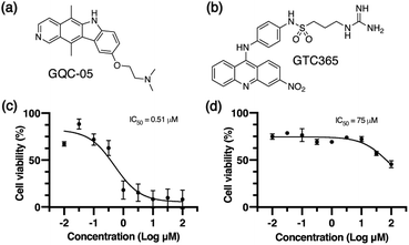
We used two small molecule ligands, GQC-05 and GTC365, to stabilise G4s in MCF-7 human mammary epithelial adenocarcinoma cells and investigated the potential for G4 stabilisation to alter chromatin organisation and subsequent gene expression. GQC-05 (NSC338258, Fig. 1a) is an ellipticine analogue that binds with varying affinities to different G4-forming sequences, stabilising a range of G4 topologies.33 On the other hand, GTC365 (Fig. 1b) specifically binds to the higher-order G4 structure observed in the hTERT promoter via dual-motif targeting for the G4 and the mismatched duplex stem loop.34We first evaluated the effects of 72 h incubation with each of the two G4 ligands on the viability of MCF-7 cells by an MTS assay (Fig. 1c and d). IC50 values for GQC-05 and GTC365 were found to be 0.51 μM and 75 μM, respectively, indicative of a higher toxicity associated with the broad targeting of GQC-05. It has previously been shown that treatment with G4-stabilising ligands can induce DNA damage,35 representing a mechanism whereby the broad-targeting G4 ligand, GQC-05, may cause greater G4 stabilisation than GTC365, resulting in increased DNA damage and higher toxicity. For further experiments, cells were incubated with the respective ligand at a concentration less than the IC50 for 72 h.
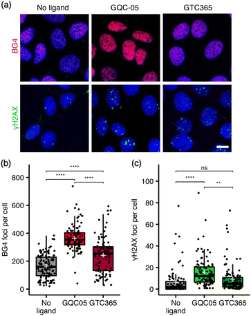
Next, the relationship between ligand targeting, G4 stabilisation and the induction of DNA damage was investigated via immunocytochemistry. G4s were identified with the structure-specific, single-chain variable fragment (scFv) BG4 antibody,36 and DNA damage identified using an antibody against γH2AX, a marker for DNA double-strand breaks.37 The broad-targeting G4 ligand, GQC-05, resulted in a significant increase in both γH2AX and BG4 foci counts, while the hTERT G4-targeting ligand, GTC365, resulted in a smaller increase in BG4 foci and no significant change in DNA damage (Fig. 2). Interestingly, GTC365 increased the mean BG4 foci count by 78 relative to the untreated control, suggesting it causes stabilisation of G4s other than the hTERT promoter G4 either directly or via downstream effects. The stabilisation of more G4s and greater induction of DNA damage by GQC-05 compared to GTC365 demonstrates that the induction of DNA double-stranded breaks is related to the extent of G4 stabilisation and thus the targeting specificity of the ligand, contributing to the higher toxicity of GQC-05. Our results mirror the findings of De Magis et al. where treatment with G4 stabilising ligands in vivo led to an increase in γH2AX foci, compared to non-G4 binders.35
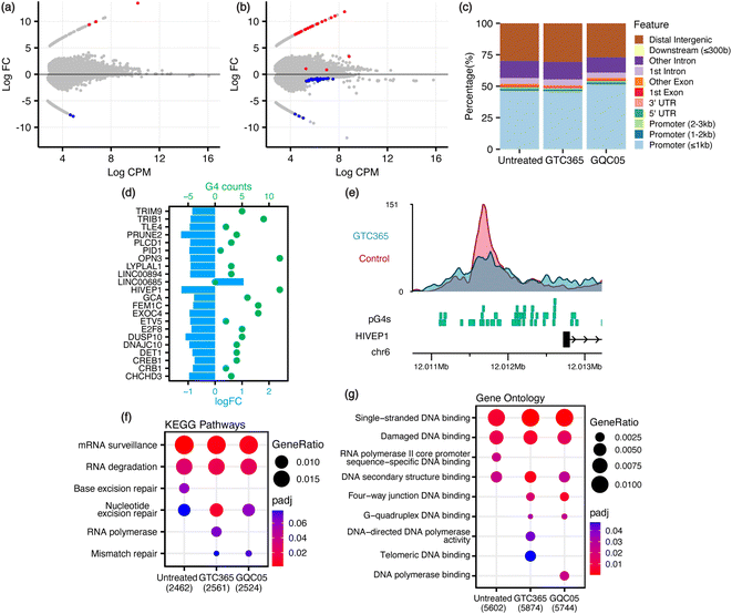
Given that remodelling of the chromatin landscape is a fundamental feature in the regulation of DNA damage response, we hypothesised that GQC-05 treatment would induce a significant shift in the global chromatin landscape, as opposed to GTC-365 which is expected to have more targeted effects. ATAC-seq analysis of ligand-treated MFC-7 cells showed differential chromatin accessibility in both GQC-05- and GTC365-treated samples. GQC-05 treatment resulted in a greater global shift of chromatin accessibility than GTC365, with accessibility of the promoters increasing to 51.7% of total peaks, 5% higher than the untreated control (46.6%) and GTC365 (46.2%) (Fig. 3 and S1, S2†). This is consistent with the well-characterised enrichment of G4-forming sites in gene promoter regions10,38 and parallels the observed extent of ligand-induced G4 stabilisation, as GQC-05 also caused a larger increase in BG4 foci counts than GTC365. Incubation with GTC365 induced 61 significant differentially accessible regions (DARs), defined as regions with false discovery rate (FDR) < 0.1 to allow for the largest range of genomic interactions to be identified. Of these 61 DARs, 22 were located in gene promoter regions. On the other hand, GQC-05 treatment resulted in only 5 DARs (Fig. 3a, b and Table S1†). This suggests that the stabilisation of specific G4s by GTC365 causes targeted alterations to chromatin accessibility that are consistent across the entire population of cells, resulting in significant changes for the target regions with minimal global implications. GQC-05, however, broadly targets a range of G4s, resulting in global shifts in accessibility towards gene promoters with few regions consistently affected across the entire cell population, resulting in fewer statistically significant DARs.
To explore the role of G4s in regulating chromatin accessibility changes, predicted G4 forming sequences (pG4s) within the genome were identified using the G4Hunter algorithm.38 The pG4 and ATAC-seq datasets were intersected to identify pG4s within promoter regions exhibiting differential accessibility (Fig. 3d, e and S3†). For GTC365, there is a strong correlation between pG4s and modified accessibility, with 21 (95.5%) of the 22 promoter DARs having both reduced accessibility and at least one pG4 (mean 5, range 1–12, median 4). For comparison, of all the ATAC-seq peaks detected in gene promoter regions, only 80.9% contained a pG4. This corresponds to P = 0.058 (binomial) for at least 21 out of 22 randomly selected regions to contain a pG4. The single DAR without a pG4 has increased accessibility. In contrast, 25 out of 39 non-promoter DARs had increased accessibility and 14 were reduced (Fig. S4†). Importantly, only 8 of the non-promoter DARs contained a pG4, and 7 of these 8 had reduced accessibility, consistent with the reduced accessibility observed for pG4-containing promoter DARs. Of note, GTC365 is reported to be specific for the hTERT promoter G4 via dual-motif targeting of the G4 and the mismatched duplex stem-loop,34,39 and thus the reduced accessibility of these pG4-containing promoter and non-promoter regions is likely associated with the G4-stabilising effects of GTC365. GQC-05 treatment resulted in only 5 DARs, with none located within gene promoters. The prevalence of pG4s in promoter regions with differential accessibility indicates that these pG4s may contribute to the modified chromatin landscape. Endogenous G4s are more commonly found in regions with high chromatin accessibility and are associated with highly transcribed genes,18 however ligand-induced stabilisation of G4s is predominately associated with transcriptional repression.10,15,40,41 Therefore, it is not surprising that ligand-induced G4 stabilisation with GTC365 results in a decrease in chromatin accessibility in gene promoters and in non-promoter regions containing a pG4.
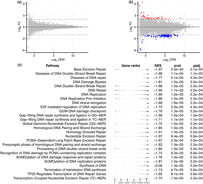
To characterise the effects on the transcriptome and downstream biological processes, RNA-sequencing was conducted on ligand-treated MCF-7 cells, with significant differentially expressed genes (DEGs) defined using the threshold FDR < 0.05 and |log2![[thin space (1/6-em)]](https://www.rsc.org/images/entities/char_2009.gif) FC| > 2 to identify genes that are maximally altered by the ligands. GQC-05 did not cause any significant differential expression, similar to the low number of DARs induced by this broad-targeting G4 ligand. GTC365 treatment resulted in 162 DEGs (Fig. 4a, b, S1 and Table S2†), with 40 genes upregulated and 122 downregulated, consistent with the reported transcriptional suppression by ligand-induced stabilisation of G4s.40,41 This further supports the hypothesis that the higher specificity of GTC365 causes consistent stabilisation of the same G4s across the entire population of cells, resulting in a strong effect on accessibility of these sites and altered gene expression, while the broad-targeting G4 ligand, GQC-05, results in less consistent stabilisation across the population of cells. This is illustrated by the levels of hTERT expression, with GTC365 causing a 6.5-fold decrease in expression due to the specificity of the ligand for the hTERT promoter G4, while the broad-targeting G4 ligand, GQC-05, had negligible effects (Table S2†). Interestingly, despite the reported specificity of GTC365 for the hTERT G4, we observed differential expression of numerous other genes, that may be a result of off-target binding or downstream effects of ligand binding.
FC| > 2 to identify genes that are maximally altered by the ligands. GQC-05 did not cause any significant differential expression, similar to the low number of DARs induced by this broad-targeting G4 ligand. GTC365 treatment resulted in 162 DEGs (Fig. 4a, b, S1 and Table S2†), with 40 genes upregulated and 122 downregulated, consistent with the reported transcriptional suppression by ligand-induced stabilisation of G4s.40,41 This further supports the hypothesis that the higher specificity of GTC365 causes consistent stabilisation of the same G4s across the entire population of cells, resulting in a strong effect on accessibility of these sites and altered gene expression, while the broad-targeting G4 ligand, GQC-05, results in less consistent stabilisation across the population of cells. This is illustrated by the levels of hTERT expression, with GTC365 causing a 6.5-fold decrease in expression due to the specificity of the ligand for the hTERT promoter G4, while the broad-targeting G4 ligand, GQC-05, had negligible effects (Table S2†). Interestingly, despite the reported specificity of GTC365 for the hTERT G4, we observed differential expression of numerous other genes, that may be a result of off-target binding or downstream effects of ligand binding.
 | ||
| Fig. 4 RNA-seq analysis for GQC-05- and GTC365-treated MCF-7 cells. MD plots for (a) GQC-05 and (b) GTC365 treatments with differentially expressed genes in red (upregulated) and blue (downregulated). (c) Gene set enrichment analysis (GSEA) of the GTC365 RNA-seq data, filtered to show pathways associated with DNA associated processes. The complete data is provided in Table S3 and Fig. S5.† The distribution of genes associated with each pathway is indicated (gene ranks), as is the normalised enrichment score (NES) and adjusted p-value. | ||
The lack of differential gene expression induced by GQC-05 was unexpected as it stabilises a wide range of G4s within cells and has been associated with differential gene expression over shorter time periods and at higher concentrations.33,42 However, for 72 h treatment as in this work, GQC-05 has relatively high toxicity and was therefore used at a low concentration to maintain cell viability. At this concentration, it is likely that the number of G4-ligand complexes formed is insufficient to stabilise all possible targets in each cell, with over 700![[thin space (1/6-em)]](https://www.rsc.org/images/entities/char_2009.gif) 000 genomic G4-forming sequences having been experimentally validated.9 Additionally, Brown et al. demonstrated that for the MYC promoter G4, for which GQC-05 has the highest reported affinity, the GQC-05
000 genomic G4-forming sequences having been experimentally validated.9 Additionally, Brown et al. demonstrated that for the MYC promoter G4, for which GQC-05 has the highest reported affinity, the GQC-05![[thin space (1/6-em)]](https://www.rsc.org/images/entities/char_2009.gif) :
:![[thin space (1/6-em)]](https://www.rsc.org/images/entities/char_2009.gif) G4 stoichiometry is 2
G4 stoichiometry is 2![[thin space (1/6-em)]](https://www.rsc.org/images/entities/char_2009.gif) :
:![[thin space (1/6-em)]](https://www.rsc.org/images/entities/char_2009.gif) 1, indicative of two ligand binding sites per G4 for complete stabilisation.33 The binding constants for these two binding sites are 0.1 μM and 1.43 μM, so both the IC50 (0.51 μM) and the concentration used in this study (0.3 μM) will not result in consistent G4 stabilisation across cells. Consequently, different subsets of G4s may be stabilised in each cell, resulting in a reduced average effect on the genomic architecture and gene expression that may be better identified via single-cell studies rather than bulk RNA- or ATAC-seq. While higher doses of GQC-05 have been shown to induce transcriptomic modifications over short treatment periods,33,42 GQC-05 cytotoxicity varies significantly between cell types, and the lower dose treatments have different effects on gene expression in different cell lines.33,43
1, indicative of two ligand binding sites per G4 for complete stabilisation.33 The binding constants for these two binding sites are 0.1 μM and 1.43 μM, so both the IC50 (0.51 μM) and the concentration used in this study (0.3 μM) will not result in consistent G4 stabilisation across cells. Consequently, different subsets of G4s may be stabilised in each cell, resulting in a reduced average effect on the genomic architecture and gene expression that may be better identified via single-cell studies rather than bulk RNA- or ATAC-seq. While higher doses of GQC-05 have been shown to induce transcriptomic modifications over short treatment periods,33,42 GQC-05 cytotoxicity varies significantly between cell types, and the lower dose treatments have different effects on gene expression in different cell lines.33,43
To explore the biological processes potentially affected by treatment with each ligand, pathway analysis was conducted on the ATAC- and RNA-seq results. Kyoto Encyclopedia of Genes and Genomes (KEGG) pathway analysis and Gene Ontology (GO) enrichment analysis was conducted on genes containing ATAC-seq peaks in the promoter region, and Gene Set Enrichment Analysis (GSEA) performed on all genes identified via RNA-seq, ranked by their expression levels. Many pathways were identified as differentially enriched by the two ligands, including several associated with DNA and RNA processes (Fig. 3f, g, 4c, S5 and Table S3†). Of note, GO analysis identified differential enrichment of GO terms associated with G-quadruplex binding and DNA polymerase activity in the ligand-treated samples, consistent with the stabilisation of G4s and their documented interactions with polymerases.44–46 Enrichment of these terms indicates that G4 stabilisation promotes a cellular response to regulate interactions with these structures. Furthermore, KEGG pathway analysis and GSEA demonstrate that both GTC365 and GQC-05 alter nucleotide excision- and DNA repair-related pathways, and GTC365 also caused differential enrichment of many pathways associated with RNA polymerase and DNA damage response, replication, and synthesis. As is expected for molecules that directly interact with DNA to modulate the chromatin landscape, there is a strong cellular response to both ligands, with many pathways involved in the maintenance of chromatin significantly affected. These results are in accordance with the immunocytochemistry experiments that demonstrated the ligands affect genomic stability and structure. GTC365 also alters several pathways related to cell cycle progression, which is halted in response to DNA damage and has also been associated with G4 stabilisation.47 It remains unclear whether these pathways are modified due to the ligands directly altering the accessibility and expression of the related genes, or if these pathways are stimulated as part of a biological response to the ligand treatment.
To explore if the transcriptomic effects of G4 stabilisation are due to altered accessibility of specific transcription factor (TF) binding sites, we conducted HOMER analysis on the ATAC-seq peaks to identify enriched TF binding motifs.48 Similar motifs were found to be enriched in each dataset, including the control (Tables S4 and S5†). Analysis of the 61 DARs from the GTC365-treated cells also identified these same motifs, suggesting that modified nucleosome occupancy of specific transcription factor binding motifs is not the primary mechanism by which these G4-stabilising ligands alter transcription. Instead, the transcriptional alterations may be a result of the interactions of stabilised G4s with DNA damage response pathways, or G4 structural effects. Additionally, the transcriptional effects of G4 ligands have previously been associated with altered helicase activity49 and their role as transcription factor binding sites,17,18 which can be impaired by G4 stabilisation.41
Conclusions
In this work we have demonstrated that treatment of MCF-7 cells with G4-stabilising ligands alters the genomic landscape, transcriptome, and DNA damage response. Modifications in chromatin accessibility are observed to correspond to the extent of G4 stabilisation and targeting specificity of the G4 ligands. GTC365, which has been reported to target a specific G4, induced consistent, significant differential accessibility of certain regions of the genome, while the broad-targeting G4 ligand, GQC-05, caused general shifts in the chromatin landscape that increased accessibility of gene promoter regions. We identified novel GTC365-induced decreases in promoter accessibility that agrees with the documented transcriptional repression associated with ligand-induced G4-stabilisation. Unlike GQC-05, GTC365 caused significant differential gene expression with the majority of altered genes being downregulated. This represents the dual possibility of small molecule G4-stabilising ligands as therapeutic agents to target G4s in diseases, inducing either specific or global effects depending on the design of the molecule.Data availability
The raw data reported in this paper are available at the NCBI GEO repository. Processed data files are included in the ESI.†Author contributions
A. O. and J. J. K. performed cell culture experiments and A. O. prepared samples for sequencing. N. B. L. conducted sequencing analysis and bioinformatic predictions. N. M. S., K. S. I. and C. W. E. developed the concept. All authors discussed and analysed results and contributed to the manuscript.Conflicts of interest
There are no conflicts to declare.Acknowledgements
This research was supported by the Australian Research Council (ARC), the National Health and Medical Research Council (NHMRC) and Cancer Council WA. The authors acknowledge the contributions of Melanie Grigg and Dr Dave Tang. We thank Prof. Laurence H. Hurley of the University of Arizona and Reglagene for providing the G4 ligands. The authors acknowledge the facilities and the scientific and technical assistance of the Australian Microscopy & Microanalysis Research Facility at the Centre for Microscopy, Characterisation & Analysis, The University of Western Australia, a facility funded by the University, State and Commonwealth Governments. N. B. Lawler acknowledges the support of the Forrest Research Foundation.References
- Z. Zhong, S. Feng, S. H. Duttke, M. E. Potok, Y. Zhang, J. Gallego-Bartolomé, W. Liu and S. E. Jacobsen, Proc. Natl. Acad. Sci. U. S. A., 2021, 118, e2023347118 CrossRef CAS PubMed.
- H. Cedar and Y. Bergman, Nat. Rev. Genet., 2009, 10, 295–304 CrossRef CAS PubMed.
- T. Hashimshony, J. Zhang, I. Keshet, M. Bustin and H. Cedar, Nat. Genet., 2003, 34, 187–192 CrossRef CAS PubMed.
- Y. Wu and R. M. Brosh Jr, FEBS J., 2010, 277, 3470–3488 CrossRef CAS PubMed.
- R. Hänsel-Hertsch, M. Di Antonio and S. Balasubramanian, Nat. Rev. Mol. Cell Biol., 2017, 18, 279–284 CrossRef PubMed.
- E. Wang, R. Thombre, Y. Shah, R. Latanich and J. Wang, Nucleic Acids Res., 2021, 49, 4816–4830 CrossRef CAS PubMed.
- Y. Qin and L. H. Hurley, Biochimie, 2008, 90, 1149–1171 CrossRef CAS PubMed.
- H. Y. Zoghbi and A. L. Beaudet, Cold Spring Harbor Perspect. Biol., 2016, 8, a019497 CrossRef PubMed.
- V. S. Chambers, G. Marsico, J. M. Boutell, M. Di Antonio, G. P. Smith and S. Balasubramanian, Nat. Biotechnol., 2015, 33, 877–881 CrossRef CAS PubMed.
- R. Hänsel-Hertsch, D. Beraldi, S. V. Lensing, G. Marsico, K. Zyner, A. Parry, M. Di Antonio, J. Pike, H. Kimura, M. Narita, D. Tannahill and S. Balasubramanian, Nat. Genet., 2016, 48, 1267–1272 CrossRef PubMed.
- A. Verma, K. Halder, R. Halder, V. K. Yadav, P. Rawal, R. K. Thakur, F. Mohd, A. Sharma and S. Chowdhury, J. Med. Chem., 2008, 51, 5641–5649 CrossRef CAS PubMed.
- J. Spiegel, S. Adhikari and S. Balasubramanian, Trends Chem., 2020, 2, 123–136 CrossRef CAS PubMed.
- J. L. Huppert and S. Balasubramanian, Nucleic Acids Res., 2007, 35, 406–413 CrossRef CAS PubMed.
- S.-Q. Mao, A. T. Ghanbarian, J. Spiegel, S. Martínez Cuesta, D. Beraldi, M. Di Antonio, G. Marsico, R. Hänsel-Hertsch, D. Tannahill and S. Balasubramanian, Nat. Struct. Mol. Biol., 2018, 25, 951–957 CrossRef CAS PubMed.
- A. Siddiqui-Jain, C. L. Grand, D. J. Bearss and L. H. Hurley, Proc. Natl. Acad. Sci. U. S. A., 2002, 99, 11593–11598 CrossRef CAS PubMed.
- S. Balasubramanian, L. H. Hurley and S. Neidle, Nat. Rev. Drug Discovery, 2011, 10, 261–275 CrossRef CAS PubMed.
- J. Spiegel, S. M. Cuesta, S. Adhikari, R. Hänsel-Hertsch, D. Tannahill and S. Balasubramanian, Genome Biol., 2021, 22, 117 CrossRef CAS PubMed.
- S. Lago, M. Nadai, F. M. Cernilogar, M. Kazerani, H. Domíniguez Moreno, G. Schotta and S. N. Richter, Nat. Commun., 2021, 12, 3885 CrossRef CAS PubMed.
- E. Mendes, I. M. Aljnadi, B. Bahls, B. L. Victor and A. Paulo, Pharmaceuticals, 2022, 15, 300 CrossRef CAS PubMed.
- S. L. Palumbo, S. W. Ebbinghaus and L. H. Hurley, J. Am. Chem. Soc., 2009, 131, 10878–10891 CrossRef CAS PubMed.
- N. Kosiol, S. Juranek, P. Brossart, A. Heine and K. Paeschke, Mol. Cancer, 2021, 20, 40 CrossRef CAS PubMed.
- J. A. Kretzmann, K. L. Irving, N. M. Smith and C. W. Evans, NAR Cancer, 2021, 3, zcab048 CrossRef PubMed.
- R. Rodriguez, K. M. Miller, J. V. Forment, C. R. Bradshaw, M. Nikan, S. Britton, T. Oelschlaegel, B. Xhemalce, S. Balasubramanian and S. P. Jackson, Nat. Chem. Biol., 2012, 8, 301–310 CrossRef CAS PubMed.
- R. Camarillo, S. Jimeno and P. Huertas, Front. Genet., 2021, 12, 742434 CrossRef CAS PubMed.
- J. Stadler and H. Richly, Int. J. Mol. Sci., 2017, 18, 1715 CrossRef PubMed.
- C. Dinant, A. B. Houtsmuller and W. Vermeulen, Epigenet. Chromatin, 2008, 1, 9 CrossRef PubMed.
- A. Harrod, K. A. Lane and J. A. Downs, DNA Repair, 2020, 93, 102919 CrossRef CAS PubMed.
- G. Kollárovič, C. E. Topping, E. P. Shaw and A. L. Chambers, Nucleic Acids Res., 2020, 48, 1872–1885 CrossRef PubMed.
- A. M. Fleming, Y. Ding and C. J. Burrows, Proc. Natl. Acad. Sci. U. S. A., 2017, 114, 2604–2609 CrossRef CAS PubMed.
- S. Roychoudhury, S. Pramanik, H. L. Harris, M. Tarpley, A. Sarkar, G. Spagnol, P. L. Sorgen, D. Chowdhury, V. Band, D. Klinkebiel and K. K. Bhakat, Proc. Natl. Acad. Sci. U. S. A., 2020, 117, 11409–11420 CrossRef CAS PubMed.
- R. Linke, M. Limmer, S. A. Juranek, A. Heine and K. Paeschke, Int. J. Mol. Sci., 2021, 22, 12599 CrossRef CAS PubMed.
- K. G. Zyner, A. Simeone, S. M. Flynn, C. Doyle, G. Marsico, S. Adhikari, G. Portella, D. Tannahill and S. Balasubramanian, Nat. Commun., 2022, 13, 142 CrossRef CAS PubMed.
- R. V. Brown, F. L. Danford, V. Gokhale, L. H. Hurley and T. A. Brooks, J. Biol. Chem., 2011, 286, 41018–41027 CrossRef CAS PubMed.
- H.-J. Kang, Y. Cui, H. Yin, A. Scheid, W. P. D. Hendricks, J. Schmidt, A. Sekulic, D. Kong, J. M. Trent, V. Gokhale, H. Mao and L. H. Hurley, J. Am. Chem. Soc., 2016, 138, 13673–13692 CrossRef CAS PubMed.
- A. De Magis, S. G. Manzo, M. Russo, J. Marinello, R. Morigi, O. Sordet and G. Capranico, Proc. Natl. Acad. Sci. U. S. A., 2019, 116, 816–825 CrossRef CAS PubMed.
- G. Biffi, D. Tannahill, J. McCafferty and S. Balasubramanian, Nat. Chem., 2013, 5, 182–186 CrossRef CAS PubMed.
- L.-J. Mah, A. El-Osta and T. C. Karagiannis, Leukemia, 2010, 24, 679–686 CrossRef CAS PubMed.
- A. Bedrat, L. Lacroix and J.-L. Mergny, Nucleic Acids Res., 2016, 44, 1746–1759 CrossRef PubMed.
- J. H. Song, H.-J. Kang, L. A. Luevano, V. Gokhale, K. Wu, R. Pandey, H.-H. Sherry Chow, L. H. Hurley and A. S. Kraft, Cell Chem. Biol., 2019, 26, 1110–1121.e4 CrossRef CAS PubMed.
- D. Rhodes and H. J. Lipps, Nucleic Acids Res., 2015, 43, 8627–8637 CrossRef CAS PubMed.
- C. Li, H. Wang, Z. Yin, P. Fang, R. Xiao, Y. Xiang, W. Wang, Q. Li, B. Huang, J. Huang and K. Liang, Genome Res., 2021, 31, 1546–1560 CrossRef PubMed.
- D. A. Flusberg, N. F. Rizvi, V. Kutilek, C. Andrews, P. Saradjian, C. Chamberlin, P. Curran, B. Swalm, S. Kattar, G. F. Smith, P. Dandliker, E. B. Nickbarg and J. O'Neil, SLAS Discovery, 2019, 24, 142–157 CrossRef CAS PubMed.
- J. J. Montoya, M. A. Turnidge, D. H. Wai, A. R. Patel, D. W. Lee, V. Gokhale, L. H. Hurley, R. J. Arceci, C. Wetmore and D. O. Azorsa, BMC Cancer, 2019, 19, 1251 CrossRef CAS PubMed.
- K. N. Estep, T. J. Butler, J. Ding and R. M. Brosh, Curr. Med. Chem., 2019, 26, 2881–2897 CrossRef CAS PubMed.
- S. Cogoi, M. Paramasivam, A. Membrino, K. K. Yokoyama and L. E. Xodo, J. Biol. Chem., 2010, 285, 22003–22016 CrossRef CAS PubMed.
- K. Usdin and K. J. Woodford, Nucleic Acids Res., 1995, 23, 4202–4209 CrossRef CAS PubMed.
- J. J. King, K. L. Irving, C. W. Evans, R. V. Chikhale, R. Becker, C. J. Morris, C. D. Peña Martinez, P. Schofield, D. Christ, L. H. Hurley, Z. A. E. Waller, K. S. Iyer and N. M. Smith, J. Am. Chem. Soc., 2020, 142, 20600–20604 CrossRef CAS PubMed.
- S. Heinz, C. Benner, N. Spann, E. Bertolino, Y. C. Lin, P. Laslo, J. X. Cheng, C. Murre, H. Singh and C. K. Glass, Mol. Cell, 2010, 38, 576–589 CrossRef CAS PubMed.
- O. Mendoza, A. Bourdoncle, J.-B. Boulé, R. M. Brosh and J.-L. Mergny, Nucleic Acids Res., 2016, 44, 1989–2006 CrossRef CAS PubMed.
Footnotes |
| † Electronic supplementary information (ESI) available. See DOI: https://doi.org/10.1039/d3sc02533k |
| ‡ These authors contributed equally. |
| § Present address: Rockefeller University, New York, NY, USA. |
| This journal is © The Royal Society of Chemistry 2023 |


