
Unraveling the composition of each atomic layer in the MXene/MAX phase structure – identification of oxycarbide, oxynitride, and oxycarbonitride subfamilies of MXenes†
Paweł Piotr
Michałowski

Łukasiewicz Research Network–Institute of Microelectronics and Photonics, Warsaw, Poland. E-mail: pawel.michalowski@imif.lukasiewicz.gov.pl
First published on 19th July 2024
Abstract
MXenes, the largest known family of 2D materials, are known for their complicated structure consisting of many different elements. Their properties can be finely tuned by precise engineering of the composition of each atomic layer. Thus it is necessary to further develop the secondary ion mass spectrometry (SIMS) technique which can unambiguously identify each element with atomic precision. The newly established protocol of deconvolution and calibration of the SIMS data enables layer-by-layer characterization of MAX phase and MXene samples with ±1% accuracy. Such precision is particularly important for samples that consist of several different transition metals in their structure. This confirms that most MXenes contain a substantial amount of oxygen in the X layers, thus enabling the identification of oxycarbide, oxynitride, and oxycarbonitride subfamilies of these materials. It can also be applied for under- and over-etched samples and to determine the exact composition of termination layers. Generally, the SIMS technique may provide invaluable support in the synthesis and optimization of MAX phase and MXene studies.
New conceptsSecondary ion mass spectrometry (SIMS) has to date been the only analytical technique that can unambiguously identify all elements, starting with hydrogen, with atomic depth resolution. Such precision is invaluable for the characterization of MAX phase and MXene samples which may consist of many different elements, including hydrogen, carbon, nitrogen, and oxygen which are difficult to detect with other techniques with atomic resolution. The newly established deconvolution and calibration protocol for secondary ion mass spectrometry enables layer-by-layer analysis of the MXene composition with ±1% accuracy. Given the complexity of the structure of MXenes, SIMS can complement a vast amount of research threads. For the M layer, it is particularly useful for the analysis of materials with several different transition metals, particularly high entropy samples – theoretical studies suggest that their distribution may not be fully random, and SIMS may provide experimental verification. It can be used to quantify oxygen in the X layers, thus identifying oxycarbide, oxynitride, and oxycarbonitride subfamilies of MXenes. It can be applied to evaluate the etching procedures of the MAX phase and potentially help with the synthesis of new materials. Quantification of the termination layer composition enables finely tuning the materials for various applications. |
More than a decade after the initial discovery, the rate at which new MXene materials are synthesized rapidly increases.1 Notably, solid-solution materials, consisting of more than one type of element in the M2,3 and X4,5 layers, are emerging. The structure of recently discovered high entropy MXenes is even more complicated as they may contain four or even more different transition metals in the same layer.6,7 The properties of these materials and their possible applications are strongly related to their composition. Secondary ion mass spectrometry (SIMS) is currently the only available characterization technique that can reveal the composition of each atomic layer and identify different atoms, including light elements such as hydrogen, carbon, nitrogen, and oxygen.8 However, the addition of new constituents in the atomic layer decreases the accuracy of the concentration estimation and renders the technique qualitative only. Thus it is of crucial importance to establish a procedure for the deconvolution of the SIMS data which will enable quantitative analysis of the composition of each layer.
It is important to emphasize that in the previously reported method of the SIMS profiling of MXene particles, only the analysis of the termination layers was quantitative.8 The composition of the M and X layers was only estimated based on the arbitrary assumed linear relation between signal intensity and concentration. However, not all signals were found suitable for such analysis. The major obstacle was the necessity to register numerous signals, some of which were just probed (lower integration time). Imperfections of this method can be immediately spotted when the intensity of carbon peaks is compared for pure carbide and oxycarbide materials. Even though it was estimated that for oxycarbide materials up to 30% of the X layer is composed of oxygen, the intensity of carbon peaks was identical for both types of the sample as shown in Fig. S1 (ESI†).
However, it has been noticed that when only a carbon signal is registered there is a clear difference between carbides and oxycarbides as shown in Fig. S2 (ESI†). For the former, the peaks are uniform and almost 50% more intense than registered in the multiple-signal mode. For oxycarbides, these peaks are also more intense but a clear fluctuation can be observed. Importantly, these peaks are always less intense than those for the pure carbide material. It has led to the conclusion that in the standard multiple-signal mode the carbon signal is truncated at a specific intensity and thus it is impossible to distinguish carbides and oxycarbides from the analysis of the C signal only.
Thus the multi-signal mode has been recalibrated by decreasing the beam energy, increasing the integration time, and rearranging the extraction parameters, as described in the section “Recalibration of the measurement parameters” of the ESI.† As shown in Fig. S2 and S3 (ESI†), the results of single- and multiple-signal modes are identical and thus it is possible to perform the proper quantification of the profiles.
It is important to emphasize that, despite the Gaussian-like shape, it is not possible to model the experimental data with a single function. Particularly, the edges of peaks are not properly simulated which can be problematic for overlapping peaks, as shown in Fig. S4 (ESI†).
Thus, a different calibration method has been proposed. Based on measurements on MAX and MXene samples with varying compositions (different elements in M, A, X, and Tx layers), it has been experimentally noted that the sputtering rate is independent of the actual elemental composition and depends on the type of layer only. The sputtering rate scales as 1![[thin space (1/6-em)]](https://www.rsc.org/images/entities/char_2009.gif) :0.6:1:0.3 for the M, A, X, and Tx layers, respectively. From any depth profile (example shown in Fig. S3, ESI†), it can be noted that a single MAX period is fully registered using 36, 56, and 76 data points for M2AX, M3AX2, and M4AX3 configurations, respectively. Interestingly, these values remain the same for MXenes where the A-layer is replaced by two termination layers Tx. A simple consideration therefore reveals that the span of a layer is 10, 6, 10, and 3 data points for the M, A, X, and Tx layers, respectively.
:0.6:1:0.3 for the M, A, X, and Tx layers, respectively. From any depth profile (example shown in Fig. S3, ESI†), it can be noted that a single MAX period is fully registered using 36, 56, and 76 data points for M2AX, M3AX2, and M4AX3 configurations, respectively. Interestingly, these values remain the same for MXenes where the A-layer is replaced by two termination layers Tx. A simple consideration therefore reveals that the span of a layer is 10, 6, 10, and 3 data points for the M, A, X, and Tx layers, respectively.
Each of these peaks can therefore be deconvoluted and represented as a set of 10/6/10/3 Gaussian functions for the M, A, X, and Tx layers, respectively, represented by the formula:
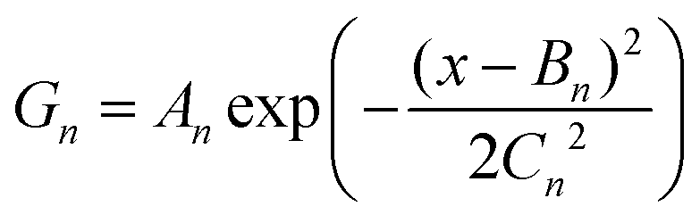
The position of each Gaussian (parameter Bn) is fixed at consecutive data points and the other two parameters are determined numerically to provide the best fit for the real data, as shown in Fig. S5 (ESI†). It is important to notice that fitting has revealed that the parameter Cn equals 3.07 ± 0.01 for all registered signals, layers, and elements. This is not surprising as the blurring of the signal is directly related to the SIMS mixing effect caused by the bombardment of a sample with cesium ions and not the properties of individual layers. It can therefore be concluded that in the ideal case when no mixing effect is present the parameter Cn should equal 0 and the Gaussian functions should be transformed into step-functions with the width of a single data point and the height related to the parameter An. This final deconvolution step is shown in Fig. S6 (ESI†).
Thus, the depth profile that consists of Gaussian-like peaks is finally transformed into a set of box-like features, each representing the composition of a single atomic layer. The final calibration step is based on a simple assumption that the sum of all elements present in a layer is equal to 100%. Given that the ionization probability of each element in the SIMS experiment is different, proportionality factors need to be determined to calibrate the intensity of each signal into the concentration, following the formula:
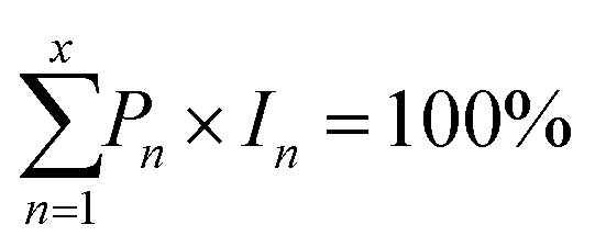
The proportionality factors have been determined numerically using no less than 300 data points for each element and each layer. An interesting observation has been made that these parameters depend only on the type of element and are identical for different layers (for example, the proportionality parameter for oxygen in X, A, and Tx layers) and different samples (for example, the proportionality parameter for oxygen in the X layer determined for Ti2CTx, Ti2NTx, and Ti3CNTx samples). It can be concluded that it strongly validates the proposed deconvolution and calibration procedure.
Fig. 1 presents a final transformation of the as-registered depth profile of a Cr2TiAlC2 MAX phase9 sample into a fully calibrated profile presenting the exact composition of each atomic layer. Fig. S7 and S8/S9 (ESI†) present a similar transformation for a Ti3C2Tx MXene sample. It is important to emphasize that the sum of all components for each data point equals 100% and the error is about 1% which confirms the validity of the proposed calibration method.
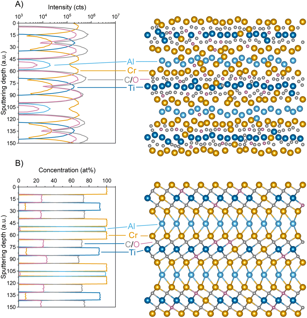 | ||
| Fig. 1 A comparison between the as-registered (part A) and the calibrated (part B) depth profiles of the Cr2TiAlC2 MAX sample. The as-registered data enable the identification of each layer but the estimation of its composition is available only for simple structures. The calibration procedure ensures that the composition of each layer can be precisely quantified with a relatively low error of about 1%. | ||
The precision of the established method enables the exploration of various research problems related to the properties of each type of atomic layer in MAX/MXene samples. For the M layer, the most relevant research is about multiple transition metals MAX and MXenes where two or more different metals are used in the synthesis process. They can be arranged in in- and out-of-plane ordered and solid–solution structures.10,11Fig. 1 confirms the partial ordering as outer layers are composed exclusively of chromium. The inner layer is a solid solution of titanium and chromium and the exact composition can be determined. Cr concentration in the inner layer is 8.08 ± 0.62% which is about 20% lower than the initial estimation (10%).8
Similar analysis is even more crucial for the high-entropy MXenes which may consist of several transition metals.6,7 Theoretical studies predict that their distribution may not be fully random and some elements may have a stronger tendency to occupy the outer layers.12 The newly established procedure for SIMS profiling will be able to verify these suggestions and provide quantitative answers.
Last but not least, some recent studies show the benefit of over-etching MXene samples.13 Given that the proportionality factors are determined on high-quality samples with negligible amounts of vacancies, the method can reveal a deficiency in some layers. Fig. S10 (ESI†) presents an over-etched Ti3C2Tx MXene sample. It is important to note that the concentration of Ti in the outer layers is about 90–95%, whereas the inner layer has not been affected by the etching process. In this case, the quantification procedure leads to a situation where the sum of all components in a layer is lower than 100% and the difference can be attributed to the presence of vacancies. It is important to emphasize that the procedure is not suitable for studying low-concentration vacancies if their amount is lower than the uncertainty of the method (±1%).
For the A layer (see Fig. 1), it becomes apparent that some residual oxygen can be found, typically 0–2 at%. There are, however, samples with much higher concentrations (Fig. S11 (ESI†) shows a profile of the Ti3AlC2 MAX sample with 11–14% oxygen in the A layer). It can be hypothesized that such a large amount of oxygen may have a significant impact on the kinetics of the etching process but a definitive answer requires further studies.
Recent reports show the potential benefits of an under-etched Ti2AlC sample that has been used for hydrogen storage purposes.14 Fig. S12 (ESI†) presents a profile of such an under-etched Ti2AlC MAX sample where the concentration of aluminum in the A layer is decreased to about 10–15%. At the same time, the formation of two distinct termination layers with different compositions can be observed. It is important to emphasize that the atomic resolution is still preserved for such experiments and the remainder of the A-layer (6 data points width) is superimposed on two termination layers (each with 3 data points width). The sum of all components for each data point remains 100 ± 1%.
It is worth noting that most MXenes are derived from the MAX phase precursors where the A layer is aluminum. However, to date MAX phases with 28 different elements have been synthesised.15 In most cases, the selective etching processes have not yet been optimized. The proposed SIMS profiling method may therefore provide some important insight into the structure of such unsuccessfully etched samples and help identify the products of the reaction.
For the X layer, the most revolutionary has been the discovery that most MXenes are actually oxycarbides and not carbides as was assumed.8 Fig. S13 and S14 (ESI†) show that substantial amounts of oxygen can also be found in the X layers in nitride and carbonitride materials and thus the existence of new subfamilies of MXenes, namely oxynitrides and oxycarbonitrides, can be definitively confirmed.
The precision of the proposed method enables a direct comparison of the oxygen concentration in the X layers in MXenes and their MAX phase precursors, as shown in Fig. 2. 363 carbides (black symbols), 101 nitrides (red symbols) and 52 carbonitrides (blue symbols) with varying transition metals and structures have been analyzed and they all followed the exact same trend. An almost perfectly linear relation has been observed, which implies that for most cases the etching process does not impact the oxygen concentration in MXenes. This property is directly inherited from the MAX phase precursors. Only for the MAX phase samples that already contained a lot of oxygen (>30%), the resulting MXene sample may contain a few percent more of this element in the X layer. Most probably, these MAX phase samples contained a significant amount of defects and incorporated even more oxygen during the etching process.
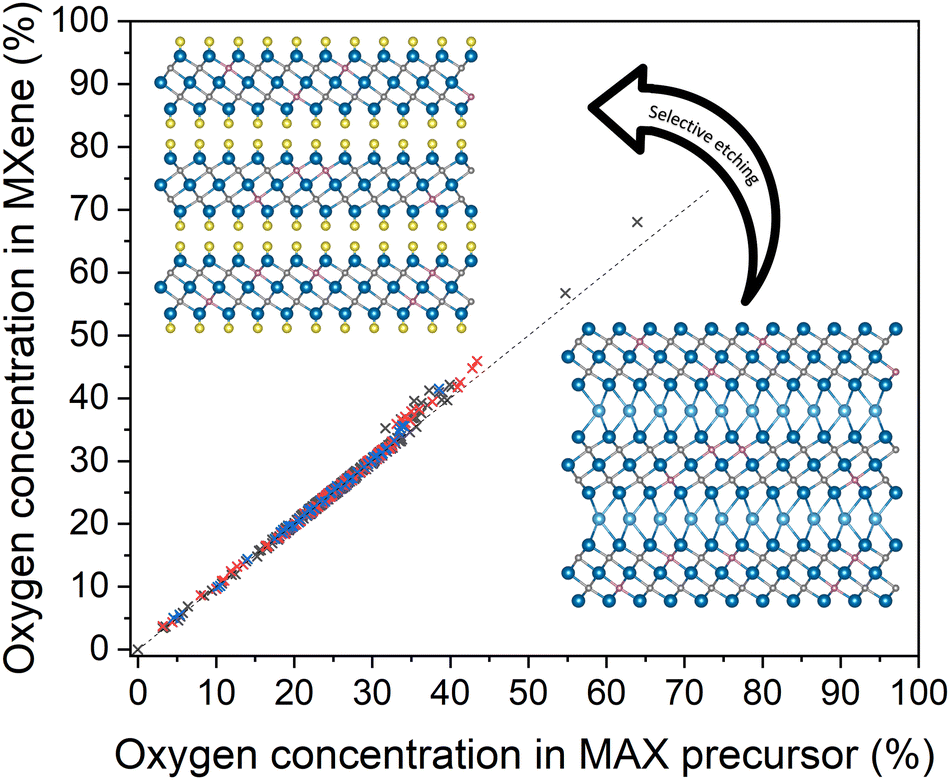 | ||
| Fig. 2 Oxygen concentration in the X layers for various carbide (black symbols), nitride (red symbols), and carbonitride (blue symbols) MXenes as a function of the oxygen concentration in the X layers in their parent MAX phase sample. For most samples, a very distinct 1:1 relation can be observed, indicating that the concentration of oxygen in the X layers in the MXene is directly inherited from the MAX phase precursor – as shown in the schematic drawings. Only MAX phase samples that already contained a lot of oxygen (>30%) may attract even more of this element during/after the etching process. The SIMS analysis therefore reveals that most MXenes are actually oxycarbides, oxynitrides and oxycarbonitrides. | ||
Interestingly, among these samples, only a pure carbide material has been found. In this case, the MAX precursor has been synthesised with an excess of aluminum,16 which led to the formation of a pure carbide material. The proposed method enables verification of whether the same approach can be used to obtain pure nitride and carbonitride MAX phase/MXene samples.
It is very important to emphasize that for all samples presented in Fig. 2 oxygen has been unintentionally incorporated into the structure. The discovery of the oxycarbide subfamily of MXenes8 has opened up a discussion about the possibility of precise control of the oxygen incorporation and its potential impact on the properties of the sample. Theoretical predictions show that a stable Ti3C2−xOx MXene may exist even for x = 1.5.17 Total replacement of oxygen (i.e. x = 2) leads to the distortion of the material and changing the ABC stacking to ABA.
A different study, initiated by the discovery of the oxycarbide MXenes, predicts the possibility of the existence of two-dimensional V2O3 as an oxygen-terminated V2O MOene (transition metal oxide).18 Empirical proofs have not yet been delivered but the method that can precisely quantify the amount of oxygen in each atomic layer of the structure will provide an invaluable aid in these experiments.
Last but not least, some recent experiments show that the formation of some complicated MAX and MXene structures, namely M5AX4 and resulting M5X4Tx, can be stabilized by the intentional addition of metal oxides as precursors during the synthesis.3 Thus, with the SIMS technique, it will be possible to monitor the incorporation of oxygen into the structure.
While substitution of carbon and/or nitrogen atoms with oxygen has gained a lot of attention, the impact of vacancies in the X layer on the properties of MXenes has been reported.19–22 Similarly to the M layer, the proposed method enables quantification of such defects with a similar limitation – their concentration cannot be lower than the uncertainty of the method (±1%).
For the termination layer analysis, it is important to emphasize that in the previously established method8 it was a completely different mode of measurement, i.e. during the acquisition of the data related to the termination layers all information about the core structure of MXene was lost. The newly established method rectifies this problem and enables simultaneous characterization of all layers, as shown in Fig. S8, S9, and S11 (ESI†). Thus, research related to precise engineering of the termination layer may be supplemented by the SIMS technique.
Until now, SIMS has been seen as the only analytical technique used for MXene/MAX phase characterization that can unambiguously identify each element from the periodic table, starting with hydrogen with monoatomic-layer precision. The timely established devolution and calibration procedure enables a fully quantitative analysis of the composition of each atomic layer with 1% accuracy. Such precision may be invaluable for further development and application of these innovative two-dimensional materials.
Author contributions
P. P. M.: conceptualization, data curation, formal analysis, funding acquisition, investigation, methodology, validation, visualization, writing – original draft, writing – review & editing.Data availability
All relevant data are available from the author on reasonable request and/or are included within the article and the ESI.†Conflicts of interest
There are no conflicts to declare.Acknowledgements
This work was supported by the National Science Centre (NCN) within SONATA 14 2018/31/D/ST5/00399 and the National Centre for Research and Development (NCBR) within LIDER XII LIDER/8/0055/L-12/20/NCBR/2021 projects. The author is grateful to Professor Yury Gogotsi, Drexel University, for attracting his attention to this subject and many helpful discussions, as well as to his students and postdocs for providing MAX and MXene samples for this study.References
- B. Anasori and Y. Gogotsi, Graphene 2D Mater., 2022, 7, 75–79 CrossRef
.
- Z. He, L. Yao, W. Guo, N. Sun, F. Wang, Y. Wang, R. Wang and F. Wang, Adv. Funct. Mater., 2023, 33, 2305251 CrossRef CAS
- M. Downes, C. E. Shuck, R. W. Lord, M. Anayee, M. Shekhirev, R. J. Wang, T. Hryhorchuk, M. Dahlqvist, J. Rosen and Y. Gogotsi, ACS Nano, 2023, 17, 17158–17168 CrossRef CAS PubMed
- X. Tang, Y. Zhu, D. Duan, X. Zhai, Y. Xia, T. Xu, Q. Liu, H. Zhang and M. Zhou, Adv. Funct. Mater., 2023, 33, 2305965 CrossRef CAS
- T. Zhang, C. E. Shuck, K. Shevchuk, M. Anayee and Y. Gogotsi, J. Am. Chem. Soc., 2023, 145, 22374–22383 CrossRef CAS PubMed
- S. K. Nemani, B. Zhang, B. C. Wyatt, Z. D. Hood, S. Manna, R. Khaledialidusti, W. Hong, M. G. Sternberg, S. K. R. S. Sankaranarayanan and B. Anasori, ACS Nano, 2021, 15, 12815–12825 CrossRef CAS PubMed
- Z. Du, C. Wu, Y. Chen, Z. Cao, R. Hu, Y. Zhang, J. Gu, Y. Cui, H. Chen, Y. Shi, J. Shang, B. Li and S. Yang, Adv. Mater., 2021, 33, 2101473 CrossRef CAS PubMed
- P. P. Michałowski, M. Anayee, T. S. Mathis, S. Kozdra, A. Wójcik, K. Hantanasirisakul, I. Jóźwik, A. Piatkowska, M. Możdżonek, A. Malinowska, R. Diduszko, E. Wierzbicka and Y. Gogotsi, Nat. Nanotechnol., 2022, 17, 1192–1197 CrossRef PubMed
- K. Hantanasirisakul, B. Anasori, S. Nemsak, J. L. Hart, J. Wu, Y. Yang, R. V. Chopdekar, P. Shafer, A. F. May, E. J. Moon, J. Zhou, Q. Zhang, M. L. Taheri, S. J. May and Y. Gogotsi, Nanoscale Horiz., 2020, 5, 1557–1565 RSC
-
J. Rosen, M. Dahlqvist, Q. Tao and L. Hultman, In- and Out-of-Plane Ordered MAX Phases and Their MXene Derivatives, ed. B. Anasori and Y. Gogotsi, Springer International Publishing, Cham, 2019, pp. 37–52 Search PubMed
- W. Hong, B. C. Wyatt, S. K. Nemani and B. Anasori, MRS Bull., 2020, 45, 850–861 CrossRef
- Z. Leong, H. Jin, Z. M. Wong, K. Nemani, B. Anasori and T. L. Tan, Chem. Mater., 2022, 34, 9062–9071 CrossRef CAS
- S. Wang, Y. Liu, Y. Liu and W. Hu, Chem. Eng. J., 2023, 452, 139512 CrossRef CAS
- S. Liu, J. Liu, X. Liu, J. Shang, L. Xu, R. Yu and J. Shui, Nat. Nanotechnol., 2021, 16, 331–336 CrossRef CAS PubMed
- M. Dahlqvist, M. W. Barsoum and J. Rosen, Mater. Today, 2024, 72, 1–24 CrossRef
- T. S. Mathis, K. Maleski, A. Goad, A. Sarycheva, M. Anayee, A. C. Foucher, K. Hantanasirisakul, C. E. Shuck, E. A. Stach and Y. Gogotsi, ACS Nano, 2021, 15, 6420–6429 CrossRef CAS PubMed
- J. D. Gouveia and J. R. Gomes, Surf. Interfaces, 2024, 46, 103920 CrossRef CAS
- J. Xie, L. Yan, J. Wang, G. Wang, Z. Sun, L. Zhou, J. Yang and H. Dong, Int. J. Hydrogen Energy, 2024, 58, 1587–1595 CrossRef CAS
- J. D. Gouveia and J. R. B. Gomes, Phys. Rev. Mater., 2022, 6, 024004 CrossRef CAS
- E. Marquis, F. Benini, B. Anasori, A. Rosenkranz and M. C. Righi, Nano Convergence, 2023, 10, 16 CrossRef CAS PubMed
- Y. Xue, S. Chao, M. Xu, Q. Wu, Q. Zhang, Y.-J. Liu, F. Wu, L. Liu, M. S. Javed and W. Zhang, Energy Storage Mater., 2024, 71, 103558 CrossRef
- M. M. Hassan, J. Islam, W. R. Sajal, M. N. H. Noman and M. A. Rahman, Heliyon, 2024, 10, e25913 CrossRef CAS PubMed
Footnote |
| † Electronic supplementary information (ESI) available. See DOI: https://doi.org/10.1039/d4nh00151f |
| This journal is © The Royal Society of Chemistry 2024 |