
 Open Access Article
 This Open Access Article is licensed under a
 Creative Commons Attribution 3.0 Unported Licence
Reconceptualising mucoadhesion for future medicines†
Michael T.
Cook
 * and
David
Shorthouse
* and
David
Shorthouse
UCL School of Pharmacy, University College London, 29-39 Brunswick Square, London, WC1N 1AX, UK. E-mail: Michael.T.Cook@UCL.ac.uk
First published on 10th September 2024
Abstract
The field of mucoadhesion has grown from a niche interest to a central consideration for the optimisation of mucosal medicines. As new therapies progress through development pipelines there are constantly emerging conditions which would benefit from the ability to target prolonged residence at mucosal sites. As such, there continues to be expansive investigation into mucoadhesion and the design of novel mucoadhesive materials for dosage form design. In this perspective piece, we give consideration to the recent progress in the field of mucoadhesive materials and make suggestion for reconsideration of current focus. Opinion on risks around current approaches to the development of mucoadhesive materials are described. Furthermore, challenges with translation are discussed, focussing on sensitisation and incompatibilities. Finally, the state of data in this field is critically assessed with a focus to in vitro–in vivo correlation and the formulation state space. It is intended that this manuscript challenges some important areas currently under investigation in the field.
Introduction
Mucoadhesion is defined as the adhesive interaction of a material, usually a polymeric solid, gel, colloid, or liquid, with a mucosal membrane. The primary focus of mucoadhesion centres on the concept that increased adhesion to a mucosal membrane enables improved therapeutic effects.1,2 When a mucoadhesive material is imbibed with an active pharmaceutical ingredient (API) the retention of this dosage form on a mucosal membrane, in principle, allows prolonged local effects and a greater duration over which absorption across the mucosal membrane can occur, thus positively impacting absolute bioavailability.3 A related concept is “mucopenetration”, whereby dosage forms, typically nanoparticles, are designed so that they do not exhibit strong attractive interaction with mucosal layers, but rather diffuse through secretory mucous with little impediment. This allows dosage forms to transport to epithelial layers rapidly, and often avoid some clearance mechanisms, thereby enhancing retention.4 These mucous interactions are an important area to achieve clinical impacts. For example, nasal bioavailability of current macromolecular therapeutics is typically between 0.5–5% due in part to low retention on the nasal mucosa.5 In principle, increased duration of API localisation to this mucosal site increases the time over which systemic absorption can occur, giving benefits such as: enabling nasal administration of peptides that otherwise are not usefully bioavailable, reducing dose requirements, and enhancing therapeutic effects. In another example, the efficacy of mucoadhesive dosage forms has been demonstrated for local administration to the oral mucosa for the treatment of Lichen Planus.6 In this study an electrospun poly(vinylpyrrolidone)/Eudragit RS100 film was found to adhere to the oral mucosa for ca. 2 h, which allowed sustained local administration of clobetasol-17-propionate.7 Clinical trial of the system demonstrated an improved treatment of the condition relative to placebo.6 Clearly there are valuable applications and opportunities for mucoadhesive materials that could enable new therapies or improve current ones through reformulation into adhesive dosage forms. Mucopenetration may be preferable to mucoadhesion, in the case of nanomedicines, to allow direct contact of dosage form with epithelial cells, however this article will focus on perspectives on mucoadhesives.4,8Over the last 15 years the field of mucoadhesion has exploded, with >600 papers related to the subject published in 2023 alone on the Scopus database. Whilst the importance of generating materials with effective retention on mucosal membranes has been known for many years, this recent surge in interest appears to be driven by factors including improved understanding of mechanisms of mucoadhesion,9 new routes to effective modification of hydrophilic polymers,10 the recent innovations in stimuli-responsive materials,11 and 21st Century medicines which are poorly orally bioavailable. The mechanisms underpinning mucoadhesion have been described elsewhere,1,2,12,13 but can be simplified herein to relate to physical and chemical processes occurring between a polymeric excipient in the dosage form and the mucous gel layer coating the epithelium of the mucosa. There is, of course, nuance in this paradigm, particularly in the heterogeneity of mucosal surfaces which range from secretory mucous-rich surfaces such as the lining of the stomach, to those which do not have this gel layer, such as the outside of the eye. Physical mechanisms proposed for mucoadhesion include processes such as entanglement of polymer chains with the mucous gel layer and wetting (spreading due to interfacial forces) to the membrane. Chemical adhesion processes were, in the early generations of mucoadhesive materials, related to non-covalent associations such as hydrogen-bonding,14 electrostatic interactions9 or hydrophobic effects with the mucin glycoprotein constituent of mucous.15 However, subsequent generations of mucoadhesive materials have focussed on chemical modification of polymers to promote covalent bond formation between macromolecule and mucin, typically at cysteine residues near the termini of the targeted glycoprotein. Considering recent focus on this class of mucoadhesive which forms covalent bonds with mucous, there is a need to critically consider the potential for translation of these novel excipients.
In this perspective article we aim to review the current state of the field in mucoadhesion research with a critical eye on current limitations we suggest be considered by the next generation of studies in the area. This piece provides the author's views on important paradigm shifts and reprioritisation that should occur in selected aspects of the field. For a more comprehensive overview of the area, readers are directed to recent reviews in the area.16,17 There is enormous potential in the site-specific retention of medicines to challenging mucosal membranes. This piece aims to provide insight into the pharmaceutical considerations as novel mucoadhesive medicines are developed, considering the materials used in design, preclinical testing regimens, and data within the current literature base upon which future knowledge will be built. Focus is given to novel mucoadhesive materials, which the author's recommend require careful consideration.
Mucoadhesion through “reactive mucoadhesives”: a high risk approach?
In recent years the scientific community has focussed heavily on the generation of new mucoadhesive materials, aiming to increase their covalent adhesion to biological membranes to generate increasingly retentive formulations. For the purposes of classifying these materials as distinct from those which interact via non-covalent interactions, we refer to these materials as “reactive mucoadhesives”. An enormous amount of research has been conducted on “Thiomers”, thiol-group-bearing polymers which are capable of disulfide bridging with cysteine residues on mucin.18,19 Common approaches for generation of thiomers include carbodiimide coupling of cysteine or other thiol-containing molecules onto the backbone of a preformed polymer, typically an existing pharmaceutical excipient.20 This approach was expanded to “preactivated thiomers”, in which the thiol group is protected to stabilise it prior to reaction with mucous to cause adhesion.21 Furthermore, publications report generation of thiomers from protected-thiol-bearing monomers, which are deprotected to yield the thiol after polymerisation,22 to avoid chain transfer during propagation of the monomer.23 Other approaches to thiol-thiol reactivity for enhancing mucoadhesion have also been reported.10 The literature base is broad, but alternative strategies include acrylate,24 methacrylate,25 maleimide,10,26–28 aldehyde,25,29 boronic acid,22 crotonic acid,25 catechol,30 and N-hydroxy(sulfo)succinimide31 modification to promote covalent bonding to mucin (Fig. 1). Overall, these approaches either form reversible covalent associations (such as thiol-disulfide or boronic acid reactivity) or covalent linkages which are not likely to be reverse in vivo (e.g. Michael acceptors such as maleimide or acrylates and N-hydroxysuccinimide linking). This approach has shown sizable improvements in retention on a wide range of mucosal membranes including the eye, buccal pouch, gastrointestinal tract, urinary bladder, and nasal mucosa. However, we believe there is sizable risk in using reactive mucoadhesives for the development of new pharmaceuticals. | ||
| Fig. 1 (A) Schematic diagram of MUC2 as an exemplar secretory mucin showing the heavily glycosylated PTS region and cysterin-rich domains at the termini with which reactive mucoadhesives are often designed to interact. (B) Exemplar structure of selected reactive mucoadhesives, with a polymer backbone (black) adorned by pendant groups (red) capable of reacting with mucosal membranes. (C) Mechanisms for covalent binding of reactive adhesives to mucin components, believed to drive mucoadhesion. Thiol-disulfide reaction occurs between thiols/protected thiols (termed “preactivated thiomers”)32 and cysteine, thiol-Michael addition for (meth)acrylated materials and (e.g.) cysteine,33 thiol–ene click between maleimide-derived polymers and cysteine,28 amide-coupling for NHS-ester polymers with lysine residues,31 Schiff-base formation of aldehyde reaction with amine moieties in mucin and boronation of 1,2-diols, such as found in sialic acid residues.10,22,29,34,35 Please note that these reactions are hypothesized, and may not proceed with the specificity shown herein. The image of amino acid distribution in MUC2 is adapted from Gallego, et al., Nat. Commun., 2023, 14(1) under a Creative Commons (CC) Attribution 4.0 International License. | ||
We argue that the current paradigm on the design of these materials is such that success in generating a reactive mucoadhesive is mechanistically directly linked to step one of the adverse outcome pathway for sensitisation, requiring a significant paradigm shift in this field. The covalent attachment of reactive molecules, termed “haptens” to endogenous proteins is well-established to potentially cause immunological responses.36 Binding of the hapten to endogenous proteins may be followed by a sequence of keratinocyte activation, dendritic cell activation, and proliferation of antigen-specific T cells. The Organisation for Economic Cooperation and Development (OECD) state that in the adverse outcome pathway for skin sensitisation, “the molecular initiating event (i.e. the first key event), is the covalent binding of electrophilic substances to nucleophilic centres in skin proteins”.37 We believe the covalent-associations reported for current-generation mucoadhesive materials present a high risk of leading to sensitisation with prolonged use. Indeed, the FDA perspective on current screening for sensitisation potential includes assays that specifically use cysteine reactivity as an endpoint for sensitisation in the Direct Peptide Reactivity Assay (DPRA).38 These DPRA assays have a reported accuracy of 80% in discriminating sensitisers from non-sensitisers with a sensitivity and specificity of 80% and 77%, respectively.37,39,40In chemico assay for sensitisation is described by the OECD.37 In brief, the assay involves mixing the test substance with cysteine- and lysine-containing synthetic peptides and incubating the solution for 24 h. The mixture is then assayed by HPLC and the depletion of cysteine- and lysine-containing peptides determined. Reducing the concentration of unreacted peptide by ca. 6% is an indicator of potential sensitisation.37 In this context, this reactivity with cysteine could even be an indicator of the success of the reactive mucoadhesive with the binding to this amino acid in mucin a target for adhesion.
Considering the potential for sensitisation of reactive mucoadhesives, the question that must be asked is “how can this risk be mitigated?” Indeed, many working in the area have made considerable effort to assess safety of reactive mucoadhesives in vitro and in vivo,21,41,42 and there is a regulatory need to determine many toxicological endpoints, as described in the International Pharmaceutical Excipient Council's (IPEC) tiered toxicity testing programme it recommends for excipients.43 Furthermore, there is a consideration for what is practical for most research groups in this area and 3Rs considerations for toxicity assessment where many sensitisation tests are performed in vivo.44 It is our position that the reactive mucoadhesives, however, have significant sensitisation potential and that this risk should be mitigated early in the development pipeline. As such, it is suggested that the further key events along the adverse outcome pathway for sensitisation be assessed to mitigate risk. OECD TG 442D evaluates keratinocyte activation, the second key event in skin sensitisation.45 OECD TG 442E focuses on dendritic cell activation, the third key event.46 Lastly, T-cell proliferation, the fourth key event, is indirectly measured through the murine Local Lymph Node Assay.47 These assays provide standardised testing options for evaluating sensitisation potential along the adverse outcome pathway, however there are challenges to their adoption. The group require access to cell culture facilities and know-how in the area. Furthermore, access to cell lines is a barrier as they are, in the case of KeratinoSens and LuSens (OECD TG 442D),45 not widely commercially available. Collaboration between pharmaceutical scientists with expert toxicologists is a potential route to evaluating this risk.
There is hope for reactive mucoadhesives due to the successful delivery of commercialised products, although there are limited examples. PureRegen gel is a disulfide cross-linked hyaluronic acid, licensed by the Food and Drug Administration of the Phillipines48 as a medical device to reduce epithelial adhesions post nasal surgery. In this case the cross-linking prior to application does not directly classify it as a reactive mucoadhesive, in our view. Ocular Bandage Gel by Eye produced by EyeGate (now Kiora) is another example of disulfide cross-linked hyaluronic acid and has been used as an ocular bandage.49 It does not have full regulatory approval. Lacrimera is a thiolated chitosan for treatment of dry eye syndrome produced by Croma,19,50 again regulated as a class 3 medical device. It is currently unavailable for purchase. To the authors’ knowledge there are no reactive mucoadhesives with regulatory approval in medicines, only as a device. There are several examples of successful clinical studies involving thiomers,19 but not other reactive mucoadhesives to our knowledge. It may be that the reversibility of some reactive mucoadhesives (e.g. boronic acids, thiomers) have reduced sensitisation potential relative to irreversible bond formation (e.g. Michael acceptors), however this has not been directly studied.
Reactive mucoadhesives also have a high risk in incompatibility with other pharmaceutical excipients or APIs. Thiomers have potential for oxidation/reduction via intra-/inter-molecular reaction as single components.51 They also have the potential to act as Michael donors in their own right, as well as a plethora of other reactivities.52 Maleimide/acrylate functionality are reactive via nucleophilic Michael-type addition, which can proceed under ambient conditions.53 A further limitation is the inherent protein reactivity that drives their function. Therapeutic cells, proteins, and peptides are all likely to undergo modification in the presence of reactive mucoadhesives, which are designed specifically to react with mucin proteins. This severely limits potential use-cases, as many 21st century medicines, including all biologics, fall into these categories, and so are highly likely to react to this category of mucoadhesive. Should there be a reaction leading to ligation of the mucoadhesive polymer to the therapeutic entity, this will subsequently impact on factors including functionality, liberation, pharmacokinetics, and, as a result, regulatory status of the drug.
The aim of mucoadhesion research is principally in the development of more efficacious treatments for disease. The ideal test of the efficacy of any new mucoadhesive medicines or devices is in human trials, however justifying these trials for novel mucoadhesive excipients requires significant toxicological assessment, and raising capital for such studies is challenging. These toxicological risks may be substantially reduced for mucosal dosage forms consisting of excipients with a history of use in pharmaceuticals, however this makes it difficult to innovate in such a crowded space, raising challenges around protection of intellectual property. However, until such a time that there are significantly expedited routes for novel excipients into the medicines this latter approach will remain the most likely to yield positive impacts on patients.
Refocussing on the patient; how can clinical behaviours be predicted?
A big challenge with translation to clinical study is the prediction of in vivo behaviours for novel medicines. The literature base describing in vivo mucoadhesion in both humans and laboratory animals is small,54,55 and given the diversity of dosage forms and routes of administration there is a large challenge in predicting in vivo performance. Additionally, there is a further philosophical question in which in vivo endpoints are most important; retention, drug liberation, pharmacodynamics and pharmacokinetics will all affect therapeutic outcomes and establishing meaningful prediction of in vivo data is extremely challenging. In vitro–in vivo correlation (IVIVC), or the mathematical relationship between in vitro (or ex vivo) and in vivo behaviour, is highly desirable to reduce costs and accelerate medicines development.56 However, well-established IVIVC models for human mucosal retention do not exist. The community has attempted to standardise testing methods for mucoadhesives,57 but the approaches are still disparate – recently Bayer grouped these approaches into 10 different experimental approaches.16 In our view, this diversity in testing approaches is driven largely by necessity given the diversity of dosage forms, from tablets to liquids and disperse systems.There are some reported efforts to establish IVIVC for mucoadhesives. Correlation between buccal retention of solid dosage forms in a dissolution apparatus-based retention test on porcine musosa and human volunteers has been studied. In this small study, it appears that there is an over-prediction of retention time by the ex vivo retention test but a linear relationship between the two (Fig. 2A).58 The same approach was used by the group for physical hydrogels, yielding a weaker linear relationship (Fig. 2B).59 Comparison between human and porcine intestinal mucosa has been studied ex vivo, which found no significant difference between the tissues under a specific tensile testing approach, but with large intra-subject variability (Fig. 2C and D).60 The large error present in all of these experiments (either induced by biological or experimental variability) demonstrates how unreliable these assessment methods may be for determining the clinical use of these materials. The most widely used techniques for adhesion assessment are the use of tensile detachment methods, which measure forces needed to detach a solid dosage form from mucosa, and bespoke flow-through methods, which run physiologically relevant fluid over a dosage form applied typically to ex vivo mucosa.61 There are well-established issues with current in vitro testing methods in terms of standardisation of both instruments and conditions.57,62 Furthermore, the subsequent IVIVC relating in vitro/ex vivo measurements to in vivo performance in laboratory animals is questionable. For example, it has been found that tensile detachment ex vivo does not predict tensile detachment forces directly measured in vivo in rats via a gastric cannula.63 We call on the field to establish improved databases of human data which will allow improvements in IVIVC, as well as allow the benchmarking of the disparate in vitro techniques used by the community.
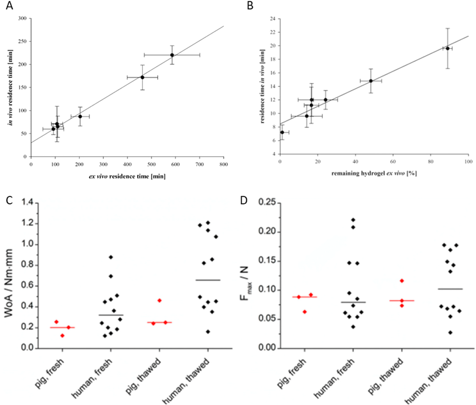 | ||
| Fig. 2 Top: IVIVC between ex vivo residence time of solid oral dosage forms (A) and hydrogels (B) on buccal porcine mucosa and human volunteers. Bottom: comparison between the work of adhesion (WoA) (C) and force of adhesion (Fmax) (D) in fresh and thawed human and pig intestine. (A) Adapted with permission from Baus, et al., Mol. Pharm., 2019, 16(6), 2719–2727. Copyright 2019 American Chemical Society. (B) Adapted with permission from Baus, et al., Eur. J. Pharm. Biopharm., 2019, 142, 498–505. Copyright 2019 Elsevier. (C) and adapted from Müller, et al., Pharmaceutics 2023, 15(6), 1740, under a CC BY 4.0 License. | ||
Material exploration: making the most of what we have
Aside from issues with translatability and relevance of measured data on mucoadhesive properties, much of the advancements in the field have been through the development and manipulation of a very small number of related materials. Whilst it is tempting to state that we should explore completely novel materials and chemistries with the aim of producing more clinically viable mucoadhesion properties, it is more sensible to focus on materials that have known low toxicities and good biocompatibility.To explore the current landscape of materials used for mucoadhesion, we collected information from the literature by searching the ISI Web of Science with the terms “mucoadhes* AND tensile AND (tablet or solid)” in January 2024. This search returned 88 results, which we used to collect a database of 101 published materials (ESI†) that have been assessed for mucoadhesion. These materials cover a wide range of base polymers (Fig. 3A), chemistry types, and tissues on which they are assessed. We find that, where available, comparison of Work of Adhesion (WoA), and Force of Adhesion (FoA) gives a good, though not perfect, correlation (Pearson r = 0.730)(Fig. 3B) – indicating that these measurements are not necessarily directly comparable.
 | ||
| Fig. 3 Properties of published mucoadhesive polymers. (A) Proportions of base polymer units used in 101 published polymers assessed for mucoadhesion. (B) Correlation between force of adhesion (N), and work of adhesion (N mm−1) for 101 published polymers. (C) Total landscape of unexplored (grey) and explored (blue) mucoadhesive materials using all possible combinations of published base polymer units and parameters. FAMD – Factor Analysis of Mixed Data. | ||
Building on this we determined a landscape of potential mucoadhesive materials based purely on the state space available from currently published materials (Fig. 3C). In brief, this analysis considers all possible combinations of existing polymers and derivatisations. We note this analysis doesn't take into account feasibility of creating materials, but assumes the ranges of already published properties are combinable. Highlighting the currently explored materials on this landscape shows that published materials are limited to an extremely small subset of this state space, and that there is huge potential in exploring currently unexplored regions of this map, particularly through complex mixtures of excipient. This may enable a balance to be struck between novelty (and therefore patentability), and biocompatibility without introducing materials with an unknown biocompatibility profile and new chemistries.
Conclusions: refocussing the field
Having brought our view that there are limitations in current studies of mucoadhesive polymers – namely that focus on reactive mucoadhesives is high risk, testing for adhesion is not necessarily the best measure for assessing usefulness, clinical efficacy is hard to predict from in vitro experiments, and that current work has focussed on exploring a small number of available material materials, we suggest a refocussing of the field is necessary.We recommend that reactive mucoadhesives are inherently high-risk for incompatibility issues and sensitisation. Sensitisation risks should be evaluated as a priority through collaboration with expert toxicologists who are most likely to have access to the required facilities and materials. We also recommend that the risk of these materials would be best justified by enabling new therapies on severe health conditions which are not possible to successfully treat with the current excipients available, to give clinical and economic justification for their trial in human subjects.
Assessments of these materials should be adapted for maximising clinical/biological relevance. Considering the lack of IVIVC data using laboratory animals and large anatomical differences in mucosal sites compared to humans which will impact retention,64 it is suggested that researchers focus on well-recognised in vitro/ex vivo testing regimen, for example tensile testing and flow-through assessments,65,66 for rank order determinations and then accelerate towards pilot study in humans, contributing both to effective medicines development and yielding important data for establishment of future IVIVC. This data may then be used to validate in vitro models. When dosage forms are composed of existing pharmaceutical excipients, these studies require lower costs (through avoidance of toxicological regimes) and have fewer ethical considerations. As such, we recommend the generation of high quality IVIVC using these established excipients. Pilot studies demonstrating enhanced retention in vivo may then support larger clinical studies determining pharmacokinetics/pharmacodynamics of the medicines. There are synthetic alternatives to tissue which can be used in these testing regimes where it is difficult to source, which has been shown to be equivalent to ex vivo tissue.61,67–69 There is potential, therefore, to entirely remove the need for ex vivo tissue from preclinical evaluation of mucoadhesives if there is sufficient human data to validate.
We also suggest that more can be made from the existing materials – whilst most published materials are based on one of a small number of well characterised and biocompatible components, the landscape of materials that have been assessed is extremely limited. Exploring further combinations of materials and processes in this landscape will significantly enhance our understanding of this complex environment. Recent developments in data science make exploration of complex mixtures increasingly feasible.
Data availability
No primary research results, software or code have been included and no new data were generated as part of this review. All data used in construction of figures is included as ESI.†Conflicts of interest
The authors have no conflicts of interest to declare.Acknowledgements
The authors thank Dr Sarah Gould at Charles River Laboratories and Dr Grant Bradley at Makevale for their useful discussions concerning the manuscript. Dr Bradley is a further acknowledged for first raising the potential issues of sensitisation described.References
- V. V. Khutoryanskiy, Macromol. Biosci., 2011, 11, 748–764 CrossRef PubMed
.
- J. D. Smart, Adv. Drug Delivery Rev., 2005, 57, 1556–1568 CrossRef PubMed
- M. T. Cook and V. V. Khutoryanskiy, Int. J. Pharm., 2015, 495, 991–998 CrossRef PubMed
- P. Schattling, E. Taipaleenmäki, Y. Zhang and B. Städler, Macromol. Biosci., 2017, 17, 1700060 CrossRef
- M. D. Donovan and Y. Huang, Adv. Drug Delivery Rev., 1998, 29, 147–155 CrossRef
- M. T. Brennan, L. S. Madsen, D. P. Saunders, J. J. Napenas, C. McCreary, R. N. Riordain, A. M. L. Pedersen, S. Fedele, R. J. Cook, R. Abdelsayed, M. T. Llopiz, V. Sankar, K. Ryan, D. A. Culton, Y. Akhlef, F. Castillo, I. Fernandez, S. Jurge, A. R. Kerr, C. McDuffie, T. McGaw, A. Mighell, T. P. Sollecito, T. Schlieve, M. Carrozzo, A. Papas, T. Bengtsson, I. Al-Hashimi, L. Burke, N. W. Burkhart, S. Culshaw, B. Desai, J. Hansen, P. Jensen, T. Menné, P. B. Patel, M. Thornhill, N. Treister and T. Ruzicka, J. Oral Pathol. Med., 2022, 51, 86–97 CrossRef PubMed
- H. E. Colley, Z. Said, M. E. Santocildes-Romero, S. R. Baker, K. D'Apice, J. Hansen, L. S. Madsen, M. H. Thornhill, P. V. Hatton and C. Murdoch, Biomaterials, 2018, 178, 134–146 CrossRef
- N. Vasquez-Martínez, D. Guillen, S. A. Moreno-Mendieta, S. Sanchez and R. Rodríguez-Sanoja, Polymers, 2023, 15, 1615 CrossRef PubMed
- I. A. Sogias, A. C. Williams and V. V. Khutoryanskiy, Biomacromolecules, 2008, 9, 1837–1842 CrossRef
- R. P. Brannigan and V. V. Khutoryanskiy, Macromol. Biosci., 2019, 19, 1–11 CrossRef PubMed
- M. A. C. Stuart, W. T. S. Huck, J. Genzer, M. Müller, C. Ober, M. Stamm, G. B. Sukhorukov, I. Szleifer, V. V. Tsukruk, M. Urban, F. Winnik, S. Zauscher, I. Luzinov and S. Minko, Nat. Mater., 2010, 9, 101–113 CrossRef
- N. A. Peppas and Y. Huang, Adv. Drug Delivery Rev., 2004, 56, 1675–1687 CrossRef PubMed
- A. Sosnik, J. das Neves and B. Sarmento, Prog. Polym. Sci., 2014, 39, 2030–2075 CrossRef
- H. Hsein, G. Garrait, E. Beyssac and V. Hoffart, Colloids Surf., B, 2015, 136, 799–808 CrossRef PubMed
- R. Bansil and B. S. Turner, Curr. Opin. Colloid Interface Sci., 2006, 11, 164–170 CrossRef
- I. S. Bayer, Adv. Mater. Interfaces, 2022, 9, 2200211 CrossRef CAS
- R. Kumar, T. Islam and M. Nurunnabi, J. Controlled Release, 2022, 351, 504–559 CrossRef CAS PubMed
- A. Bernkop-Schnürch and A. Greimel, Am. J. Drug Delivery, 2005, 3, 141–154 CrossRef
- S. Bonengel and A. Bernkop-Schnürch, J. Controlled Release, 2014, 195, 120–129 CrossRef CAS
- K. Albrecht and A. Bernkop-Schnürch, Nanomedicine, 2007, 2, 41–50 CrossRef CAS PubMed
- J. Iqbal, G. Shahnaz, S. Dünnhaupt, C. Müller, F. Hintzen and A. Bernkop-Schnürch, Biomaterials, 2012, 33, 1528–1535 CrossRef CAS
- O. M. Kolawole, W. M. Lau and V. V. Khutoryanskiy, J. Pharm. Sci., 2019, 108, 3046–3053 CrossRef
- M. T. Cook, S. A. Schmidt, E. Lee, W. Samprasit, P. Opanasopit and V. V. Khutoryanskiy, J. Mater. Chem. B, 2015, 3, 6599–6604 RSC
- M. Davidovich-Pinhas and H. Bianco-Peled, J. Mater. Sci.: Mater. Med., 2010, 21, 2027–2034 CrossRef PubMed
- E. O. Shatabayeva, D. B. Kaldybekov, L. Ulmanova, B. A. Zhaisanbayeva, E. A. Mun, Z. A. Kenessova, S. E. Kudaibergenov and V. V. Khutoryanskiy, Biomacromolecules, 2024, 25, 1612–1628 CrossRef PubMed
- D. B. Kaldybekov, P. Tonglairoum, P. Opanasopit and V. V. Khutoryanskiy, Eur. J. Pharm. Sci., 2019, 143, 24–34 Search PubMed
- N. Sahatsapan, B. Pamornpathomkul, T. Rojanarata, T. Ngawhirunpat, R. Poonkhum, P. Opanasopit and P. Patrojanasophon, J. Drug Deliv. Sci. Technol., 2022, 69, 103173 CrossRef CAS
- P. Tonglairoum, R. P. Brannigan, P. Opanasopit and V. V. Khutoryanskiy, J. Mater. Chem. B, 2016, 4, 6581–6587 RSC
- E. E. Brotherton, T. J. Neal, D. B. Kaldybekov, M. J. Smallridge, V. V. Khutoryanskiy and S. P. Armes, Chem. Sci., 2022, 13, 6888–6898 RSC
- S. Choi, J. Jeon, Y. Bae, Y. Hwang and S.-W. Cho, Adv. Funct. Mater., 2023, 33, 2303043 CrossRef CAS
- C. Menzel, M. Hauser, A. Frey, M. Jelkmann, F. Laffleur, S. K. Götzfried, R. Gust and A. Bernkop-Schnürch, Eur. J. Pharm. Biopharm., 2019, 139, 161–167 CrossRef CAS PubMed
- H. E. Friedl, S. Dünnhaupt, C. Waldner and A. Bernkop-Schnürch, Biomaterials, 2013, 34, 7811–7818 CrossRef CAS
- M. Davidovich-Pinhas and H. Bianco-Peled, Acta Biomater., 2011, 7, 625–633 CrossRef CAS PubMed
- X. Zhu, C. Borchers, R. J. Bienstock and K. B. Tomer, Biochemistry, 2000, 39, 11194–11204 CrossRef CAS PubMed
- S. J. Hunter, M. H. Abu Elella, E. C. Johnson, L. Taramova, E. E. Brotherton, S. P. Armes, V. V. Khutoryanskiy and M. J. Smallridge, J. Colloid Interface Sci., 2023, 651, 334–345 CrossRef CAS PubMed
- E. Sakamoto, Y. Katahira, I. Mizoguchi, A. Watanabe, Y. Furusaka, A. Sekine, M. Yamagishi, J. Sonoda, S. Miyakawa, S. Inoue, H. Hasegawa, K. Yo, F. Yamaji, A. Toyoda and T. Yoshimoto, Biology, 2023, 12, 123 CrossRef CAS PubMed
- Test No. 442C: In Chemico Skin Sensitisation : Assays addressing the Adverse Outcome Pathway key event on covalent binding to proteins, OECD Publishing, Paris, 2021, DOI:10.1787/9789264229709-en
- A. M. Avila, I. Bebenek, J. A. Bonzo, T. Bourcier, K. L. D. Bruno, D. B. Carlson, J. Dubinion, I. Elayan, W. Harrouk, S. L. Lee, D. L. Mendrick, J. C. Merrill, J. Peretz, E. Place, M. Saulnier, R. L. Wange, J. Yao, D. Zhao and P. C. Brown, Regul. Toxicol. Pharmacol., 2020, 114, 104662 CrossRef
- S. Hoffmann, N. Kleinstreuer, N. Alépée, D. Allen, A. M. Api, T. Ashikaga, E. Clouet, M. Cluzel, B. Desprez, N. Gellatly, C. Goebel, P. S. Kern, M. Klaric, J. Kühnl, J. F. Lalko, S. Martinozzi-Teissier, K. Mewes, M. Miyazawa, R. Parakhia, E. van Vliet, Q. D. Zang and D. Petersohn, Crit. Rev. Toxicol., 2018, 48, 344–358 CrossRef
- Test No. 442C: In Chemico Skin Sensitisation : Direct Peptide Reactivity Assay (DPRA), OECD Publishing, Paris, 2015, DOI:10.1787/9789264229709-en
- S. Choi, J. Jeon, Y. Bae, Y. Hwang and S. W. Cho, Adv. Funct. Mater., 2023, 44(33), 2303043 CrossRef
- X. N. Shan, S. Aspinall, D. B. Kaldybekov, F. Buang, A. C. Williams and V. V. Khutoryanskiy, ACS Appl. Polym. Mater., 2021, 3, 5882–5892 CrossRef
- C. C. DeMerlis, J. M. Goldring, R. Velagaleti, W. Brock and R. Osterberg, Pharm. Technol., 2009, 33, 72–82 Search PubMed
- BS EN ISO 10993-10:2023, Biological evaluation of medical devices, British Standards Institution, London, 2023 Search PubMed
- OECD, Test No. 442D: In Vitro Skin Sensitisation, in OECD Guidelines for the Testing of Chemicals, Section 4: Health Effect, 2022, DOI:10.1787/9789264229822-en
- OECD, Test No. 442E: In Vitro Skin Sensitisation, in OECD Guidelines for the Testing of Chemicals, Section 4: Health Effect, 2023, DOI:10.1787/9789264264359-en
- OECD, Test No. 429: In Skin Sensitisation, in OECD Guidelines for the Testing of Chemicals, Section 4: Health Effect, 2023, DOI:10.1787/20745788
- Food and Drug Administration of the Phillipines , PUREREGEN GEL SINUS NASAL AND SINUSDRESSING Registration Number MDR-10963C, https://verification.fda.gov.ph/medical_devicesview.php?showdetail=®istration_number=MDR-10963C, (accessed 15.12.2023, 2023).
- D. S. Durrie, D. Wolsey, V. Thompson, C. Assang, B. Mann and B. Wirostko, J. Cataract Refractive Surg., 2018, 44, 369–375 CrossRef PubMed
- M. Messina and H. S. Dua, Int. Ophthalmol., 2019, 39, 693–696 CrossRef
- L. B. Poole, Free Radical Biol. Med., 2015, 80, 148–157 CrossRef CAS
- D. P. Nair, M. Podgórski, S. Chatani, T. Gong, W. X. Xi, C. R. Fenoli and C. N. Bowman, Chem. Mater., 2014, 26, 724–744 CrossRef CAS
- N. B. Cramer, J. P. Scott and C. N. Bowman, Macromolecules, 2002, 35, 5361–5365 CrossRef CAS
- C. Woertz, M. Preis, J. Breitkreutz and P. Kleinebudde, Eur. J. Pharm. Biopharm., 2013, 85, 843–853 CrossRef CAS
- S. L. Cook, S. Woods, L. Methven, J. K. Parker and V. V. Khutoryanskiy, Mucoadhesive polysaccharides modulate sodium retention, release and taste perception, Food Chem., 2018, 240, 482–489 CrossRef CAS
- V. F. Patel, F. Liu and M. B. Brown, J. Controlled Release, 2012, 161, 746–756 CrossRef CAS
- J. Bassi da Silva, S. B. D. S. Ferreira, A. V. Reis, M. T. Cook and M. L. Bruschi, Polymers, 2018, 10, 1–19 CrossRef
- R. A. Baus, M. F. Haug, C. Leichner, M. Jelkmann and A. Bemkop-Schnürch, Mol. Pharm., 2019, 16, 2719–2727 CrossRef
- R. A. Baus, F. Zahir-Jouzdani, S. Dünnhaupt, F. Atyabi and A. Bernkop-Schnürch, Eur. J. Pharm. Biopharm., 2019, 142, 498–505 CrossRef PubMed
- L. Müller, C. Rosenbaum, A. Rump, M. Grimm, F. Klammt, A. Kleinwort, A. Busemann and W. Weitschies, Pharmaceutics, 2023, 15, 1740 CrossRef
- M. T. Cook and V. V. Khutoryanskiy, Int. J. Pharm., 2015, 495, 991–998 CrossRef
- J. Bassi da Silva, S. B. D. S. Ferreira, O. de Freitas and M. L. Bruschi, Drug Dev. Ind. Pharm., 2017, 43, 1053–1070 CrossRef
- B. Laulicht, P. Cheifetz, A. Tripathi and E. Mathiowitz, J. Controlled Release, 2009, 134, 103–110 CrossRef PubMed
- F. Schneider, K. Koziolek and W. Weitschies, Pharmaceutics, 2019, 11, 416 CrossRef PubMed
- R. A. Cave, J. P. Cook, C. J. Connon and V. V. Khutoryanskiy, Int. J. Pharm., 2012, 428, 96–102 CrossRef
- J. Bassi da Silva, S. B. S. Ferreira, A. V. Reis, M. T. Cook and M. L. Bruschi, Polymers, 2018, 10, 254 CrossRef PubMed
- M. T. Cook, S. L. Smith and V. V. Khutoryanskiy, Chem. Commun., 2015, 51, 14447–14450 RSC
- R. Donahue, J. K. Sahoo, S. Rudolph, Y. Chen and D. L. Kaplan, Adv. Healthcare Mater., 2023, 12, 2300301 CrossRef
- D. J. Hall, O. V. Khutoryanskaya and V. V. Khutoryanskiy, Soft Matter, 2011, 7, 9620 RSC
Footnote |
| † Electronic supplementary information (ESI) available. See DOI: https://doi.org/10.1039/d4pm00149d |
| This journal is © The Royal Society of Chemistry 2024 |