
An esterase-activated diketopyrrolopyrrole-based theranostic prodrug for precise pyroptosis and synergistic chemo-photodynamic therapy of pancreatic cancer†
Xixin
Gu‡
a,
Kaini
Yang‡
b,
Sifan
Li
 a,
Ju
Mei
a,
Xiao-Peng
He
*a,
Wei
Chen
*b and
Jianli
Hua
*a
a,
Ju
Mei
a,
Xiao-Peng
He
*a,
Wei
Chen
*b and
Jianli
Hua
*a
aKey Laboratory for Advanced Materials, Joint International Research Laboratory for Precision Chemistry and Molecular Engineering, Feringa Nobel Prize Scientist Joint Research Center, School of Chemistry and Molecular Engineering, East China University of Science and Technology, Shanghai 200237, China. E-mail: xphe@ecust.edu.cn; jlhua@ecust.edu.cn
bKey Laboratory for Advanced Materials, Joint Int Department of Biliary-Pancreatic Surgery, Renji Hospital, School of Medicine, Shanghai Jiaotong University, Shanghai 200127, China. E-mail: chensurg@aliyun.com
First published on 7th March 2024
Abstract
Pyroptosis, a novel form of programmed cell death, has demonstrated significant antitumor effects. However, enhancing the precision of pyroptosis and improving the treatment efficacy for malignant tumors still remain formidable challenges. In this work, we propose a novel synergistic chemo-photodynamic therapy strategy that targets endogenous esterase in the tumor microenvironment (TME), aiming to achieve precise pyroptosis and facilitate the release of chemotherapeutic drugs to promote pyroptosis. An esterase-activated diketopyrrolopyrrole-based theranostic prodrug DPP-QS was synthesized by the conjugation of a chemotherapeutic drug chlorambucil, with DPP-QE characteristic of aggregation-induced emission (AIE) through a one-step nucleophilic reaction. Besides, the detection limit of DPP-QS toward esterase in vitro was 5.13 × 10⁻⁵ U mL−1. Upon cleavage by esterase endogenously expressed in pancreatic cancer cells, the released DPP-QE moiety selectively accumulated in lipid droplets (LDs) with a significantly enhanced fluorescence due to AIE, and upon subsequent exposure to light, reactive oxygen species (ROS) are generated to induce pyroptosis. Simultaneously, chlorambucil entered the nucleus of cancer cells, causing DNA damage, which further enhanced the effect of pyroptosis. In vivo experiments demonstrated that the prodrug displayed significant TME-activatable features and excellent tumor therapeutic effects through synergistic chemo-photodynamic therapy.
Introduction
Pyroptosis is a new form of programmed cell death that activates caspase-1 to cleave gasdermin-D (GSDMD).1 The cleaved GSDMD releases gasdermin-N domains (N-GSDMD) which would translocate into the cell membrane to form membrane pores, causing the cell membrane to lose its integrity, resulting in the loss of the ability of the cell membrane to control the entry and exit of substances, and ultimately leading to the dissolution of the cell membrane. In addition, compared with apoptosis, pyroptosis releases cell contents such as lactate dehydrogenase (LDH), adenosine triphosphate (ATP) and inflammatory cytokines including interleukin-1β (IL-1β) and interleukin-18 (IL-18), thereby inducing an inflammatory response.2,3 It has been confirmed recently that ROS can induce pyroptosis.4–6 However, there are few works that can accurately induce pyroptosis by distinguishing normal cells and cancer cells through fluorescence (FL) imaging guidance.7 Moreover, PDT-mediated pyroptosis usually does not damage DNA in the nucleus, which leaves the risk of cancer recurrence.Photodynamic therapy (PDT) is a new non-invasive diagnosis and treatment mode, involving photosensitizer (PS), light and endogenous molecular oxygen.8 The PS is activated by light and in the excited state, energy is transferred to surrounding oxygen, generating ROS, particularly singlet oxygen (1O2), which oxidize the surrounding biomolecules, ultimately causing cell damage or death.9 In addition, AIE PSs are frequently created for the purpose of FL imaging due to their tendency to aggregate in biological tissues while maintaining luminescence.10,11 It is important to mention that LDs are involved in lipid metabolism, membrane formation, energy production and storage, and play a crucial role in the proliferation, invasion and metastasis of cancer cells.12 Therefore, LDs can be used as important targets for PDT.13,14 However, PDT is a treatment method that is applied locally, and its effectiveness in killing tumors depends mostly on providing sufficient light irradiation to the affected area.15,16 In contrast, chemotherapy (CT) is a systemic treatment that, when combined with phototherapy, can enhance the therapeutic effects for cancer to some extent.17,18
Prodrugs with TME-responsive properties are generally prepared based on the overexpression of intracellular acidity, ROS, thiols, hypoxia, enzymes, etc.19–22 Esterase is overexpressed in cancer cells and plays a key role in malignant invasion, migration and proliferation.23 Esterase has been extensively developed as a target for various applications, including monitoring cellular activity, imaging cancer cell organelles, and releasing chemotherapeutic drugs.24–26 Recently, Zhao et al. developed an esterase-activated alkyl ester prodrug TPE-QC intended for both dual-organelle targeted imaging and chemo-photodynamic synergistic approaches for the treatment of cancer, which displayed efficient tumor microenvironment-activatable features and good tumor therapeutic effects through combinational CT and PDT.27 Generally, alkyl esters exhibit high stability and slow hydrolysis, even in the presence of esterase, while phenolic esters were reported to be readily hydrolyzable by esterases.28 Besides, the chemotherapeutic drug chlorambucil is a DNA alkylating agent that can undergo nucleophilic substitution with bases on guanine to induce DNA cross-linking in the nucleus.29–31
Diketopyrrolopyrrole (DPP) derivatives possess tunable properties, including a substantial molar extinction coefficient, favorable photostability and exceptional ability to produce ROS.14,32,33 Considering the above situation, we developed phenolic esters as a “linking bridge” between DPP-based PS and chlorambucil. The bridge can be hydrolyzed by esterase in cancer cells, and can leave in the form of para-quinone methides without any effect on the DPP-based PS or chemotherapeutic drug. Herein, we prepared a molecule SF-3 containing chlorambucil and phenolic ester groups, which can be covalently coupled to an AIE PS (DPP-QE) through a one-step nucleophilic reaction to construct an esterase-activated theranostic prodrug (DPP-QS, Scheme 1A). After modification with SF-3, the solubility of cationic compound DPP-QS in water was significantly enhanced compared to that of DPP-QE. In addition, the expression of esterase in pancreatic cancer was higher than that in normal cells, so it was selected as the research object.34 Due to the more negative transmembrane potential, DPP-QS can be better absorbed by pancreatic cancer cells rather than normal cells. Importantly, the release of the lipophilic PS (DPP-QE) by esterase hydrolysis can effectively locate LDs to help monitor FL imaging in pancreatic cancer cells rather than in normal cells and implement PDT when the fluorescence signal reaches its maximum, which improves the precise control of pyroptosis. Interestingly, a significant amount of ROS generated during PDT has the capability to induce pyroptosis or apoptosis, and heighten nucleus permeability, thus elevating the likelihood of DNA alkylating agents entering the nucleus and leading to a significant expansion of DNA damage. Once the DNA in the nucleus sustains damage, pancreatic cancer cells become less resistant to the large amount of ROS produced, thereby enhancing the effect of pyroptosis (Scheme 1B). This chemo-photodynamic therapy synergistic effect results in good feedback, lower prodrug concentration, smaller light power and shorter illumination time, which are very beneficial for the treatment of malignant tumors, such as pancreatic cancer.
 | ||
| Scheme 1 (A) Construction of prodrug DPP-QS and its reaction mechanism towards esterase. (B) The synergistic chemo-photodynamic therapy mechanism of esterase-activated theranostic prodrug DPP-QS for pancreatic cancer. | ||
Results and discussion
Design and synthesis of DPP-QS
SF-3 contains a phenolic ester group, which could be used as an esterase-activated site to release chlorambucil. Besides, DPP-QE was constructed by selecting phenyl-DPP as the acceptor core, and 4-[bis(biphenyl-4-yl)amino]phenyl (BTPA) with a rotor as the electron donor to disrupt the ‘π–π’ stacking configuration and achieve AIE performance. At the same time, we chose the quinoline group as the site of the nucleophilic substitution attack by SF-3, because the formation of the quinolinium cationic salt leads to a mild positive charge, making DPP-QS show a superior cell membrane penetration ability compared to pyridine and acridine.35 Upon reaction with esterase, the phenolic ester group of DPP-QS could be readily and selectively hydrolyzed to release chlorambucil and DPP-QE, which could improve the fluorescence and photo-sensitization due to the break of intramolecular charge transfer (ICT) from the DPP moiety to the quinolinium cation. The synthetic route of the target compound (DPP-QS) is shown in Scheme S1 (ESI†). The important intermediate SF-3 was synthesized through a series of chemical processes, involving esterification, reduction, and bromination of chlorambucil and p-hydroxybenzaldehyde. The synthesis of DPP-QE was mainly through two-step Suzuki coupling on the basis of phenyl-DPP.36DPP-QS was obtained by the nucleophilic substitution reaction of DPP-QE and SF-3. All the new compounds have been confirmed via1H NMR, 13C NMR, and HRMS (Fig. S1–S18, ESI†).Photophysical properties
First, the AIE feature of DPP-QE and DPP-QS was investigated in EtOH/hexane mixtures with increasing hexane fractions (fH). The absorption spectra of DPP-QE in ethanol and hexane were obtained, as shown in Fig. S19 (ESI†). DPP-QE showed weak fluorescence in pure ethanol, but when fH reached 99%, the fluorescence intensity at 625 nm increased by 190 times (Fig. 1A and B). Furthermore, the DLS analysis confirmed the formation of DPP-QE nanoaggregates with an average diameter of 435.9 nm, which was also confirmed by the SEM (Fig. S20, ESI†). Moreover, the solid-state fluorescence quantum yield (ΦF) of DPP-QE is 22.4%. However, DPP-QS hardly emits light in EtOH/hexane mixtures, and showed only ΦF of 1.0% in the solid-state. The above findings demonstrated that DPP-QE has the AIE properties.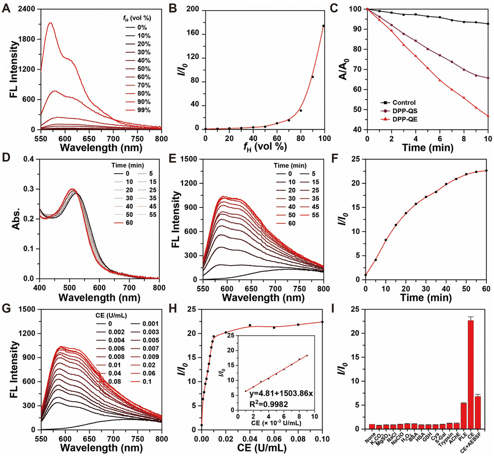 | ||
| Fig. 1 (A) FL spectra and (B) plots of relative FL emission intensity (I/I0) of DPP-QE (10 μM) in EtOH/hexane mixtures with increasing hexane fractions (fH) from 0% to 99%. λex = 488 nm. (C) Decomposition rates of DPBF (30 μM) with the irradiation time. (D) Absorption spectra, (E) FL spectra and (F) plots of relative FL emission intensity (I/I0) of DPP-QS (10 μM) after the addition of 0.01 U mL−1 CE at different times. λex = 514 nm. (G) FL spectra and (H) plots of relative FL emission intensity (I/I0) of DPP-QS (10 μM) treated with different CE concentrations (0 to 0.01 U mL−1) for 60 minutes. λex = 514 nm. Inset: Linear plots of relative FL emission intensity at 625 nm treated with different CE concentrations (0.001 to 0.009 U mL−1). (I) Fluorescence responses of DPP-QS (10 μM) to various biologically relevant substances. λex = 514 nm. Data were expressed as mean ± standard error (n = 3). | ||
Second, the ability of the two PSs to generate ROS was measured using the ROS (primarily 1O2) indicator DPBF.33,37 In comparison to DPP-QS, DPP-QE displayed a more pronounced decline in the absorption peak at 410 nm subsequent to persistent irradiation at 30 mW cm−2 of 530 nm laser for 10 minutes (Fig. 1C and Fig. S21, ESI†). It was shown that both DPP-QS and DPP-QE could rapidly produce ROS after radiation, but the ROS production of DPP-QE was higher than that of DPP-QS. This may be due to the enhanced ICT process, which makes it more difficult for electrons to move from the excited state to the triplet state.14 The data above suggested that DPP-QE had stronger fluorescence intensity and 1O2 production ability compared with DPP-QS, which may aid in the development of a responsive PS.
Emission enhancement and mechanism of esterase activation
In order to evaluate the reactivity of DPP-QS to esterase, cholesterol esterase (CE, from hog pancreas) was selected as the main test object, and the optical properties of DPP-QS in solution (PBS/THF = 9![[thin space (1/6-em)]](https://www.rsc.org/images/entities/char_2009.gif) :1, pH 7.4) were detected at 37 °C. As shown in Fig. 1D, it can be seen that as the reaction time is increased, the esterase (0.01 U mL−1) caused a gradual blue shift of the maximum absorbance of the system from 525 nm to 510 nm and also the light absorbing capacity increased. At the same time, the fluorescent intensity measurement revealed a shift of the maximum emission wavelength towards the blue end of the spectrum from 730 nm to around 600 nm, as well as a 22.6 times higher intensity at 625 nm (Fig. 1E and F). The phenomenon above demonstrated that the phenolic ester group of DPP-QS can be hydrolyzed to release DPP-QE, which increased significantly the fluorescence due to the breaking of the ICT process from the DPP moiety to the quinolinium cation. It is notable that the rate of emission enhancement at 625 nm showed a good linear relationship (R2 = 0.9982) with esterase (0.001 to 0.009 U mL−1), and the detection limit (LOD = 3δ/k) was 5.13 × 10⁻⁵ U mL−1 (Fig. 1G and H). As shown in Fig. 1I, compared with CE, negligible emission enhancement was observed when nonspecific inorganic salts (K2CO3, MgSO4, and NaCl), reactive oxygen species (ClO− and H2O2), common amino acids [L-cysteine (Cys), reduced glutathione (GSH)] or other types of enzymes [including human serum albumin (HSA), bovine serum albumin (BSA), β-galactosidase (β-Gal), trypsin, acetylcholinesterase (AChE)] were added into the DPP-QS solution with fixed concentration. Additionally, porcine liver esterase (PLE) was also selected to explore the responsiveness of the phenolic ester group. The fluorescence signal at 625 nm was only enhanced by 5 times, which was far less than that of CE. To further verify the promotion of the DPP-QS emission by CE, a comparison was made using the esterase inhibitor AEBSF.24,27 It was found that AEBSF effectively inhibited the CE response, resulting in only 7 times increase in the fluorescence intensity. The aforementioned tests indicated that DPP-QS exhibits substantial sensitivity to esterase response. Moreover, both HPLC (Fig. S22, ESI†) and HRMS (Fig. S23, ESI†) analyses demonstrated the successful liberation of DPP-QE and chlorambucil from DPP-QS through esterase catalyzed enzymatic cleavage reactions. The aforementioned examination demonstrated that DPP-QS exhibited reliable selectivity and substantial sensitivity for esterase, especially cholesterol esterase from hog pancreas.
:1, pH 7.4) were detected at 37 °C. As shown in Fig. 1D, it can be seen that as the reaction time is increased, the esterase (0.01 U mL−1) caused a gradual blue shift of the maximum absorbance of the system from 525 nm to 510 nm and also the light absorbing capacity increased. At the same time, the fluorescent intensity measurement revealed a shift of the maximum emission wavelength towards the blue end of the spectrum from 730 nm to around 600 nm, as well as a 22.6 times higher intensity at 625 nm (Fig. 1E and F). The phenomenon above demonstrated that the phenolic ester group of DPP-QS can be hydrolyzed to release DPP-QE, which increased significantly the fluorescence due to the breaking of the ICT process from the DPP moiety to the quinolinium cation. It is notable that the rate of emission enhancement at 625 nm showed a good linear relationship (R2 = 0.9982) with esterase (0.001 to 0.009 U mL−1), and the detection limit (LOD = 3δ/k) was 5.13 × 10⁻⁵ U mL−1 (Fig. 1G and H). As shown in Fig. 1I, compared with CE, negligible emission enhancement was observed when nonspecific inorganic salts (K2CO3, MgSO4, and NaCl), reactive oxygen species (ClO− and H2O2), common amino acids [L-cysteine (Cys), reduced glutathione (GSH)] or other types of enzymes [including human serum albumin (HSA), bovine serum albumin (BSA), β-galactosidase (β-Gal), trypsin, acetylcholinesterase (AChE)] were added into the DPP-QS solution with fixed concentration. Additionally, porcine liver esterase (PLE) was also selected to explore the responsiveness of the phenolic ester group. The fluorescence signal at 625 nm was only enhanced by 5 times, which was far less than that of CE. To further verify the promotion of the DPP-QS emission by CE, a comparison was made using the esterase inhibitor AEBSF.24,27 It was found that AEBSF effectively inhibited the CE response, resulting in only 7 times increase in the fluorescence intensity. The aforementioned tests indicated that DPP-QS exhibits substantial sensitivity to esterase response. Moreover, both HPLC (Fig. S22, ESI†) and HRMS (Fig. S23, ESI†) analyses demonstrated the successful liberation of DPP-QE and chlorambucil from DPP-QS through esterase catalyzed enzymatic cleavage reactions. The aforementioned examination demonstrated that DPP-QS exhibited reliable selectivity and substantial sensitivity for esterase, especially cholesterol esterase from hog pancreas.
In vitro cell imaging
Following the outcomes of esterase-activated emission enhancement in vitro, the research progressed to examining the implementation of DPP-QS in cell imaging through confocal laser scanning microscopy (CLSM). When DPP-QS was incubated with the Mia PaCa-2 cells for 90 minutes, the fluorescence signal was enhanced, which gradually illuminated the cytoplasm (Fig. 2A, Fig. S24 and S26A, ESI†). In addition, in Mia PaCa-2 cells, the fluorescence signal decreased with the increase of AEBSF concentration, which confirmed that the esterase in Mia PaCa-2 cells had a significant contribution to the hydrolysis of DPP-QS (Fig. 2A, Fig. S25 and S26B, ESI†). As shown in Fig. 2B, the Pearson's correlation coefficient between DPP-QS and cell membrane tracer DiO in Mia PaCa-2 cells decreased from 0.70 to 0.27, which was attributed to the electrostatic attraction and protein transport between the positively charged DPP-QS and the negatively charged cancer cell membrane. As shown in Fig. 2C and Fig. S27 (ESI), the Pearson's correlation coefficient between DPP-QS and the LDs-tracker BODIPY 505/515 in Mia PaCa-2 cells increased from 0.15 to 0.90, indicating that a large amount of DPP-QE (clogP = 14.6) was produced and localized in LDs. The Pearson's correlation coefficients between DPP-QS and BODIPY 505/515 in HUVEC and NIH/3T3 cells were only 0.26 and 0.31, respectively, because of the low expression of endogenous esterase and the decrease of DPP-QS hydrolysis in normal cells. The results showed that cancer cells were more likely to take up DPP-QS due to the more negative transmembrane potential on the cell membrane. Then the lipophilic compound DPP-QE is released by DPP-QS, which accumulates and lights up LDs. Importantly, we can monitor drug release by observing the change of fluorescence signals in cancer cells. When the fluorescence signal reaches the strongest, it indicates that the drug release is completed and reminds the implementation of PDT.
 | ||
| Fig. 2 (A) CLSM images of Mia PaCa-2 cells stained with DPP-QS under different treatment times. (B) Co-location of DPP-QS and DiO in Mia PaCa-2 cells at different times. (C) Co-location of DPP-QS and BODIPY 505/515 in different cells at 90 minutes. Scale bar: 10 μm. | ||
Cell viability and cancer cell ablation
To assess whether DPP-QS has the potential to synergistically inhibit cancer cell proliferation, the cytotoxicity of the compound was evaluated using the CCK-8 assay under both dark and light irradiation conditions. DPP-QS exhibited a typical, dose-dependent cytotoxicity towards Mia PaCa-2 cells in the dark (Fig. 3A). However, DPP-QS also exhibited dark toxicity on normal HUVEC and NIH/3T3 cells, likely due to the prolonged incubation time (Fig. S28, ESI†). Surprisingly, DPP-QS significantly inhibited the proliferation of Mia PaCa-2 cells under light conditions. When Mia PaCa-2 cells were treated with 2.5 μM DPP-QS, the cell survival rate was close to 0% even after 10 minutes of illumination. Additionally, Mia PaCa-2 cells treated with Calcein-AM and propidium iodide (PI) staining under different conditions were selected and the above results were also confirmed by CLSM (Fig. 3B). | ||
| Fig. 3 (A) Cell viability curves of Mia PaCa-2 cells at different treatment times. Data were expressed as mean ± standard error (n = 4). (B) Live/dead staining images of Mia PaCa-2 cells at different treatment times. Scale bar = 100 μm. (C) DCFH-DA fluorescence CLSM image of Mia PaCa-2 cells at different treatment times. Scale bar = 10 μm. (D) CLSM image of Mia PaCa-2 cells at different illumination times. Scale bar = 10 μm. (E) SEM image of Mia PaCa-2 cells at different treatment times. Scale bar = 10 μm. (F) γ-H2AX immunofluorescence in Mia PaCa-2 cells at different treatment times. Scale bar = 10 μm. (G) Schematic diagram of DPP-QS synergistic chemotherapy and photodynamic therapy based on esterase activation. L1 refers to white light irradiation (30 mW cm−2) for 5 minutes, and L2 refers to white light irradiation (30 mW cm−2) for 10 minutes. | ||
Based on the promising results of the cytotoxicity experiments, the effects of DPP-QS on PDT and CT were investigated in Mia PaCa-2 cells. DCFH-DA was used as an indicator to investigate the ability of DPP-QE, which was produced by esterase hydrolysis of DPP-QS to produce ROS in Mia PaCa-2 cells. As shown in Fig. 3C, with increasing illumination time, the green fluorescence signal within the cytoplasm gradually increased in brightness. Additionally, the cell experienced swelling and the plasma membrane underwent vesiculation, with the bubbles gradually enlarging with the irradiation duration (Fig. 3D). Scanning electron microscopy (SEM) was used to observe more clear changes in cell morphology. With the prolongation of illumination time, the cell morphology changed, the nucleus and cytoplasm were separated, and about 1 μm bubble-like protrusions were seen in the cytoplasm (Fig. 3E, shown by the red arrow). The above results are very similar to the pyroptosis phenomenon reported in the literature.2,4
Subsequently, to confirm whether DPP-QS is capable of producing chlorambucil following esterase hydrolysis, and causing DNA cross-linking in the nucleus, γ-H2AX immunofluorescence was employed to assess the degree of DNA damage within the nucleus.38,39 As shown in Fig. 3F, under dark conditions, the green fluorescence signal in the cells increased as the concentration of DPP-QS increased, showing a typical concentration-dependent effect on DNA damage and could produce the effect of direct administration of chlorambucil. In addition, under light conditions, the green fluorescence signal within the nucleus increased, while some nuclei were incomplete, and the nucleus shifted towards the center of the cytoplasm, undergoing degradation and ultimately shrinking (Fig. 3F, shown by the red arrow). The results showed that the ROS generated during PDT could enhance the damage of the nucleus by chlorambucil, which was attributed to the fact that ROS could enhance the permeability of the nucleus.
In summary, the experimental findings indicated that ROS generated near LDs cause pyroptosis and changes in nuclear permeability, allowing chlorambucil to better enter the nucleus for DNA cross-linking (Fig. 3G). On the other hand, once chlorambucil has caused DNA damage, cancer cells cannot repair the damage caused by ROS in time, which enhances the effect of pyroptosis and leads to rapid death of cancer cells. Therefore, even a minimal drug concentration (1.25 μM) and exposure to white light (30 mW cm−2) for 10 minutes can result in almost complete cell ablation.
Mechanistic studies of cancer cell ablation
There were a variety of small organic molecules that can cause cell pyroptosis by activating ROS, but the majority are PSs that specifically target and anchor to cell membranes. In order to verify whether LD-targeted PS can also induce pyroptosis, the ELISA method and western blot analysis were used. Only a small amount of LDH, ATP, IL-18, and IL-1β were detected in the cell supernatant without light, and the release was significantly increased after a certain amount of light was present, indicating that ROS is the main cause of pyroptosis (as shown in Fig. 4A, B, D, and E). As shown in Fig. 4C, Fig. S29 and S30 (ESI†), both the bands of cleaved caspase-1 and cleaved N-terminal GSDMD in the DPP-QS + L2 group were more forceful than those in the DPP-QS + L1 group, while there was very little related signal in the unilluminated state of the DPP-QS group and the chlorambucil group. These above results indicated that ROS generated in close proximity to LDs are capable of activating caspase-1, resulting in GSDMD drilling, IL-1β and IL-18 separation, and ATP and LDH release (Fig. 4F). | ||
| Fig. 4 The effects of different conditions on the release of LDH (A), ATP (B), IL-1β (D) and IL-18 (E) in Mia Paca-2 cells. (C) Western blot of cleaved N-terminal GSDMD and cleaved caspase-1 in Mia Paca-2 cells at different treatment times. (F) Schematic illustration of LD-targeted PDT-induced pyroptosis. (G) Flow cytometry profiles for Mia PaCa-2 cells costained with Annexin V-FITC/PI at different treatment times. Group 1: control; Group 2: chlorambucil; Group 3: DPP-QS; Group 4: DPP-QS + L1; Group 5: DPP-QS + L2. L1 refers to white light irradiation (30 mW cm−2) for 5 minutes and L2 refers to white light irradiation (30 mW cm−2) for 10 minutes. (A)–(D) Comparison with the control group by Student's t-test (****p < 0.0001, ***p < 0.001, **p < 0.01, *p < 0.05, ns: p > 0.05, n = 3). | ||
When pyroptosis occurs, simultaneous apoptosis frequently takes place within the cells. To investigate the presence of apoptotic pathways in Mia PaCa-2 cells under different treatment conditions, Annexin-FITC/PI staining flow cytometry was utilized. As shown in Fig. 4G, the cells treated with the chlorambucil group and the DPP-QS group can undergo a certain degree of apoptosis, and the proportion of apoptosis was 11.1% and 8.69%, due to the chemotherapeutic effect of chlorambucil. And the percentage of apoptosis induced by the DPP-QS + L1 group and the DPP-QS + L2 group was 50% and 62.6%, respectively, demonstrating a positive synergistic effect between photodynamic therapy and chemotherapy. Apparently, DPP-QS has the ability to induce both pyroptosis and apoptosis in Mia PaCa-2 cells, which could be instrumental in advancing the in vivo treatment of pancreatic cancer.
In vivo theranostic application
Based on the excellent LD-targeted imaging ability and therapeutic effects of DPP-QS in Mia PaCa-2 cells, the feasibility of FL image-guided tumor ablation in vivo was investigated. The hemolysis of blood cells to DPP-QS was first evaluated and found to have a good biosafety at a concentration of 0.1 mM (Fig. S31, ESI†). As shown in Fig. 5A and Fig. S32 (ESI†), by means of peritumoral injection, DPP-QS was administered to Mia PaCa-2 tumor-bearing mice, and after 2 hours, fluorescence signals emerged at the tumor site. After 6 hours of injection, the fluorescence intensity of the tumor site reached the maximum, which was 6.74 times that at 0 hour, indicating that DPP-QS had better esterase hydrolysis in the tumor site. After 24 hours of injection, a robust fluorescence signal remained visible at the tumor site, indicating successful accumulation and retention of DPP-QS at the site of action. After 48 hours of injection, the fluorescence signal basically disappeared, indicating that DPP-QS can be digested by organisms. These results indicated that DPP-QS can penetrate adjacent skin tissue resulting in a weak fluorescence signal, but the expression of esterase in normal tissue is not high and therefore there is no intense fluorescence signal. Additionally, it was found that there was almost no fluorescence signal in the heart, spleen, lungs and kidneys, and a weak fluorescence signal is observed in the liver, which represented only 14% of the tumor (Fig. S33A and B, ESI†). The results showed that DPP-QS has good specificity for tumor recognition and can be used as a real-time fluorescence visualization diagnostic tool for accurate diagnosis. | ||
| Fig. 5 (A) FL imaging of DPP-QS by peritumoral injection in Mia PaCa-2 tumor-bearing mice. (B) Tumor treatment program by peritumoral injection. (C) Tumor photos and (D) tumor weight from the end of the treatment. (E) The average weight and (F) relative tumor volume of Mia PaCa-2 tumor-bearing mice during treatment within 12 days. (G) H&E staining, TUNEL staining and Ki67 staining of tumor slides from Mia PaCa-2 tumor-bearing mice from the end of the treatment. Scale bars = 50 μm. (D), (E) and (F) Comparison with the PBS group by Student's t-test (****p <0.0001, ***p < 0.001, **p < 0.01, *p < 0.05, ns: p > 0.05, and n = 4). | ||
Based on the accurate detection of DPP-QS positive tumor-specific responses, the potential therapeutic applications of DPP-QS in treating tumor were investigated. The Mia PaCa-2 tumor-bearing mice after 12 days of cultivation were randomly divided into five groups, four in each group. PBS, PBS + L, DPP-QS, chlorambucil, and DPP-QS + L were grouped and treated, and the relative volume of the tumors was monitored. Peritumoral injection was performed once in every 3 days and laser irradiation (white light, 30 mW cm−2, 8 minutes) was performed 6 hours later (Fig. 5B). After 12 days of treatment, there was no significant weight loss in each group (Fig. 5D and Fig. S34, ESI†). H&E staining revealed no pathological changes or abnormalities during the histological examination of the main organs, demonstrating the good biocompatibility and biosafety of DPP-QS (Fig. S35, ESI†). The average tumor weight and exemplary tumor photos verified that both DPP-QS and chlorambucil groups displayed comparable tumor inhibitory performance, suggesting that DPP-QS achieved a favorable chemotherapeutic outcome (Fig. 5C, E and F).
Additionally, the DPP-QS + L group exhibited unparalleled efficacy in suppressing tumors. As shown in Fig. 5G, H&E staining demonstrated that only the group treated with DPP-QS + L exhibited a substantial amount of cancer cells that had lost cellular integrity. Furthermore, the DPP-QS + L group displayed the most pronounced immunofluorescence red signal in TUNEL staining, implying that apoptosis had the highest occurrence. In the Ki67 staining, the DPP-QS + L group showed the smallest amount of brown area, suggesting a significant inhibition of cell proliferation and consequently deceleration in tumor growth. The above results indicated that DPP-QS is an efficient esterase-activated theranostic prodrug, which can be enriched at the tumor sites and monitored through FL imaging, and shows an excellent chemo-photodynamic synergistic therapeutic effect, which requires only a lower drug concentration, lower light power and shorter illumination time.
Conclusion
In summary, we developed a new synergistic chemo-photodynamic strategy based on the overexpression of esterase in the tumor microenvironment to improve the precision of PDT-mediated pyroptosis, and enhanced the effect by introducing the CT effect to cause DNA damage in the nucleus. And an esterase-activated theranostic prodrug, DPP-QS, was selectively cleaved into a chemotherapeutic and another photoactive moiety, thus inducing DNA damage and imaging-guided ROS generation leading to pyroptosis. Thanks to the overly expressed esterase in cancer cells, the cytotoxic effect of DPP-QS was minimal for healthy cells. More interestingly, the cationic nature of the prodrug further promoted its accumulation in cancer cells rather than healthy cells. Besides, the photoconverted ROS also improved the permeability of the cell nucleus, which then facilitated the entry of chlorambucil into the nucleus to cause DNA damage. This chemotherapeutic effect makes up for the deficiency that pyroptosis generally does not damage the nucleus. In vivo experiments showed that DPP-QS could enrich in the tumor region of mice bearing pancreatic cancer cell line-derived xenograft within 6 hours, while a minimal fluorescence signal was observed in other organs. The prodrug inhibited tumor growth at a low drug concentration and a minimized light power and irradiation time (0.1 mM DPP-QS, 30 mW cm−2 white light for 8 min). This synergistic chemo-photodynamic therapy strategy helps to achieve more precise pyroptosis and promote the treatment of malignant tumors.Author contributions
J. H. conceived and directed the project. W. C. provided guidance for in vivo experiments. X.-P. H. provided guidance for the experimental arrangement and manuscript writing. J. M. provided guidance for the manuscript writing. X. G. participated in most of the experiments and data analysis, including compound synthesis and characterization, cell uptake investigation, western blot analysis, imaging experiments, in vivo experiments, and manuscript writing. K. Y. participated in the in vivo experiments. Dr Sifan Li participated in the design and synthesis of compounds.Conflicts of interest
There are no conflicts to declare.Acknowledgements
This work was supported by the National Natural Science Foundation of China (21788102, 22271093, 21971064 and 82102942), the Shanghai Municipal Science and Technology Major Project (Grant No. 2018SHZDZX03), the Fundamental Research Funds for the Central Universities (222201717003 and 50321101918001) and the Programme of Introducing Talents of Discipline to Universities (B16017). All of the animal experimental procedures were performed according to the protocols approved by the Animal Care and Use Renji Hospital, School of Medicine, Shanghai Jiaotong University (license no. RJ2023-131A).Notes and references
- I. Jorgensen, M. Rayamajhi and E. A. Miao, Programmed cell death as a defence against infection, Nat. Rev. Immunol., 2017, 17, 151–164 CrossRef CAS PubMed.
- J. Ding, K. Wang, W. Liu, Y. She, Q. Sun, J. Shi, H. Sun, D.-C. Wang and F. Shao, Pore-forming activity and structural autoinhibition of the gasdermin family, Nature, 2016, 535, 111–116 CrossRef CAS PubMed.
- P. Broz and V. M. Dixit, Inflammasomes: mechanism of assembly, regulation and signalling, Nat. Rev. Immunol., 2016, 16, 407–420 CrossRef CAS PubMed.
- M. Wu, X. Liu, H. Chen, Y. Duan, J. Liu, Y. Pan and B. Liu, Activation of Pyroptosis by Membrane-Anchoring AIE Photosensitizer Design: New Prospect for Photodynamic Cancer Cell Ablation, Angew. Chem., Int. Ed., 2021, 60, 9093–9098 CrossRef CAS PubMed.
- X. Su, W. Wang, Q. Cao, H. Zhang, B. Liu, Y. Ling, X. Zhou and Z. Mao, A Carbonic Anhydrase IX (CAIX)- Anchored Rhenium(I) Photosensitizer Evokes Pyroptosis for Enhanced Anti-Tumor Immunity, Angew. Chem., Int. Ed., 2022, 61, e202115800 CrossRef CAS PubMed.
- P. Lu, X. Liu, X. Chu, F. Wang and J.-H. Jiang, Membrane-tethered activation design of a photosensitizer boosts systemic antitumor immunity via pyroptosis, Chem. Sci., 2023, 14, 2562–2571 RSC.
- L. Chen, X. Ma, W. Liu, Q. Hu and H. Yang, Targeting Pyroptosis through Lipopolysaccharide-Triggered Noncanonical Pathway for Safe and Efficient Cancer Immunotherapy, Nano Lett., 2023, 23, 8725–8733 CrossRef CAS.
- Z. Zhou, J. Song, L. Nie and X. Chen, Reactive oxygen species generating systems meeting challenges of photodynamic cancer therapy, Chem. Soc. Rev., 2016, 45, 6597–6626 RSC.
- M. Kolarikova, B. Hosikova, H. Dilenko, K. Barton-Tomankova, L. Valkova, R. Bajgar, L. Malina and H. Kolarova, Photodynamic therapy: Innovative approaches for antibacterial and anticancer treatments, Med. Res. Rev., 2023, 43, 717–774 CrossRef CAS.
- F. Hu, S. Xu and B. Liu, Photosensitizers with Aggregation-Induced Emission: Materials and Biomedical Applications, Adv. Mater., 2018, 30, 1801350 CrossRef.
- X. Cai and B. Liu, Aggregation-Induced Emission: Recent Advances in Materials and Biomedical Applications, Angew. Chem., Int. Ed., 2020, 59, 9868–9886 CrossRef CAS PubMed.
- C. M. Fader Kaiser, P. S. Romano, M. C. Vanrell, C. A. Pocognoni, J. Jacob, B. Caruso and L. R. Delgui, Biogenesis and Breakdown of Lipid Droplets in Pathological Conditions, Front. Cell Dev. Biol., 2022, 9, 826248 CrossRef.
- T. Liang, D. Wen, G. Chen, A. Chan, Z. Chen, H. Li, Z. Wang, X. Han, L. Jiang, J. Zhu and Z. Gu, Adipocyte-Derived Anticancer Lipid Droplets, Adv. Mater., 2021, 33, 2100629 CrossRef CAS.
- X. Li, W. Xu, Z. Yang, S. Li, X. Gu, T. Yuan, C. Li, Y. Wang and J. Hua, A lipid droplet-targeted multifunctional AIE-active fluorescent probe for hydrogen peroxide detection and imaging-guided photodynamic therapy, Sens. Actuators, B, 2023, 375, 132892 CrossRef CAS.
- X. Zhao, J. Liu, J. Fan, H. Chao and X. Peng, Recent progress in photosensitizers for overcoming the challenges of photodynamic therapy: from molecular design to application, Chem. Soc. Rev., 2021, 50, 4185–4219 RSC.
- J. Park, Y.-K. Lee, I.-K. Park and S. R. Hwang, Current Limitations and Recent Progress in Nanomedicine for Clinically Available Photodynamic Therapy, Biomedicines, 2021, 9, 85 CrossRef CAS PubMed.
- C. Glorieux, L. Cui, P. Zeng, X. Xia and P. Huang, Diverse effects of chemotherapeutic agents on immune cell function and implications in immunochemotherapy, Cancer Commun., 2021, 41, 432–435 CrossRef.
- C. Yan, Y. Zhang and Z. Guo, Recent progress on molecularly near-infrared fluorescent probes for chemotherapy and phototherapy, Coord. Chem. Rev., 2021, 427, 213556 CrossRef CAS.
- H.-H. Han, H.-M. Wang, P. Jangili, M. Li, L. Wu, Y. Zang, A. C. Sedgwick, J. Li, X.-P. He, T. D. James and J. S. Kim, The design of small-molecule prodrugs and activatable phototherapeutics for cancer therapy, Chem. Soc. Rev., 2023, 52, 879–920 RSC.
- L. Liu, F. Liu, D. Liu, W. Yuan, M. Zhang, P. Wei and T. Yi, A Smart Theranostic Prodrug System Activated by Reactive Oxygen Species for Regional Chemotherapy of Metastatic Cancer, Angew. Chem., Int. Ed., 2022, 61, e202116807 CrossRef CAS PubMed.
- J. Mei and H. Tian, Most recent advances on enzyme-activatable optical probes for bioimaging, Aggregate, 2021, 2, e32 CrossRef CAS.
- Y. Zhang, H. Cui, R. Zhang, H. Zhang and W. Huang, Nanoparticulation of Prodrug into Medicines for Cancer Therapy, Adv. Sci., 2021, 8, 2101454 CrossRef CAS PubMed.
- N. Qiu, X. Liu, Y. Zhong, Z. Zhou, Y. Piao, L. Miao, Q. Zhang, J. Tang, L. Huang and Y. Shen, Esterase-Activated Charge-Reversal Polymer for Fibroblast-Exempt Cancer Gene Therapy, Adv. Mater., 2016, 28, 10613–10622 CrossRef CAS PubMed.
- J. Wang, W. Xu, Z. Yang, Y. Yan, X. Xie, N. Qu, Y. Wang, C. Wang and J. Hua, New Diketopyrrolopyrrole-Based Ratiometric Fluorescent Probe for Intracellular Esterase Detection and Discrimination of Live and Dead Cells in Different Fluorescence Channels, ACS Appl. Mater. Interfaces, 2018, 10, 31088–31095 CrossRef CAS PubMed.
- R. Zhang, G. Niu, Z. Liu, J. H. C. Chau, H. Su, M. M. S. Lee, Y. Gu, R. T. K. Kwok, J. W. Y. Lam and B. Z. Tang, Single AIEgen for multiple tasks: Imaging of dual organelles and evaluation of cell viability, Biomaterials, 2020, 242, 119924 CrossRef CAS.
- Y. Zhang, S. Wang, N. Zhang, X. Wang, Q. Zan, L. Fan, X. Yu, S. Shuang and C. Dong, Three birds with one stone: a single AIEgen for dual-organelle imaging, cell viability evaluation and photodynamic cancer cell ablation, Mater. Chem. Front., 2022, 6, 333–340 RSC.
- J. Zhuang, N. Li, Y. Zhang, B. Li, H. Wen, X. Zhang, T. Zhang, N. Zhao and B. Z. Tang, Esterase-Activated Theranostic Prodrug for Dual Organelles-Targeted Imaging and Synergetic Chemo-Photodynamic Cancer Therapy, CCS Chem., 2022, 4, 1028–1043 CrossRef CAS.
- H. Dong, L. Pang, H. Cong, Y. Shen and B. Yu, Application and design of esterase-responsive nanoparticles for cancer therapy, Drug Delivery, 2019, 26, 416–432 CrossRef CAS PubMed.
- S. M. Rink, M. S. Solomon, M. J. Taylor, S. B. Rajur, L. W. McLaughlin and P. B. Hopkins, Covalent structure of a nitrogen mustard-induced DNA interstrand cross-link: an N7-to-N7 linkage of deoxyguanosine residues at the duplex sequence 5'-d(GNC), J. Am. Chem. Soc., 1993, 115, 2551–2557 CrossRef CAS.
- D. Fu, J. A. Calvo and L. D. Samson, Balancing repair and tolerance of DNA damage caused by alkylating agents, Nat. Rev. Cancer, 2012, 12, 104–120 CrossRef CAS PubMed.
- A.-M. Chiorcea-Paquim and A. M. Oliveira-Brett, Electrochemistry of chemotherapeutic alkylating agents and their interaction with DNA, J. Pharm. Biomed. Anal., 2023, 222, 115036 CrossRef CAS PubMed.
- Y. Gao, G. Feng, T. Jiang, C. Goh, L. Ng, B. Liu, B. Li, L. Yang, J. Hua and H. Tian, Biocompatible Nanoparticles Based on Diketo-Pyrrolo-Pyrrole (DPP) with Aggregation-Induced Red/NIR Emission for In Vivo Two-Photon Fluorescence Imaging, Adv. Funct. Mater., 2015, 25, 2857–2866 CrossRef CAS.
- X. Cheng, C. Zhang, K. Shen, H. Liu, C. Bai, Q. Ding, M. Guan, J. Wu, Z. Tian, D. Chen, L. Cai, X. Hong and Y. Xiao, Novel diketopyrrolopyrrole NIR-II fluorophores and DDR inhibitors for in vivo chemo-photodynamic therapy of osteosarcoma, Chem. Eng. J., 2022, 446, 136929 CrossRef CAS.
- M. Capello, M. Lee, H. Wang, I. Babel, M. H. Katz, J. B. Fleming, A. Maitra, H. Wang, W. Tian, A. Taguchi and S. M. Hanash, Carboxylesterase 2 as a Determinant of Response to Irinotecan and Neoadjuvant FOLFIRINOX Therapy in Pancreatic Ductal Adenocarcinoma, J. Natl. Cancer Inst., 2015, 107, 8 CrossRef PubMed.
- Y. Li, J. Zhuang, Y. Lu, N. Li, M. Gu, J. Xia, N. Zhao and B. Z. Tang, High-Performance Near-Infrared Aggregation-Induced Emission Luminogen with Mitophagy Regulating Capability for Multimodal Cancer Theranostics, ACS Nano, 2021, 15, 20453–20465 CrossRef CAS PubMed.
- T. G. Hwang, G. R. Han, J. M. Lee, J. W. Lee, H. M. Kim, D. Hwang, S. K. Kim and J. P. Kim, Fluorescence Quenching of 4,4′-Dimethoxytriphenylamine-Substituted Diketopyrrolopyrrole via Intramolecular Photoinduced Electron Transfer, J. Phys. Chem. C, 2019, 123, 24263–24274 CrossRef CAS.
- P. Carloni, E. Damiani, L. Greci, P. Stipa, F. Tanfani, E. Tartaglini and M. Wozniak, On the use of 1,3-diphenylisobenzofuran (DPBF). Reactions with carbon and oxygen centered radicals in model and natural systems, Res. Chem. Intermed., 1993, 19, 395–405 CrossRef CAS.
- Y. Zou, D. Huang, S. He, X. Song, W. Liu, W. Sun, J. Du, J. Fan and X. Peng, Cooperatively enhanced photothermal-chemotherapy via simultaneously downregulating HSPs and promoting DNA alkylation in cancer cells, Chem. Sci., 2023, 14, 1010–1017 RSC.
- J. Yuan, Q. Zhou, S. Xu, Q. Zuo, W. Li, X. Zhang, T. Ren, L. Yuan and X. Zhang, Enhancing the Release Efficiency of a Molecular Chemotherapeutic Prodrug by Photodynamic Therapy, Angew. Chem., Int. Ed., 2022, 61, e202206169 CrossRef CAS PubMed.
Footnotes |
| † Electronic supplementary information (ESI) available. See DOI: https://doi.org/10.1039/d4qm00052h |
| ‡ X. G. and K. Y. contributed equally to this work. |
| This journal is © the Partner Organisations 2024 |