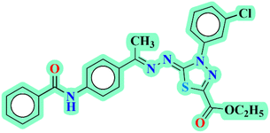
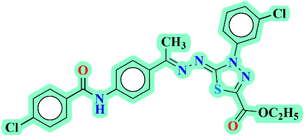
 Open Access Article
Design, synthesis, and evaluation of novel thiadiazole derivatives as potent VEGFR-2 inhibitors: a comprehensive in vitro and in silico study†
Ibrahim H. Eissa *a,
Walid E. Elgammal
 b,
Hazem A. Mahdy
 a,
Susi Zarac,
Simone Carradori
 c,
Dalal Z. Huseind,
Maymounah N. Alharthi
 e,
Ibrahim M. Ibrahimf,
Eslam B. Elkaeed*g,
Hazem Elkady
 a and
Ahmed M. Metwaly
 *h
*h
aDepartment of Pharmaceutical Medicinal Chemistry & Drug Design, Faculty of Pharmacy (Boys), Al-Azhar University, Cairo, 11884, Egypt. E-mail: Ibrahimeissa@azhar.edu.eg
bChemistry Department, Faculty of Science, Al-Azhar University, Nasr City, Cairo 11751, Egypt
cDepartment of Pharmacy, “G. d'Annunzio” University of Chieti-Pescara, Chieti, 66100, Italy
dDepartment of Chemistry, Faculty of Science, New Valley University, El-Kharja 72511, Egypt
eDepartment of Chemistry, College of Science, Princess Nourah bint Abdulrahman University, P.O. Box 84428, Riyadh 11671, Saudi Arabia
fDepartment of Biophysics, Faculty of Science, Cairo University, Giza 12613, Egypt
gDepartment of Pharmaceutical Sciences, College of Pharmacy, AlMaarefa University, P.O. Box 71666, Riyadh 11597, Saudi Arabia. E-mail: ekaeed@um.edu.sa
hDepartment of Pharmacognosy and Medicinal Plants, Faculty of Pharmacy (Boys), Al-Azhar University, Cairo 11884, Egypt. E-mail: ametwaly@azhar.edu.eg
First published on 6th November 2024
Abstract
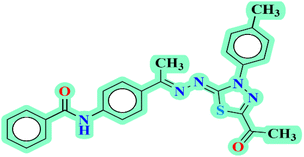
Objective: This study aims to investigate the potential of designed 2,3-dihydro-1,3,4-thiadiazole derivatives as anti-proliferative agents targeting VEGFR-2, utilizing a multidimensional approach combining in vitro and in silico analyses. Methods: The synthesized derivatives were evaluated for their inhibitory effects on MCF-7 and HepG2 cancer cell lines. Additionally, VEGFR-2 inhibition was assessed. Further investigations into the cellular mechanisms were conducted to elucidate the effects of 20b (N-(4-((E)-1-(((Z)-5-Acetyl-3-(p-tolyl)-1,3,4-thiadiazol-2(3H)-ylidene)hydrazono) ethyl) phenyl) benzamide) on cell cycle arrest and apoptosis induction. Furthermore, computational investigations, including molecular docking, MD simulations, DFT calculations, MM-GBSA, PCAT, and ADMET predictions were conducted. Results: Compound 20b emerged as a standout candidate with significantly lower IC50 values of 0.05 μM and 0.14 μM for MCF-7 and HepG2 cell lines, respectively. It exhibited notable VEGFR-2 inhibition (0.024 μM), surpassing the efficacy of sorafenib (0.041 μM). Compound 20b demonstrated cancer-specific targeting potential with a high selectivity index in normal WI-38 cells (IC50 0.19 μM). Mechanistic studies revealed its ability to arrest the cell cycle of MCF-7 cells and induce apoptosis (total apoptosis 34.47%, early apoptosis 18.48%, and late apoptosis 15.99%), supported by upregulated caspase-8 (3.42-fold) and caspase-9 (5.44-fold) expression. Additionally, 20b arrested the cell cycle of MCF-7 cells at the %G0-G1 phase. Computational investigations provided insights into its molecular interactions with VEGFR-2, contributing to the rational design and understanding of its pharmacological profile. Conclusions: Compound 20b presents as a promising anti-proliferative agent targeting VEGFR-2. Also, this comprehensive investigation underscores the potential of 2,3-dihydro-1,3,4-thiadiazole derivatives as promising candidates for further development in anti-cancer research.
1. Introduction
The number of new cancer cases is anticipated to exceed 35 million by 2050, representing a 77% rise from the estimated 20 million cases in 2022.1 These statistics highlight the substantial worldwide impact of cancer, emphasizing the necessity for ongoing and extensive scientific research to create innovative treatment methods. Angiogenesis, involving Vascular Endothelial Growth Factor (VEGF) and its receptor VEGFR-2, is pivotal in the development of both cancer and cardiovascular diseases.2,3 VEGFR-2 functions include promoting vessel development and regulating the proliferation, migration, and survival of endothelial cells in vasculogenesis and angiogenesis.4 Ongoing research has identified a significant correlation between increased VEGFR-2 expression and resistance of the anti-cancer drugs, heightened angiogenesis, and reduced apoptosis.5 Accordingly, targeting VEGFR-2 has been introduced as a promising therapeutic approach.6 Anti-VEGFR-2 drugs are designed to inhibit VEGFR-2, a receptor tyrosine kinase crucial for angiogenesis in tumor cells.7,8 While these inhibitors effectively treat various cancers, they often come with serious side effects, including hypertension, nephropathy,9 proteinuria,10 cardiac ischemia,11 reversible posterior leukoencephalopathy syndrome,12 and potential links to germline polymorphisms.13 Consequently, the development of new anti-VEGFR-2 candidates holds potential to enhance treatment efficacy, patient's health, and advance the standard of cancer care.The integrated approach, combining computational (in silico) and experimental (in vitro) methods, serves to validate the efficacy of active anticancer compounds while laying the groundwork for future enhancements and practical applications.14 This harmonious blend of approaches not only affirms the compounds' effectiveness in laboratory settings but also guides the refinement and optimization of their design.15 The insights gained from this integrated strategy hold the potential of targeted cancer therapies, emphasizing the promise of new derivatives as a notable class of compounds with substantial anticancer activity.16 In essence, the study's combination of experimental and computational methods advances these findings towards practical applications, offering hope for the development of more effective anticancer treatments.
Our research teamwork has previously discovered a variety of potential anti-VEGFR-2 compounds exhibiting anticancer properties including quinolones,17 isatins,18 nicotinamides,19,20 thiazolidines,21 pyridines,22 theobromines,23–27 naphthalenes,28 and indoles.29 Thiadiazole derivatives have gained recognition in medicinal chemistry due to their diverse pharmacological actions, including anticancer and anti-inflammatory properties.30–33
This study introduces new 2,3-dihydro-1,3,4-thiadiazole derivatives, with a specific focus on compound 20b, identified as a potent VEGFR-2 inhibitor through molecular docking studies.
1.1 Rationale
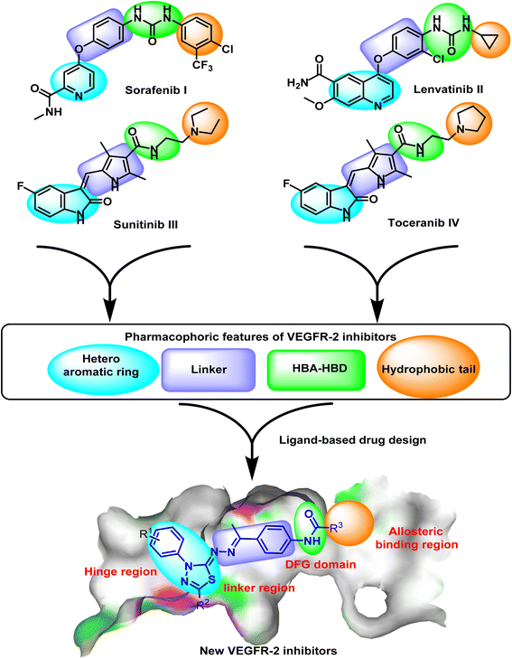
Many drugs have been approved by the FDA-approved as VEGFR-2 inhibitors for the treatment of several types of cancers.34 Sorafenib I,35 lenvatinib II,36 sunitinib III,37 and toceranib IV (ref. 38) are well-known examples of FDA-approved VEGFR-2 inhibitors (Fig. 1). VEGFR-2 inhibitors are characterized by four essential pharmacophoric features crucial for effective binding with the ATP binding site of VEGFR-2. These features are as follows: (i) a hetero-aromatic system: This component is designed to occupy the hinge region, ensuring a specific arrangement within the molecular structure for optimal interaction;39 (ii) a linker moiety: Positioned to orient into the region between the DFG domain and the hinge region, this linker serves as a critical element facilitating the proper spatial alignment of the inhibitor within the active site.;40 (iii) a pharmacophore moiety: Comprising at least one H-bond donor and one H-bond acceptor group, this pharmacophore is strategically positioned to occupy the DFG domain. It plays a pivotal role in establishing hydrogen bond interactions, enhancing the inhibitor's affinity for the target;41 (iv) a terminal hydrophobic moiety: Positioned to occupy the allosteric hydrophobic pocket of the ATP binding site, this terminal hydrophobic component contributes to the overall stability and specificity of the inhibitor's binding42–44 (Fig. 1).Depending on the ligand-based drug design approach, a new series of 2,3-dihydro-1,3,4-thiadiazole derivatives was designed as VEGFR-2 inhibitors. The designed compounds involve 2,3-dihydro-1,3,4-thiadiazole moiety as a hetero-aromatic system that can occupy the hinge region. In addition, the 1-phenylethylidenehydrazine moiety was utilized as a linker group. Furthermore, the amide group was utilized as a pharmacophore moiety to occupy the DFG domain. Finally, different substituted phenyl rings were used as hydrophobic tails of the designed molecules (Fig. 1).
2. Results and discussion
2.1. Chemistry
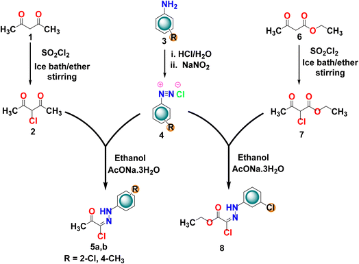
Schemes 1–3 describes the pathways that have been followed to synthesize the desired derivatives. At first, the starting intermediates 5a,b and 8 were meticulously synthesized. This involved subjugating the 1,3-diketones (acetylacetone 1 and/or ethyl acetoacetate 6) to a chlorination reaction using sulfuryl dichloride (SO2Cl2).45 Subsequently, the resulting products (2 and 7, respectively) were allowed to react with the appropriate diazonium salts 4 of aromatic amines 3 to produce the key intermediates 5a,b and 8. The diazonium salts were obtained from the reaction of the substituted aromatic amines 3 with a solution of sodium nitrite in the presence of aqueous hydrochloric acid in an ice bath46 (Scheme 1).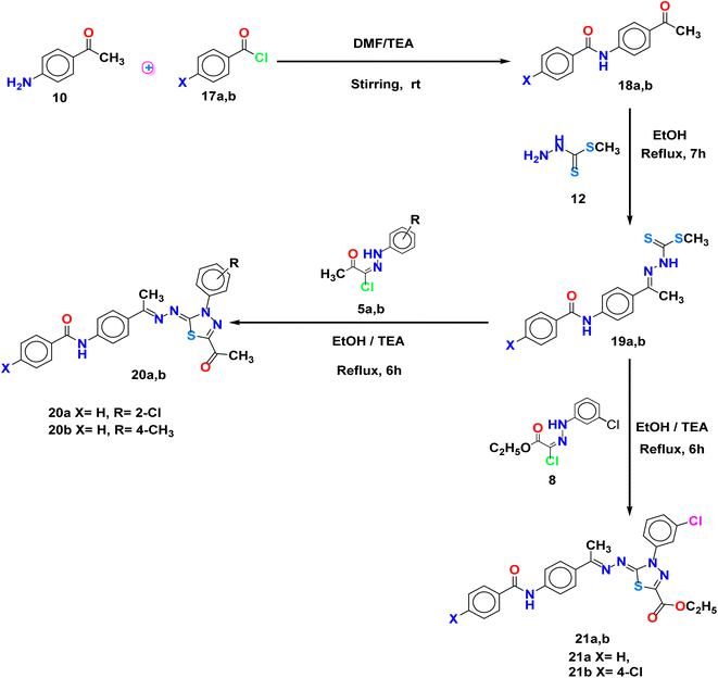 | ||
| Scheme 3 A classical method for the synthesis of acetyl and ester of 2,3-dihydro-1,3,4-thiadiazole derivatives 20a,b and 21a,b. | ||
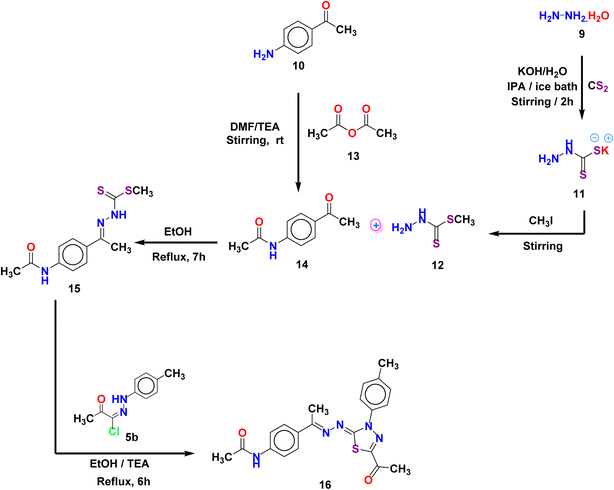
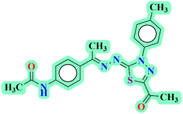
On the other hand, Scheme 2 illustrates the synthesis of compound 16. The first step entailed synthesizing N-(4-acetylphenyl)acetamide 14, as previously documented.47 The accomplishment was made through the reaction of 4-aminoacetophenone 10 with acetic acid anhydride 13 as the acetylating agent in DMF utilizing trimethylamine (TEA) as a base. Afterward, compound 14 underwent reflux with methyl hydrazinecarbodithioate 12,48 in pure ethanol, synthesizing the desired compound 15. This compound was then reacted with (E)-2-oxo-N-(p-tolyl)propanehydrazonoyl chloride 5b in ethanol to produce the corresponding 2,3-dihydro-1,3,4-thiadiazoles derivative 16.
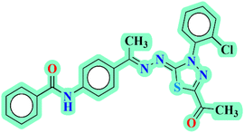
Likewise, the benzoylation of 4-aminoacetophenone 10 using (un)substituted benzoyl chloride 17a,b under identical acylation conditions resulted in the formation of the isolated products 18a,b. Subsequently, compounds 18a,b were condensed with methyl hydrazinecarbodithioate 12 in absolute ethanol to yield compounds 19a,b, which were obtained in their pure form through recrystallization from a mixture of methanol and dichloromethane. Eventually, the hetrocyclization of compounds 19a,b was achieved by the reaction with substituted hydrazonoyl chlorides 5a,b and/or 8 in a heated solution of EtOH/TEA to give 20a,b and 21a,b, respectively.
2.2. Biology
While the IC50 values provide valuable insights into the potency of the compounds, it is advisable to conduct additional studies to gain a more comprehensive understanding of their anti-proliferative efficacy. In conclusion, the presented IC50 values suggest that compound 20b and its derivatives hold promise as potent anti-proliferative agents, warranting further investigation.
Results in Table 3 showed that compound 20b showed moderate to weak inhibition values against the four investigated protein kinases in comparison to the selected reference kinase inhibitors. For its activity against CDK8, compound 20b showed IC50 value of 0.266 ± 0.058 μM, compared to Cortistatin A (IC50 = 0.032 ± 0.004 μM). Regarding its activity against PIK3α, it showed IC50 value of 2.143 ± 0.095 in comparison to PIK-90 (IC50 = 2.143 ± 0.095 μM). Additionally, compound 20b demonstrated weak inhibitory activities against BRAF (IC50 = 5.841 ± 0.250 μM) and EGFR (IC50 = 1.431 ± 0.085 μM) compared to those of Vemurafenib (IC50 = 0.347 ± 0.029 μM) and Erlotinib (IC50 = 0.005 ± 0.001 μM). From these results, we can found that compound 20b has no selectivity against the tested four kinases but it has a great selectivity against VEGFR-2 receptor.
| Comp. | CDK8 IC50 (μM) (mean ± SD) | PIK3α IC50 (μM) (mean ± SD) | BRAF IC50 (μM) (mean ± SD) | EGFR IC50 (μM) (mean ± SD) |
|---|---|---|---|---|
| 20b | 0.266 ± 0.058 | 2.143 ± 0.095 | 5.841 ± 0.250 | 1.431 ± 0.085 |
| Cortistatin A | 0.032 ± 0.004 | — | — | — |
| PIK-90 | — | 0.033 ± 0.008 | — | — |
| Vemurafenib | — | — | 0.347 ± 0.029 | — |
| Erlotinib | — | — | — | 0.005 ± 0.001 |
2.2.5.1 Cell cycle analysis. The cell cycle distribution percentages for MCF-7 cells treated with compound 20b and untreated MCF-7 cells were conducted by flow cytometry and were presented in Table 5 and Fig S1.† The cell cycle distribution analysis reveals notable alterations induced by compound 20b in MCF-7 cells. Specifically, there is an increase in the percentage of cells in the G0–G1 phase from 62.37% (untreated MCF-7) to 69.25% (compound 20b). This suggests that compound 20b may impede cell cycle progression by inducing cell cycle arrest at the G0–G1 phase. Concomitantly, there is a decrease in the percentage of cells in the S phase from 23.91% (untreated MCF-7) to 18.76% (compound 20b), indicating a potential reduction in DNA synthesis. Moreover, there is a slight decrease in the G2/M phase to be 11.99% (compound 20b) instead of 13.72% (untreated MCF-7), suggesting a modest impact on the transition to mitosis. These changes in cell cycle distribution are indicative of the regulatory effects of compound 20b on the cell cycle dynamics of MCF-7 cells. The observed arrest at the G0–G1 phase suggests that the compound may interfere with key checkpoints, preventing cells from progressing into the synthesis and mitotic phases. Such cell cycle modulation is a crucial aspect of anti-proliferative mechanisms, as disruption of cell cycle progression can lead to reduced cell proliferation and increased susceptibility to apoptosis.
| Sample | Cell cycle distribution (%) | ||
|---|---|---|---|
| % G0-G1 | % S | % G2/M | |
| 20b | 69.25 | 18.76 | 11.99 |
| Control | 62.37 | 23.91 | 13.72 |
The findings underscore the potential of compound 20b as a cell cycle regulator in MCF-7 cells, demonstrating its ability to influence the delicate balance of cell cycle phases. Overall, the observed alterations in cell cycle distribution suggest that compound 20b holds promise as a candidate for further exploration in the development of targeted therapies against cancer cells.
2.2.5.2 Apoptosis assay. The flow cytometry assay was performed to evaluate the apoptosis analysis in MCF-7 cells, comparing untreated MCF-7 cells to those treated with compound 20b. The apoptosis analysis (Table 6 and Fig. S2†) reveals a remarkable difference in the percentage of apoptotic cells between untreated MCF-7 cells and those treated with compound 20b. Specifically, total apoptosis after treatment with compound 20b is substantially higher at 34.47% compared to 0.85% in the untreated cells. This increase is indicative of the promising pro-apoptotic effect exerted by compound 20b.
| Total | Early apoptosis | Late apoptosis | Necrosis | |
|---|---|---|---|---|
| Compound 20b | 38.55 | 18.48 | 15.99 | 4.14 |
| Control | 2.28 | 0.61 | 0.24 | 1.43 |
Breaking down the stages of apoptosis, the early apoptosis percentage increases from 0.61% in the untreated MCF-7 cells to a notable 18.48% after treatment. Concurrently, the percentage of cells in late apoptosis rises from 0.24% to 15.99%. These findings collectively suggest that compound 20b induces both early and late apoptosis, indicating a multifaceted impact on the apoptotic pathways of MCF-7 cells. In contrast, the percentage of cells undergoing necrosis, a form of cell death associated with cellular damage, is relatively low but still increases from 1.43% in the untreated MCF-7 cells to 4.14% with compound 20b. This suggests that compound 20b may trigger a modest degree of necrosis in addition to its pronounced apoptotic effects.
The substantial increase in apoptosis, particularly in the early and late stages, highlights the potent cytotoxic effects of compound 20b on the MCF-7 cells. Apoptosis is a fundamental process that eliminates damaged or abnormal cells, and the observed increase suggests that compound 20b may have significant potential as an inducer of programmed cell death in cancer cells. These findings underscore the importance of apoptosis induction as a key mechanism in the anti-proliferative activity of compound 20b and position it as a promising candidate for further exploration in anti-cancer drug discovery.
2.2.5.3 Reverse transcription polymerase chain reaction (RT-PCR) studies. Table 7 presents the results of the RT-PCR analysis, indicating the fold change in the expression levels of caspase-8 and caspase-9 in MCF-7 cells treated with compound 20b compared to untreated control. In detail, the RT-PCR results demonstrate a substantial increase in the expression levels of both caspase-8 and caspase-9 in MCF-7 cells treated with compound 20b compared to the untreated control. The fold change for caspase-8 is 3.4176, indicating a more than 3-fold increase, while caspase-9 shows an even higher fold change of 5.4353.
| RT-PCR (fold change) | ||
|---|---|---|
| Caspase-8 | Caspase-9 | |
| Compound 20b | 3.4176 | 5.4353 |
| Control | 1 | 1 |
Caspase-8 and caspase-9 are integral components of the apoptotic pathway, playing crucial roles in initiating and executing programmed cell death. The upregulation of these caspases in response to compound 20b treatment confirms a pronounced activation of apoptotic pathways in MCF-7 cells. This alignment between apoptotic marker upregulation and increased apoptosis supports the notion that compound 20b exerts its anti-proliferative effects through the induction of apoptosis in MCF-7 cells. Apoptosis induction, especially through the activation of key caspases, is a crucial mechanism in eliminating cancer cells. The specific targeting of apoptotic pathways aligns with the compound's overall impact on cell cycle distribution and apoptosis percentages, providing a coherent picture of its anti-proliferative properties.
2.3. Molecular docking study
MOE (Molecular Operating Environment, version 2019) software was utilized to conduct molecular docking simulations to explore potential drug binding patterns within the active site of VEGFR-2. The docking calculations employed the crystal structure of VEGFR-2 (PDB ID: 2OH4), with sorafenib, a known effective drug, serving as the reference compound.In this section, we chose compound 20b as the template control for the docking investigation since it has the strongest anti-proliferative activity against the tested cell lines. According to Fig. 2, compound 20b was discovered to have been inserted into the deep cleft of the ATP binding site in the hinge region forming stable hydrophobic bonds with Cys917. The terminal amide also enabled the insertion of compound 20b into the DFG motif region by forming two crucial hydrogen bonds between Glu883 and Asp 1044 at distances of 2.16 and 1.88, respectively.
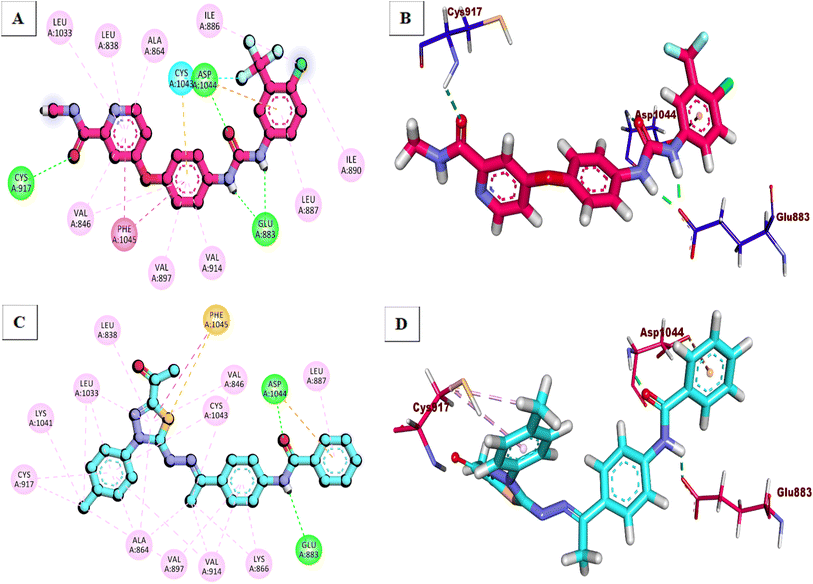 | ||
| Fig. 2 Binding of the tested compounds against VEGFR-2. (A) 2D of sorafenib, (B) 3D of sorafenib, (C) 2D of compound 20b and (D) 3D of compound 20b. | ||
2.4. MD simulation
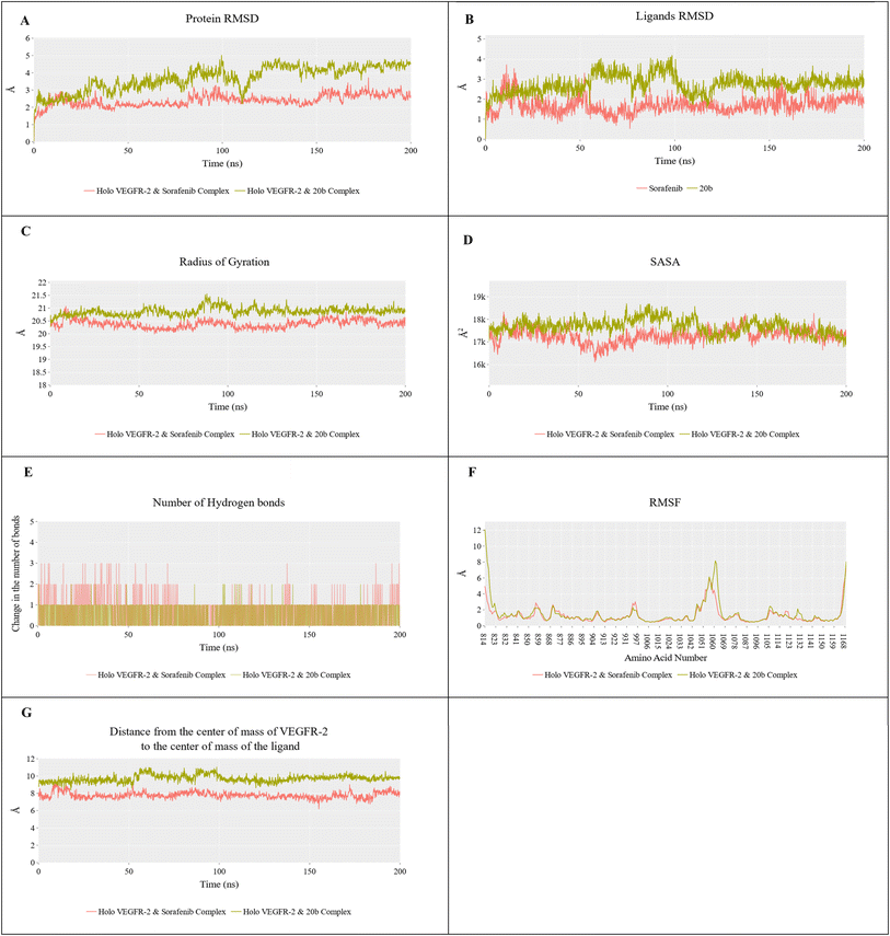
During the simulation (200 ns), the root-mean-square deviation (RMSD) values for the reference system (VEGFR-2_sorafenib) exhibited stability, maintaining a consistent value of approximately 2.5 Å (indicated by the red line). In contrast, the RMSD values for the VEGFR-2_20b system (represented by the dark green line) initially showed an upward trend during the first 120 ns of the simulation. After this period, the values stabilized at around 4.2 Å (Fig. 3A). For the compound 20b alone (dark green line), the RMSD showed a slight increasing trend in the initial 100 ns before reaching a stable value of approximately 2.9 Å (Fig. 3B). On the other side, sorafenib (red line) showed a more stable profile throughout the simulation, with an average RMSD of about 1.5 Å, indicating stronger binding stability. Furthermore, Fig. 3C illustrates a subtle difference in the radius of gyration's average (RoG) between the two systems. The VEGFR-2_sorafenib system had a lower RoG value of around 20.4 Å, compared to the VEGFR-2_20b system, which had an RoG of approximately 20.9 Å. This suggests that the protein-ligand complex with sorafenib is slightly more compact than with compound 20b. Additionally, as shown in Fig. 3D, both systems exhibited similar average solvent-accessible surface area (SASA) values, each around 17![[thin space (1/6-em)]](https://www.rsc.org/images/entities/char_2009.gif) 500 Å2, indicating comparable exposure of the protein surfaces to the solvent. Fig. 3E provides insights into the hydrogen bonding patterns during the simulation. Compound 20b consistently maintained nearly a hydrogen bond throughout the 200 ns of the simulation duration, whereas sorafenib fluctuated between forming one and two hydrogen bonds. In terms of structural stability, the root-mean-square fluctuation (RMSF) profiles revealed that both systems maintained overall structural integrity with minimal deviations. The primary difference was observed in the oscillations of the C-alpha atoms within the Ile1042 loop. In the reference system (VEGFR-2_sorafenib), the RMSF reached a maximum of 6 Å, whereas in the VEGFR-2_20b system, it increased to about 8 Å (Fig. 3F), indicating slightly greater flexibility in the latter. Additionally, the average distance between the protein's center of mass and the ligand was stable in both systems. For compound 20b, this distance was approximately 9.3 Å, suggesting a stable interaction, though it was slightly greater than the reference compound, sorafenib, by about 1.5 Å (Fig. 3G). This difference in distance further highlights the slightly more stable binding conformation of sorafenib within the active site of VEGFR-2. Overall, these results show that both VEGFR-2-ligand complexes remained stable structurally, with minor differences in dynamic behavior and interaction patterns.
500 Å2, indicating comparable exposure of the protein surfaces to the solvent. Fig. 3E provides insights into the hydrogen bonding patterns during the simulation. Compound 20b consistently maintained nearly a hydrogen bond throughout the 200 ns of the simulation duration, whereas sorafenib fluctuated between forming one and two hydrogen bonds. In terms of structural stability, the root-mean-square fluctuation (RMSF) profiles revealed that both systems maintained overall structural integrity with minimal deviations. The primary difference was observed in the oscillations of the C-alpha atoms within the Ile1042 loop. In the reference system (VEGFR-2_sorafenib), the RMSF reached a maximum of 6 Å, whereas in the VEGFR-2_20b system, it increased to about 8 Å (Fig. 3F), indicating slightly greater flexibility in the latter. Additionally, the average distance between the protein's center of mass and the ligand was stable in both systems. For compound 20b, this distance was approximately 9.3 Å, suggesting a stable interaction, though it was slightly greater than the reference compound, sorafenib, by about 1.5 Å (Fig. 3G). This difference in distance further highlights the slightly more stable binding conformation of sorafenib within the active site of VEGFR-2. Overall, these results show that both VEGFR-2-ligand complexes remained stable structurally, with minor differences in dynamic behavior and interaction patterns.
Fig. 4 illustrates the various components of the predicted binding free energy, calculated using the MM-GBSA method. The binding energy for molecule 20b is −3.9 kcal mol−1, in contrast to sorafenib's average of −43.3 kcal mol−1, indicating significant differences in the components influencing binding. The van der Waals bondings are almost identical for both systems, averaging around −50 kcal mol−1. However, electrostatic interactions are more favorable for sorafenib compared to 20b (−17.92 kcal mol−1 versus +0.91 kcal mol−1). This contributes to the lower total binding energy of 20b relative to the reference compound. Additionally, the solvation energy (Egb) for 20b is less favorable than for sorafenib.
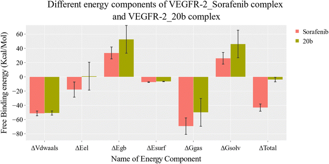 | ||
| Fig. 4 The energetic components of the MM-GBSA method and their respective values for the two systems. The bars represent the standard deviations of these values. | ||
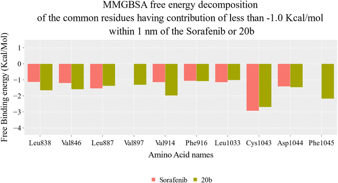
To determine the contribution of each amino acid within 1 nm of each ligand, a decomposition analysis was performed (Fig. 5). Six amino acids showed a more significant share to the binding affinity of 20b compared to sorafenib:
Leu838: 1.65 kcal mol−1 (20b) versus 1.13 kcal mol−1 (sorafenib).
Val846: −1.58 kcal mol−1 (20b) versus 1.2 kcal mol−1 (sorafenib).
Val897: −1.31 kcal mol−1 (20b) versus −0.96 kcal mol−1 (sorafenib).
Val914: −1.97 kcal mol−1 (20b) versus −1.14 kcal mol−1 (sorafenib).
Phe916: −1.08 kcal mol−1 (20b) versus −1.06 kcal mol−1 (sorafenib).
Asp1044: −1.46 kcal mol−1 (20b) versus −1.41 kcal mol−1 (sorafenib).
Phe1045: −2.17 kcal mol−1 (20b) versus −0.86 kcal mol−1 (sorafenib).
These values highlight the enhanced binding energy of compound 20b with these specific amino acids compared to sorafenib, providing insights into the molecular interactions that contribute to the improved binding of 20b in comparison to sorafenib.
To get more information about the Protein–Ligand Interaction Fingerprints (ProLIF) and Principal Component Analysis (PCA), see Fig. S3–S10.†
2.5. DFT studies
One of the most popular and reliable techniques for estimating molecular systems as well as their electronic characteristics is the DFT/B3LYP approach. The molecular system of compound 20b is selected due to its remarkable reactivity as conducted practically. Using the Gaussian 09 package, the basis set 6-31G with double zeta plus polarization (d,p) was applied through the density functional theory (DFT/B3LYP) approach to optimize the molecular structure of 20b. The chemical structure was plotted using Gauss View 05 and the optimized output file was analyzed using Multiwfn, GaussSum, and AIMAll programs. Based on the global reactivity parameters and band gap energy, the reactivity of the selected compound is predicted. The optimized geometry with atom numbering is presented in Fig. 6a. Compound 20b is a polar molecule as its calculated dipole moment was found to be 7.257 Debye (Table 8), which indicates a higher tendency to interact with targets (amino acids). The high polarity of a given molecule increases the potential interatomic forces such as hydrogen bonding and dipole–dipole interactions between the selected molecule and the amino acids.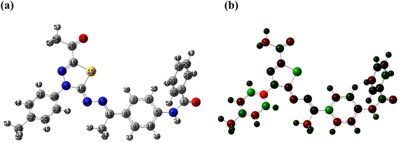 | ||
| Fig. 6 The entire optimized geometry (a), the distribution of Mullikan atomic charges (b). These results were obtained using B3LYP/6-31+G(d,p). | ||
| IP | EA | μ (eV) | χ (eV) | η (eV) | σ (eV) | ω (eV) | Dm (Debye) | TE (eV) | ΔNmax | ΔE (eV) |
|---|---|---|---|---|---|---|---|---|---|---|
| 5.684 | 2.476 | −4.080 | 4.080 | 1.604 | 0.624 | 13.348 | 7.254 | −49717.3 | 2.544 | −13.348 |
Mulliken charge analysis provides an approximation of the arrangement of electrons for each atom inside a molecule and then it affects the dipole moment, molecular polarizability, and electronic structure. Hence, it can provide an understanding of the chemical behavior of the studied molecule as reactive sites can be identified. The designed candidate, 20b, in Fig. 6b, shows that sulfur and N17 are acceptors while C21 is the most acceptor site in the molecule. On the other hand, all oxygen and nitrogen (except for N17) are donors because they are negative sites and C20 has the highest negative charge. To get more information about HOMO, LUMO, the total density of states (TDOs), and the quantum theory of atoms in molecule (QTAIM) for compound 20b, see Fig. S11A–C.†
2.6. In silico ADMET analysis
Table 9 presents the in silico ADMET results of the new thiadiazole compounds, which offer important information on their possible pharmacokinetic behavior. All compounds except compound 16 have very low levels of blood–brain barrier permeability, which indicates minimal penetration ability into the CNS. Regarding the aqueous solubility, compound 16 showed a low level of solubility whilst the rest of compounds were predicted to have very low levels, indicating that methods to improve their water solubility could be required for efficient delivery. Compound 20b showed a good amount of absorption, indicating a high probability of systemic availability after treatment. All compounds are anticipated to be non-inhibitors with regard to the CYP2D6 interaction, suggesting a decreased probability of metabolic interactions with medications that are metabolized by this enzyme. Finally yet importantly, all substances, including sorafenib, should bind to plasma proteins >90% of the time. This could have an impact on how the synthesized derivatives are distributed and eliminated.| Compound | BBB level | Solubility level | Absorption level | CYP2D6 prediction | PPB prediction |
|---|---|---|---|---|---|
| 16 | 2 | 2 | 0 | False | True |
| 20a | 4 | 1 | 1 | ||
| 20b | |||||
| 21a | 2 | ||||
| 21b | |||||
| Sorafenib | 0 |
2.7. In silico toxicity analysis
Table 10 reports a comprehensive evaluation of compounds (16, 20a, 20b, 21a, 21b) and the reference compound Sorafenib, with a focus on carcinogenic potency, mutagenicity, and toxicity profiles. In terms of carcinogenic potency (TD50), compound 20b exhibits a value of 18.94, placing it within the range of the experimental compounds and slightly below Sorafenib (19.23). Notably, all experimental compounds are classified as non-carcinogens, except for sorafenib, which is identified as a single carcinogen.| Compound | Carcinogenicity TD50 (Mouse)a | Mouse- female FDA | DTP | Rat oral LD50b | Rat chronic LOAELb | Dermal irritancy | Ocular irritancy |
|---|---|---|---|---|---|---|---|
| a Unit: mg kg−1 per day.b Unit: g kg−1. | |||||||
| 16 | 24.8533 | Non-carcinogen | Non-toxic | 0.663451 | 0.0754607 | Non-irritant | Mild |
| 20a | 8.42209 | 0.703374 | 0.0790792 | ||||
| 20b | 18.9369 | 0.457358 | 0.0906188 | ||||
| 21a | 17.6022 | 0.902703 | 0.0589522 | ||||
| 21b | 9.60638 | 0.896764 | 0.0336175 | ||||
| Sorafenib | 19.2359 | Single-carcinogen | Toxic | 0.822583 | 0.00482816 | ||
A crucial aspect of compound evaluation is its mutagenicity, and in this context, compound 20b is recognized as mutagenic, indicating a potential to induce mutations. Regarding toxicity, all experimental compounds, including 20b, are categorized as non-toxic, suggesting a favorable safety profile. However, sorafenib is labeled as toxic, indicating a potential for adverse effects. Further insights are provided through assessments such as rat oral LD50 and chronic LOAEL, where compound 20b exhibits a lower oral LD50 of 0.457358. Additionally, a higher chronic LOAEL of 0.0906188, indicates a safe potential for long-term use. Notably, all compounds, including 20b, are classified as non-irritants for both skin and ocular irritation, enhancing their safety profile in these regards. In conclusion, compound 20b's distinct non-carcinogenic potency and the comparison with sorafenib underscores the general safety. While the current data provide valuable insights, additional in vivo assessments and clinical trials are imperative for a comprehensive understanding of the safety and efficacy of compound 20b for potential therapeutic applications.
3. Conclusion
In conclusion, the comprehensive investigation presented in this paper highlights the promising potential of compound 20b as a potent anti-proliferative agent. The compound demonstrates significant inhibitory effects on MCF-7 and HepG2 cancer cell lines, with notably low IC50 values, suggesting enhanced potency compared to reference compounds. Importantly, the selectivity index values underscore the preferential inhibitory activity of 20b on cancer cells over normal WI-38 cells, laying the foundation for potential therapeutic applications. The mechanistic insights provided by the study revealed that compound 20b exerted its effects through multiple ways. The substantial inhibition of VEGFR-2, as evidenced by the low IC50 value, suggests a potential disruption of angiogenesis, while alterations in cell cycle distribution and the induction of apoptosis further contribute to its anti-proliferative and cytotoxic effects on cancer cells. Moreover, the upregulation of key apoptotic markers, caspase-8 and caspase-9, as observed in RT-PCR analysis, provides molecular evidence supporting the compound's pro-apoptotic effects. Incorporating in silico analyses enhanced our understanding of compound 20b's molecular interactions with VEGFR-2, aiding in its rational design and pharmacological profile comprehension. Overall, the findings presented in this study underscore the importance of compound 20b as a lead compound with multifaceted anti-proliferative properties, encouraging further analyses.4. Experimental
4.1. Chemistry
4.1.2.1 N-(4-((E)-1-(((Z)-5-Acetyl-3-(p-tolyl)-1,3,4-thiadiazol-2(3H)-ylidene)hydrazono)ethyl) phenyl) acetamide 16. Yellow powder (yield, 74%); m.p. = 201–202 °C. Comprehensive data, including detailed information from FT-IR, 1H NMR, 13C NMR and EI-Ms can be found in the ESI.†
4.1.3.1 N-(4-((E)-1-(((Z)-5-Acetyl-3-(2-chlorophenyl)-1,3,4-thiadiazol-2(3H)-ylidene)hydrazono) ethyl) phenyl)benzamide 20a. Pale yellow powder (yield, 70%); m.p. = 190–192 °C. Comprehensive data, including detailed information from FT-IR, 1H NMR, 13C NMR and EI-Ms can be found in the ESI.†
4.1.3.2 N-(4-((E)-1-(((Z)-5-Acetyl-3-(p-tolyl)-1,3,4-thiadiazol-2(3H)-ylidene)hydrazono) ethyl) phenyl) benzamide 20b. Yellow powder (yield, 78%); m.p. = 185–187 °C. Comprehensive data, including detailed information from FT-IR, 1H NMR, 13C NMR and EI-Ms can be found in the ESI.†
4.1.3.3 Ethyl (Z)-5-(((E)-1-(4-benzamidophenyl)ethylidene)hydrazono)-4-(3-chlorophenyl)-4,5-dihydro-1,3,4-thiadiazole-2-carboxylate 21a. Yellowish white powder (yield, 73%); m.p. = 230–232 °C. Comprehensive data, including detailed information from FT-IR, 1H NMR, 13C NMR and EI-Ms can be found in the ESI.†
4.1.3.4 Ethyl (Z)-5-(((E)-1-(4-(4-Chlorobenzamido)phenyl)ethylidene)hydrazono)-4-(3-chlorophenyl)-4,5-dihydro-1,3,4-thiadiazole-2-carboxylate 21b. Yellow powder (yield, 75%); m.p. = 203–205 °C. Comprehensive data, including detailed information from FT-IR, 1H NMR, 13C NMR and EI-Ms can be found in the ESI.†
4.2. Biological evaluation
4.3. In silico studies
Data availability
All data regarding the presented work was included in the main manuscript and the ESI.†Conflicts of interest
The authors verify that they have no conflicts of interest associated with this publication involving any party or among the co-authors.Acknowledgements
This research was funded by Princess Nourah bint Abdulrahman University Researchers Supporting Project number (PNURSP2024R481), Princess Nourah bint Abdulrahman University, Riyadh, Saudi Arabia. The authors would like to thank AlMaarefa University, Riyadh, Saudi Arabia, for supporting this research.References
- W. H. Organization, Global cancer burden growing, amidst mounting need for services, https://www.who.int/news/item/01-02-2024-global-cancer-burden-growing--amidst-mounting-need-for-services, accessed 1 February 2024.
- M. Shibuya, J. Biochem., 2013, 153, 13–19 CrossRef CAS PubMed.
- L. Claesson-Welsh and M. Welsh, J. Intern. Med., 2013, 273, 114–127 CrossRef CAS PubMed.
- X. Wang, A. M. Bove, G. Simone and B. Ma, Front. Cell Dev. Biol., 2020, 8, 599281 CrossRef PubMed.
- K. Cheng, C. F. Liu and G. W. Rao, Curr. Med. Chem., 2021, 28, 2540–2564 CAS.
- T. A. Farghaly, W. A. Al-Hasani and H. G. Abdulwahab, Expert Opin. Ther. Pat., 2021, 31, 989–1007 CrossRef CAS PubMed.
- E. B. Elkaeed, R. G. Yousef, H. Elkady, A. B. Mehany, B. A. Alsfouk, D. Z. Husein, I. M. Ibrahim, A. M. Metwaly and I. H. Eissa, J. Biomol. Struct. Dyn., 2023, 41, 7986–8001 CrossRef CAS PubMed.
- K. Czaja, J. Kujawski, P. Śliwa, R. Kurczab, R. Kujawski, A. Stodolna, A. Myślińska and M. K. Bernard, Int. J. Mol. Sci., 2020, 21, 4793 CrossRef CAS PubMed.
- J. Versmissen, K. M. Mirabito Colafella, S. L. W. Koolen and A. H. J. Danser, Cardiovasc. Res., 2019, 115, 904–914 CrossRef CAS PubMed.
- R. M. Hanna, E. A. Lopez, H. Hasnain, U. Selamet, J. Wilson, P. N. Youssef, N. Akladeous, S. Bunnapradist and M. B. Gorin, Clin. Kidney J., 2019, 12, 92–100 CrossRef CAS PubMed.
- H. Abdel-Qadir, J. L. Ethier, D. S. Lee, P. Thavendiranathan and E. Amir, Cancer Treat. Rev., 2017, 53, 120–127 CrossRef CAS PubMed.
- X. Li, J. Chai, Z. Wang, L. Lu, Q. Zhao, J. Zhou and F. Ju, OncoTargets Ther., 2018, 11, 4407–4411 CrossRef PubMed.
- T. M. Sissung, C. J. Peer, N. Korde, S. Mailankody, D. Kazandjian, D. J. Venzon, O. Landgren and W. D. Figg, Cancer Chemother. Pharmacol., 2017, 80, 217–221 CrossRef CAS PubMed.
- P. Chunarkar-Patil, M. Kaleem, R. Mishra, S. Ray, A. Ahmad, D. Verma, S. Bhayye, R. Dubey, H. N. Singh and S. Kumar, Biomedicines, 2024, 12, 201 CrossRef CAS PubMed.
- K. Balasubramanian, Reference Module in Biomedical Sciences, 2021 Search PubMed.
- R. Bozorgpour, S. Sheybanikashani and M. Mohebi, arXiv, 2023, preprint, arXiv:2310.19950, 2023.
- M. S. Taghour, H. Elkady, W. M. Eldehna, N. El-Deeb, A. M. Kenawy, A. E. Abd El-Wahab, E. B. Elkaeed, B. A. Alsfouk, A. M. Metwaly and I. H. Eissa, J. Biomol. Struct. Dyn., 2022, 1–16 Search PubMed.
- E. B. Elkaeed, M. S. Taghour, H. A. Mahdy, W. M. Eldehna, N. M. El-Deeb, A. M. Kenawy, B. A. Alsfouk, M. A. Dahab, A. M. Metwaly and I. H. Eissa, J. Enzyme Inhib. Med. Chem., 2022, 37, 2191–2205 CrossRef CAS PubMed.
- E. B. Elkaeed, R. G. Yousef, H. Elkady, I. M. Gobaara, B. A. Alsfouk, D. Z. Husein, I. M. Ibrahim, A. M. Metwaly and I. H. Eissa, Molecules, 2022, 27, 4606 CrossRef CAS PubMed.
- R. G. Yousef, A. Elwan, I. M. Gobaara, A. B. Mehany, W. M. Eldehna, S. A. El-Metwally, B. A. Alsfouk, E. B. Elkaeed, A. M. Metwaly and I. H. Eissa, J. Enzyme Inhib. Med. Chem., 2022, 37, 2206–2222 CrossRef CAS PubMed.
- H. Elkady, A. A. Abuelkhir, M. Rashed, M. S. Taghour, M. A. Dahab, H. A. Mahdy, A. Elwan, H. A. Al-Ghulikah, E. B. Elkaeed, I. M. Ibrahim, D. Z. Husein, A. Metwaly and I. H. Eissa, Comput. Biol. Chem., 2023, 107, 107958 CrossRef CAS PubMed.
- R. G. Yousef, H. Elkady, E. B. Elkaeed, I. M. Gobaara, H. A. Al-Ghulikah, D. Z. Husein, I. M. Ibrahim, A. M. Metwaly and I. H. Eissa, Molecules, 2022, 27, 7719 CrossRef CAS PubMed.
- I. H. Eissa, R. G. Yousef, H. Elkady, E. B. Elkaeed, A. A. Alsfouk, D. Z. Husein, I. M. Ibrahim, M. A. Elhendawy, M. Godfrey and A. M. Metwaly, Comput. Biol. Chem., 2023, 107, 107953 CrossRef CAS PubMed.
- H. Elkady, W. E. Elgammal, H. A. Mahdy, S. Zara, S. Carradori, D. Z. Husein, A. A. Alsfouk, I. M. Ibrahim, E. B. Elkaeed and A. M. Metwaly, Comput. Biol. Chem., 2024, 108221 CrossRef CAS PubMed.
- I. H. Eissa, R. G. Yousef, H. Elkady, E. B. Elkaeed, A. A. Alsfouk, D. Z. Husein, I. M. Ibrahim, M. M. Radwan and A. M. Metwaly, ChemistryOpen, 2023, 12, e202300066 CrossRef CAS PubMed.
- H. A. Mahdy, H. Elkady, M. S. Taghour, A. Elwan, M. A. Dahab, M. A. Elkady, E. G. Elsakka, E. B. Elkaeed, B. A. Alsfouk and I. M. Ibrahim, Future Med. Chem., 2023, 15, 1233–1250 CrossRef CAS PubMed.
- M. A. Dahab, H. A. Mahdy, H. Elkady, M. S. Taghour, A. Elwan, M. A. Elkady, E. G. Elsakka, E. B. Elkaeed, A. A. Alsfouk and I. M. Ibrahim, J. Biomol. Struct. Dyn., 2024, 42, 4214–4233 CrossRef CAS PubMed.
- E. B. Elkaeed, R. G. Yousef, H. Elkady, A. B. Mehany, B. A. Alsfouk, D. Z. Husein, I. M. Ibrahim, A. M. Metwaly and I. H. Eissa, J. Biomol. Struct. Dyn., 2022, 1–16 Search PubMed.
- E. B. Elkaeed, R. G. Yousef, H. Elkady, I. M. Gobaara, A. A. Alsfouk, D. Z. Husein, I. M. Ibrahim, A. M. Metwaly and I. H. Eissa, Processes, 2022, 10, 1391 CrossRef CAS.
- M. Szeliga, Pharmacol. Rep., 2020, 72, 1079–1100 CrossRef CAS PubMed.
- U. A. Atmaram and S. M. Roopan, Appl. Microbiol. Biotechnol., 2022, 106, 3489–3505 CrossRef CAS PubMed.
- S. Janowska, A. Paneth and M. Wujec, Molecules, 2020, 25, 1–41 CrossRef PubMed.
- A. Ricci, M. Gallorini, D. Del Bufalo, A. Cataldi, I. D’Agostino, S. Carradori and S. Zara, Molecules, 2022, 27, 1–20 CrossRef PubMed.
- L. Huang, Z. Huang, Z. Bai, R. Xie, L. Sun and K. Lin, Future Med. Chem., 2012, 4, 1839–1852 CrossRef CAS PubMed.
- W. Tang, Z. Chen, W. Zhang, Y. Cheng, B. Zhang, F. Wu, Q. Wang, S. Wang, D. Rong and F. Reiter, Signal Transduction Targeted Ther., 2020, 5, 87 CrossRef PubMed.
- L. J. Scott, Drugs, 2015, 75, 553–560 CrossRef CAS PubMed.
- L. Q. Chow and S. G. Eckhardt, J. Clin. Oncol., 2007, 25, 884–896 CrossRef CAS PubMed.
- C. R. Prakash, P. Theivendren and S. Raja, Pharmacol. Pharm., 2012, 3, 62–71 CrossRef CAS.
- K. Lee, K.-W. Jeong, Y. Lee, J. Y. Song, M. S. Kim, G. S. Lee and Y. Kim, Eur. J. Med. Chem., 2010, 45, 5420–5427 CrossRef CAS PubMed.
- V. A. Machado, D. Peixoto, R. Costa, H. J. Froufe, R. C. Calhelha, R. M. Abreu, I. C. Ferreira, R. Soares and M.-J. R. Queiroz, Bioorg. Med. Chem., 2015, 23, 6497–6509 CrossRef CAS PubMed.
- Z. Wang, N. Wang, S. Han, D. Wang, S. Mo, L. Yu, H. Huang, K. Tsui, J. Shen and J. Chen, PLoS One, 2013, 8, e68566 CrossRef CAS PubMed.
- J. Dietrich, C. Hulme and L. H. Hurley, Bioorg. Med. Chem., 2010, 18, 5738–5748 CrossRef CAS PubMed.
- Q.-Q. Xie, H.-Z. Xie, J.-X. Ren, L.-L. Li and S.-Y. Yang, J. Mol. Graphics Modell., 2009, 27, 751–758 CrossRef CAS PubMed.
- R. N. Eskander and K. S. Tewari, Gynecol. Oncol., 2014, 132, 496–505 CrossRef CAS PubMed.
- R. Verhé, L. De Buyck, N. De Kimpe, A. De Rooze and N. Schamp, Bull. Soc. Chim. Belg., 1978, 87, 143–152 CrossRef.
- S. R. Donohue, R. F. Dannals, C. Halldin and V. W. Pike, J. Med. Chem., 2011, 54, 2961–2970 CrossRef CAS PubMed.
- L. Yurttaş, Y. Özkay, G. Akalın-Çiftçi and Ş. Ulusoylar-Yıldırım, J. Enzyme Inhib. Med. Chem., 2014, 29, 175–184 CrossRef PubMed.
- D. L. Klayman, J. F. Bartosevich, T. S. Griffin, C. J. Mason and J. P. Scovill, J. Med. Chem., 1979, 22, 855–862 CrossRef CAS PubMed.
- W. E. Elgammal, H. Elkady, H. A. Mahdy, D. Z. Husein, A. A. Alsfouk, B. A. Alsfouk, I. M. Ibrahim, E. B. Elkaeed, A. M. Metwaly and I. H. Eissa, RSC Adv., 2023, 13, 35853–35876 RSC.
- T. Mosmann, J. Immunol. Methods, 1983, 65, 55–63 CrossRef CAS PubMed.
- F. Denizot and R. Lang, J. Immunol. Methods, 1986, 89, 271–277 CrossRef CAS PubMed.
- M. Thabrew, R. D. Hughes and I. G. Mcfarlane, J. Pharm. Pharmacol., 1997, 49, 1132–1135 CrossRef CAS PubMed.
- A. R. Kotb, A. E. Abdallah, H. Elkady, I. H. Eissa, M. S. Taghour, D. A. Bakhotmah, T. M. Abdelghany and M. A. El-Zahabi, RSC Adv., 2023, 13, 10488–10502 RSC.
- S. A. El-Metwally, H. Elkady, M. Hagras, E. B. Elkaeed, B. A. Alsfouk, A. S. Doghish, I. M. Ibrahim, M. S. Taghour, D. Z. Husein and A. M. Metwaly, Future Med. Chem., 2023, 15, 2065–2086 CrossRef CAS PubMed.
- Y. M. Suleimen, R. A. Jose, G. K. Mamytbekova, R. N. Suleimen, M. Y. Ishmuratova, W. Dehaen, B. A. Alsfouk, E. B. Elkaeed, I. H. Eissa and A. M. Metwaly, Plants, 2022, 11, 2072 CrossRef CAS PubMed.
- M. J. Abraham, T. Murtola, R. Schulz, S. Páll, J. C. Smith, B. Hess and E. Lindahl, SoftwareX, 2015, 1, 19–25 CrossRef.
- A. Amadei, A. B. Linssen and H. J. Berendsen, Proteins: Struct., Funct., Bioinf., 1993, 17, 412–425 CrossRef CAS PubMed.
- D. Z. Husein, R. Hassanien and M. Khamis, Asian J. Chem., 2021, 11, 27027–27041 CAS.
- D. Studio, Accelrys, 2008, 420, 1–2 Search PubMed.
- A. M. Metwaly, E. B. Elkaeed, B. A. Alsfouk, A. M. Saleh, A. E. Mostafa and I. H. Eissa, Plants, 2022, 11(14), 1–19 CrossRef PubMed.
Footnote |
| † Electronic supplementary information (ESI) available. See DOI: https://doi.org/10.1039/d4ra04158e |
| This journal is © The Royal Society of Chemistry 2024 |

