 Open Access Article
Creative Commons Attribution 3.0 Unported Licence
Strain-affected ferroelastic domain walls in RbMnFe charge-transfer materials undergoing collective Jahn–Teller distortion
Marius Hervé *ab,
Shintaro Akagi
 c,
Laurent Guérin
 ab,
Leland B. Gee
 d,
Ryan D. Ribson
 d,
Matthieu Chollet
 d,
Marco Cammarata
 e,
Shuntaro Nagashima
 c,
Shin-ichi Ohkoshi
 bf,
Hiroko Tokoro
 bc and
Eric Collet
 *abg
*abg
aUniv Rennes, CNRS, IPR (Institut de Physique de Rennes) – UMR 6251, 35000 Rennes, France. E-mail: marius.herve@univ-rennes.fr; eric.collet@univ-rennes.fr
bCNRS, Univ Rennes, DYNACOM (Dynamical Control of Materials Laboratory) – IRL 2015, The University of Tokyo, 7-3-1 Hongo, Tokyo 113-0033, Japan
cDepartment of Materials Science, Faculty of Pure and Applied Sciences, University of Tsukuba, 1-1-1 Tennodai, Tsukuba, Ibaraki 305-8577, Japan
dLinac Coherent Light Source, SLAC National Accelerator Laboratory, Menlo Park, CA, USA
eESRF – The European Synchrotron, 71 Avenue des Martyrs, CS40220, 38043 Grenoble Cedex 9, France
fDepartment of Chemistry, School of Science, The University of Tokyo, 7-3-1 Hongo, Bunkyo-ku, Tokyo 113-0033, Japan
gInstitut Universitaire de France (IUF), 75231 Paris, France
First published on 4th November 2024
Abstract
Many rubidium manganese hexacyanoferrate materials, with the general formula RbxMn[Fe(CN)6](x+2)/3·zH2O, exhibit diverse charge-transfer-based functionalities due to the bistability between a high temperature MnII(S = 5/2)FeIII(S = 1/2) cubic phase and a low-temperature MnIII(S = 2)FeII(S = 0) tetragonal phase. The collective Jahn–Teller distortion on the Mn sites is responsible for the cubic-to-tetragonal ferroelastic phase transition, which is associated with the appearance of ferroelastic domains. In this study, we use X-ray diffraction to reveal the coexistence of 3 types of ferroelastic tetragonal domains and estimate the spatial extension of the strain around the domain walls, which represents about 30% of the volume of the crystal.
Introduction
Many materials exhibit a ferroelastic phase transition,1–3 where the symmetry-breaking between different crystalline systems induces a spontaneous strain in the low symmetry phase. Ferroelastic materials show rich properties, with important applications in memory, multifunctional and novel controllable devices. In this respect, the cubic–tetragonal ferroelastic distortion was deeply investigated in many systems,4–9 with detailed analysis of the associated volume and tetragonal distortion strains. When such a symmetry breaking occurs, ferroelastic domains usually form,10 separated by a domain wall. The domain wall represents a twin interface where a multitude of properties can emerge.11There are many switchable magnetic and molecular-based materials, which exhibit ferroelastic phase transitions coupled to an electronic bistability, such as spin-crossover (SCO) or charge-transfer (CT) in cyanide-bridge heterobimetallic materials.12–20 The effect of this coupling between both phenomena was rationalized within the frame of Collet's approach of the Landau theory of phase transition,21 which successfully explained the simultaneous or the sequential occurrence of electronic state switching and symmetry-breaking phenomena, the occurrence of hysteretic behaviour, and the emergence of various types of functions,22–33 including magnetoelectric (ME) effects and magnetic-field-induced spin state trapping (MIESST).34–40 The role of the ferroelastic domain walls and their dynamics was also scrutinized in Mn-based spin-crossover materials.12–15 It is now well understood that local properties can change around the deformed domain walls, giving rise for example to an electric polarization in an otherwise centrosymmetric system. The concept of functional domain walls opens new potential avenues for device designs.11
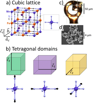
Rubidium manganese hexacyanoferrate materials (Fig. 1a), with composition RbxMn[Fe(CN)6](x+2)/3·zH2O, exhibit CT phase transitions, between MnII(S = 5/2)FeIII(S = 1/2) high temperature (HT) cubic phase and MnIII(S = 0)FeII(S = 0) low-temperature (LT) tetragonal phase, characterized by a wide thermal hysteresis.19,20,41,42 A Landau theory study revealed that this phase transition results from the coupling to the volume strain of the CT instability and the ferroelastic cubic–tetragonal phase transition associated with the collective Jahn–Teller distortion around the MnIII sites.43 Crystallographic studies investigated the important lattice changes between the HT cubic phase (F![[4 with combining macron]](https://www.rsc.org/images/entities/char_0034_0304.gif) 3m, ac ≃ 10.5 Å) and the LT phase described in the non-conventional F
3m, ac ≃ 10.5 Å) and the LT phase described in the non-conventional F![[4 with combining macron]](https://www.rsc.org/images/entities/char_0034_0304.gif) 2m tetragonal space group, at ≃ 10.0 Å, ct ≃ 10.5 Å).43,44 The cubic → tetragonal ferroelastic distortion is associated with the loss of the three-fold rotational symmetry of the cubic lattice. The structural instability occurs at the Γ point of the Brillouin zone and the Jahn–Teller symmetry-breaking order parameter belongs to the bidimensional E representation of the F
2m tetragonal space group, at ≃ 10.0 Å, ct ≃ 10.5 Å).43,44 The cubic → tetragonal ferroelastic distortion is associated with the loss of the three-fold rotational symmetry of the cubic lattice. The structural instability occurs at the Γ point of the Brillouin zone and the Jahn–Teller symmetry-breaking order parameter belongs to the bidimensional E representation of the F![[4 with combining macron]](https://www.rsc.org/images/entities/char_0034_0304.gif) 3m point group. Fig. 1b shows the cubic → tetragonal distortion, associated with the Jahn–Teller deformation of the MnIIIN6 core along the ct axis in the tetragonal phase. This collective distortion can equally occur along the principal directions ac, bc or cc of the initially cubic lattice.6,9,10,45 Therefore, 3 equivalent ferroelastic tetragonal domains can form in the LT phase.
3m point group. Fig. 1b shows the cubic → tetragonal distortion, associated with the Jahn–Teller deformation of the MnIIIN6 core along the ct axis in the tetragonal phase. This collective distortion can equally occur along the principal directions ac, bc or cc of the initially cubic lattice.6,9,10,45 Therefore, 3 equivalent ferroelastic tetragonal domains can form in the LT phase.
Unexpected spectroscopic changes also occur in the LT phase. IR spectroscopy revealed a broad IR band (≃50 cm−1) around 2100 cm−1, attributed to the splitting of the C–N stretching mode, while a sharp (≃10 cm−1) band is observed in the HT phase.46,47 Optical spectroscopy revealed a broad and intense band around 500 nm,48–50 identified as a Mn-centred d–d transition, which is supposed to be weak in the LT D4h symmetry. This absorption band plays a key role for driving photoinduced phase transition in these materials.17,44,47,48 The origin of these unusual spectroscopic features was however not clearly explained so far. Hereafter we use single crystal and powder X-ray diffraction (XRD) to study the ferroelastic domains in the LT phase and estimate the spatial extension of the lattice deformation around the domain walls.
Experimental
Sample preparation
Single crystals of RbMn[Fe(CN)6] (hereafter referred to as RbMnFe) were synthesized by gradually diffusing 2 mL of an aqueous solution of MnCl2·4H2O (1.0 mol dm−3) and 2 mL of an aqueous solution of K3[Fe(CN)6] (1.0 mol dm−3) into 100 mL of an aqueous solution of RbCl (0.5 mol dm−3) over one week at 40 °C. RbMnFe is known to exhibit a wide thermal hysteresis between ≃230 K and ≃300 K.51,52 Rb0.94Co0.06Mn0.94[Fe(CN)6]0.98·0.2H2O, referred to hereafter as RbMn0.94Co0.06Fe, was synthesized according to the procedure reported in previous works,17 where additional characterization data can be found. Magnetic measurements on the powder indicate a broad bistability regime around room temperature (T↓ = 253 K, T↑ = 328 K), between the LT and HT phases. The powder sample was either cooled below T↓ to generate the LT phase, or warmed above T↑ to generate the HT phase. It was then investigated at room temperature (293 K), where both phases are stable. Two identical batches of powder sample were prepared and analysed separately using two different powder X-ray diffraction techniques.Single crystal X-ray diffraction
We used single-crystal X-ray diffraction to study the RbMnFe sample. The data were collected in the HT phase at 300 K and in the LT phase at 100 K. Single-crystal X-ray diffraction was conducted using a Rigaku XtaLAB Synergy-R/DW with monochromatic Mo Kα radiation (λ = 0.70930 Å). We used paratone N oil to mount the single crystal (50 μm large) on a Micro MountsTM holder (Fig. 1c). The data analysis and the reciprocal space reconstruction was performed with CrisAlisPro software of Rigaku.Conventional powder X-ray diffraction of RbMn0.94Co0.06Fe
Conventional powder X-ray diffraction data of the RbMn0.94Co0.06Fe sample were collected using a Rigaku Ultima-IV diffractometer with Cu Kα radiation (λ = 1.5418 Å, scanning range: 10–70°, scanning speed: 1° min−1, sampling width: 0.02°). The powder sample consists of plate-shaped crystals, with an average size of 0.9 ± 0.3 μm (Fig. 1d). The LT phase was prepared by cooling the powder in HT phase with liquid nitrogen. After the cooling, the LT phase was sustained at room temperature due to the existence of the thermal hysteresis over room temperature. The attribution of the Bragg peaks was based on previous analysis using Rietveld refinement.17Streaming powder X-ray diffraction of RbMn0.94Co0.06Fe
In the second approach, XRD measurements were performed at the XPP beamline of the LCLS X-ray Free-Electron Laser (X-FEL), using monochromatic X-rays (λ = 1.890 Å, ΔE/E ∼ 10−4).53,54 We used the streaming powder technique, which makes it possible to study ultra-fast and persistent photoinduced phase transitions.17,23 The RbMn0.94Co0.06Fe crystals were dispersed in ethanol with a 1![[thin space (1/6-em)]](https://www.rsc.org/images/entities/char_2009.gif) :
:![[thin space (1/6-em)]](https://www.rsc.org/images/entities/char_2009.gif) 90 crystal-solvent weight ratio, and the solution was streamed through a 75 μm free-flowing jet to interact with the X-ray beam. The diffracted intensity was collected shot-to-shot using an epix10k2M detector55 in the transmission geometry, at the repetition rate of the LCLS X-FEL (120 Hz). Each 2D image was azimuthally integrated using pyFAI56 and normalized on the X-ray intensity. A typical number of 10
90 crystal-solvent weight ratio, and the solution was streamed through a 75 μm free-flowing jet to interact with the X-ray beam. The diffracted intensity was collected shot-to-shot using an epix10k2M detector55 in the transmission geometry, at the repetition rate of the LCLS X-FEL (120 Hz). Each 2D image was azimuthally integrated using pyFAI56 and normalized on the X-ray intensity. A typical number of 10![[thin space (1/6-em)]](https://www.rsc.org/images/entities/char_2009.gif) 000 shots are subsequently averaged, over an acquisition time of about 100 s. The resulting pattern extends in the [0.5 Å−1; 3.6 Å−1] Q-range. It contains a broad scattering contribution due to the solvent, which is removed by subtracting the scattering pattern of pure ethanol measured in the same conditions. The shape of the diffraction peak(s) is then analysed following the same procedure as for static powder XRD.
000 shots are subsequently averaged, over an acquisition time of about 100 s. The resulting pattern extends in the [0.5 Å−1; 3.6 Å−1] Q-range. It contains a broad scattering contribution due to the solvent, which is removed by subtracting the scattering pattern of pure ethanol measured in the same conditions. The shape of the diffraction peak(s) is then analysed following the same procedure as for static powder XRD.
Results and discussion
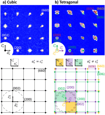
Fig. 2 shows single crystal XRD data, characteristic of the cubic → tetragonal phase transition of RbMn[Fe(CN)6]. The phase transition is associated with a symmetry breaking from HT cubic phase with F![[4 with combining macron]](https://www.rsc.org/images/entities/char_0034_0304.gif) 3m space group (aHT ≈ 10.5 Å) to LT non-conventional F
3m space group (aHT ≈ 10.5 Å) to LT non-conventional F![[4 with combining macron]](https://www.rsc.org/images/entities/char_0034_0304.gif) 2m space group (aLT ≈ 10.0 Å, cLT ≈ 10.5 Å). In the cubic phase (Fig. 2a), the diffracted intensity in the (h0l) reciprocal plane, with cubic reciprocal lattice vectors
2m space group (aLT ≈ 10.0 Å, cLT ≈ 10.5 Å). In the cubic phase (Fig. 2a), the diffracted intensity in the (h0l) reciprocal plane, with cubic reciprocal lattice vectors  , corresponds to sharp Bragg peaks on the node where the h, k and l indices have same parity (F cell). The diffracted patterns are the same in the (0kl) and (hk0) planes due to cubic symmetry. In the LT phase (Fig. 2b), the lengths of the lattice vectors differ due to the tetragonal distortion
  . We can see in Fig. 2b a splitting of the Bragg peaks due to the formation of the ferroelastic domains, for which the tetragonal distortion defining the
  axis can equally occur along the HT axes
  ,
,  or
or  . The diffraction pattern in LT phase (Fig. 2b), corresponding to the (h0l) plane of HT phase (Fig. 2a), is then characteristic of the superposition of the diffracted signal from the 3 tetragonal domains with different orientations of the
  axis in the LT phase: a square
  lattice (yellow, with
  perpendicular to the figure), and two rectangular
  and
and  lattices with
  vertical (green) or horizontal (purple). Consequently, the (h0h) Bragg peaks split in 3 peaks. We can also observe an anisotropic broadening of the Bragg peaks in the LT phase, with some diffuse scattering in-between the peaks of the different domains. This is particularly clear for the (606) Bragg peaks for example. This peak broadening is characteristic of non-uniform lattice strains.57,58
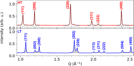
Fig. 3 shows the XRD powder diffraction patterns along the scattering vector Q of RbMn0.94Co0.06Fe, measured at room temperature in the HT and LT phases with conventional powder XRD. The most important changes in the diffraction pattern, due to the ferroelastic distortion in the LT phase, correspond to the splitting along scattering vector Q of the Bragg peaks (h00) and (00h), and of the Bragg peaks (hh0) and (h0h), which are initially equivalent in the HT phase. For instance, Fig. 2b shows that the (200) and (002) Bragg peaks are no longer located on the same diffraction ring defined by |Q| in the LT phase since |Q(200)| ≠ |Q(002)|. Compared to previous studies,42,59 the splitting of the (002) and (200) peaks, indexed the LT F![[4 with combining macron]](https://www.rsc.org/images/entities/char_0034_0304.gif) 2m space group, corresponds to the splitting of the (200) and (110) peaks indexed the LT I
2m space group, corresponds to the splitting of the (200) and (110) peaks indexed the LT I![[4 with combining macron]](https://www.rsc.org/images/entities/char_0034_0304.gif) m2 space group. A similar splitting is observed for different Bragg peaks. In addition, due to lattice contraction, the peaks shift to higher Q in the LT phase. Fig. 3 shows asymmetric Bragg peaks in the LT phase, compared to HT, with diffuse scattering in between the peaks. As mentioned above, this is due to the lattice strains, also observed in single crystal XRD with the diffuse scattering around the Bragg peaks.
m2 space group. A similar splitting is observed for different Bragg peaks. In addition, due to lattice contraction, the peaks shift to higher Q in the LT phase. Fig. 3 shows asymmetric Bragg peaks in the LT phase, compared to HT, with diffuse scattering in between the peaks. As mentioned above, this is due to the lattice strains, also observed in single crystal XRD with the diffuse scattering around the Bragg peaks.
While conventional Rietveld refinement can be used to characterize uniform strain in powder patterns, it is usually inadequate to describe highly-anisotropic strains, that give asymmetric Bragg peaks.60 Instead, we used an approach similar to the one presented by Daniels et al. to characterize the domain structures from diffraction profiles in tetragonal ferroelastic ceramics.61 We analysed the shape of each peak or group of peaks separately by individual fitting, using a combination of split pseudo-Voigt peak profiles with a polynomial background:
where PVX(Q) (X = L, R) is the usual combination of a Lorentzian and a Gaussian profile with a common half width at half maximum σX and a mixing fraction αX ∈ [0; 1]:
Finally, ensuring continuity at the peak centre Q0 imposes:
As such, SPV profiles enable the analysis of asymmetry in the peak shape profiles, by extracting separately the half widths and the peak shape on both sides of the peaks. In the following, the reported values of the parameters correspond to the best fit of the data, and the associated uncertainties are the standard deviations of the least-square fit. In the case of (111), (220) and (400) Bragg peaks, we had to consider a weak additional signal due to an impurity, the passive Rb2MnII[FeII(CN)6]·3.5H2O crystals (Fm![[3 with combining macron]](https://www.rsc.org/images/entities/char_0033_0304.gif) m a = 10.185(6) Å), known to form during the synthesis.17 This impurity does not exhibit CT nor ferroelastic phase transition and we used the same contribution from this impurity in the analysis of the signal of HT and LT states at 293 K.
m a = 10.185(6) Å), known to form during the synthesis.17 This impurity does not exhibit CT nor ferroelastic phase transition and we used the same contribution from this impurity in the analysis of the signal of HT and LT states at 293 K.
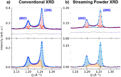
The top panel of Fig. 3 shows the least-square fit of all the Bragg peaks for the HT phase, where the SPV fit yields good results with quite symmetric profiles. For example, the fit of the (200) Bragg peak provides quite similar values for L/R sides: σL = 4.90(0.04) × 10−3 Å−1 vs. σR = 3.88(0.03) × 10−3 Å−1 and αL = 0.69(0.02) vs. αR = 0.45(0.02). The same fitting procedure was applied to the LT phase. We focus first our attention on the (002) and (200) peaks. The top panel of Fig. 4a shows the fit with 2 SPVs, which is in good agreement with the XRD data. In this LT phase, the peak profiles are however strongly asymmetric, as characterized by the fitting parameters.
For the (002) Bragg peak, we obtained σL = 6.7(0.3) × 10−3 Å−1 vs. σR = 10.3(0.3) × 10−3 Å−1 and αL = 0.59(0.07) vs. αR = 1 and for the (200) Bragg peak σL = 7.51(0.08) × 10−3 Å−1 vs. σR = 4.39(0.08) × 10−3 Å−1 and αL = 1 vs. αR = 0.68(0.03). Compared to the HT phase, both LT peaks are broader (σLTL,R > σHTL,R), and display a significant L/R asymmetry: the width of the peaks is larger in between the two Bragg peaks compared to their outer part (σR(002) > σL(002) and σL(200) > σR(200)). The inner part of the two peaks also have a more pronounced Lorentzian character, as αX reaches its maximum value (1). This fitting procedure thus maps the non-negligible intensity observed between the two peaks in the LT phase, in agreement with Fig. 2b. This asymmetry of the powder diffraction peaks is well understood from the single crystal data in Fig. 2b, as diffuse scattering is observed at |Q| in between |Q(200)| and |Q(002)|. The present fit shows that this intensity cannot be explained by a simple isotropic broadening of the peaks as observed in other ferroelastic materials.
We can highlight the asymmetry of the LT (002) and (200) peaks by comparing their profiles with the more symmetric one of the HT (200) peak. The LT peaks were fitted with the SPV profile of the HT phase, with widths σL,R and fractions αL,R fixed to the HT values and refined amplitudes and centres. The result is shown in Fig. 4a, where the red-shaded areas corresponds to fit of the peaks with the HT profiles. The differential between the experimental XRD pattern and the result from this fit is shown by the red curve. It is clear that the parameters of the sharp HT Bragg peaks poorly reproduce the LT data, as only the maximum of each peak is well described. The differential curve between the fit and the experimental data highlights the strong XRD signal between the (002) and (200) Bragg peaks, their asymmetry and broadening. Interestingly, a differential signal is also observed on the outer part of the two Braggs (i.e., below 1.20 Å−1 and above 1.26 Å−1).
Thus, the LT Bragg peaks are globally broadened compared to the HT phase and the signal observed in between (002) and (200) is characteristic of lattice strains in the LT phase, due to the formation of ferroelastic domains walls in the crystal.
Indeed, the strong asymmetry of the (200) and (002) Bragg peaks is due to the fact that tetragonal crystals have 90° {101}-type ferroelastic domain walls.62–65 There is an important strain due to the difference between the LT lattice parameters, with at ≃ 10.0 Å and ct ≃ 10.5 Å. The different possible orientations of the {101}-type domain walls are shown in Fig. 5.
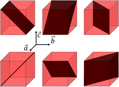 | ||
| Fig. 5 Domain walls in a cubic lattice with tetragonal distortion, corresponding to {101} planes or symmetry-equivalent. | ||
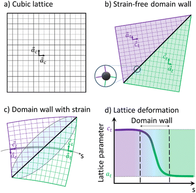
Fig. 6 shows a 2D representation of the formation of two ferroelastic domains. Compared to the regular HT cubic lattice (Fig. 6a), tetragonal domains can form in the LT phase, separated by a wall. The geometrically strain-free twinning angle only depends on the ct/at ratio (Fig. 6b). However, due to constraints during the cubic-to-tetragonal phase transition and the development of the domains, this ideal twinning angle is not achieved. This is especially true in this polymeric crystal as the metallic sites, including the ones on the wall, are stabilized in D4h symmetry (Fig. 1b). The sites on the wall for the strain-free twinning strongly deviates from D4h symmetry as shown by the inset in Fig. 6b, which costs too much energy. Therefore, the lattice has to deform as shown in Fig. 6c, which results in strain fields in the vicinity of domain walls. The lattice constant changes gradually from ct to at as we cross the domain wall along the path s shown in Fig. 6c, which represents the Mn–N–C–Fe bridges of the lattice. This change of lattice parameter is schematically shown in Fig. 6d. These strain fields around the wall, corresponding to a gradient of lattice parameter, result in the broadening of the Bragg peaks and diffuse scattering around some of them. The volume of material close to the domain wall that is affected by this strain field depends on the elastic modulus of the material. In the powder pattern, the signal between (002) and (200) is a direct signature of this strain field and the shape and amplitude of this inner signal reflects the strains extending around the domain walls.
To further characterize the gradient of lattice parameters around the domain walls, we performed a strain analysis, with an approach similar to the one proposed by Daniels et al.61 In the hypothetical absence of gradient, both (002) and (200) peaks would display symmetric profiles. Thus, we artificially constructed symmetrized peak profiles Ssymm(Q), using the fit parameters of the outer parts of the two peaks, which is not affected by the gradient:
| Ssymm(002)(Q) = AL(002)·PVL(002)(Q) |
| Ssymm(200)(Q) = AR(200)·PVR(200)(Q) |
The X-ray diffraction signal affected by the strain of the gradient can then be calculated by subtracting the symmetrized peak profiles:
| Sstrain(Q) = S(Q) − [Ssymm(002)(Q) + Ssymm(200)(Q)] |
The resulting signal is shown as the orange curve in Fig. 4a. As can be seen, the signal spreads over the whole Q range between the two peaks. Its shape is similar to the expected strain distribution around domain walls, as discussed in the literature for other ferroelastic phase transitions.61 A maximum is observed on the upper Q limit, that can be attributed to difference in the peak multiplicity between (h00) and (00h) Bragg peaks in the tetragonal lattice. Furthermore, this analysis enables to estimate the total volume of the crystals that is affected by the strain, by integrating Sstrain(Q) and comparing it to the total integrated signal. In the present case, our fit indicates that the strain-affected volume represents ∼30% of the crystalline volume. This means that the formation of the ferroelastic domain walls induces important strains, which extend over about one third of each crystal.
Additionally, the width of the outer part of (002) and (200) peaks brings additional signature of the presence of ferroelastic domains. The outer parts of these peaks are indeed not affected by the gradient of the domain walls, and their broadening compared to the HT model cannot be related to lattice strain from ct to at. Instead, the larger width is due to the small size of the ferroelastic domains forming during the HT-to-LT phase transition. Indeed, as domains are created in the LT phase, the coherence length of the diffraction signal decreases from the crystal size in the HT phase to the domain size in the LT phase. The formation of small domains increases the width of the peaks, according to the Scherrer equation:
We can therefore estimate the domain size from the broadening of (002) and (200) Bragg peaks relative to the HT phase, by considering that the width of HT (200) peak is mainly due to the IRF and that only the small size of the domains is responsible for the peak broadening in the LT phase. In this way, the increase of width of the LT (002) and (200) peaks, compared to HT (200) peak, gives ΔFWHMθ(002) and ΔFWHMθ(200). Given that we considered pseudo-Voigt peak profiles, ΔFWHMθ was estimated as a mixture of Gaussian- and Lorentzian-corrected FWHMs:
These signatures of ferroelastic domains were further confirmed by streaming powder X-ray diffraction measurements on RbMn0.94Co0.06Fe at the XPP beamline at LCLS (Fig. 4b). Similar trends are measured, with asymmetric peak shapes and peak broadening. Slight differences are visible in the shape of the strain-affected signal, whose maximum is less pronounced, and with a strain-affected volume of ∼20%. This difference, compared to Fig. 4a, may be due to the batch used or to the different thermal cycling of the samples during the experiments. Nonetheless, the overall signatures of the strain around the ferroelastic domain walls are clearly observed.
A similar influence of the strain around the domain walls is also expected for other Bragg peaks. Fig. 7 shows the fit of (111), (202)/(220), (113)/(311)/(222) and (004)/(400) peaks, and their comparison with the corresponding HT model. The profile of the (111) peak matches the HT model, as indicated by a negligible differential. This is also true for the (222) peak. On the other hand, (202)/(220), (113)/(311) and (004)/(400) all display a behaviour similar to (002)/(200): strong differential scattering intensity is observed between the peaks, as well as broadening on the outer part of the peaks. These three groups of peaks indeed correspond to peak splitting due to the ferroelastic phase transition, as (002)/(200). The gradient imposed by the domain walls during the transition thus affects the corresponding lattice constant. However, compared to the seminal case of (002)/(200) splitting, further strain analysis is difficult for these peaks, since their broadening can be affected on both left and right sides by the strain around the domain walls. For instance, (202) is affected by the aLT → cLT gradient on the left side, and by the cLT → aLT gradient on the right side. In the case of (111) and (222), the good fit with the HT model shows that the strain field is weaker along these crystalline directions, as expected for {101}-type ferroelastic domain walls.
Conclusions
This X-ray diffraction study of the ferroelastic phase transition from the cubic MnIIFeIII HT to the tetragonal MnIIIFeII LT phase of RbMnFe materials reveals the formation of ferroelastic domains walls during the cooperative Jahn–Teller distortion around the Mn sites. Three types of ferroelastic tetragonal domains, with ct axis pointing in all three possible directions, coexist in the LT phase, as revealed by single-crystal X-ray diffraction. Our analysis shows that the spatial extension of the strain around the domain walls represents ≃30% of the volume. Because of these distortions of the polymeric Mn–N–C–Fe network, the local symmetry of the ligand field of the metal ions around the wall deviates from D4h, as shown in Fig. 6c. In this figure, a single domain wall is represented. However, as explained in Fig. 5, different domain walls with different orientations can form. Along the path s in Fig. 6c, the ct axis points up, while the at axis points down. However, around a symmetry-equivalent wall the ct axis will point down, while the at axis will points up. In average, ct and at axes are horizontal. The angular distribution of these axes broadens transversally the Bragg peaks (Fig. 2b). Our analysis, evidences that a large fraction of the sample (≃30% of the volume) is affected by the lattice strain around the domain wall. This translates through the appearance of scattering signal in-between Bragg peaks, such as (002)/(200) for example. Such a diffuse signal was observed in previous experiments, but strain analysis was never performed so far. Our strain analysis makes it possible to understand the unexpected spectroscopic changes occurring in the LT phase. On the one hand, a broad IR band (≃50 cm−1) of the C–N stretching mode is observed in the LT phase, compared to the sharp IR band (≃10 cm−1) in the HT phase.47 This broadening results from both a splitting of this IR band due to the symmetry-breaking in the tetragonal phase and a broadening of these bands due to the strain field around the wall. On the other hand, a broad and intense optical band, which has a strong Mn-centred d–d character, appears around 500 nm in the LT phase,48 which is not expected for Mn sites in D4h symmetry. Our results show that about 30% of the Mn sites are in a strong strain field. Consequently, the associated lattice strain, bending the Mn–N–C–Fe bridges, activates this optical transition. The strain field around domain walls plays therefore an important role for the optical control of the magnetic properties of RbMnFe materials, as the Mn-centred d–d transition is also used to drive photomagnetic properties.17,44,47,48 This highlights the important consequences of the formation of a domain wall. The present strain analysis is thus complementary to the structural information obtained though, e.g., Rietveld refinements. Our next goal is to extend this study to the time domain, to understand the evolution of lattice strains during ultrafast and persistent photoinduced phase transition.17 This family of multifunctional materials19 has good chemical stability and durability, and does not contain any banned elements, making it a promising candidate for practical applications, such as barocaloric effects,66 photomagnetism,20,41,67 or Ferroelectricity.68 It was also shown that the application of an electric field above a threshold value leads to a transition from the high- to the low-temperature phase69 and consequently changes in their magnetic, optical, and electronic properties. This can open the way for novel electro-optical devices, and here again the domain walls may also play an important role in the process.Data availability
The powder X-ray diffraction data and their fit are available at Zenodo https://doi.org/10.5281/zenodo.13366596.Author contributions
Marius Hervé: methodology, data curation, formal analysis, writing – original draft; Shintaro Akagi: resources, data curation, investigation, formal analysis; Laurent Guérin, formal analysis; Leland Gee: data curation; Ryan Ribson: data curation; Matthieu Chollet: data curation; Marco Cammarata: data curation, funding acquisition; Shuntaro Nagashima: data curation, investigation, formal analysis; Shin-ichi Ohkoshi: funding acquisition, supervision; Hiroko Tokoro: resource, data curation, funding acquisition, supervision; Eric Collet: funding acquisition, data curation, investigation, conceptualization, supervision, writing – original draft, review & editing. All authors critically reviewed and revised the manuscript draft and approved the final version for submission.Conflicts of interest
There are no conflicts to declare.Acknowledgements
The authors thank S. Zerdane, G. Privault, for technical assistance during data collection at LCLS. The authors gratefully acknowledge the Agence Nationale de la Recherche for financial support under grant ANR-19-CE30-0004 ELECTROPHONE, ANR-19-CE07-0027 SMAC, ANR-19-CE29-0018 MULTICROSS. E. C. thanks University Rennes and Fondation Rennes 1 for funding. This work was carried out in the frame of a JST FOREST Program (JPMJFR213Q), a JSPS Grant-in-Aid for Scientific Research (B) (22H02046), Scientific Research (A) (20H00369), a JST SPRING program (JPMJSP2124), an Organization for Design the Future at University of Tsukuba, Quantum Leap Flagship Program (Q-LEAP, Grant Number JPMXS0118068681) by MEXT, and CNRS-University of Tokyo “Excellence Science” Joint Research Program. Use of the Linac Coherent Light Source, SLAC National Accelerator Laboratory, is supported by the US Department of Energy, Office of Science, Office of Basic Energy Sciences under contract no. DE-AC02-76SF00515.References
- E. K. Salje, Phase Transitions in Ferroelastic and Co-elastic Crystals, Cambridge University Press, Cambridge, 1991 Search PubMed.
- E. K. H. Salje, Annu. Rev. Mater. Res., 2012, 42, 265–283 CrossRef.
- M. A. Carpenter, E. K. H. Salje and A. Graeme-Barber, Eur. J. Mineral., 1998, 10, 621–691 CrossRef.
- S. C. Tarantino, M. Giannini, M. A. Carpenter and M. Zema, Iucrj, 2016, 3, 354–366 CrossRef PubMed.
- M. A. Carpenter and E. K. Salje, Eur. J. Mineral., 1998, 10, 693 CrossRef.
- V. Janovec, V. Dvořák and J. Petzelt, Czech. J. Phys. B, 1975, 25, 1362–1396 CrossRef.
- T. Yamanaka, T. Mine, S. Asogawa and Y. Nakamoto, Phys. Rev. B: Condens. Matter Mater. Phys., 2009, 80, 134120 CrossRef.
- D. C. Palmer, E. K. H. Salje and W. W. Schmahl, Phys. Chem. Miner., 1989, 16, 714–719 CrossRef.
- J. D. Axe and Y. Yamada, Phys. Rev. B: Condens. Matter Mater. Phys., 1981, 24, 2567–2569 CrossRef CAS.
- V. Janovec and J. Privratska, International Tables for Crystallography, 2006, D, 449–505 Search PubMed.
- E. K. H. Salje, J. Appl. Phys., 2020, 128, 164104 CrossRef CAS.
- V. B. Jakobsen, E. Trzop, E. Dobbelaar, L. C. Gavin, S. Chikara, X. Ding, M. Lee, K. Esien, H. Müller-Bunz, S. Felton, E. Collet, M. A. Carpenter, V. S. Zapf and G. G. Morgan, J. Am. Chem. Soc., 2022, 144, 195–211 CrossRef CAS.
- E. Dobbelaar, V. B. Jakobsen, E. Trzop, M. Lee, S. Chikara, X. Ding, H. Müller-Bunz, K. Esien, S. Felton, M. A. Carpenter, E. Collet, G. G. Morgan and V. S. Zapf, Angew. Chem., Int. Ed., 2022, 61, e202114021 CrossRef CAS PubMed.
- V. B. Jakobsen, S. Chikara, J.-X. Yu, E. Dobbelaar, C. T. Kelly, X. Ding, F. Weickert, E. Trzop, E. Collet, H.-P. Cheng, G. G. Morgan and V. S. Zapf, Inorg. Chem., 2021, 60, 6167–6175 CrossRef CAS PubMed.
- V. B. Jakobsen, E. Trzop, L. C. Gavin, E. Dobbelaar, S. Chikara, X. Ding, K. Esien, H. Müller-Bunz, S. Felton, V. S. Zapf, E. Collet, M. A. Carpenter and G. G. Morgan, Angew. Chem., Int. Ed., 2020, 59, 13305–13312 CrossRef CAS PubMed.
- S. Chikara, J. Gu, X. G. Zhang, H.-P. Cheng, N. Smythe, J. Singleton, B. Scott, E. Krenkel, J. Eckert and V. S. Zapf, Nat. Commun., 2019, 10, 4043 CrossRef.
- M. Hervé, G. Privault, E. Trzop, S. Akagi, Y. Watier, S. Zerdane, I. Chaban, R. G. Torres Ramírez, C. Mariette, A. Volte, M. Cammarata, M. Levantino, H. Tokoro, S. Ohkoshi and E. Collet, Nat. Commun., 2024, 15, 267 CrossRef.
- S. Ohkoshi, H. Tokoro and E. Collet, Chimia Rep., 2019, 22, 498–507 CrossRef CAS.
- H. Tokoro and S. Ohkoshi, Bull. Chem. Soc. Jpn., 2015, 88, 227–239 CrossRef.
- S. Ohkoshi and H. Tokoro, Acc. Chem. Res., 2012, 45, 1749–1758 CrossRef CAS.
- E. Collet and G. Azzolina, Phys. Rev. Mater., 2021, 5, 044401 CrossRef CAS.
- E. Collet, G. Azzolina, J. Jeftić and M.-H. Lemée-Cailleau, Adv. Phys.: X, 2023, 8, 2161936 Search PubMed.
- M. Reinhard, K. Kunnus, K. Ledbetter, E. Biasin, D. B. Zederkof, R. Alonso-Mori, T. B. van Driel, S. Nelson, M. Kozina, O. J. Borkiewicz, M. Lorenc, M. Cammarata, E. Collet, D. Sokaras, A. A. Cordones and K. J. Gaffney, ACS Nano, 2024, 18, 15468–15476 CrossRef CAS PubMed.
- F. J. Valverde-Muñoz, R. G. Torres Ramírez, A. Ulhe, E. Trzop, M. Dutta, C. Das, P. Chakraborty and E. Collet, CrystEngComm, 2023, 25, 3588–3597 RSC.
- R. G. Torres Ramírez, E. Trzop and E. Collet, Dalton Trans., 2024, 53, 10159–10167 RSC.
- K. W. Törnroos, M. Hostettler, D. Chernyshov, B. Vangdal and H.-B. Bürgi, Chem.–Eur. J., 2006, 12, 6207–6215 CrossRef PubMed.
- S. Lakhloufi, P. Guionneau, M. H. Lemée-Cailleau, P. Rosa and J. F. Létard, Phys. Rev. B: Condens. Matter Mater. Phys., 2010, 82, 132104 CrossRef.
- V. A. Money, J. Elhaïk, I. Radosavljevic Evans, M. A. Halcrow and J. A. K. Howard, Dalton Trans., 2004, 65–69, 10.1039/B311262B.
- G. A. Craig, J. Sánchez Costa, O. Roubeau, S. J. Teat and G. Aromí, Chem.–Eur. J., 2011, 17, 3120–3127 CrossRef PubMed.
- A. Djemel, O. Stefanczyk, M. Marchivie, E. Trzop, E. Collet, C. Desplanches, R. Delimi and G. Chastanet, Chem.–Eur. J., 2018, 24, 14760–14767 CrossRef PubMed.
- S. Floquet, N. Guillou, P. Négrier, E. Rivière and M.-L. Boillot, New J. Chem., 2006, 30, 1621–1627 RSC.
- B. Weber, E. S. Kaps, J. Obel, K. Achterhold and F. G. Parak, Inorg. Chem., 2008, 47, 10779–10787 CrossRef.
- Y.-Y. Zhu, C.-W. Liu, J. Yin, Z.-S. Meng, Q. Yang, J. Wang, T. Liu and S. Gao, Dalton Trans., 2015, 44, 20906–20912 RSC.
- Y. Otsuki, S. Kimura, S. Awaji and M. Nakano, AIP Adv., 2019, 9, 085219 CrossRef.
- M. Nakano, G.-e. Matsubayashi and T. Matsuo, Phys. Rev. B: Condens. Matter Mater. Phys., 2002, 66, 212412 CrossRef.
- J. Olguín, Coord. Chem. Rev., 2020, 407, 213148 CrossRef.
- N. A. Spaldin and R. Ramesh, Nat. Mater., 2019, 18, 203–212 CrossRef CAS.
- S.-W. Cheong and M. Mostovoy, Nat. Mater., 2007, 6, 13–20 CrossRef CAS PubMed.
- N. A. Spaldin and M. Fiebig, Science, 2005, 309, 391–392 CrossRef CAS PubMed.
- X. Zhang, W. H. Xu, W. Zheng, S. Q. Su, Y. B. Huang, Q. Shui, T. Ji, M. Uematsu, Q. Chen, M. Tokunaga, K. Gao, A. Okazawa, S. Kanegawa, S. Q. Wu and O. Sato, J. Am. Chem. Soc., 2023, 145, 15647–15651 CrossRef CAS PubMed.
- H. Tokoro, T. Matsuda, T. Nuida, Y. Moritomo, K. Ohoyama, E. D. L. Dangui, K. Boukheddaden and S. Ohkoshi, Chem. Mater., 2008, 20, 423–428 CrossRef CAS.
- H. Tokoro, K. Nakagawa, K. Imoto, F. Hakoe and S. Ohkoshi, Chem. Mater., 2012, 24, 1324–1330 CrossRef CAS.
- G. Azzolina, R. Bertoni, C. Ecolivet, H. Tokoro, S. Ohkoshi and E. Collet, Phys. Rev. B: Condens. Matter Mater. Phys., 2020, 102, 134104 CrossRef CAS.
- G. Azzolina, R. Bertoni, C. Mariette, M. Cammarata, E. Trzop, C. Ecolivet, M. Sander, M. Levantino, H. Tokoro, K. Imoto, M. Yoshikiyo, S. Ohkoshi and E. Collet, J. Mater. Chem. C, 2021, 9, 6773–6780 RSC.
- P. Toledano, M. M. Fejer and B. A. Auld, Phys. Rev. B: Condens. Matter Mater. Phys., 1983, 27, 5717–5746 CrossRef CAS.
- H. Tokoro, A. Namai, M. Yoshikiyo, R. Fujiwara, K. Chiba and S. Ohkoshi, Sci. Rep., 2018, 8, 63 CrossRef PubMed.
- G. Privault, M. Hervé, N. Godin, R. Bertoni, S. Akagi, J. Kubicki, H. Tokoro, S. Ohkoshi, M. Lorenc and E. Collet, Angew. Chem., Int. Ed., 2024, e202408284 CAS.
- G. Azzolina, H. Tokoro, K. Imoto, M. Yoshikiyo, S. Ohkoshi and E. Collet, Angew. Chem., Int. Ed., 2021, 60, 23267–23273 CrossRef CAS.
- S. Ohkoshi, T. Nuida, T. Matsuda, H. Tokoro and K. Hashimoto, J. Mater. Chem., 2005, 15, 3291 RSC.
- E. D. Loutete-Dangui, E. Codjovi, H. Tokoro, P. R. Dahoo, S. Ohkoshi and K. Boukheddaden, Phys. Rev. B: Condens. Matter Mater. Phys., 2008, 78, 014303 CrossRef.
- S. Ohkoshi, H. Tokoro, M. Utsunomiya, M. Mizuno, M. Abe and K. Hashimoto, J. Phys. Chem. B, 2002, 106, 2423–2425 CrossRef.
- H. Tokoro, S. Ohkoshi, T. Matsuda and K. Hashimoto, Inorg. Chem., 2004, 43, 5231–5236 CrossRef PubMed.
- M. Chollet, R. Alonso-Mori, M. Cammarata, D. Damiani, J. Defever, J. T. Delor, Y. Feng, J. M. Glownia, J. B. Langton, S. Nelson, K. Ramsey, A. Robert, M. Sikorski, S. Song, D. Stefanescu, V. Srinivasan, D. Zhu, H. T. Lemke and D. M. Fritz, J. Synchrotron Radiat., 2015, 22, 503–507 CrossRef PubMed.
- C. Antolini, V. Sosa Alfaro, M. Reinhard, G. Chatterjee, R. Ribson, D. Sokaras, L. Gee, T. Sato, P. L. Kramer, S. L. Raj, B. Hayes, P. Schleissner, A. T. Garcia-Esparza, J. Lim, J. T. Babicz, A. H. Follmer, S. Nelson, M. Chollet, R. Alonso-Mori and T. B. van Driel, Molecules, 2024, 29, 2323 CrossRef.
- T. B. van Driel, S. Nelson, R. Armenta, G. Blaj, S. Boo, S. Boutet, D. Doering, A. Dragone, P. Hart, G. Haller, C. Kenney, M. Kwaitowski, L. Manger, M. McKelvey, K. Nakahara, M. Oriunno, T. Sato and M. Weaver, J. Synchrotron Radiat., 2020, 27, 608–615 CrossRef.
- G. Ashiotis, A. Deschildre, Z. Nawaz, J. P. Wright, D. Karkoulis, F. E. Picca and J. Kieffer, J. Appl. Crystallogr., 2015, 48, 510–519 CrossRef.
- B. D. Cullity, Elements of X-Ray Diffraction, Addison-Wesley, Reading, MA, USA, 2nd edn, 1978 Search PubMed.
- H. Boysen, Z. Kristallogr., 2005, 220, 726–734 Search PubMed.
- G. Azzolina, E. Collet, C. Mariette, M. Cammarata, E. Trzop, M. Sander, M. Levantino, K. Nakagawa, H. Tokoro, S. Ohkoshi and R. Bertoni, Eur. J. Inorg. Chem., 2019, 2019, 3142–3147 CrossRef CAS.
- G. Tutuncu, M. Motahari, M. R. Daymond and E. Ustundag, Acta Mater., 2012, 60, 1494–1502 CrossRef CAS.
- J. E. Daniels, J. L. Jones and T. R. Finlayson, J. Phys. D: Appl. Phys., 2006, 39, 5294 CrossRef CAS.
- J. Sapriel, Phys. Rev. B: Condens. Matter Mater. Phys., 1975, 12, 5128–5140 CrossRef CAS.
- S. H. Curnoe and A. E. Jacobs, Phys. Rev. B: Condens. Matter Mater. Phys., 2000, 62, R11925–R11928 CrossRef CAS.
- T. J. Parker and J. C. Burfoot, J. Phys. D: Appl. Phys., 1969, 2, 1168 CrossRef.
- G. Arlt and P. Sasko, J. Appl. Phys., 1980, 51, 4956–4960 CrossRef CAS.
- S. Ohkoshi, K. Nakagawa, M. Yoshikiyo, A. Namai, K. Imoto, Y. Nagane, F. Jia, O. Stefanczyk, H. Tokoro, J. Wang, T. Sugahara, K. Chiba, K. Motodohi, K. Isogai, K. Nishioka, T. Momiki and R. Hatano, Nat. Commun., 2023, 14, 8466 CrossRef CAS.
- H. Tokoro, T. Matsuda, K. Hashimoto and S. Ohkoshi, J. Appl. Phys., 2005, 97, 10M508 CrossRef.
- S. Ohkoshi, H. Tokoro, T. Matsuda, H. Takahashi, H. Irie and K. Hashimoto, Angew. Chem., Int. Ed., 2007, 46, 3238–3241 CrossRef CAS PubMed.
- T. Mahfoud, G. Molnár, S. Bonhommeau, S. Cobo, L. Salmon, P. Demont, H. Tokoro, S.-I. Ohkoshi, K. Boukheddaden and A. Bousseksou, J. Am. Chem. Soc., 2009, 131, 15049–15054 CrossRef CAS PubMed.
| This journal is © The Royal Society of Chemistry 2024 |

