
Dithiocarbazate–Zn(II) complexes for photodynamic therapy and chemotherapy against lung cancer†
JunGang
Deng‡
a,
YouRu
Wu‡
a,
AiLi
Li‡
a,
WeiPing
Pan
a,
LiXia
Hou
a,
DaQi
Wu
a,
ZhenLei
Zhang
b,
Feng
Yang
*b and
Yi
Gou
 *ac
*ac
aThe Laboratory of Respiratory Disease, Affiliated Hospital of Guilin Medical University, Guilin 541001, Guangxi, China. E-mail: sanchuanok@126.com
bSchool of Chemistry and Pharmaceutical Sciences, State Key Laboratory for Chemistry and Molecular Engineering of Medicinal Resources, Guangxi Normal University, Guilin, 541004, P. R. China. E-mail: fyang@mailbox.gxnu.edu.cn
cDepartment of Respiratory Medicine, Affiliated Hospital of Guilin Medical University, Guilin 541001, Guangxi, China
First published on 20th September 2023
Abstract
Combining photodynamic therapy with chemotherapy is a promising strategy for treating tumors. Herein, a series of new dithiocarbazate–Zn(II) complexes, including [ZnII(L1)2] Zn1, [ZnII(L2)2] Zn2, [ZnII(L3)2] Zn3, and [ZnII(L4)2] Zn4, have been synthesized for chemotherapy and photodynamic therapy of lung cancer. These complexes can be used for live-cell fluorescence-based imaging, are photostable, and exhibit hemolysis comparable to cisplatin. Among them, complex Zn3 shows good chemotherapeutic ability, and its chemotherapeutic mechanism relies on multiple pathways. At the same time, complex Zn3 generates superoxide anion radicals (˙O2−) under light irradiation, leading to cancer cell death through photodynamic therapy. Its remarkable anticancer effect is achieved through multimodal photodynamic therapy/chemotherapy. This study paves the way for the use of novel Zn(II) complexes as anticancer agents, providing new strategies for multimodal cancer therapy.
Introduction
Lung cancer is a highly prevalent type of cancer and a significant barrier to increasing life expectancy in every country in the world.1,2 According to estimates from the GLOBOCAN in 2020, lung cancer is the leading cause of cancer death (18.0%) and the second most frequently diagnosed cancer (11.4%) in the world, with nearly 1.8 million deaths and 2.2 million new cancer cases annually.3 Globally, the cancer burden is projected to double by 2050, led by lung cancer.4 Moreover, the 5-year relative survival rate for lung cancer patients after diagnosis is only 10%–20% in most countries.3,5 Various therapies against lung cancer including surgery, radiotherapy, chemotherapy, photodynamic therapy, and immunotherapy have been developed. However, because of the high heterogeneity of lung cancer, the efficacy of monotherapy is often limited.6,7 Therefore, multimodal therapies that combine different therapeutic approaches have attracted considerable interest; they utilize different therapeutic mechanisms that can act synergistically, resulting in higher therapeutic efficiency.8,9 In particular, the combination of photodynamic therapy and chemotherapy is promising for the treatment of lung cancer.10–14Photodynamic therapy is a minimally invasive treatment modality with few side effects.15 In photodynamic therapy, photosensitizers are accumulated in tumors and generate reactive oxygen species (ROS) upon irradiation with light, thereby inducing tumor regression.16 After photodynamic therapy, a series of therapeutic effects occur, such as damage or permeabilization of the tumor vasculature, tumor cell apoptosis, tumor cell necrosis, and immune system response activation to induce tumor regression.17 Moreover, photodynamic therapy can be repeated on the same site without a decrease in efficacy.18 However, photodynamic therapy as a monotherapy often results in incomplete tumor killing because of limited light penetration in tissues, disease recurrence caused by cells that survived photodynamic therapy, tumor hypoxia, and tumor migration.14,19,20 Drug chemotherapy can effectively eradicate tumors, but often causes side effects such as drug resistance and toxicity, thereby severely reducing the quality of life of patients.21,22 Several studies demonstrated that the combination of photodynamic therapy with chemotherapy shows multifold advantages: (1) photodynamic therapy can be used to treat the tumor tissue locally with minimal damage to adjacent tissues, whereas chemotherapy can eliminate surviving tumor cells, especially in deep or large tumors that are difficult to penetrate with lasers; (2) photodynamic therapy reduces tumor solid pressure, reduces extravascular barriers, and permeabilizes tumor vessels, which may enhance the accumulation of chemotherapeutic drugs in the tumor, thereby improving the therapeutic effect; (3) the ROS generated during photodynamic therapy can directly damage lysosomes and endosomes to promote the delivery of intracellular drugs to targets such as mitochondria, nucleus, and microtubules; (4) DNA damage from combined photodynamic therapy and chemotherapy cannot be easily repaired by drug-resistant DNA-repairing enzymes.22–28 To date, many studies have attempted to realize the synergistic chemo-photodynamic therapy through the chemical linking or nanoloading of photosensitizers and anticancer drugs together to obtain higher therapeutic effects.14,21,29–40 However, chemical linking often relies on the use of additional toxic chemicals. Nanoloading generally requires complex operations and often causes toxicity problems because of the accumulation of nanomaterials in vivo. Therefore, designing and developing a molecule with both chemotherapeutic and photodynamic effects seems a promising strategy for tumor therapy.
Recently, Zn(II)-based complexes have attracted attention owing to their photophysical properties and chemotherapeutic effects.41–46 Dithiocarbazinates and their derivatives, as a class of Schiff base ligands with multiple pharmaceutical and biological activities, can coordinate with a variety of metal ions to form versatile metal complexes.47 Herein, we propose a series of dithiocarbazate–Zn(II) complexes (Scheme 1) as potential photodynamic therapy and chemotherapy agents for lung cancer treatment. Porphyrins, phthalocyanines, and chlorins constitute the basic structures of many photodynamic therapeutic agents, which are not only difficult to synthesize and purify but also suffer from other limitations such as aggregation and poor stability in biological fluids.46 In this work, dithiocarbazate–Zn(II) complexes were easily synthesized and purified, showing good water stability and low hemolysis. Among them, complex Zn3 exhibited good chemotherapeutic and photodynamic activities. The mechanisms of the photodynamic and chemotherapeutic effects of complex Zn3 were investigated using various methods. The mechanism of photodynamic action can be mainly divided into two types: type I and type II.48 In the type II process, O2 obtains energy directly from the excited photosensitizer, resulting in the formation of ROS. For the type I process, ROS are obtained by direct transfer of hydrogen atoms or electrons when the photosensitizer interacts with the substrate, thus avoiding the large consumption of oxygen.49 Thus, type I photoreactivity may lead to strong cytotoxic effects under low-oxygen conditions, which is beneficial for overcoming tumor hypoxia.49–51 However, hypoxic-active type I photosensitizers remain an underexplored research area and are still rarely reported.52 Interestingly, complex Zn3 can cause cellular damage by exploiting type I processes. In addition, studies on the chemotherapeutic mechanism of action of complex Zn3 revealed that Zn3 suppresses cancer cells by triggering multiple mechanisms. Our results provide new insights into the development and utilization of Zn(II)-based complexes and the treatment of lung cancer.
 | ||
| Scheme 1 Synthesis of complexes Zn1–Zn4. | ||
Results and discussion
Characterization of the Zn(II) complexes
The dithiocarbazate Schiff-base ligands HL1–HL4 and complex Zn1 were synthesized using the previously reported methods.53–56 Complexes Zn2–Zn4 were prepared by reacting 1 equivalent of Zn(II) with 2 equivalents of ligands in CH3OH. All Zn(II) complexes were characterized using X-ray crystallography. The obtained crystallographic information is summarized in Tables S1 and S2.†X-ray crystallography shows that these Zn(II) complexes have similar coordination geometries but exhibit two distinct crystal systems, triclinic (Zn1) and monoclinic (Zn2–Zn4). As shown in Fig. 1, all Zn(II) complexes are in mononuclear six-coordination form, showing distorted octahedral coordination geometry. One S atom, one imine N atom, and one pyridine N atom from one ligand and one imine N atom from another ligand occupy the basal coordination sites. The coordination sites at the axial positions are occupied by one S atom and one pyridine N atom from the second ligand. Similar Zn(II)-based complexes have been previously reported; in these complexes, both ligands can both be deprotonated (thiol form) at the thioamide N atom or both in the neutral thione form.43,57–60 Herein, two Schiff-base ligands were coordinated in their deprotonated form via the S/N/N donor atoms, forming four 5-membered bichelate rings around the Zn(II) center. The Zn–S, Zn–Nimine, and Zn–Npyridine bond lengths and the angles with Zn atom as the vertex are within the range reported for other similar Zn(II) complexes.61–65 In general, average Zn–Nimine bond lengths (2.113(2)–2.128(2) Å) are lower than the Zn–Npyridine bond lengths (2.212(2)–2.376(2) Å).
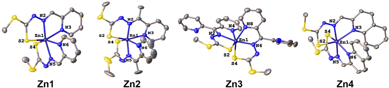 | ||
| Fig. 1 ORTEP views of the complexes Zn1–Zn4 with displacement ellipsoids drawn at the 30% probability level. All H atoms are omitted for clarity. | ||
Although these Zn(II) complexes have similar structures, only Zn1, Zn3, and Zn4 complexes show significant π⋯π interactions in the solid state. As presented in Fig. S1,† complex Zn1 is linked into a dimeric structure by π⋯π interactions between pyridine rings, with an angle of 0°, shift distance of 1.672 Å, and centroid–centroid distance of 3.854 Å. Adjacent monomer units in complex Zn3 are linked into a 1D zigzag chain by π⋯π interactions between the pyridine rings, with an angle of 171.201°, shift distance of 1.741 Å, and centroid–centroid distance of 3.793 Å (Fig. S2†). In complex Zn4, the adjacent monomer units are linked into a dimeric structure by π⋯π stacking between the aromatic rings, with the angle of 0°, shift distance of 1.498 Å, and centroid–centroid distance of 3.737 Å (Fig. S3†).
To determine the nature of the aforementioned π⋯π stacking interactions in these complexes, energy decomposition analysis based on the classical molecular force field (EDA-FF) was performed. The EDA-FF method can decompose the weak interactions of these system into repulsion interactions, electrostatic interactions and dispersion interactions. As depicted in Table S3,† the EDA-FF results show the same trend, that is, the dispersion interaction accounted for approximately 60%, and the electrostatic interaction accounted for the least (about 15%). This indicates that the attractive dispersion interactions dominate in whole π⋯π stacking interactions.
Photophysical properties
The UV-vis absorption spectra of complexes Zn1–Zn4 in methanol were obtained (Fig. 2A). The bands at 300–500 nm were primarily assigned to spin-allowed ligand-centered n → π* and/or π → π* transitions. To gain insight into the properties of charge transfer between Zn and the ligands in the UV-vis absorption spectra, the time-dependent density functional theory (TD-DFT) calculations were also performed for these Zn(II) complexes. Based on the calculation results (Table S4†), the relatively bands at 300–500 nm were primarily attributed to ligand-to-ligand charge-transfer transitions, followed by a minor contribution from Zn-to-ligand charge-transfer transitions. | ||
| Fig. 2 (A) Absorption spectra of complexes Zn1–Zn4 (30 μM) measured in methanol. (B) The photostability of complexes Zn1–Zn4 (30 μM) under continuous LED irradiation at 470 nm. (C) Fluorescence spectra of complexes Zn1–Zn4 (10 μM) measured in methanol followed by excitation at 470 nm. | ||
The photostability of complexes Zn1–Zn4 was examined by monitoring the UV-vis absorption spectra of their methanol solutions under continuous LED light irradiation at 470 nm for 30 min. No significant changes are observed in the absorption properties of all complexes throughout the experiment (Fig. 2B), demonstrating their photostability.
Upon excitation at 470 nm, complexes Zn1–Zn4 exhibit green emission (Fig. 2C). The emission lifetimes of complexes Zn1–Zn4 fall in the range of 2–5 ns, indicating the fluorescent nature of the emission. These complexes exhibit fluorescence in the green region of the visible spectrum, suggesting that they may be applied for cellular fluorescence imaging. Next, the fluorescence imaging properties of complexes Zn1–Zn4 in living lung cancer A549 cells were investigated using confocal microscopy. The cells were incubated with the complexes (10 μM) for various times. As shown in Fig. 3, these Zn(II) complexes are efficiently taken up by the cells and can be used for cellular imaging because fluorescent signals are clearly observed in living cells. It was previously reported that the position of an Ir complex luminescence does not change over time in the cell (luminescence is always observed throughout the cell).66 Interestingly, our results show that these Zn(II) complexes primarily accumulated in the cytoplasm within the first 60 min and partly migrated to the nucleus during further incubation (120 min).
 | ||
| Fig. 3 Confocal fluorescence images of living A549 cells incubated with complexes Zn1–Zn4 (10 μM) in real time (15, 30, 60, 120 min); λex = 488 nm, λem = 520–570 nm. Scale bar: 100 μm. | ||
Hemocompatibility assay
The interaction between human blood components, especially human erythrocytes, and drugs is an important phenomenon.61 The effects of these Zn(II) complexes on blood were hence assessed through a hemolysis assay using human red blood cells. The hemolytic behavior of these Zn(II) complexes was studied at different concentrations up to 40 μM using Triton X-100 as a positive control.67 According to ISO/Technical Report 7406, the allowable level of hemolysis of biological materials needs to be less than 5%. All Zn(II) complexes exhibited negligible red hemoglobin release (% hemolysis <5%) in a concentration range of 0–40 μM (Fig. S4†), indicating excellent blood compatibility. The hemolytic activity of cisplatin, a commonly used clinical metal-based anticancer drug, was also used as a reference. All Zn(II) complexes showed hemolysis comparable to cisplatin.In vitro cytotoxicity
To study the efficacy of the Zn(II) Zn1–Zn4 complexes as anticancer agents, their cytotoxicity toward human lung carcinoma cells (A549, A549/DDP, and Calu-1) in the dark and upon irradiation (photocytotoxicity) at 470 nm (15 min, 20 mW cm−2) was investigated. Complexes Zn1, Zn3, and Zn4 cause cell death in all investigated lung cancer cell lines with IC50 values in the range of 4.55–16.78 μM in the dark. Interestingly, upon exposure to light, all complexes, especially complexes Zn2 and Zn3, show higher anticancer activity (Table 1), indicating that these complexes are highly phototoxic.| Complex | Cell growth inhibition, IC50 ± SD (μM) | |||
|---|---|---|---|---|
| A549 | A549/DDP | Calu-1 | ||
| Zn1 | Dark | 13.36 ± 1.47 | 13.98 ± 0.74 | 16.42 ± 0.81 |
| Light | 5.12 ± 0.31 | 5.87 ± 0.57 | 9.07 ± 1.79 | |
| Zn2 | Dark | >20 | >20 | >20 |
| Light | 0.36 ± 0.11 | 0.29 ± 0.04 | 0.24 ± 0.05 | |
| Zn3 | Dark | 4.55 ± 0.75 | 5.57 ± 0.41 | 8.63 ± 0.71 |
| Light | 0.26 ± 0.05 | 0.25 ± 0.01 | 0.22 ± 0.03 | |
| Zn4 | Dark | 14.66 ± 1.23 | 15.06 ± 1.18 | 16.78 ± 1.48 |
| Light | 7.63 ± 0.56 | 7.44 ± 0.38 | 9.48 ± 0.87 | |
Having confirmed a close relationship between ROS and phototoxicity, we investigated ROS generation inside the A549 cells after irradiation. Cells treated with complexes Zn1–Zn4 and a ROS probe in the dark showed little fluorescence. In contrast, a remarkable increase in ROS production (strong red fluorescence) occurred when light irradiation followed the treatment with these Zn(II) complexes (Fig. S5†), which confirms that these Zn(II) complexes are promising photodynamic therapy agents because ROS generation is the main mechanism of photodynamic therapy-induced cell death.68 Overall, these results show that complexes Zn1, Zn3, and Zn4 can be used for both chemotherapy and photodynamic therapy of tumors. Tumor recurrence from surviving cells after photodynamic therapy is a significant concern, which is partly due to the limited diffusion distance and short half-life of ROS in cells.14,18,69 Chemotherapy can eliminate surviving tumor cells after photodynamic therapy, thereby reducing the possibility of cancer recurrence.22 At the same time, photodynamic therapy has shown the potential to enhance the efficacy of chemotherapy.24 Thus, agents with both photodynamic and chemotherapeutic properties can ablate cancer cells more effectively.
Zn3 in 3D multicellular tumor spheroids
After evaluating the cytotoxicity on cell monolayers, the cytotoxic effect of complex Zn3 with good chemotherapeutic and photodynamic therapy capabilities was investigated in 3D multicellular tumor spheroids. Notably, 3D tumor spheroids are closer than monolayer cell models to tumor models for clinical treatment; thus, they are a widely used tissue model for assessing drug delivery.70,71 3D tumor spheroids can also replicate certain pathological elements of solid tumors such as spatial architecture and hypoxia at the tumor center.72,73 Consequently, 3D A549 multicellular tumor spheroids were incubated with complex Zn3. One of the reasons why many of the investigated anticancer drugs fail during translation from cancer monolayers to in vivo models is impaired drug delivery due to drug penetration through the extracellular barrier. The penetration of the complex was first analyzed via Z-stack imaging microscopy. After 12 h of incubation, fluorescence signals are observed at each slice depth (Fig. S6†), indicating complete penetration of the complex in the 3D tumor spheroids.We further investigated the potential antisolid tumor activity of complex Zn3 in 3D A549 multicellular tumor spheroids. The 3D tumor spheres in the control group were very dense and had a smooth surface. The 3D spheres treated with complex Zn3 became loose, with cell leakage in the dark (Fig. S7†), suggesting that complex Zn3 also exhibits a chemotherapeutic effect in 3D tumor spheroids. The 3D tumor spheres treated with complex Zn3 followed by irradiation with 470 nm light exhibit significant surface disintegration, with more cells lost from the spheroid than in the dark, and loss of 3D structure at day 3 (Fig. 4). In addition, these results indicate that the photodynamic therapy capabilities of complex Zn3 are superior to its chemotherapeutic capabilities because photodynamic therapy did more damage to 3D spheres at lower doses and shorter times than chemotherapy.
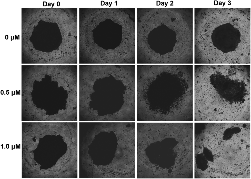 | ||
| Fig. 4 Representative pictures of 3D A549 tumor spheroids at different times before (0 h) and after they were exposed once to 470 nm-LEDs. The spheroids were incubated without or with complex Zn3 at the indicated concentrations for 24 h. They were then illuminated in PBS with LEDs (470 nm, 20 mW cm−2) for 15 min. After this exposure, tumor spheroids were suspended in fresh medium and imaged. After 24 h, 48 h and 72 h, the second, third and fourth irradiation steps were performed as described above. | ||
Photodynamic mechanism of Zn3
The photodynamic mechanisms are currently generally divided into type I and type II pathways, both inducing the production of ROS.74 Type II photodynamic pathway is manifested as the generation of singlet oxygen 1O2 through direct energy transfer. The photochemical type I pathway is more complex than the type II pathway and involves the transfer of photo-induced electrons of biological substrates, which after several steps generate free radical species such as hydrogen peroxide (H2O2), peroxynitrite anion (ONOO−), superoxide anion radical (˙O2−), and hydroxyl radical (˙OH). To identify the specific ROS generated after the treatment with complex Zn3, A549 cells were incubated with several selective ROS scavengers, including NaN3 as a 1O2 scavenger, D-mannitol as a ˙OH scavenger, sodium pyruvate as an H2O2 scavenger, tiron as an ˙O2− scavenger, and ebselen as an ONOO− scavenger.68 Upon light excitation, the cells treated with complex Zn3 in the presence of NaN3/D-mannitol/sodium pyruvate/ebselen exhibit cell morphologies similar to that of cells treated with complex Zn3 alone, which ruled out the possibility that 1O2, ˙OH, H2O2, and ONOO− are involved in the photodynamic effect (Fig. 5). Interestingly, in the presence of tiron, the intracellular production of ROS was significantly inhibited, which suggested the generation of ˙O2− in the cells after light irradiation in the presence of complex Zn3. Low O2 concentrations in solid tumors remain an obstacle for the use of classical photosensitizers because photodynamic therapy requires molecular oxygen to initiate cell death.50,75 In contrast to type II photoreactivity, type I photoreactivity can still lead to strong cytotoxicity under hypoxic conditions.52 Therefore, complex Zn3 is promising for the photodynamic therapy of tumors because it utilizes the type I pathway, which is beneficial for overcoming the problem of tumor hypoxia. | ||
| Fig. 5 Representative image of ROS assay with A549 cells exposed to light (470 nm, 20 mW cm−2). Cells were pre-incubated with specific ROS scavengers (NaN3/D-mannitol/sodium pyruvate/ebselen/tiron) and then treated with complex Zn3 (2 h in the dark followed by 15 min under the irradiation). | ||
Chemotherapeutic mechanisms of Zn3
In view of the good chemotherapeutic capabilities (in the dark) of complex Zn3, its chemotherapeutic mechanism has also been carefully studied. Four-dimensional data-independent acquisition (4D-DIA) proteomic analysis is a fast, efficient, and sensitive method that can help elucidate potential anticancer mechanisms of drugs at the protein level.76,77 Therefore, we performed 4D-DIA proteomic analysis in the diaPASEF mode to understand the details of the chemotherapeutic mechanism of complex Zn3.The total number of identified peptides is 86![[thin space (1/6-em)]](https://www.rsc.org/images/entities/char_2009.gif) 379, and the corresponding total number of quantified proteins is 7897. Among these quantified proteins, 1065 proteins were found to be significantly differentially expressed based on the following criteria: FC > 1.5 or FC < 0.6667 and P < 0.05. Among these differentially expressed proteins (DEPs), 520 were upregulated and 545 were downregulated (Fig. S8†). Subcellular localization studies (Fig. S9†) show that different numbers of DEPs are mainly concentrated in the nucleus (358), cytoplasm (213), plasma membrane (156), and mitochondria (120).
379, and the corresponding total number of quantified proteins is 7897. Among these quantified proteins, 1065 proteins were found to be significantly differentially expressed based on the following criteria: FC > 1.5 or FC < 0.6667 and P < 0.05. Among these differentially expressed proteins (DEPs), 520 were upregulated and 545 were downregulated (Fig. S8†). Subcellular localization studies (Fig. S9†) show that different numbers of DEPs are mainly concentrated in the nucleus (358), cytoplasm (213), plasma membrane (156), and mitochondria (120).
The Kyoto Encyclopedia of Genes and Genomes (KEGG) pathway analysis can help systematically understand cell biological processes, disease mechanisms or characteristics, and drug action mechanisms. We thus performed KEGG pathway enrichment analysis based on DEPs to elucidate the pathways of action significantly affected by complex Zn3. As shown in Fig. 6, the 20 most important pathways cover organismal systems (complement and coagulation cascades and protein digestion and absorption), environmental information processing (cytokine–cytokine receptor interaction, ECM–receptor interaction, PI3K-Akt signaling pathway, and HIF-1 signaling pathway), genetic information processing (mismatch repair, fanconi anemia pathway, base excision repair, DNA replication, and homologous recombination), human diseases (microRNAs in cancer, small cell lung cancer, malaria, pathways in cancer, and toxoplasmosis), cellular processes (p53 signaling pathway and mitophagy-animal), and metabolism (glycosaminoglycan biosynthesis and sphingolipid metabolism) were considered. Among these KEGG pathways, the greatest number of differentially expressed proteins (43) is involved in pathways in cancer, of which 20 are upregulated and 23 are downregulated. Moreover, among these KEGG pathways, those closely related to cell proliferation, death, and migration, including the p53 signaling pathway, ECM–receptor interaction, cytokine–cytokine receptor interaction, PI3K-Akt signaling pathway, and HIF-1 signaling pathway, are jointly involved in the composition of pathways in cancer (Fig. S10†). Details on the differential proteins involved in these KEGG pathways are listed in Table S5.† Therefore, we further validated these pathways through western blotting and flow cytometry.
 | ||
| Fig. 6 KEGG pathway enrichment analysis (top 20) of complex Zn3 treated A549 cells. The abscissa represents the enrichment factor, and the ordinate indicates a significantly enriched KEGG pathway. The color of the point changes from blue to red to represent the change of P-value from large to small. The size of the point represents the number of DEPs annotated for the corresponding function. | ||
Dysregulation of the p53 signaling pathway was detected in almost all tumors, thus the development of therapeutic drugs targeting the p53 signaling pathway has attracted considerable attention.78 Activation of the p53 signaling pathway primarily leads to cell cycle arrest, or apoptosis.79 We, therefore, investigated the effect of complex Zn3 on apoptosis and cell cycle of A549 cells. As shown in Fig. 7A, complex Zn3 induced dose-dependent apoptosis of A549 cells. To further confirm that complex Zn3 can induce apoptosis, we conducted western blotting analyses to monitor the expression levels of cleaved caspase-3 and cleaved PARP-1. As shown in Fig. 7B, the levels of cleaved caspase-3 and cleaved PARP-1 were upregulated, which further indicated that the apoptosis program was executed. In addition, the effect of complex Zn3 on the cell cycle was monitored through flow cytometry. Flow cytometric analysis reveals that complex Zn3 arrested the cell cycle at the G1 phase (Fig. 7C).
 | ||
| Fig. 7 (A) Apoptotic effects of complex Zn3 on A549 cells. (B) Western blot analysis of the protein expression levels of cleaved caspase-3, PARP-1, cleaved PARP-1, ITGA5, MMP-9, CDKN1A, HIF1A, LTBR, and GAPDH in A549 cells. (C) The cell cycle distribution histograms of A549 cells treated with complex Zn3. | ||
ECM–receptor interactions enable direct or indirect control of cellular activities such as adhesion, migration, and proliferation.80,81 The KEGG study showed that the ECM–receptor interaction pathway is disturbed by the upregulation of ITGA5, COL4A1, COL4A2, ITGA2, and LAMB3 as well as the downregulation of AGRN, LAMA5, LAMB1, LAMC1, SDC1, HSPG2, DAG1, and LAMA3. ITGA5, as an important protein in the ECM–receptor interaction pathway, plays a crucial role in different tumor cell subpopulations and cell-to-cell interactions.82,83 Western blotting revealed that the treatment of A549 cells causes dose-dependent upregulation of the ITGA5 level (Fig. 7B), which agrees with the proteomics results. In addition, given that the ECM–receptor interaction plays a key role in cell migration,84 the effect of complex Zn3 on the migration of A549 cells was investigated through wound-healing assays and western blotting. As shown in Fig. 8, the cells rapidly migrated, and the gap significantly narrowed in the control group. In contrast, fewer migrated cells and larger gaps are observed after the addition of complex Zn3. We also used western blotting to investigate the expression of extracellular matrix protein MMP-9 that can promote tumor cell migration.85 Complex Zn3 inhibited the expression of MMP-9 (Fig. 7B), which matches the results of the wound-healing assays.
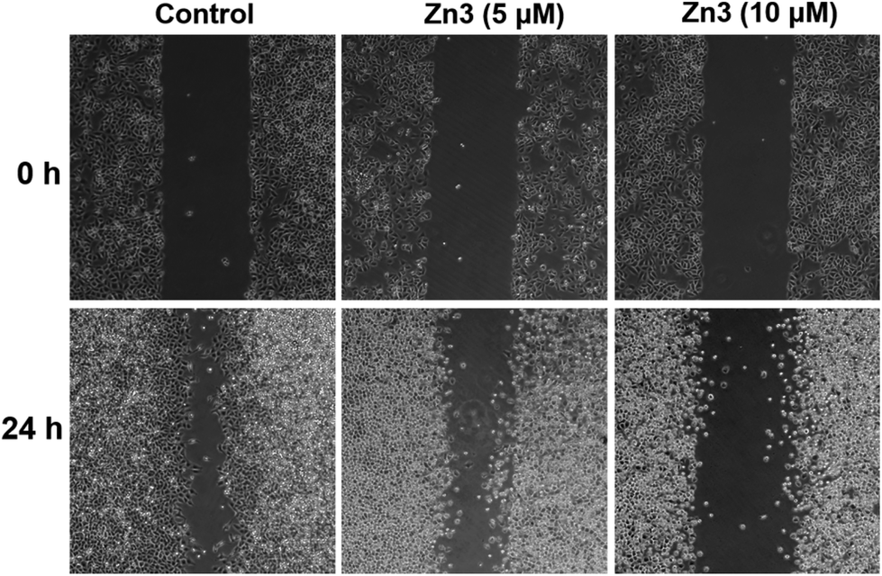 | ||
| Fig. 8 Antimigratory effect of on A549 cells by wound healing assay. | ||
In addition, LTBR protein in cytokine–cytokine receptor interaction, CDKN1A protein in PI3K-Akt signaling pathway and LTBR, CDKN1A and HIF1A proteins in HIF-1 signaling pathway were selected to further verify their expression by western blotting. Western blotting revealed that the treatment of A549 cells causes a dose-dependent increase in the expression of CDKN1A and HIF1A as well as a dose-dependent downregulation in the expression of LTBR (Fig. 7B). The western blotting data are consistent with the proteomics results: complex Zn3 activates the cytokine–cytokine receptor interaction, PI3K-Akt signaling pathway, and HIF-1 signaling pathway.
Collectively, these results suggest that complex Zn3 exerts chemotherapeutic effects by regulating multiple pathways, such as pathways in cancer, p53 signaling pathway, and ECM–receptor interactions.
Conclusions
A series of fluorescent N-heterocyclic dithiocarbazate–zinc(II) complexes were prepared, and their anti-lung cancer properties were assessed. These Zn(II) complexes exhibit excellent blood compatibility and can be used for live-cell imaging. Among them, complex Zn3 exhibited good chemotherapeutic and photodynamic therapy capabilities in both monolayer cancer cell models and 3D A549 multicellular tumor spheroids. Very importantly, under visible light irradiation, treatment with complex Zn3 generates type I ROS in living cells, superoxide anion radicals (˙O2−), which can overcome the O2-dependence of conventional photosensitizers, thus suggesting promising applications in photodynamic therapy against hypoxic tumors. Moreover, the studies of chemotherapeutic mechanisms demonstrated that complex Zn3 kills lung cancer cells through multiple pathways. Overall, these findings indicate that complex Zn3 can simultaneously express photodynamic and chemotherapeutic effects, which provides a simple method for the multimodal treatment of cancer and extends the horizon of anticancer research on zinc complexes.Data availability
The data supporting the study is available within the ESI.†Author contributions
JunGang Deng, YouRu Wu and AiLi Li: investigation, data curation, writing – original draft. WeiPing Pan: investigation, data curation. LiXia Hou: data curation. DaQi Wu: data curation. ZhenLei Zhang: software, methodology. Feng Yang: supervision, writing – review & editing. Yi Gou: supervision, resources, conceptualization, writing – review & editing.Conflicts of interest
There are no conflicts to declare.Acknowledgements
We thank these projects 22007023, 22267004, 2020GXNSFBA238003 and 2022GXNSFGA035003. We also sincerely thank Peng-Fei Yao (Baise University) and Jin-Long Li (Nantong University) for their help in the work.References
- J. Li, L. Zhu and H. F. Kwok, Nanotechnology-based approaches overcome lung cancer drug resistance through diagnosis and treatment, Drug Resistance Updates, 2023, 66, 100904 CrossRef CAS PubMed.
- I. Toumazis, M. Bastani, S. S. Han and S. K. Plevritis, Risk-Based lung cancer screening: A systematic review, Lung Cancer, 2020, 147, 154–186 CrossRef PubMed.
- H. Sung, J. Ferlay, R. L. Siegel, M. Laversanne, I. Soerjomataram, A. Jemal and F. Bray, Global Cancer Statistics 2020: GLOBOCAN Estimates of Incidence and Mortality Worldwide for 36 Cancers in 185 Countries, CA Cancer J. Clin., 2021, 71, 209–249 CrossRef PubMed.
- R. Nooreldeen and H. Bach, Current and Future Development in Lung Cancer Diagnosis, Int. J. Mol. Sci., 2021, 22, 8661 CrossRef CAS PubMed.
- C. Allemani, T. Matsuda, V. Di Carlo, R. Harewood, M. Matz, M. Nikšić, A. Bonaventure, M. Valkov, C. J. Johnson, J. Estève, O. J. Ogunbiyi, E. S. G. Azevedo, W. Q. Chen, S. Eser, G. Engholm, C. A. Stiller, A. Monnereau, R. R. Woods, O. Visser, G. H. Lim, J. Aitken, H. K. Weir and M. P. Coleman, Global surveillance of trends in cancer survival 2000–14 (CONCORD-3): analysis of individual records for 37513025 patients diagnosed with one of 18 cancers from 322 population-based registries in 71 countries, Lancet, 2018, 391, 1023–1075 CrossRef PubMed.
- F. Wu, J. Fan, Y. He, A. Xiong, J. Yu, Y. Li, Y. Zhang, W. Zhao, F. Zhou, W. Li, J. Zhang, X. Zhang, M. Qiao, G. Gao, S. Chen, X. Chen, X. Li, L. Hou, C. Wu, C. Su, S. Ren, M. Odenthal, R. Buettner, N. Fang and C. Zhou, Single-cell profiling of tumor heterogeneity and the microenvironment in advanced non-small cell lung cancer, Nat. Commun., 2021, 12, 2540 CrossRef CAS PubMed.
- A. Crous and H. Abrahamse, Photodynamic therapy of lung cancer, where are we?, Front. Pharmacol., 2022, 13, 932098 CrossRef CAS PubMed.
- W. H. Chen, C. X. Yang, W. X. Qiu, G. F. Luo, H. Z. Jia, Q. Lei, X. Y. Wang, G. Liu, R. X. Zhuo and X. Z. Zhang, Multifunctional theranostic nanoplatform for cancer combined therapy based on gold nanorods, Adv. Healthcare Mater., 2015, 4, 2247–2259 CrossRef CAS PubMed.
- Z. Yang, Y. Dai, C. Yin, Q. Fan, W. Zhang, J. Song, G. Yu, W. Tang, W. Fan, B. C. Yung, J. Li, X. Li, X. Li, Y. Tang, W. Huang, J. Song and X. Chen, Activatable Semiconducting Theranostics: Simultaneous Generation and Ratiometric Photoacoustic Imaging of Reactive Oxygen Species In Vivo, Adv. Mater., 2018, 30, 1707509 CrossRef PubMed.
- A. El-Hussein, S. L. Manoto, S. Ombinda-Lemboumba, Z. A. Alrowaili and P. Mthunzi-Kufa, A Review of Chemotherapy and Photodynamic Therapy for Lung Cancer Treatment, Anticancer Agents Med. Chem., 2021, 21, 149–161 CrossRef CAS PubMed.
- M. Kimura, K. Miyajima, M. Kojika, T. Kono and H. Kato, Photodynamic Therapy (PDT) with Chemotherapy for Advanced Lung Cancer with Airway Stenosis, Int. J. Mol. Sci., 2015, 16, 25466–25475 CrossRef CAS PubMed.
- X. Shi, X. Yang, M. Liu, R. Wang, N. Qiu, Y. Liu, H. Yang, J. Ji and G. Zhai, Chondroitin sulfate-based nanoparticles for enhanced chemo-photodynamic therapy overcoming multidrug resistance and lung metastasis of breast cancer, Carbohydr. Polym., 2021, 254, 117459 CrossRef CAS PubMed.
- C. Yue, Y. Yang, C. Zhang, G. Alfranca, S. Cheng, L. Ma, Y. Liu, X. Zhi, J. Ni, W. Jiang, J. Song, J. M. de la Fuente and D. Cui, ROS-Responsive Mitochondria-Targeting Blended Nanoparticles: Chemo- and Photodynamic Synergistic Therapy for Lung Cancer with On-Demand Drug Release upon Irradiation with a Single Light Source, Theranostics, 2016, 6, 2352–2366 CrossRef CAS PubMed.
- P. Thapa, M. Li, M. Bio, P. Rajaputra, G. Nkepang, Y. Sun, S. Woo and Y. You, Far-Red Light-Activatable Prodrug of Paclitaxel for the Combined Effects of Photodynamic Therapy and Site-Specific Paclitaxel Chemotherapy, J. Med. Chem., 2016, 59, 3204–3214 CrossRef CAS PubMed.
- F. Yang, M. Xu, X. Chen and Y. Luo, Spotlight on porphyrins: Classifications, mechanisms and medical applications, Biomed. Pharmacother., 2023, 164, 114933 CrossRef CAS PubMed.
- M. Kolarikova, B. Hosikova, H. Dilenko, K. Barton-Tomankova, L. Valkova, R. Bajgar, L. Malina and H. Kolarova, Photodynamic therapy: Innovative approaches for antibacterial and anticancer treatments, Med. Res. Rev., 2023, 43, 717–774 CrossRef CAS PubMed.
- M. Yang, H. Zhao, Z. Zhang, Q. Yuan, Q. Feng, X. Duan, S. Wang and Y. Tang, CO/light dual-activatable Ru(ii)-conjugated oligomer agent for lysosome-targeted multimodal cancer therapeutics, Chem. Sci., 2021, 12, 11515–11524 RSC.
- P. Agostinis, K. Berg, K. A. Cengel, T. H. Foster, A. W. Girotti, S. O. Gollnick, S. M. Hahn, M. R. Hamblin, A. Juzeniene, D. Kessel, M. Korbelik, J. Moan, P. Mroz, D. Nowis, J. Piette, B. C. Wilson and J. Golab, Photodynamic therapy of cancer: an update, CA Cancer J. Clin., 2011, 61, 250–281 CrossRef PubMed.
- J. Sun, K. Du, J. Diao, X. Cai, F. Feng and S. Wang, GSH and H(2) O(2) Co-Activatable Mitochondria-Targeted Photodynamic Therapy under Normoxia and Hypoxia, Angew. Chem., Int. Ed., 2020, 59, 12122–12128 CrossRef CAS PubMed.
- X. Li, X. H. Peng, B. D. Zheng, J. Tang, Y. Zhao, B. Y. Zheng, M. R. Ke and J. D. Huang, New application of phthalocyanine molecules: from photodynamic therapy to photothermal therapy by means of structural regulation rather than formation of aggregates, Chem. Sci., 2018, 9, 2098–2104 RSC.
- L. Menilli, C. Milani, E. Reddi and F. Moret, Overview of Nanoparticle-Based Approaches for the Combination of Photodynamic Therapy (PDT) and Chemotherapy at the Preclinical Stage, Cancers, 2022, 14, 4462 CrossRef CAS PubMed.
- E. Chang, J. Bu, L. Ding, J. W. H. Lou, M. S. Valic, M. H. Y. Cheng, V. Rosilio, J. Chen and G. Zheng, Porphyrin-lipid stabilized paclitaxel nanoemulsion for combined photodynamic therapy and chemotherapy, J. Nanobiotechnol., 2021, 19, 154 CrossRef CAS PubMed.
- G. Yang, J. Tian, C. Chen, D. Jiang, Y. Xue, C. Wang, Y. Gao and W. Zhang, An oxygen self-sufficient NIR-responsive nanosystem for enhanced PDT and chemotherapy against hypoxic tumors, Chem. Sci., 2019, 10, 5766–5772 RSC.
- C. Y. Sun, Z. Cao, X. J. Zhang, R. Sun, C. S. Yu and X. Yang, Cascade-amplifying synergistic effects of chemo-photodynamic therapy using ROS-responsive polymeric nanocarriers, Theranostics, 2018, 8, 2939–2953 CrossRef CAS PubMed.
- C. Shi, M. Li, Z. Zhang, Q. Yao, K. Shao, F. Xu, N. Xu, H. Li, J. Fan, W. Sun, J. Du, S. Long, J. Wang and X. Peng, Catalase-based liposomal for reversing immunosuppressive tumor microenvironment and enhanced cancer chemo-photodynamic therapy, Biomaterials, 2020, 233, 119755 CrossRef CAS PubMed.
- H. S. Jung, J. Han, H. Shi, S. Koo, H. Singh, H. J. Kim, J. L. Sessler, J. Y. Lee, J. H. Kim and J. S. Kim, Overcoming the Limits of Hypoxia in Photodynamic Therapy: A Carbonic Anhydrase IX-Targeted Approach, J. Am. Chem. Soc., 2017, 139, 7595–7602 CrossRef CAS PubMed.
- S. Zhen, X. Yi, Z. Zhao, X. Lou, F. Xia and B. Z. Tang, Drug delivery micelles with efficient near-infrared photosensitizer for combined image-guided photodynamic therapy and chemotherapy of drug-resistant cancer, Biomaterials, 2019, 218, 119330 CrossRef CAS PubMed.
- S. Jiang, M. Xiao, W. Sun, D. Crespy, V. Mailänder, X. Peng, J. Fan and K. Landfester, Synergistic Anticancer Therapy by Ovalbumin Encapsulation-Enabled Tandem Reactive Oxygen Species Generation, Angew. Chem., Int. Ed., 2020, 59, 20008–20016 CrossRef CAS PubMed.
- M. Aghajanzadeh, M. Zamani, F. Rajabi Kouchi, J. Eixenberger, D. Shirini, D. Estrada and F. Shirini, Synergic Antitumor Effect of Photodynamic Therapy and Chemotherapy Mediated by Nano Drug Delivery Systems, Pharmaceutics, 2022, 14, 322 CrossRef CAS PubMed.
- M. Majerník, R. Jendželovský, J. Vargová, Z. Jendželovská and P. Fedoročko, Multifunctional Nanoplatforms as a Novel Effective Approach in Photodynamic Therapy and Chemotherapy, to Overcome Multidrug Resistance in Cancer, Pharmaceutics, 2022, 14, 1075 CrossRef PubMed.
- N. Kwon, H. Kim, X. Li and J. Yoon, Supramolecular agents for combination of photodynamic therapy and other treatments, Chem. Sci., 2021, 12, 7248–7268 RSC.
- M. Bio, P. Rajaputra, G. Nkepang and Y. You, Far-red light activatable, multifunctional prodrug for fluorescence optical imaging and combinational treatment, J. Med. Chem., 2014, 57, 3401–3409 CrossRef CAS PubMed.
- R. Liang, S. You, L. Ma, C. Li, R. Tian, M. Wei, D. Yan, M. Yin, W. Yang, D. G. Evans and X. Duan, A supramolecular nanovehicle toward systematic, targeted cancer and tumor therapy, Chem. Sci., 2015, 6, 5511–5518 RSC.
- X. Li, Z. Guo, T. Zhao, Y. Lu, L. Zhou, D. Zhao and F. Zhang, Filtration Shell Mediated Power Density Independent Orthogonal Excitations-Emissions Upconversion Luminescence, Angew. Chem., Int. Ed., 2016, 55, 2464–2469 CrossRef CAS PubMed.
- G. Yang, J. Tian, C. Chen, D. Jiang, Y. Xue, C. Wang, Y. Gao and W. Zhang, An oxygen self-sufficient NIR-responsive nanosystem for enhanced PDT and chemotherapy against hypoxic tumors, Chem. Sci., 2019, 10, 5766–5772 RSC.
- G. Yu, B. Zhu, L. Shao, J. Zhou, M. L. Saha, B. Shi, Z. Zhang, T. Hong, S. Li, X. Chen and P. J. Stang, Host-guest complexation-mediated codelivery of anticancer drug and photosensitizer for cancer photochemotherapy, Proc. Natl. Acad. Sci. U. S. A., 2019, 116, 6618–6623 CrossRef CAS PubMed.
- Y. Shao, B. Liu, Z. Di, G. Zhang, L. D. Sun, L. Li and C. H. Yan, Engineering of Upconverted Metal-Organic Frameworks for Near-Infrared Light-Triggered Combinational Photodynamic/Chemo-/Immunotherapy against Hypoxic Tumors, J. Am. Chem. Soc., 2020, 142, 3939–3946 CrossRef CAS PubMed.
- M. Yang, H. Zhao, Z. Zhang, Q. Yuan, Q. Feng, X. Duan, S. Wang and Y. Tang, CO/light dual-activatable Ru(ii)-conjugated oligomer agent for lysosome-targeted multimodal cancer therapeutics, Chem. Sci., 2021, 12, 11515–11524 RSC.
- J. Karges, T. Yempala, M. Tharaud, D. Gibson and G. Gasser, A Multi-action and Multi-target Ru(II) -Pt(IV) Conjugate Combining Cancer-Activated Chemotherapy and Photodynamic Therapy to Overcome Drug Resistant Cancers, Angew. Chem., Int. Ed., 2020, 59, 7069–7075 CrossRef CAS PubMed.
- J. Yuan, Q. H. Zhou, S. Xu, Q. P. Zuo, W. Li, X. X. Zhang, T. B. Ren, L. Yuan and X. B. Zhang, Enhancing the Release Efficiency of a Molecular Chemotherapeutic Prodrug by Photodynamic Therapy, Angew. Chem., Int. Ed., 2022, 61, e202206169 CrossRef CAS PubMed.
- X. Tian, S. Hussain, C. de Pace, L. Ruiz-Pérez and G. Battaglia, Zn(II) Complexes for Bioimaging and Correlated Applications, Chem. – Asian J., 2019, 14, 509–526 CrossRef CAS PubMed.
- M. Porchia, M. Pellei, F. Del Bello and C. Santini, Zinc Complexes with Nitrogen Donor Ligands as Anticancer Agents, Molecules, 2020, 25, 5814 CrossRef CAS PubMed.
- A. E. Stacy, D. Palanimuthu, P. V. Bernhardt, D. S. Kalinowski, P. J. Jansson and D. R. Richardson, Zinc(II)-Thiosemicarbazone Complexes Are Localized to the Lysosomal Compartment Where They Transmetallate with Copper Ions to Induce Cytotoxicity, J. Med. Chem., 2016, 59, 4965–4984 CrossRef CAS PubMed.
- M. Pellei, F. Del Bello, M. Porchia and C. Santini, Zinc coordination complexes as anticancer agents, Coord. Chem. Rev., 2021, 445, 214088 CrossRef CAS.
- F. Dumur, Zinc complexes in OLEDs: An overview, Synth. Met., 2014, 195, 241–251 CrossRef CAS.
- J. Karges, U. Basu, O. Blacque, H. Chao and G. Gasser, Polymeric Encapsulation of Novel Homoleptic Bis(dipyrrinato) Zinc(II) Complexes with Long Lifetimes for Applications as Photodynamic Therapy Photosensitisers, Angew. Chem., Int. Ed., 2019, 58, 14334–14340 CrossRef CAS PubMed.
- Y. Gou, X. Jia, L. X. Hou, J. G. Deng, G. J. Huang, H. W. Jiang and F. Yang, Dithiocarbazate-Fe(III), -Co(III), -Ni(II), and -Zn(II) Complexes: Design, Synthesis, Structure, and Anticancer Evaluation, J. Med. Chem., 2022, 65, 6677–6689 CrossRef CAS PubMed.
- P. Mroz, A. Yaroslavsky, G. B. Kharkwal and M. R. Hamblin, Cell death pathways in photodynamic therapy of cancer, Cancers, 2011, 3, 2516–2539 CrossRef CAS PubMed.
- X. Li, L. Chen, M. Huang, S. Zeng, J. Zheng, S. Peng, Y. Wang, H. Cheng and S. Li, Innovative strategies for photodynamic therapy against hypoxic tumor, Asian J. Pharm. Sci., 2023, 18, 100775 CrossRef PubMed.
- X. Li, N. Kwon, T. Guo, Z. Liu and J. Yoon, Innovative Strategies for Hypoxic-Tumor Photodynamic Therapy, Angew. Chem., Int. Ed., 2018, 57, 11522–11531 CrossRef CAS PubMed.
- V. Novohradsky, G. Vigueras, J. Pracharova, N. Cutillas, C. Janiak, H. Kostrhunova, V. Brabec, J. Ruiz and J. Kasparkova, Molecular superoxide radical photogeneration in cancer cells by dipyridophenazine iridium(III) complexes, Inorg. Chem. Front., 2019, 6, 2500–2513 RSC.
- R. Bevernaegie, B. Doix, E. Bastien, A. Diman, A. Decottignies, O. Feron and B. Elias, Exploring the Phototoxicity of Hypoxic Active Iridium(III)-Based Sensitizers in 3D Tumor Spheroids, J. Am. Chem. Soc., 2019, 141, 18486–18491 CrossRef CAS PubMed.
- S. Cheng-Yong, Y. Xiao-Ping, K. Bei-Sheng, Y. Kai-Bei, T. Ye-Xiang and M. T. Cong-Wai, Syntheses and Crystal Structures of Methyl 2-Pyridylmethylidenehydrazinecarbodithioate and of Its Copper(II) Complex: Supramolecular Arrays Exhibiting pseudo-Helical Arrangement Controlled by Hydrogen Bonding or S⋯S Contact, Bull. Chem. Soc. Jpn., 1999, 72, 2217–2222 CrossRef.
- M. A. Ali, A. H. Mirza, M. H. S. A. Hamid and P. V. Bernhardt, Diphenyltin(IV) complexes of the 2-quinolinecarboxaldehyde Schiff bases of S-methyl- and S-benzyldithiocarbazate (Hqaldsme and Hqaldsbz): X-ray crystal structures of Hqaldsme and two conformers of its diphenyltin(IV) complex, Polyhedron, 2005, 24, 383–390 CrossRef.
- M. T. Basha, J. D. Chartres, N. Pantarat, M. A. Ali, A. H. Mirza, D. S. Kalinowski, D. R. Richardson and P. V. Bernhardt, Heterocyclic dithiocarbazate iron chelators: Fe coordination chemistry and biological activity, Dalton Trans., 2012, 41, 6536–6548 RSC.
- A. H. Mirza, M. H. S. A. Hamid, S. Aripin, M. R. Karim, M. Arifuzzaman, M. A. Ali and P. V. Bernhardt, Synthesis, spectroscopy and X-ray crystal structures of some zinc(II) and cadmium(II) complexes of the 2-pyridinecarboxaldehyde Schiff bases of S-methyl- and S-benzyldithiocarbazates, Polyhedron, 2014, 74, 16–23 CrossRef CAS.
- N. C. Kasuga, K. Sekino, M. Ishikawa, A. Honda, M. Yokoyama, S. Nakano, N. Shimada, C. Koumo and K. Nomiya, Synthesis, structural characterization and antimicrobial activities of 12 zinc(II) complexes with four thiosemicarbazone and two semicarbazone ligands, J. Inorg. Biochem., 2003, 96, 298–310 CrossRef CAS PubMed.
- D. Kovala-Demertzi, P. N. Yadav, J. Wiecek, S. Skoulika, T. Varadinova and M. A. Demertzis, Zinc(II) complexes derived from pyridine-2-carbaldehyde thiosemicarbazone and (1E)-1-pyridin-2-ylethan-1-one thiosemicarbazone. Synthesis, crystal structures and antiproliferative activity of zinc(II) complexes, J. Inorg. Biochem., 2006, 100, 1558–1567 CrossRef CAS PubMed.
- L. Cheng, L.-M. Zhang and J.-Q. Wang, Bis(pyridine-2-carbaldehyde thio-semi-carbazone)zinc(II) dinitrate dihydrate, Acta Crystallogr., Sect. E: Struct. Rep. Online, 2010, 66, m1340 CrossRef CAS PubMed.
- M. X. Li, C. L. Chen, D. Zhang, J. Y. Niu and B. S. Ji, Mn(II), Co(II) and Zn(II) complexes with heterocyclic substituted thiosemicarbazones: synthesis, characterization, X-ray crystal structures and antitumor comparison, Eur. J. Med. Chem., 2010, 45, 3169–3177 CrossRef CAS PubMed.
- Y. Gou, X. Jia, L. X. Hou, J. G. Deng, G. J. Huang, H. W. Jiang and F. Yang, Dithiocarbazate-FeIII, -CoIII, -NiII, and -ZnII Complexes: Design, Synthesis, Structure, and Anticancer Evaluation, J. Med. Chem., 2022, 65, 6677–6689 CrossRef CAS PubMed.
- M. A. Ali, A. H. Mirza, M. Nazimuddin, R. Ahmed, L. R. Gahan and P. V. Bernhardt, Synthesis and characterization of mono- and bis-ligand zinc(II) and cadmium(II) complexes of the di-2-pyridylketone Schiff base of S-benzyl dithiocarbazate (Hdpksbz) and the X-ray crystal structures of the [Zn(dpksbz)2] and [Cd(dpksbz)NCS]2 complexes, Polyhedron, 2003, 22, 1471–1479 CrossRef.
- C. d. Q. O. Cavalcante, D. d. S. Arcanjo, G. G. d. Silva, D. M. d. Oliveira and C. C. Gatto, Solution and solid behavior of mono and binuclear zinc(II) and nickel(II) complexes with dithiocarbazates: X-ray analysis, mass spectrometry and cytotoxicity against cancer cell lines, New J. Chem., 2019, 43, 11209–11221 RSC.
- M. X. Li, L. Z. Zhang, C. L. Chen, J. Y. Niu and B. S. Ji, Synthesis, crystal structures, and biological evaluation of Cu(II) and Zn(II) complexes of 2-benzoylpyridine Schiff bases derived from S-methyl- and S-phenyldithiocarbazates, J. Inorg. Biochem., 2012, 106, 117–125 CrossRef CAS PubMed.
- A. B. Beshir, S. K. Guchhait, J. A. Gascón and G. Fenteany, Synthesis and structure–activity relationships of metal–ligand complexes that potently inhibit cell migration, Bioorg. Med. Chem. Lett., 2008, 18, 498–504 CrossRef CAS PubMed.
- P. Zhang, H. Huang, S. Banerjee, G. J. Clarkson, C. Ge, C. Imberti and P. J. Sadler, Nucleus-Targeted Organoiridium-Albumin Conjugate for Photodynamic Cancer Therapy, Angew. Chem., Int. Ed., 2019, 58, 2350–2354 CrossRef CAS PubMed.
- M. K. M. Subarkhan and R. Ramesh, Ruthenium(II) arene complexes containing benzhydrazone ligands: synthesis, structure and antiproliferative activity, Inorg. Chem. Front., 2016, 3, 1245–1255 RSC.
- V. Novohradsky, A. Rovira, C. Hally, A. Galindo, G. Vigueras, A. Gandioso, M. Svitelova, R. Bresolí-Obach, H. Kostrhunova, L. Markova, J. Kasparkova, S. Nonell, J. Ruiz, V. Brabec and V. Marchán, Towards Novel Photodynamic Anticancer Agents Generating Superoxide Anion Radicals: A Cyclometalated Ir(III) Complex Conjugated to a Far-Red Emitting Coumarin, Angew. Chem., Int. Ed., 2019, 58, 6311–6315 CrossRef CAS PubMed.
- R. Lindberg-Larsen, H. Sølvsten and K. Kragballe, Evaluation of recurrence after photodynamic therapy with topical methylaminolaevulinate for 157 basal cell carcinomas in 90 patients, Acta Derm.-Venereol., 2012, 92, 144–147 CrossRef PubMed.
- H. Lu and M. H. Stenzel, Multicellular Tumor Spheroids (MCTS) as a 3D In Vitro Evaluation Tool of Nanoparticles, Small, 2018, 14, e1702858 CrossRef PubMed.
- K. H. Lee and T. H. Kim, Recent Advances in Multicellular Tumor Spheroid Generation for Drug Screening, Biosensors, 2021, 11, 445 CrossRef CAS PubMed.
- J. Friedrich, C. Seidel, R. Ebner and L. A. Kunz-Schughart, Spheroid-based drug screen: considerations and practical approach, Nat. Protoc., 2009, 4, 309–324 CrossRef CAS PubMed.
- E. C. Costa, A. F. Moreira, D. de Melo-Diogo, V. M. Gaspar, M. P. Carvalho and I. J. Correia, 3D tumor spheroids: an overview on the tools and techniques used for their analysis, Biotechnol. Adv., 2016, 34, 1427–1441 CrossRef PubMed.
- S. Kwiatkowski, B. Knap, D. Przystupski, J. Saczko, E. Kędzierska, K. Knap-Czop, J. Kotlińska, O. Michel, K. Kotowski and J. Kulbacka, Photodynamic therapy - mechanisms, photosensitizers and combinations, Biomed. Pharmacother., 2018, 106, 1098–1107 CrossRef PubMed.
- A. Pinto, Y. Mace, F. Drouet, E. Bony, R. Boidot, N. Draoui, I. Lobysheva, C. Corbet, F. Polet, R. Martherus, Q. Deraedt, J. Rodríguez, C. Lamy, O. Schicke, D. Delvaux, C. Louis, R. Kiss, A. V. Kriegsheim, C. Dessy, B. Elias, J. Quetin-Leclercq, O. Riant and O. Feron, A new ER-specific photosensitizer unravels (1)O2-driven protein oxidation and inhibition of deubiquitinases as a generic mechanism for cancer PDT, Oncogene, 2016, 35, 3976–3985 CrossRef CAS PubMed.
- M. Chen, P. Zhu, Q. Wan, X. Ruan, P. Wu, Y. Hao, Z. Zhang, J. Sun, W. Nie and S. Chen, High-Coverage Four-Dimensional Data-Independent Acquisition Proteomics and Phosphoproteomics Enabled by Deep Learning-Driven Multidimensional Predictions, Anal. Chem., 2023, 95, 7495–7502 CrossRef CAS PubMed.
- S. Jia, R. Wang, K. Wu, H. Jiang and Z. Du, Elucidation of the Mechanism of Action for Metal Based Anticancer Drugs by Mass Spectrometry-Based Quantitative Proteomics, Molecules, 2019, 24, 581 CrossRef PubMed.
- J. Huang, Current developments of targeting the p53 signaling pathway for cancer treatment, Pharmacol. Ther., 2021, 220, 107720 CrossRef CAS PubMed.
- K. Suntharalingam, T. C. Johnstone, P. M. Bruno, W. Lin, M. T. Hemann and S. J. Lippard, Bidentate ligands on osmium(VI) nitrido complexes control intracellular targeting and cell death pathways, J. Am. Chem. Soc., 2013, 135, 14060–14063 CrossRef CAS PubMed.
- A. van der Flier and A. Sonnenberg, Function and interactions of integrins, Cell Tissue Res., 2001, 305, 285–298 CrossRef CAS PubMed.
- I. Canobbio, C. Balduini and M. Torti, Signalling through the platelet glycoprotein Ib-V–IX complex, Cell. Signalling, 2004, 16, 1329–1344 CrossRef CAS PubMed.
- H. Zhu, G. Wang, H. Zhu and A. Xu, ITGA5 is a prognostic biomarker and correlated with immune infiltration in gastrointestinal tumors, BMC Cancer, 2021, 21, 269 CrossRef CAS PubMed.
- F. Pantano, M. Croset, K. Driouch, N. Bednarz-Knoll, M. Iuliani, G. Ribelli, E. Bonnelye, H. Wikman, S. Geraci, F. Bonin, S. Simonetti, B. Vincenzi, S. S. Hong, S. Sousa, K. Pantel, G. Tonini, D. Santini and P. Clézardin, Integrin alpha5 in human breast cancer is a mediator of bone metastasis and a therapeutic target for the treatment of osteolytic lesions, Oncogene, 2021, 40, 1284–1299 CrossRef CAS PubMed.
- Y. Bao, L. Wang, L. Shi, F. Yun, X. Liu, Y. Chen, C. Chen, Y. Ren and Y. Jia, Transcriptome profiling revealed multiple genes and ECM-receptor interaction pathways that may be associated with breast cancer, Cell. Mol. Biol. Lett., 2019, 24, 38 CrossRef PubMed.
- S. Mondal, N. Adhikari, S. Banerjee, S. A. Amin and T. Jha, Matrix metalloproteinase-9 (MMP-9) and its inhibitors in cancer: A minireview, Eur. J. Med. Chem., 2020, 194, 112260 CrossRef CAS PubMed.
Footnotes |
| † Electronic supplementary information (ESI) available. CCDC 2265560–2265562. For ESI and crystallographic data in CIF or other electronic format see DOI: https://doi.org/10.1039/d3qi01777j |
| ‡ These authors contributed equally to this work. |
| This journal is © the Partner Organisations 2023 |