Gadolinium(III) based nanoparticles for T1-weighted magnetic resonance imaging probes
Chang-Tong Yang
*,
Parasuraman Padmanabhan
and
Balázs Z. Gulyás
Lee Kong Chian School of Medicine, Nanyang Technological University, 59 Nanyang Drive, Singapore 636921. E-mail: yangct@ntu.edu.sg
First published on 20th June 2016
Abstract
The development of magnetic resonance imaging (MRI) contrast agents with delicate sensitivity and advanced functionalities has recently received extensive interest. Nanoparticle MRI contrast agents have been synthesized for various potential applications because of their unique properties, such as large surface area, surface modifications for multifunction, contrast enhancement, and conjugation with biomolecules for therapeutic and diagnostic applications. This review summarized the recent progress on Gd-based nanoparticles as T1-weighted MRI contrast agents including inorganic crystalline Gd(III) nanoparticles and Gd(III) chelate-grafted macromolecular nanoparticles. The recent development of Gd(III)-based nanoparticle as multimodal contrast agents including T1-weighted MRI/computed X-ray (CT) and T1-weighted MRI/optical were also included.
Introduction
Magnetic resonance imaging (MRI) is a complementary molecular imaging modality that provides images with high spatial resolution and anatomical details without exposure to radioisotopes such as positron emission tomography (PET) or single-photon emission computed tomography (SPECT).1–3 Contrast agents have been introduced to differentiate the hydrogen nuclei situated at diverse environment under an applied magnetic field, highlight the part of interest by means of contrast difference resulted from the alternation of the signal intensity.4–6 Gd(III) possesses seven half-filled f orbitals unpaired electrons, leading to a favorable large magnetic moment (spin-only μeff = 7.94 BM) and the spin of which perturbs the proton relaxation in water results in an efficient shortening of longitudinal relaxation times and increase the MR signal intensity. Therefore, Gd(III)-based agents are the constituent of most MRI contrast agents which provide with positive signals and perform as high efficiency T1-weighted agents as they have ideal physicochemical, pharmacological properties such as thermodynamic and kinetic stability, good water solubility, high relaxivity, in vivo stability, low toxicity, etc.7–10Nanomaterials form a convenient platform that can carry highly specific targeting biomaterials, antibodies, drugs and combine different clinically relevant properties in a single unit for bioimaging probes.11 Nanomaterial-based MRI contrast agents have been synthesized and developed to increase MRI sensitivity, and to adjust the biodistribution of the contrast agents for tissue-targeting purpose.12–29 Novel nanomaterials MRI contrast agents with a therapeutic component as theranostic nanomedicines have been reviewed for monitoring drug delivery, drug release and drug efficacy.30
Gadolinium is a highly toxic rare earth metal in the unbounded form.31 Exposure to the unchelated Gd(III) is associated with nephrogenic systemic fibrosis (NSF) which is known to inhibit the calcium channels and has considerable cardiovascular and neurologic toxicity.32–37 When the Gd(III) ions are chelated, the toxicity can be enormously reduced and the LD50 increases 100-fold after chelation.38 In 2009, the World Health Organization (WHO) issued a restriction on use of several current commercial available Gd(III) chelates contrast agents in patients with severe kidney problems, or with who have recently received a liver transplant.
The Gd(III) based nanomaterial as effective contrast agents in preclinical and clinical applications has been extensively studied. There is a need to evaluate the potential toxicity of these Gd(III) based nanomaterial, and to investigate their bioaccumulation and clearance properties.39–46 The toxic effects were evaluated through the biological interaction between Gd nanoparticles with various mouse and human cells. The studies on Gd2O3 nanoparticles injected in mice showed the particles were naturally eliminated by renal excretion after a few hours with low cytotoxicity.39 The potential toxicity of Gd based nanoparticle depends on its constituent materials, on the chemical properties of its surface coating and on the size of the particles.40 Through the surface modifications, size selection and interior chemistry the Gd2O3 nanoparticles would enhance their biocompatibility with satisfactory cytotoxicity, minimal immunotoxicity and optimal pharmacokinetics and excretion characteristics.41–46
This review focuses on development of Gd(III)-based nanoparticles contrast agents including Gd(III) containing inorganic crystalline nanoparticles and Gd(III) chelate-grafted macromolecular nanoparticles for T1-weighted MRI, multimodality imaging contrast agents. Synthetic methods, surface modification, physicochemical properties of Gd(III) containing nanoparticles for T1-weighted MRI contrast agent are summarized. This review also highlighted the functionalization of the Gd-based nanoparticle as multimodal contrast agents including T1-weighted MRI/computed X-ray (CT) and T1-weighted MRI/optical for therapeutic and diagnostic applications.
Inorganic crystalline Gd(III) nanoparticles
Inorganic crystalline Gd based compound nanoparticles are considered as a new generation T1 contrast agent because a rigid crystal environment could effectively prevent the free Gd(III) ions release from nanoparticles.47–52 These Gd based nanoparticles can be used as efficient T1-weighted contrast agents because of their large paramagnetic properties with a small r2/r1 values. The preparation of monodispersed ultra-small nanoparticles with size uniformity and large surface area is of highly importance as only the Gd close to the surface contributes to the contrast effect. Added T2 contrast materials to the T1 contrast nanoparticles would lead to T1/T2 dual contrast agent. For example, Gd(III)-labeled magnetite nanoparticles obtained by modifying Gd-DTPA chelates on the surface of T2 magnetic iron oxide nanoparticles were reported by Bae et al.53 and Yang et al.54 However, proper design of nanostructure is needed in order to separating the T1 contrast materials and T2 contrast layers to prevent the magnetic coupling between them.55Nanomaterials gadolinium oxide (Gd2O3),20–29 gadolinium fluoride (GdF3),56 inorganic fluoride nanoparticles KGdF4 and NaGdF4 (ref. 57) have been extensively explored as promising T1 contrast agents with improved T1-weighted MR contrast enhancement. Lanthanide doped inorganic fluoride KGdF4 and lanthanide doped GdPO4 nanocrystals58–70 have been investigated as potential optical/magnetic multimodal imaging probes. Several methods have been developed to synthesize small sized Gd2O3 nanoparticles for the application in MRI contrast agents.56–65 Gd2O3 nanoparticles were synthesized from three different Gd(III) precursors by refluxing each of them in tripropylene glycol under O2 flow.56 The r1 value 9.9 mM−1 s−1 can be achieved when the Gd2O3 particle diameter is 1.0 nm. The high relaxivity of the particles is possibly because several surface Gd(III) ions interact with the water proton cooperatively and thus induce the longitudinal relaxation of water proton as shown in Fig. 1. Rahman's experiments on 7T showed that there is an optimum Gd2O3 nanoparticle size of 2.3 nm with highest spin-lattice relaxation rate of the water protons.21
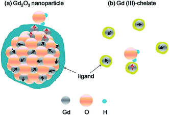 | ||
| Fig. 1 Schematic diagram showing that (a) four surface Gd(III) ions as an example cooperatively induce the longitudinal relaxation of the water proton, whereas (b) such an effect does not exist in individual Gd(III) chelates. The interacting and noninteracting Gd(III) ions with a water proton are denoted as dotted and solid arrows for their spins, respectively. The ligands are drawn arbitrarily (ref. 56). | ||
Preparation from Gd precursors in the presence of stabilizers such as polyethylene glycol or its derivatives results in stable Gd2O3 colloidal solutions with particle size ranging from 2 to 15 nm.59–62 Polyethylene glycol as a capping agent on Gd2O3 nanoparticles could prevent aggregation, and enhance the blood retention, and facilitate the cell uptake of the nanoparticles, etc. The impact of the polyethylene glycol on the relaxivities and signal intensity of ultrasmall Gd2O3 nanoparticles was investigated by Fortin et al.59 Functional Gd2O3 surface treatment with a stabilizing PEG-silane, the obtained PEG-silane–Gd2O3 exhibited enhanced proton relaxivities and high signal intensity. PEG-silane could prevent aggregation of the oxide cores and PEG chains could potentially be used as binding sites for targeting molecules. Stability, biocompatible coatings and nanocrystal functionalization of the PEG-coated Gd2O3 nanoparticles can be achieved for positive MR contrast agent. Same group also investigated the impact of agglomeration on the relaxometric properties of aqueous suspension of paramagnetic diethylene glycol (DEG) coating ultra-small Gd2O3 nanoparticles.60 Roux et al. reported ultrasmall Gd-based nanoparticles (GBNs) can be used for contrast MR enhancement and dose enhancement of X-ray microbeams.62 Since GBNs induce both a positive contrast for MRI and a radiosensitizing, when the Gd content is high enough in the tumor and low in the surrounding healthy tissue, radiosensitizing effect of GBNs can be activated by X-ray microbeams for image-guided radiotherapy. Ultrafine Gd2O3 nanoparticles were synthesized from Gd-acetate clusters inside single-wall carbon nanoborns was reported by Miyawaki et al. Decomposition of Gd(OAc)3 encapsulated in single wall carbon nanotubes produces Gd2O3 nanoparticles with a small particle size of 2.3 nm.63 The ligand-size dependent proton relaxivities of ultrasmall Gd2O3 nanoparticles was reported by Kim et al. Both of r1 and r2 values decreased with increasing ligand size.71
For the preparation of ultra-small metal oxides, the organic synthesis route has been proved to be able to produce very good particle quality with a controlled size and narrow distribution. This method includes the decomposition of metal precursors in a non-polar organic solvent with the presence of oleic acid (OA) or trioctylphosphine oxide as the capping agent.72–74 However, most studies have reported that maintaining OA on the nanoparticles surface keeps the water molecules away from the Gd2O3 core, and thus seriously affects the magnetic influence of Gd(III) to the relaxation of protons. We have adopted the organic synthesis route with an improved procedure to synthesize monodispersed ultra-small Gd2O3 nanoparticles.75 The OA capped Gd2O3 nanoparticles were phase transferred into hydrophilic through bilayer coating and ligand exchange methods (Fig. 2). In the bilayer coating method, OA was maintained by applying another surfactant layer of cetyltrimethylammonium bromide (CTAB) with its ammonium group facing outwards forming a hydrophilic surface. Through the ligand exchange, OA on Gd2O3 nanoparticles was replaced by a hydrophilic polymer polyvinyl pyrrolidone (PVP). The influence of different surface coating on Gd2O3 nanoparticles was then investigated based on their performance in reducing T1 relaxation time. After successful ligand exchange with PVP, the Gd2O3–PVP showed a high r1 value as a potential effective T1 contrast agent, while Gd2O3–OA–CTAB showed a very low r1 possibly due to the bi-layer long hydrophobic chains effectively prevent the water protons from approaching the Gd in the core of the nanoparticles. The in vitro cell viability and in vivo experiment were carried out to evaluate their cytotoxicity and application to T1-weighted MR imaging.
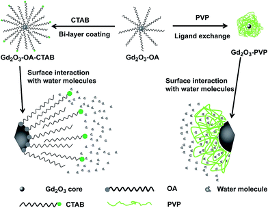 | ||
| Fig. 2 Schematic illustration of the Gd2O3 nanoparticle surface modifications and their interactions with water molecules (ref. 75). | ||
Four different sizes between 2.5 and 8.0 nm of thermodynamically stable β-NaGdF4 nanoparticles were synthesized by modifying nanoparticle growth dynamics as T1-weighted contrast agents was reported by van Veggel et al.67 The synthesized β-NaGdF4 were exchanged the oleate ligands with biocompatible polyvinyl pyrrolidone in water. The r1 relaxivity increased with decreasing particle size and with increasing the surface-to-volume ratio demonstrated the surface Gd(III) ions on the particles are mainly contributed the relaxivity enhancement (Fig. 3). Moreover, the surface Gd(III) ions on a large particle affect the relaxivity stronger than those on a small particle. The ultrasmall β-NaGdF4 nanoparticles can be doped with Yb(III)/Tm(III) as potential bimodal probes.
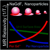 | ||
| Fig. 3 The r1 relaxivity of NaGdF4 nanoparticles depends on particles size, unit surface area, and surface-to-volume ratio (ref. 67). | ||
Hou and co-workers synthesized differently size of NaGdF4-PEG–mAb nanoprobes, NaGdF4 nanocrystals conjugated to anti-EGFR monoclonal antibody (mAb) via “click” reaction both on the maleimide residue on particle surface and on thiol group from the partly reduced anti-EGFR monoclonal antibody (mAb) (Fig. 4).76 The biocompatibility and binding specificity of NaGdF4 nanocrystals (NaGdF4-PEG–mAb) with narrow particle size distributions were evaluated through in vitro and in vivo experiments. The NaGdF4-PEG–mAb probes exhibited promising tumor-specific targeting ability and enhanced MRI contrast effects.
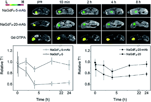 | ||
| Fig. 4 (Top) T1-weighted MR images of tumor-bearing mice acquired before and at different time points after the intravenous injections of Gd-DTPA or NaGdF4-PEG–mAb probes formed by using NaGdF4–5–mAb and NaGdF4–20–mAb, respectively. (Bottom) T1 values extracted before and after the injections of NaGdF4-PEG–mAb probes or the corresponding mother particles from the tumor sites. The color at tumors show the contrast enhancement effects of the particle probes (ref. 76). | ||
Poly-(acrylic acid)-coated gadolinium hydrated carbonate nanoparticle (GHC-1) with ultrasmall size (2.3 ± 0.1 nm) through a simple strategy was developed by Liang and co-authors (Fig. 5).77 The obtained GHC-1 showed a high longitudinal relaxivity of 34.8 mM−1 s−1 at 0.55 T and low r2/r1 ratio of 1.17, highly dispersible and stable in aqueous solution because of the hydrophilic polymer coating. The biodistribution in organs and in vivo MRI studies demonstrated that GHC-1 could be a potential candidate as a T1-weighted contrast agent. Lin's group developed the gadolinium hexanedione nanoparticles (GdH-NPs) as a cell tracking agent.78 Experiments demonstrated GdH-NPs were nontoxic for human mesenchymal stem cells (hMSCs), the GdH-NPs labeled stem cells have had better signals in cellular MRI assay. The GdH-NPs could be as a potential MRI contrast agent for stem cell tracking.
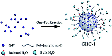 | ||
| Fig. 5 Schematic showing the synthesis of GHC-1 and its effect in inducing the longitudinal relaxation of the water proton (ref. 77). | ||
Mesoporous silica nanoparticles doped with Gd can be potentially used for MRI contrast agents because of their characteristic uniform porous structure.79–83 The advantages of mesoporous silica nanoparticles, such as easy surface modification, good biocompatibility, make them being easily functionalized for cellular labeling,84 or bioconjugation with biomolecules for drug delivery, gene therapy.85,86 But most of studies focus on their preparations and physicochemical properties. Recently Gd2O3-assembled silica nanocomposite Gd2O3@MCM-41 was reported by Li et al. as an effective targeted probe for in vivo molecular imaging of cancer with systematic pre-clinical studies (Fig. 6).87 The biocompatibility includes in vitro cell viability assessment, and in vivo study on immunotoxicity and acute toxicity test of the nanocomposite. Biodistribution and excretion studies of the nanocomposite were carried out for pharmacokinetic profile. MRI nanoprobe demonstrated a larger water proton relaxivity r1 and a better T1-weighted MR imaging when compared to Gd-DTPA.
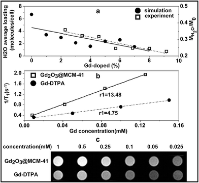 | ||
| Fig. 6 (a) The average loading H2O of simulated models (left vertical axis) versus the water adsorption of experimentally measured samples (right vertical axis) in MCM-41 silica assembled with different additions of the Gd2O3 molecule. Comparison of the proton relaxivity r1 (b) and T1-weighted phantom MR image (c) between Gd2O3@MCM-41 silica nanocomposite and the commercially available Gd-DTPA (ref. 87). | ||
Gadofullerenes are Gd-containing metallofullerenes, a new class of T1-weighted MRI contrast agents.88 Gadofullerene derivatives such as Gd@C60(OH)X, Gd@C60[C(COOH)2]10 and Gd@C82(OH)X demonstrated high relaxivity as potential MRI contrast agents.89–93 Superparamagnetic gadonanotubes derived from ultrashort single-walled carbon nanotubes in which Gd(III) have been loaded are high-performance contrast agents.63,94,95 Water-soluble gadonanotube derivative that underwent a dramatic response to pH change at physiologically relevant conditions is a pH-activated smart contrast agent (Fig. 7).94 The relaxivity of the gadonanotubes response dramatically from 40 mM−1 s−1 (pH = 8.3) to 133 mM−1 s−1 (pH = 6.7) at 37 °C, especially approximately 40 mM−1 s−1 change between pH 7.4 and 7.0. With the dramatic response around physiological pH, the gadonanotubes could be promising for the development of clinical agents for the early detection of cancer as the extracellular pH of cancerous tissues is less than 7.0.
 | ||
| Fig. 7 (a) A pictorial representation of the gadonanotubes. Small, superparamagnetic clusters of Gd(III) ions reside within the sidewall defects of the nanotube (chloride counteranions omitted for clarity). (b) r1 relaxivity (per Gd(III)) as a function of the ph for the gadonanotubes at 1.41 T and 37 °C (ref. 94). | ||
Fillmore and co-authors reported a tumor-specific peptide IL-13 peptides was conjugated with the carboxyl functionalized metallofullerene Gd3N@C80(OH)∼26(CH2CH2COOH)∼16.96 The obtained metallofullerene showed an enhanced specificity for the human glioma cells expressing IL-13Ra2. In addition, experiments demonstrated significant uptake of the targeted metallofullerene into brain tumor tissue in an orthotopic rodent model and the targeted metallofullerene could be used for delivering imaging and therapeutic agents to tumor cells.
Gd(III) chelate grafted macromolecular nanoparticles
Gadolinium complexes, Gd(III) ions chelated with a low molecular weight acyclic or cyclic ligand are the only Gd-based contrast agents used for clinic and will continue to be the most widely used.7 Macrocyclic-based gadolinium complexes have a number of advantages such as pharmacokinetics and pharmacology due to the tight binding Gd(III) to the macrocyclic chelator. While various compounds have been evaluated as MR contrast agents, the high relaxivity and stability of Gd(III) chelates are the two important requisites for the development of contrast agents.97–100 Most of these small molecular contrast agents distribute throughout the intravascular and interstitial space, and excreted rapidly via renal filtration. Both small doses and low release of free Gd(III) ions are the most concerns as they will reduce the toxicity. Some strategies have been reviewed to increasing the relaxivity of Gd(III) chelates as MRI contrast agents.97 Gd(III) chelates coated on gold nanoparticles;101–105 immobilized on core–shell inorganic–organic hybrid nanoparticles,69,106 lipoproteins,107 viral capsid,108 nanofiber,109,110 peptides;111 entrapped in zeolites,112 mesoporous silica nanoparticles,113 human mesenchymal stem cell114 are strategies to increase the relaxivity of contrast agent as nanoparticles exhibit a high relaxivity per particle.115,116 Though simply increase the overall size of the nanoparticle will lead to a corresponding increase in the relaxivity per nanoparticles, there is an optimal nanoparticle volume or molecular weight.Gold nanoparticles can be used as carriers for Gd(III) chelates with a dithiolated derivative of DTPA (DTDTPA) in which gold core of the nanoparticle coated with Gd(III) chelate.101 Gold nanoparticles with a size of 2–2.5 nm were prepared by reducing a gold salt in the presence of new chelator. The immobilization of a large number of DTDTPA-Gd chelate on each particle contribute to a very high relaxivity (r1 = 585 mM−1 s−1 compared to 3.0 mM−1 s−1 for DTPA-Gd) of the particle which make them as contrast agents for MRI. In addition, the biocompatibility of gold offers its applications in living organisms for MRI and/or hyperthermia therapy. The DTDTPA chelator could be conjugated to a biomolecules for other applications such as specific targeting. A nanoconjugate contrast agent made from covalent attachment of Gd(III) to thiolated DNA followed by surface conjugation on to gold nanostars (DNA-Gd@stars) was reported recently by Rotz et al.117 The study showed the nanoparticle shape and surface structure play an important role for relaxivity of the nanoconjugate as they affected the organization of the conjugated DNAs on the particle surface and thus to affect the water molecules in the proximity of the Gd(III) complex. (DNA-Gd@stars) improve Gd(III) delivery and maintain biocompatibility when incubation with pancreatic cancer cells. A gold nanoparticles conjugate (DNA-Gd@AuNPs), that is functionalized with deoxythymidine oligonucleotides containing Gd(III) chelates and a red fluorescent Cy3 moiety which can be used to visualize in vivo transplanted human neural stem cells was reported by Nicholls and coworkers [Fig. 8].118 Through the in vitro assays to determine cell uptake, potential cytotoxicity and cellular relaxivity, the stable DNA-Gd@AuNPs can be used to visualize the distribution of human neural stem cells by MRI with optimal imaging parameters and exhibited an improved T1 relaxivity and excellent cell uptake.
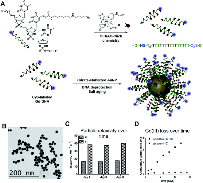 | ||
| Fig. 8 DNA-Gd@AuNP synthesis and stability. Particles consist of a gold nanoparticle core loaded with DNA to which the Gd-HPDO3A and Cy3 moieties are attached (A). Transmission electron microscopy (TEM) of conjugated DNA-Gd@Au nanoparticles for size determination (B). Particles retain their relaxivity properties at 1.41 T over more than 2 weeks (C). There is a small amount of Gd(III) loss at 37 °C, amounting to <5% of total Gd(III) over 2 weeks. However, loss is negligible (<0.4%) when particles are stored at 4 °C (D) (ref. 118). | ||
Gd-DOTA derivative conjugated self-assembling peptide amphiphiles (PACAs) form the nanofiber networks was synthesized and their in vitro MR evaluation was carried out by Meade et al.109,110 The peptide amphiphiles contains three parts: a headgroup, body, and an alkyl tail. The headgroup is made up of an epitope for specific cell interaction. The body is functional structure in which hydrogen bond formed between molecules is the driving force for fiber formation (Fig. 9). The alkyl palmitoyl tail of the PA initiated a hydrophobic collapse in an aqueous environment. Self-assembling PAs forms nanofiber, varying the position of the Gd(III) chelate with DOTA derivative on the PAs leads to the changes in the molecule's relaxivity. Novel design of MRI active supramolecular structures to noninvasively track PA gel scaffolds in vivo, provides a possible three dimensional MR images for the fate mapping of these new biomaterials. Kobayashi et al. reported Gd(III) complex Gd-EDTA (EDTA = ethylenediaminetetraacetic acid) entrapped in mesoporous silica nanoparticles Gd-EDTA/SiO2.113 (3-Aminopropyl)trimethoxysilane was introduced on the silica particles at pH 3 to make NH2/SiO2. Then immobilization of Gd-EDTA on the NH2/SiO2 particles at pH 5, the obtained Gd-EDTA/SiO2 particle colloid solution was concentrated to certain Gd concentration with centrifugation. High contrast T1-weighted MR images and high r1 relaxivity of the Gd-EDTA/SiO2 particles indicated their application to MRI.
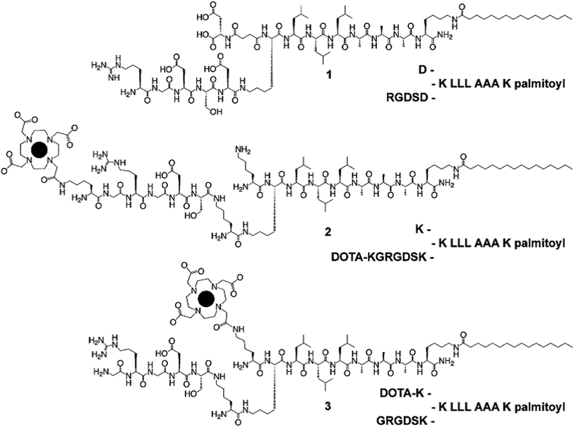 | ||
| Fig. 9 Structures of the PA molecules with the black circles representing the Gd(III) ion. (1) is an example of a filler PA and does not contain the Gd(III) chelate. (2) and (3) are the PACA molecules used in this study containing Gd(III) (ref. 110). | ||
Polymer-based nanoparticle MRI contrast agents have demonstrated optimal pharmacokinetics and blood half-life, increased tolerance to enzymatic degradation, good physical and chemical stability which improve their clinical and therapeutic value.106,119–134 Polymeric macromolecular nanoparticle contrast agents can be considered as an alternative to blood pool agents because blood pool agents have following shortcomings: enhancement of interstitial space while enhancement of blood, prolonged retention in liver and bone, cardiac toxicity, etc. Attention has been put on Gd(III) chelates conjugated onto dendrimers,119–125 liposomes,126,127 micelles,106,128–131 core cross-linked star polymers and hyperbranched polymers.132–134
Kobayashi and Brechbiel reviewed and developed the nano-sized MRI contrast agents with different dendrimer cores such as polypropylenimine diaminobutane (DAB) (Fig. 10).135 Different nano-sized molecular contrast agents in diameter behave differently in the body. Various size contrast agents up to 15 nm altered vascular wall permeability in the blood, excretion pattern, and recognition by the reticuloendothelial system. The different sizes and various properties of dendrimer-based macromolecular MRI contrast agents provide sufficient contrast enhancement for various applications.136–138 The in vitro and in vivo experiments on the dendrimer core Gd(III) chelate contrast agents demonstrated that they have much higher relaxivity compared to a single chelate unit. These contrast agents could be used for intravascular contrast-enhancing.139
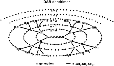 | ||
| Fig. 10 Scheme of the dendrimer core used for contrast agents (ref. 135). | ||
Dendrimers have been prepared as multifunctional nanomaterials for diagnostic and therapeutic agents.140 Gd(III) chelates conjugated to dendrimers with functional groups could be used as targeting contrast agents, as carriers for target-specific delivery of drugs. Gd-DTPA conjugated dendrimer nanoclusters were reported as a T1-weighted MRI contrast agent for tumor-targeting by Tsourkas and coworkers.141 The dendrimer nanoclusters (DNCs) were prepared by crosslinking G5 PAMAM dendrimers through a bifunctional amine-reactive crosslinker NHS-(PEG)5–NHS-(BS(PEG)5). Gd-DTPA chelates were then conjugated to DNCs and further functionalized with the folic acid for tumor-targeting and the optical imaging dye fluorescein isothiocyanate (FITC) (Fig. 11). The r1 relaxivity of Gd(III) chelate conjugated DNCs is higher than that of Gd-DTPA. In vivo studies of DNCs on folate-positive KB tumor mice showed significant contrast enhancement at 4 h to 24 h post-intravenous injection of DNCs. A tumor targeting biodegradable dendrimer MRI contrast agent FA-PEG-G2-DTPA-Gd was synthesized and evaluated for enhanced blood pool and tumor imaging by Shen and Tang groups.142,143 Both PEG chains with distal folic acid and Gd-DTPA chelates were conjugated to a polyester dendrimer. Degradable dendrimer FA-PEG-G2-DTPA-Gd (Fig. 12) at the physiological pH, hydrolysis accelerated in the presence of esterase overcame the drawbacks of long-term retention in the body, thus reduce toxicity. The FA-PEG-G2-DTPA-Gd had a high r1 relaxivity. In vivo evaluation demonstrated it had contrast enhancement in tumor and low Gd retention.143
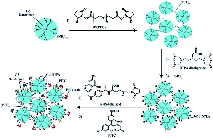 | ||
| Fig. 11 The preparation of paramagnetic targeted dendrimer nanoclusters (DNCs) (ref. 141). | ||
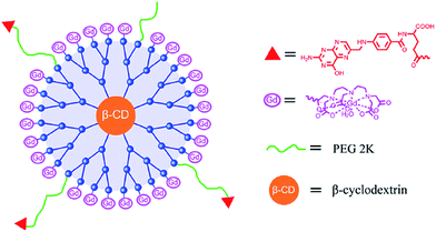 | ||
| Fig. 12 Schematic illustration of the tumor targeting dendritic MRI contrast agent (ref. 143). | ||
Polymeric micelles as a promising carrier for drug delivery have received considerable attention. Polymeric micelles that integrated multiple smart functionalities can be developed as smart nanodevices have been reviewed by Kataoka et al.144 Paramagnetic molecules such as Gd and Mn for T1-weighted MRI have been incorporated into nanodevices to enable preclinical and clinical applications.145–147 Gd-DTPA was incorporated into DACHPt-loaded micelles by utilizing the reversible complex formation between DACHPt and Gd-DTPA, leading to slow molecular reorientation and decrease the mobility with increase relaxivities (Fig. 13).144 The obtained Gd-DTPA/DACHPt-loaded micelles have a longitudinal relaxivity r1 80.5 mM−1 s−1. Animal experiments revealed that Gd-DTPA/DACHPt-loaded micelles accumulated effectively in subcutaneous murine colon carcinoma and orthotopic human pancreatic adenocarcinoma models, and specifically enhanced the signal at the tumor site for a prolonged time. Contrast-enhanced MRI exhibited that Gd-DTPA/DACHPt-loaded micelles could be applicable for the measurement of the volume of orthotopic pancreatic tumors in the abdominal cavity, and for the noninvasive evaluation of their enhanced antitumor activity.
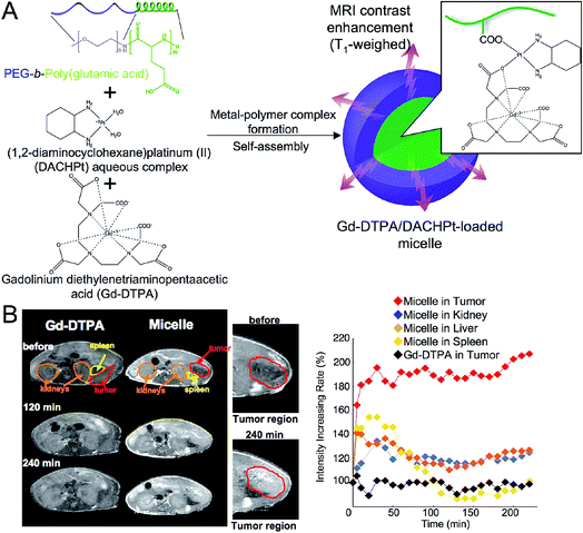 | ||
| Fig. 13 Gd-DTPA/DACHPt-loaded micelles for tracking biodistribution and therapeutic effects. (A) Scheme of Gd-DTPA/DACHPt-loaded micelles formation. (B) MRI of orthotopic pancreatic tumor bearing mice after intravenous injection of the clinically approved MRI contrast agent, Gd-DTPA, or Gd-DTPA/DACHPt-loaded micelles (ref. 144). | ||
Nanoscale micelles based on biodegradable poly(L-glutamic acid)-b-polylactide with Gd-DTPA conjugated to the shell layer (Fig. 14) was reported by Li et al. as a potential nano-sized MRI visible delivery system with a high r1 relaxivity.128 Polymeric micelles composed of polysuccinimide (PSI) derivatives incorporating methoxy-poly(ethylene glycol) (mPEG) were conjugated with DTPA-Gd, and showed better contrast on in vitro phantom compared to that of Omniscan.148 However, the pharmacokinetics of these polymeric micelles needs to be done before assessing their potential applications for in vivo MRI study. Liu et al. reported a new multifunctional pH-disintegrable micellar nanoparticles prepared from star copolymers containing asymmetrically functionalized β-cyclodextrin (β-CD) were covalently conjugated with doxorubicin (DOX), folic acid (FA), and DOTA-Gd moieties. The obtained micellar nanoparticles exhibited considerably enhanced r1 relaxivity compared to that of the small molecule counterpart. In vivo MR imaging studies found considerable uptake of micellar nanoparticles at rat kidney.149
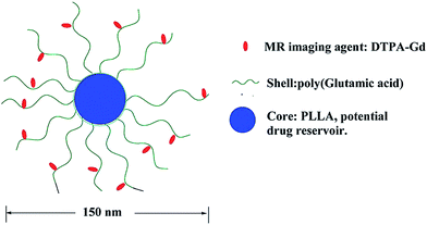 | ||
| Fig. 14 Schematic model of the micellar structure with Gd-DTPA chelate conjugated to the shell layer (ref. 128). | ||
The drawbacks for synthesis of dendrimers are challenging multistep synthesis and purification. There are issues for indirect assembly strategies to prepare micelles utilizing amphiphilic block copolymers as they are not very stable on dilution, leading to uncertainty during in vivo studies; the additional micelle cross-linking increases the inherent complexity. There is a need for the development of more versatile and efficient synthetic routes to polymeric nanoparticle MRI contrast agents such as core cross-linked star and hyperbranched polymer nanoparticles.
Gd-loaded nanoparticles (GdNPs) have been attractive as MRI theranostic applications in imaging-guided drug delivery, drug release, monitoring the treatment efficacy and personalized administration, etc.150 The ideal theranostic GdNPs should possess following properties: biodegradable and non-toxic of NPs carrier materials; efficient delivery of GdNPs to target tissue or organ; active targeting and sensitive to diagnosis; therapeutic efficacy of drug; optimal excretion pattern of Gd molecules to reduce toxicity [Fig. 15]. GdNPs as drug carriers to co-deliver the therapeutic drugs and Gd are normally lipid-based,151,152 polymeric,153 micelles,154 and silica155 nanoparticles. Nanocarrier liposomes co-encapsulated doxorubicin and Gd was reported as a dual functional diagnostic and chemotherapeutic agent, and target-specific carriers for clinical applications.151 An interesting temperature-induced liposomal system encapsulated doxorubicin and Gd-HPDO3A was developed as a theranostic agent for chemotherapeutics under MRI guidance.152 Mesoporous silica nanoparticles can be useful for theranostic applications as silica nanoparticles could gradually release therapeutic drugs and diagnostic contrast agents at the targeting area. A pH-dependent biodegradable silica nanotubes derived from Gd(OH)3 nanorods was reported by Hu et al.155 The released Gd(III) ions from the Gd(OH)3 nanorods were chelated by the pre-modified DOTA, obtained Gd-DOTA then grafted onto silica nanotubes. The Gd-DOTA grafted silica nanotubes loaded with doxorubicin exhibit enhanced T1 imaging contrast and anticancer activity.155
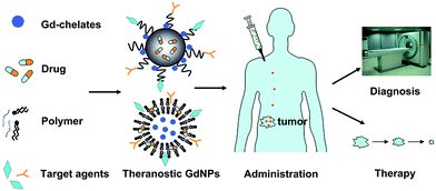 | ||
| Fig. 15 The preparation and application process of GdNPs. Gd-Chelates and therapeutic drugs were loaded in various theranostic GdNPs such as polymer-based and lipid-based nano-carriers and target agents were modified onto the GdNPs surface. After administration, GdNPs were delivered to the tumor area by passive and active targeting. An enhanced diagnostic and therapeutic was achieved simultaneously (ref. 150). | ||
Core cross-linked star and hyperbranched polymer nanoparticles were reported as MRI contrast agents by Li et al. with significant advancements in their preparations.132,133 The macromolecular architecture and precise molecular location of Gd(III) chelate has a significant impact on relaxivity and efficacy of contrast agents. When compared to the traditional micellar approach, one of the key advantages of core cross-linked star and classic hyperbranched polymer nanoparticles is the inclusion of cross-linking during the polymerization process. These polymer nanoparticles required to load hydrophobic guest molecules. Liu et al. reported that star copolymers covalently anchored with polycationic PDMA (PDMA = poly(N,N-dimethylaminoethyl methacrylate)) arms and conjugated with DOTA-Gd chelate at the lower and upper rim of toroidal β-cyclodextrin (β-CD) cores were synthesized (Fig. 16).156 The obtained cationic star copolymer could effectively bind negatively charged anionic plasmid DNA (pDNA) via electrostatic interactions, thus a dramatically increased r1 relaxivity (10.9 mM−1 s−1) was found compared to that of small molecule contrast agents. Same group recently also reported the synthesis of hyperbranched polyprodrug amphiphiles (hPAs) via reversible addition–fragmentation chain transfer (RAFT) copolymerization.134 Reduction-activatable camptothecin prodrugs and Gd-DOTA derivatives chelate conjugated onto hPAs through the click reaction in the hydrophobic cores, the nanoparticles were then decorated guanidine onto hydrophilic coronas. The hPAs nanoparticles exhibited cell penetration potency, prolonged blood circulation, synergistic activation of therapeutic efficacy and MR imaging contrast enhancement. MR imaging contrast performance on guanidine-decorated hPAs demonstrated prolonged blood circulation with a half-life up to ∼9.8 hours.134
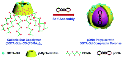 | ||
| Fig. 16 Schematic illustration of (DOTA-Gd)7-Cd-(PDMA)14 star copolymers dually acting as pDNA delivery vectors and MR imaging contrast agents (ref. 156). | ||
We presented the direct synthesis of stable Gd(III)-chelated branched copolymer nanoparticles, started from novel monomers via reversible addition–fragmentation chain transfer (RAFT) polymerization without the self-assembly techniques.157 The resulting branched copolymer nanoparticles comprise a hydrophilic corona and a hydrophobic covalently cross-linked core. This synthetic approach does not require post-polymerization modification to incorporate macrocycle functionalities. The structural advantages of branched copolymer nanoparticles have been demonstrated by encapsulated the hydrophobic dye Nile within the hydrophobic core of N2(Gd) without precipitation for three months. The stability and structural advantages of loading hydrophobic guest molecules within the nanoparticles cores, without any significant loss in relaxivity, offer the potential for simultaneous bioimaging and drug delivery. The cytotoxicity testing using HK-2 cells established their negligible toxicity profile. In vitro and in vivo MRI studies of the N2(Gd) nanoparticles showed that they have a high relaxivity and a long blood retention time. The dynamic contrast imaging on SCID mice bearing U87MG human glioma xenograft tumor demonstrated that they are promising intravascular MRI contrast agents and are able to perfuse and passively target tumor cells. The bifunctional hydrophobic cores are able to simultaneously conjugated to Gd(III) chelate and noncovalently encapsulate hydrophobic guest molecules, that combine the properties of micellar assemblies with the synthetic advantages of core cross-linked star polymers.
Bimodality probes for MRI and other imaging modalities
A series of nanostructured materials have been developed as novel multimodal bioimaging agents for applications in diagnostics and therapy as they overcome the limitations of either imaging modality used alone.158–163 The integration of MRI and fluorescence imaging (FI) techniques provides new opportunities for cancer diagnosis, drug delivery, and therapy by taking advantages of the superb spatial resolution of MRI and high sensitivity of FI.164–169 Chemistry synthesis strategies have been developed extensively for this regard. Hyeon et al. designed the multifunctional nanomedical platforms as cancer-targeted, dual modality imaging probe including MRI and optical imaging, and drug delivery.170 The multifunctional polymer nanoparticles was composed of four moieties (Fig. 17): biodegradable poly(D,L-lactic-co-glycolic acid) (PLGA) nanoparticles as vehicles for loading and release of therapeutic agents into cells; superparamagnetic magnetite nanocrystals for magnetically guided delivery and T2 MRI contrast agent and semiconductor nanoparticles (CdSe/ZnS quantum dots) for optical imaging; doxorubicin for cancer therapy; PEGylated folate for active cancer cells. Same group later prepared multifunctional nanoparticles by assembling Fe3O4 nanocrystals on uniform dye-doped mesoporous silica nanoparticles for enhanced MRI and FI dual modalities. The in vivo study showed the composite nanoparticles accumulated at the tumor site.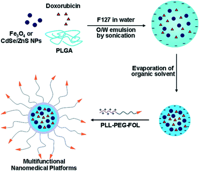 | ||
| Fig. 17 Four components in the nanoparticle and preparation route (ref. 170). | ||
Gd2O3 core embedded in a polysiloxane shell which is comprised of organic fluorophores and carboxylated PEG to form hybrid nanoparticles as contrast agent for both in vivo FI and MRI was reported by Bridot et al.171 It was found that the longitudinal relaxivity per particle enormously increases with the decrease in the particle size. Kryza and co-workers reported hybrid gadolinium oxide particles as a multimodal SPECT/MR/optical imaging and theranostic agent. The particles prepared by encapsulating Gd2O3 cores within a polysiloxane shell, which contains organic fluorophore (Cy 5), coated by a hydrophilic carboxylic layer were used for biodistribution and pharmacokinetics studies.172 Various iodine compound coated Gd2O3 nanoparticles were synthesized as T1-weighted MRI and CT dual imaging probes for cancer.173 Efforts have been made to transfer these Gd2O3 nanoparticles from hydrophobic into hydrophilic for bio-applications.174,175 Petoral et al. synthesized Tb(III)-doped ultrasmall Gd2O3 nanocrystal for combined fluorescent labelling and MRI contrast agent.176 The nanocrystals were stabilized by PEGylation. Since Tb(III) attributed to the fluorescent property, the capacity of the Tb(III)-doped nanoparticles for fluorescent labelling of living cells was investigated. A facile synthetic strategy for fabrication of rare earth Tb(III), Y(III) and Er(III)-doped Gd2O3 ultranarrow nanorods as multimodal imaging probes was reported by Das et al.177 Capping agent plays an important role in shape control during the thermolysis of the precursor complex. Oleylamine stabilized the particles and prevent the aggregation of these particles. Interactions of oleylamine on the nanoparticles lead to the formation of the extended chain, which fuse longitudinally and recrystallize to nanorods (Fig. 18). The rare earth-doped Gd2O3 nanorods demonstrated a tunable down- or up-conversion fluorescent and promising T1-weighted MRI contrast agent.
 | ||
| Fig. 18 Proposed mechanism of the nanorods formation by oriented attachment (ref. 177). | ||
Rare earth ions doped upconverting nanoparticles have extensive applications in biomedical imaging. Inorganic GdF3 or NaGdF4 nanoparticles doping with other lanthanides to integrate optical and MR contrast effect into dual modality probes have been explored.178,179 NaGdF4:Er(III),Yb(III)/NaGdF4 core/shell upconverting particles can be used as a new type of both optical and MRI bimodal probe was investigated by Hyeon.178 Without adding any other moieties, the nanoparticles exhibit multimodality on their own nanocomposites. The optical property of the particles is attributable to Er(III) or Yb(III) located on the crystal lattice of the NaGdF4 host. Chen et al. reported amine-functionalized and biocompatible KGdF4:Ln(III) nanocrystals were synthesized through a facile one-step solvothermal route179 (Fig. 19). The nanocrystals conjugated to biomolecules through amino groups on the capping ligand polyethylenimine to be functional bioprobes. The KGdF4:Ln(III) showed as a sensitive bioprobe in time-resolved fluorescence resonance energy transfer (TR-FRET) assays to quantitatively detect avidin protein. In the meantime, KGdF4 possess a relatively large longitudinal relaxivity.
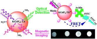 | ||
| Fig. 19 Amine-functionalized lanthanide-doped KGdF4 nanocrystals as a potential optical/magnetic multimodal bioprobe (ref. 179). | ||
Rare earth ions contained upconverting nanomaterials displayed excellent luminescence, unique MR and strong X-ray attenuation, thus they can be used as high performance contrast agents for luminescence, MR and CT imaging.180–186 Xia and co-authors reported a multifunctional lanthanide-based nanoparticle for near-infrared NIR to-NIR luminescence, CT and T1-enhanced MR trimodality in vivo imaging.180 Core@shell lanthanide-based nanoparticles doped with selected Yb and Tm, (NaLuF4:Yb(III),Tm(III)@SiO2-GdDTPA) were synthesized with NaLuF4:Yb(III),Tm(III) upconverting nanoparticles and the Gd-DTPA as the surface ligand (Fig. 20), their applications for NIR-to-NIR luminescence, CT and MRI multi-modality in vivo imaging have been established. Liu et al. reported a multifunctional up-conversion nanoprobe based on PEGylated Gd2O3:Yb(III),Er(III) nanorods for in vivo luminescence, T1-enhanced MR, and CT multi-modality imaging due to their unique electric, magnetic, and optical properties.181 Lanthanide-doped NaGdF4 upconversion nanocrystals with different crystal-phases were synthesize through a one-step controllable reaction by Cui and co-workers.182 The obtained upconversion nanocrystals are effective dual probe for in vivo luminescence imaging and CT imaging. PEG modified BaGdF5:Yb/Er upconversion nanoparticles prepared by a facile one-pot hydrothermal method as a tri-modal nanoprobe for FI, CT, and MRI was reported by Hao et al. for the first time.183 The fluorescent imaging of HeLa cell, in vivo CT images with enhanced signals of spleen of a mouse, excellent intrinsic paramagnetic property of the PEG-modified BaGdF5:Yb/Er upconversion nanoparticles indicate that the particles can be served as a tri-modal nanoprobe for FI/CT/MR bioimaging.
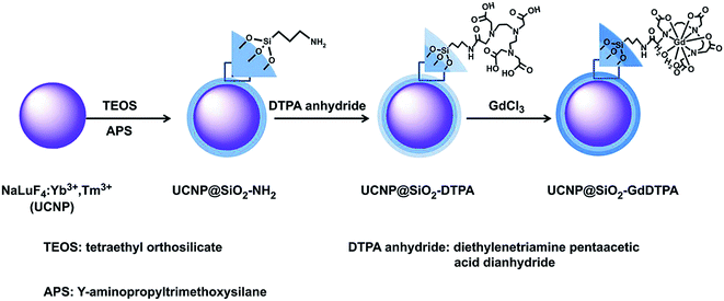 | ||
| Fig. 20 Schematic representation of the synthetic route of NaLuF4@SiO2-GdDTPA nanoparticles (ref. 180). | ||
Multifunctional Gd-loaded dendrimer-entrapped gold nanoparticles (Gd–Au DENPs) for dual CT/MR imaging was reported by Wen and co-authors.184 Amine-terminated generation 5 PAMAM dendrimers (G5·NH2) conjugated with Gd(III) complex, followed by complete acetylation of the remaining dendrimer terminal amines (Fig. 21). The r1 relaxivity and X-ray attenuation property of Gd–Au DENPs enable the nanoparticles to be used as dual CT/MRI contrast agents for in vivo imaging. In vivo biodistribution studies on major organs of rats and mice showed the Gd–Au DENPs have an extended blood circulation time. Europium-doped (GdS:Eu(III)) opto-magnetic nanoparticles prepared from sonochemistry method was reported by Jung and coworkers.185 Gadolinium sulfide constitutes the core with Gd replaced by Eu dopant. The sulfur from 1-dodecanethiol at the core also serves as a surface capping ligand which is replaced by 3-mercaptopropionic acid (MPA) for bioapplication (Fig. 22). The photophysical property is induced by Eu(III) ions dopant. The nanoparticles were internalized into the cytoplasm of breast cancer cells (SK-BR-3). The photoluminescence and paramagnetic properties of the nanoparticles enable them to be used as a dual probe for in vitro cell imaging and in vivo T1-weighted MR imaging. Strong positive contrast enhanced blood vessels and organs of mice and the confocal images of breast cancer cells containing GdS:Eu(III) nanoparticles demonstrate the dual-mode imaging capability of the GdS:Eu(III) nanoparticles.
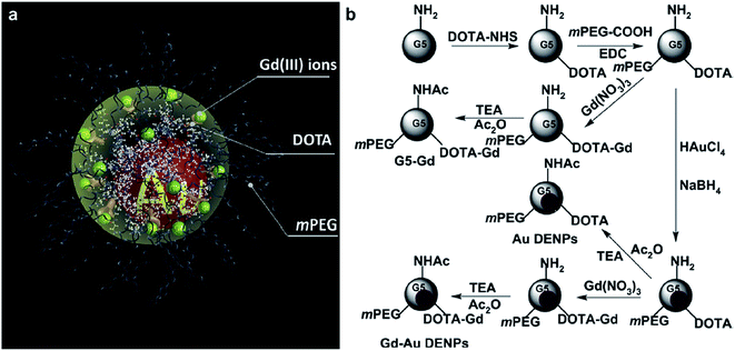 | ||
| Fig. 21 Schematic illustration of the designed nanostructure (a) and the synthesis procedure (b) of the Gd-Au DENPs. TEA and Ac2O represent triethylamine and acetic anhydride, respectively (ref. 184). | ||
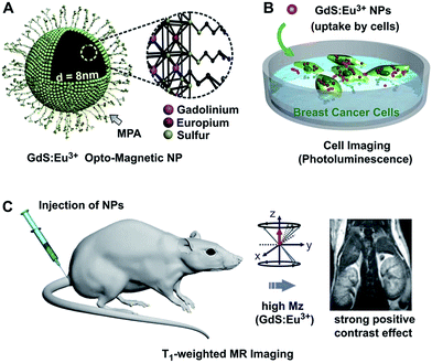 | ||
| Fig. 22 (A) Detailed structure of Eu-doped GdS (GdS:Eu(III)) opto-magnetic NPs (B) cell imaging via fluorescence emission of GdS:Eu(III) NPs at 614 nm. (C) Biocompatible GdS:Eu(III) NPs as a strong positive contrast agent for the MR imaging of blood vessels and various organs (ref. 185). | ||
Gd(III)–conjugate AuNP microcapsules was developed to visualized pancreatic islet cells for treatment of type I diabetes and enable multimodal cellular imaging of transplanted islet cells.187 Multifunctional dendrimer-entrapped Gd(III)–conjugate AuNP have been used for CT/MR dual mode imaging of tumors.188–190 Lipid Gd-DOTA functionalized AuNPs@PDA nanohybrid containing core–shell structure with the polydopamine coating gold nanoparticles as the inner core, the indocyanine green (ICG) as a phototherapeutic agent and lipids modified Gd-DO3A and lactobionic acid (LA) showed potential as theranostic agents for photothermal therapy to ablate cancer.191 Lux and coworkers reviewed the Gd-based nanoparticles developed for theranostic applications and more precisely on irradiations guided by MRI.192 Gd-based nanoparticles can act as effective radiosensitizers under different types of irradiation such as radiotherapy. These new therapeutic modalities pave the way to therapy guided by imaging and to personalized medicine. Same group also summarized the advantages of Gd-based ultrasmall nanoparticles versus molecular Gd chelates for radiotherapy guided by MRI for glioma treatment.193 Fast accumulation and strong resident time in the brain tumor of Gd-based nanoparticles was found compared to commercial chelates. The biodistribution profile is suitable for an optimal radiosensitization.
Design of Gd(III) chelate coated gold nanoparticles for both X-ray and MRI were reported by Roux et al.194 The Gd(III) chelate coated gold nanoparticles were obtained by encapsulating gold cores within a multilayered organic shell made from Gd(III) chelates bound to each other through disulfide bonds (Fig. 23). The in vitro imaging experiments demonstrated that Au@DTDTPA-Gd (DTDTPA = dithiolated derivative of diethylenetriaminepentacetic acid) nanoparticles have a positive contrast enhancement at kidney in T1-weighted images. The contrast enhancement in MR images is contributed by Gd(III) ions which are chelated by DTDTPA in the organic shell, whereas the gold core provides X-ray absorption. The Gd(III) chelate functionalized gold nanoparticles can be applied as contrast agents for both MRI and X-ray. The other examples of gold nanoparticles coated with Gd-chelate as potential CT/MRI bimodal contrast agents were reported by Kim et al.195 The chelator L = a conjugates of DTPA-bisamide (DTPA = diethylenetriaminepentacetic acid) with cysteine195 or 4-aminothiphenol.196 These well-dispersed spherical GdL@Au particles are obtained by substituting gadolinium chelate (GdL) for citrate on the gold nanoparticle surfaces. In vitro MRI showed GdL@Au nanoparticles have very high r1 relaxivity (∼104 mM−1 s−1) and the r1 relaxivity per [Gd] is as high as 10 mM−1 s−1. These particles also exhibit low cytotoxicity, indicating they can be further applied for preclinical studies (Fig. 24).
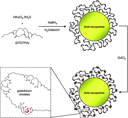 | ||
| Fig. 23 Synthesis of Au@DTDTPA-Gd nanoparticles (red circle = Gd(III) ion) (ref. 194). | ||
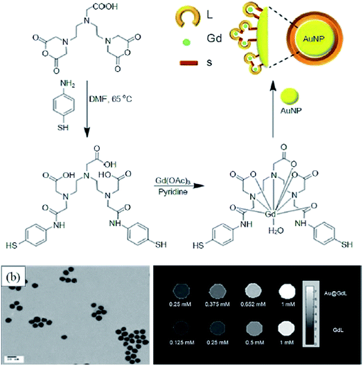 | ||
| Fig. 24 (a) Synthesis of GdL@Au, (b) Gd(III) chelate-coated gold nanoparticles, (c) r1 maps on Gd concentration in GdL@Au (ref. 195). | ||
Gd(III) complex contrast agents have been incorporated to develop dual MRI/optical imaging probe. Dendrimer-based MRI/FI dual modality nanoprobe has been prepared by covalently incorporating Gd(III) complex and organic dyes.197–201 The first dendrimer-based dual MRI-FI agent is a novel PAMAM-based nanoprobe G6-(Cy5.5)1.25(1B4M-Gd)145 which composed of PAMAM dendrimers with covalently attached Gd(III)-DTPA chelates and units of NIR fluorescent dye, Cy 5.5. It should be noted that since FI possess much higher sensitivity compared to MRI, the number of Gd(III) chelate units on the dendrimer surface must be exceed the number of fluorophore units to obtain the performance in equilibrium for both imaging modalities. In the meantime, increase concentration of dye moieties on the surface lead to partial self-quenching of the FI due to lanthanide ions located in a close proximity to the optical probe. In vivo studies of the nanoprobe by both MRI and FI modalities demonstrated that the sentinel lymph nodes in mice can be visualized by MRI and can be detectable by FI with as little as 0.5 nmol of Cy 5.5.197,198 The combination of MRI and FI of the nanoprobe could be suggested to provide the complementary information by mapping the sentinel nodes with MRI prior to surgical resection and then FI could be used for the surgeon to the appropriate sentinel nodes during surgery. Same group further reported a novel synthetic approach to prepare a biotinylated dendrimer-based MRI agent conjugated to fluorescently labeled avidin that a unique disulfide bond in the core of the Gd(III)-1B4M-DTPA chelated to G2 PAMAM dendrimer as a biotin-targeted, lectin-targeted dual MRI/FI probe.199
Core cross-linked (CCL) polymeric micelles covalently conjugated with DOTA-Gd and green-emitting fluorescent dye NBD fluorophores within pH-responsive cores were developed by Liu and co-workers. Due to pH-responsive core swelling and the associated hydrophobic–hydrophilic transition of CCL micelles, CCL micelles exhibit mildly acidic pH-triggered enhancement of signal intensities for both MR/FI imaging modalities.200 Tsien et al. reported activatable cell penetrating peptides linked to dendrimeric nanoparticle labeled with dye Cy5, and Gd(III) chelates for in vivo visualization of matrix metalloproteinase (MMP) activities by MRI and FI.201 The activatable cell penetrating peptides (ACPPs) are short polycations attached via protease-cleavable linkers to neutralizing polyanions which for in vivo fluorescence localization of active MMP-2 and MMP-9 in xenograft and transgenic tumor models. Each ACPP-conjugated dendrimers (ACPPDs) consists of a dendrimer conjugated to the polycationic segments of several ACPPs. The proteases such as MMP-2 and MMP-9 in tumors cut the linkers, releasing polyanions and leaving polycationic ACPPDs go into cells around the protease. The both Cy5 and Gd labeled “dual” ACPPD provide a good contrast for protease activity as dual probes for in vivo FI/MRI.201 However, these probes demonstrated the intrinsic shortcomings of commercial dyes, such as small Stokes shift, poor photo-stability and dye self-quenching.202
Semiconductor quantum dots (QDs) are a new class of materials for bioimaging because of their fluorescence, photostability, and narrow and tunable emission spectrum.203 QDs with a paramagnetic coating,204 Gd(III)-functionalized,205 Mn(II) doped core/shell206,207 have been reported for multifunctional probes for bioimaging. Annexin A5-functionalized QDs with a paramagnetic lipidic coating for detection of apoptotic cells with both MRI and FI in vivo was reported by van Tilborg.208 The other group also reported annexin A5-functionalized QDs with biotinylated Gd-DTPA for optical and MR imaging of cell death and platelet activation.209
Near-infrared (NIR) QDs can be used for FI to visualizing target objects in deep tissues with a low background signal.210 Jin et al. reported Gd(III)-DOTA functionalized NIR emitting QDs (CdSeTe/CdS) as a dual modal imaging probe for in vivo FI and MRI.211 Fig. 25 is a schematic representation for preparing Gd(III)-functionalized NIR-QDs. Hydrophobic CdSeTe/CdS QDs capped with trioctylphosphine (TOP), trioctylphosphine oxide (TOPO), and hexadecylamine (HAD) were replaced with a surface coating agent glutathione (GSH, a tripeptide (γ-L-glutamyl-L-cysteinylglycine)). The resulting GSH-QDs conjugated with Gd(III)-DOTA complex to add the T1-weighted MRI contrast ability.
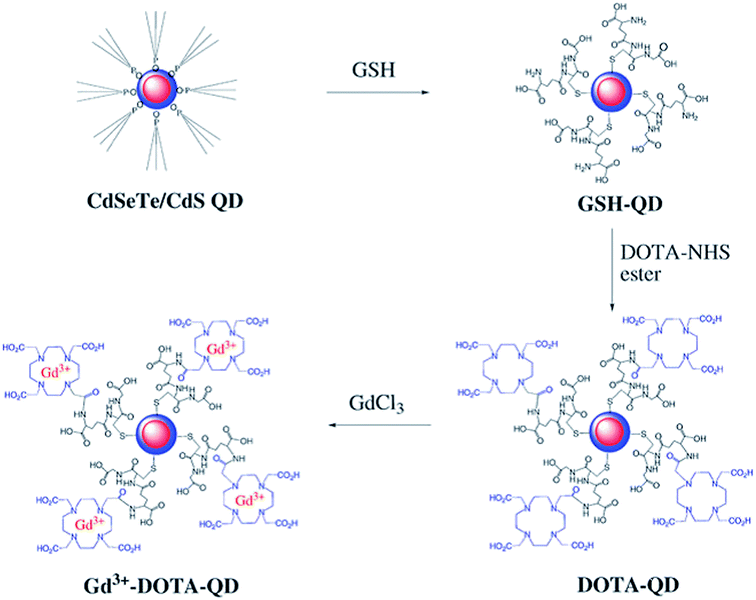 | ||
| Fig. 25 Schematic representation for the preparation of Gd(III)-DOTA functionalized CdSeTe/Cds QDs with glutathione (GSH) coating (ref. 211). | ||
Silica-based nanoparticles have been explored for molecular imaging and biomedical applications with improved biocompatibility and pharmacokinetics.212 Silica particles coated with equal amounts of both PEGylated (PEG-DSPE = 1,2-distearoyl-sn-glycero-3-phosphoethanolamine-N-[methoxy(poly(ethylene glycol))-2000]) and Gd-DTPA-based Gd-DTPA-DSA = Gd-DTPA-bis(stearylamide) lipids (Q-SiPaLCs, Fig. 26) make it hydrophilic materials as a dual probe for FI and MRI. The bio-applicable lipid coating silica particles showed an increased blood half-life time and a favourable tissue distribution profile compared to the bare silica particles. Another hybrid silica nanoparticles for multimodal imaging was reported by Lin et al.213 The hybrid silica nanoparticles containing a luminescent [Ru(bpy)3]Cl2 core (bpy = 2,2′-bipyridine) and a paramagnetic monolayer coating of a silylated Gd-DTPA chelate. The efficient uptake of these hybrid nanoparticles by monocyte cells is demonstrated with their multimodal in vitro imaging.
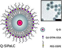 | ||
| Fig. 26 Schematic representation of a Q-SiPaLC (QD containing silica particle with a paramagnetic lipid coating). The inset shows a tem image (scale bar is 50 nm) of highly monodisperse, QD-containing, and hydrophilic silica particles (Q-Si). Schematic representations of Q-Si, Gd-DTPA-DSA, and PEG-DSPE lipids are shown in the legend (ref. 212). | ||
Biodegradable polysorbate 80-coated poly(n-butyl cyanoacrylate) (PBCA) nanoparticles can be used to deliver targeted fluorophores, which is blood–brain barrier (BBB) impermeable, into the brain to allow in vivo optical imaging of cellular and neuropathological structures, and to deliver BBB permeable MR contrast agents Gd-DTPA into the brain of living mice by using endogenous lapidated apolipoprotein E particles to facilitate BBB crossing.214 Guccione et al. reported a novel fluorescent and paramagnetically labelled polymerized gadolinium–rhodamine nanoparticles (Gd–Rd-NPs), a dual modality imaging probe, can be used for serial monitoring of cell trafficking in vivo MRI/optical imaging.215
Development of multimodal positive contrast agents is mostly based on Gd-DTPA terminated dendrimeric nanoparticles, Gd(III) chelate based lipid coating silica nanoparticles, Gd(III) chelate immobilized on QDs as mentioned previously. The potentials of Gd2O3 nanoparticles have been evaluated as multimodal contrast agents for in vivo imaging.216 The hybrid gadolinium oxide nanoparticles were prepared by encapsulating Gd2O3 cores by a polysiloxane shell containing organic fluorophores and covalent grafting of carboxylated PEG. The coating polysiloxane is porous for water molecular to contact the surface of crystalline core.
Gd(III) nanocarriers with fluorophore, strong photo-bleaching resistance and low toxicity are ideal for dual-modal imaging. Liu et al. developed a new approach to prepare Gd(III)-decorated hyperbranched polyglycerol (HPG) with a fluorescent polyhedral oligomeric silsesquioxane core (POSS-HPG-Gd) as a single molecular imaging probe for MRI/FI (Fig. 27).217 Hyperbranched molecules such as HPGs, added to hybrid nanodots composed of a rigid polyhedral oligomeric silsesquioxane (POSS) core surrounded by cationic conjugated oligoelectrolyte arms, then conjugated with DTPA-Gd to form hyperbranched architecture.218 The obtained hyperbranched molecules demonstrated excellent water solubility, low cytotoxicity and high biocompatibility. Fluorescent cell imaging of MCF-7 cancer cells demonstrated POSS-HPG has been successfully internalized into cytoplasm. Combined with MR imaging study, the POSS-HPG-Gd demonstrated a promising nanoprobe for FI/MRI. Further work from the Liu's group focused on the development of FI/MRI nanoprobes with targeting ability, such as cancer detection in live animals,219 cancer metastasis study.220
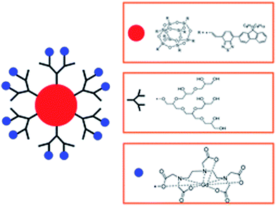 | ||
| Fig. 27 Schematic representation of POSS-HPG-Gd (ref. 217). | ||
Conclusion and outlook
Extensive research on development of Gd(III) based nanoparticle-based has been carried out to overcome the limitation of traditional Gd chelate-based contrast agents. These novel nanoparticle MRI contrast agents including inorganic crystalline Gd(III) contained nanoparticles and Gd(III) chelate-grafted macromolecular nanoparticle certainly offer delicate sensitivity and specific functionality in diagnostic and therapeutic applications such as diagnosis of cancer and metastasis. Various Gd(III) containing multifunctional nanomaterials can be used as multimodal imaging probes for introducing a paradigm shift in clinical applications.Several fundamental issues are remained, such as polar solvent-based synthetic method, surface modification for sophisticated nanoparticles, physicochemical properties such as chemical composition, shapes and sizes in order to accommodate biocompatibility and imaging properties; the major challenges in this field are functionalization of the multifunctional nanoparticles and the in vivo biological behavior of the nanoparticles such as their targeting efficiency, physiological stability and pharmacokinetics; obstacles still exist on integrated technology of combined MR and other imaging modalities for more therapeutic and diagnostic applications. All these challenges need to be addressed with an inter-disciplinary collaboration between chemist, biologist, engineer and clinician.
Acknowledgements
Authors acknowledge the support from Lee Kong Chian School of Medicine, Nanyang Technological University Start-Up Grant, Singapore. Authors thank Sachin Mishra for technical assistance and helpful comments.References
- G. Zabow, S. Dodd, J. Moreland and A. Koretsky, Nature, 2008, 453, 1058–1063 CrossRef CAS PubMed.
- R. Weissleder and U. Mahmood, Radiology, 2001, 219, 316–333 CrossRef CAS PubMed.
- W. Schima, A. Mukerjee and S. Saini, Clin. Radiol., 1996, 51, 235–244 CrossRef CAS PubMed.
- Textbook of Contrast Media, ed. P. Dawson, D. O. Cosgrove and R. G. Grainger, Isis Medical Media Ltd, Oxford, UK, 1999, section II Search PubMed.
- S. C. Jackels, Enhancement Agents for Magnetic Resonance Imaging: Fundamentals, in Pharmaceuticals in Medical Imaging, ed. D. P. Swanson, H. M. Chilton and J. H. Thrall, Macmillan, New York, 1990, p. 655 Search PubMed.
- The Chemistry of Contrast Agents in Medical Magnetic Resonance Imaging, ed. A. E. Merbach and E. Toth, John Wiley & Sons, Chichester, New York, 2001, p. 471 Search PubMed.
- P. Caravan, J. J. Ellison, T. J. McMurry and R. B. Lauffer, Chem. Rev., 1999, 99, 2293–2352 CrossRef CAS PubMed.
- S. Aime, M. Botta, M. Fasano and E. Terreno, Chem. Soc. Rev., 1998, 27, 19–29 RSC.
- S. Aime, A. Barge, C. Cabella, S. G. Crich and E. Gianolio, Curr. Pharm. Biotechnol., 2004, 5, 509–518 CAS.
- K. W.-Y. Chan and W.-T. Wong, Coord. Chem. Rev., 2006, 250, 1562–1579 CrossRef.
- Nanoplatform-Based Molecular Imaging, ed. X. Cheng, John Wiley & Sons, Inc., New Jersey, USA, 2011 Search PubMed.
- E. J. Delikatny and H. Poptani, Radiol. Clin., 2005, 43, 205–220 CrossRef.
- R. Weissleder, Science, 2006, 312, 1168–1171 CrossRef CAS PubMed.
- M. J. Sailor and J.-H. Park, Adv. Mater., 2012, 24, 3779–3802 CrossRef CAS PubMed.
- M. De, S. S. Chou, H. M. Joshi and V. P. Dravid, Adv. Drug Delivery Rev., 2011, 63, 1282–1299 CrossRef CAS PubMed.
- D. J. Heeger and D. Ress, Nat. Rev. Neurosci., 2002, 3, 142–151 CrossRef CAS PubMed.
- P. R. Seevinck, L. H. Deddens and R. M. Dijkhuizen, Angiogenesis, 2010, 13, 101–111 CrossRef PubMed.
- D. E. Sosnovik, M. Nahrendorf and R. Weissleder, Basic Res. Cardiol., 2008, 103, 122–130 CrossRef CAS PubMed.
- J. L. Major and T. J. Meade, Acc. Chem. Res., 2009, 42, 893–903 CrossRef CAS PubMed.
- F. Hu, H. M. Joshi, V. P. Dravid and T. J. Meade, Nanoscale, 2010, 2, 1884–1891 RSC.
- A. Taylor, K. M. Wilson, P. Murray, D. G. Fernig and R. Levy, Chem. Soc. Rev., 2012, 41, 2707–2717 RSC.
- P. W. Goodwill, E. U. Saritas, L. R. Croft, T. N. Kim, K. M. Krishnan, D. V. Schaffer and S. M. Conolly, Adv. Mater., 2012, 24, 3870–3877 CrossRef CAS PubMed.
- C. Fang and M. Zhang, J. Mater. Chem., 2009, 19, 6258–6266 RSC.
- C. Corot, P. Robert, J.-M. Idée and M. Port, Adv. Drug Delivery Rev., 2006, 58, 1471–1504 CrossRef CAS PubMed.
- F. Hu and Y. S. Zhao, Nanoscale, 2012, 4, 6235–6243 RSC.
- Z. R. Stephen, F. M. Kievit and M. Zhang, Mater. Today, 2011, 14, 330–338 CrossRef CAS PubMed.
- N. A. Frey, S. Peng, K. Cheng and S. Sun, Chem. Soc. Rev., 2009, 38, 2532–3542 RSC.
- L. Shan, A. Chopra, K. Leung, W. C. Eckelman and A. E. Menkens, J. Nanopart. Res., 2012, 14, 1122 CrossRef.
- E. Terreno, D. D. Castelli, A. Viale and S. Aime, Chem. Rev., 2010, 110, 3019–3042 CrossRef CAS PubMed.
- T. Lammers, S. Aime, W. Hennink, G. Strom and F. Kiessling, Acc. Chem. Res., 2011, 44, 1029–1038 CrossRef CAS PubMed.
- K. T. Rim, K. H. Koo and J. S. Park, Saf. Health Work, 2013, 4, 12–26 CrossRef CAS PubMed.
- T. Leiner and W. Kucharczyk, J. Magn. Reson. Imaging, 2009, 30, 1233–1235 CrossRef PubMed.
- P. Marckmann, L. Skov, K. Rossen, A. Dupont, M. B. Damholt, J. G. Heaf and H. S. Thomsen, J. Am. Soc. Nephrol., 2006, 17, 2359–2362 CrossRef PubMed.
- T. Grobner, Nephrol., Dial., Transplant., 2006, 21, 1104–1108 CrossRef CAS PubMed.
- J. M. Caillé, B. Lemanceau and B. Bonnemain, Am. J. Neuroradiol., 1983, 4, 1041–1042 Search PubMed.
- J. G. Penfield and R. F. Reilly, Nat. Clin. Pract. Nephrol., 2007, 3, 654–668 Search PubMed.
- P. H. Kuo, E. Kanal, A. K. Abu-Alfa and S. E. Cowper, Radiology, 2007, 242, 647–649 CrossRef PubMed.
- J. C. Bousquet, S. Saini, D. D. Stark, P. F. Hahn, M. Nigam, J. Wittenberg and J. T. Ferrucci Jr, Radiology, 1988, 166, 693–698 CrossRef CAS PubMed.
- C. Bouzigues, T. Gacoin and A. Alexandrou, ACS Nano, 2011, 5, 8488–8505 CrossRef CAS PubMed.
- N. Lewinski, V. Colvin and R. Drezek, Small, 2008, 4, 26–49 CrossRef CAS PubMed.
- L. Hernandez-Adame, N. Cortez-Espinosa, D. P. Portales-Pérez, C. Castillo, W. Zhao, Z. N. Juarez, L. R. Hernandez, H. Bach and G. Palestino, J. Biomed. Mater. Res., Part B, 2016 DOI:10.1002/jbm.b.33577.
- X. Tian, F. Yang, C. Yang, Y. Peng, D. Chen, J. Zhu, F. He, L. Li and X. Chen, Int. J. Nanomed., 2014, 9, 4043–4053 CrossRef PubMed.
- M. I. Setyawati, P. K. Khoo, B. H. Eng, S. Xiong, X. Zhao, G. K. Das, T. T. Tan, J. S. Loo, D. T. Leong and K. W. Ng, J. Biomed. Mater. Res., Part A, 2013, 101, 633–640 CrossRef PubMed.
- B. C. Heng, G. K. Das, X. Zhao, L.-L. Ma, T. T. Tan, K. W. Ng and J. S. Loo, Biointerphases, 2010, 5, FA88–FA97 CrossRef PubMed.
- H. Liu, G. Jia, S. Chen, H. Ma, Y. Zhao, J. Wang, C. Zhang, S. Wang and J. Zhang, RSC Adv., 2015, 5, 73601–73611 RSC.
- M. Longmire, P. L. Choyke and H. Kobayashi, Nanomedicine, 2008, 3, 703–717 CrossRef CAS PubMed.
- F. Hu and Y. S. Zhao, Nanoscale, 2012, 4, 6235–6243 RSC.
- H. B. Na, I. C. Song and T. Hyeon, Adv. Mater., 2009, 21, 2133–2148 CrossRef CAS.
- H. B. Na and T. Hyeon, J. Mater. Chem., 2009, 19, 6267–6273 RSC.
- S. H. Lee, B. H. Kim, H. B. Na and T. Hyeon, Wiley Interdiscip. Rev.: Nanomed. Nanobiotechnol., 2014, 6, 196–209 CAS.
- F. Chen, W. Bu, S. Zhang, X. Liu, J. Liu, H. Xing, Q. Xiao, L. Zhou, W. Peng, L. Wang and J. Shi, Adv. Funct. Mater., 2011, 21, 4285–4294 CrossRef CAS.
- D. Zhu, F. Liu, L. Ma, D. Liu and Z. Wang, Int. J. Mol. Sci., 2013, 14, 10591–10607 CrossRef PubMed.
- K. H. Bae, Y. B. Kim, Y. Lee, J. Hwang, H. Park and T. G. Park, Bioconjugate Chem., 2010, 21, 505–512 CrossRef CAS PubMed.
- H. Yang, Y. Zhang, Y. Sun, A. Dai, X. Shi, D. Wu, F. Wu, F. Li, H. Hu and S. Yang, Biomaterials, 2011, 32, 4584–4593 CrossRef CAS PubMed.
- J.-S. Choi, J.-H. Lee, T.-H. Shin, H.-T. Song, E. Y. Kim and J. Cheon, J. Am. Chem. Soc., 2010, 132, 11015–11017 CrossRef CAS PubMed.
- J. Y. Park, M. J. Baek, E. S. Choi, S. Woo, J. H. Kim, T. J. Kim, J. C. Jung, K. S. Chae, Y. Chang and G. H. Lee, ACS Nano, 2009, 3, 3663–3669 CrossRef CAS PubMed.
- A. T. M. A. Rahman, P. Majewski and K. Vasilev, Contrast Media Mol. Imaging, 2013, 8, 92–95 CrossRef PubMed.
- G. Azizian, N. Riyahi-Alam, S. Haghgoo, H. R. Moghimi, R. Zohdiaghdam, B. Rafiei and E. Gorji, Nanoscale Res. Lett., 2012, 7, 549–555 CrossRef PubMed.
- M.-A. Fortin, R. M. Petoral Jr, F. Söderland, A. Klasson, M. Engström, T. Veres, P.-O. Käll and K. Uvdal, Nanotechnology, 2007, 18, 5501–5509 CrossRef.
- L. Faucher, Y. Gossuin, A. Hocq and M. A. Fortin, Nanotechnology, 2011, 22, 295103 CrossRef PubMed.
- M. Engström, A. Klasson, H. Pedersen, C. Vahlberg, P.-O. Käll and K. Uvdal, Magma, 2006, 19, 180–186 CrossRef PubMed.
- G. Le Duc, I. Miladi, C. Alric, P. Mowat, E. Bräuer-Krisch, A. Bouchet, E. Khalil, C. Billotey, M. Janier, F. Lux, T. Epicier, P. Perriat, S. Roux and O. Tillement, ACS Nano, 2011, 5, 9566–9574 CrossRef CAS PubMed.
- J. Miyawaki, M. Yudasaka, H. Imai, H. Yorimitsu, H. Isobe, E. Nakamura and S. Lijima, J. Phys. Chem. B, 2006, 110, 5179–5181 CrossRef CAS PubMed.
- L. Faucher, A.-A. Guay-Begin, J. Lagueux, M.-F. Cote, E. Petitclerc and M.-A. Fortin, Contrast Media Mol. Imaging, 2011, 6, 209–218 CAS.
- M. Engström, A. Klasson, H. Pedersen, C. Vahlberg, P.-O. mKäll and K. Uvdal, Magn. Reson. Mater. Phys., Biol. Med., 2006, 19, 180–186 CrossRef PubMed.
- F. Evanics, P. R. Diamente, F. van Veggle, G. J. Stannisz and R. S. Prosser, Chem. Mater., 2006, 18, 2499–2505 CrossRef CAS.
- N. J. J. Johnson, W. Oakden, G. J. Stanisz, R. S. Prosser and F. C. J. M. van Veggel, Chem. Mater., 2011, 23, 3714–3722 CrossRef CAS.
- H. Hifumi, S. Yamaoka, A. Tanimoto, T. Akatsu, Y. Shindo, A. Honda, D. Citterio, K. Oka, S. Kuribayashi and K. Suzuki, J. Mater. Chem., 2009, 19, 6393–6399 RSC.
- H. Hifumi, S. Yamaoka, A. Tanimoto, D. Citterio and K. Suzuki, J. Am. Chem. Soc., 2006, 128, 15090–15091 CrossRef CAS PubMed.
- Y.-S. Yoon, B.-I. Lee, K. S. Lee, H. Heo, J. H. Lee, S.-H. Byeon and I. S. Lee, Chem. Commun., 2010, 46, 3654–3656 RSC.
- C. R. Kim, J. S. Baeck, Y. Chang, J. E. Bae, K. S. Chae and G. H. Lee, Phys. Chem. Chem. Phys., 2014, 16, 19866–19873 RSC.
- D. Kim, N. Lee, M. Park, B. H. Kim, K. An and T. Hyeon, J. Am. Chem. Soc., 2009, 131, 454–455 CrossRef CAS PubMed.
- F. Wang, Y. Han, C. S. Lim, Y. Lu, J. Wang, J. Xu, H. Chen, C. Zhang, M. Hong and X. Liu, Nature, 2010, 263, 1061–1065 CrossRef PubMed.
- R. Kooler, P. Schapotschnokow, C. de Mello Donega, T. J. H. Vlugt and A. Meijerink, ACS Nano, 2008, 2, 1703–1704 CrossRef PubMed.
- J. Fang, P. Chandrasekharan, X.-L. Liu, Y. Yang, Y.-B. Lv, C.-T. Yan and J. Ding, Biomaterials, 2014, 35, 1636–1642 CrossRef CAS PubMed.
- Y. Hou, R. Qiao, F. Fang, X. Wang, C. Dong, K. Liu, C. Liu, Z. Liu, H. Lei, F. Wang and M. Gao, ACS Nano, 2013, 7, 330–338 CrossRef CAS PubMed.
- G. Liang, L. Cao, H. Chen, Z. Zhang, S. Zhang, S. Yu, X. Shen and J. Kong, J. Mater. Chem. B, 2013, 1, 629–638 RSC.
- C.-L. Tseng, I.-L. Shih, L. Stobinski and F.-H. Lin, Biomaterials, 2010, 31, 5427–5435 CrossRef CAS PubMed.
- K. M. L. Taylor, J. S. Kim, W. J. Rieter, H. An, W. Lin and W. Lin, J. Am. Chem. Soc., 2008, 130, 2154–2155 CrossRef CAS PubMed.
- Y.-Z. Shao, L.-Z. Liu, S.-Q. Song, R.-H. Cao, H. Liu, C.-Y. Cui, X. Li, M. J. Bie and L. Li, Contrast Media Mol. Imaging, 2011, 6, 110–118 CrossRef CAS PubMed.
- G. Bhakta, R. K. Sharma, N. Gupta, S. Cool, V. Nurcombe and A. Maitra, Nanomedicine, 2011, 7, 472–479 CrossRef CAS PubMed.
- S. T. Selvan, T. T. Y Tan, D. K. Yi and N. R. Jana, Langmuir, 2010, 26, 11631–11641 CrossRef CAS PubMed.
- F. Carniato, L. Tei, W. Dastru, L. Marchese and M. Botta, Chem. Commun., 2009, 10, 1246–1248 RSC.
- J. K. Hsiao, C. P. Tsai, T. H. Chung, Y. Hung, M. Yao, H. M. Liu, C. Y. Mou, C. S. Yang, Y. C. Chen and D. M. Huang, Small, 2008, 4, 1445–1452 CrossRef CAS PubMed.
- Y.-S. Lin, S.-H. Wu, Y. Hung, Y.-H. Chou, C. Chang, M.-L. Lin, C.-P. Tsi and C.-Y. Mou, Chem. Mater., 2006, 18, 5170–5172 CrossRef CAS.
- I. Slowing, B. G. Trewyn and V. S. Y. Lin, J. Am. Chem. Soc., 2006, 128, 14792–14793 CrossRef CAS PubMed.
- Y. Shao, X. Tian, W. Hu, Y. Zhang, H. Liu, H. He, Y. Shen, F. Xie and L. Li, Biomaterials, 2012, 33, 6438–6446 CrossRef CAS PubMed.
- R. D. Bolskar, Nanomedicine, 2008, 3, 201–213 CrossRef CAS PubMed.
- B. Sitharaman, R. D. Bolskar, I. Rusakova and L. J. Wilson, Nano Lett., 2004, 4, 2373–2378 CrossRef CAS.
- R. D. Bolskar, A. F. Benedetto, L. O. Husebo, R. E. Price, E. F. Jackson, S. Wallace, L. J. Wilson and J. M. Alford, J. Am. Chem. Soc., 2003, 125, 5471–5478 CrossRef CAS PubMed.
- É. Tóth, R. D. Bolskar, A. Borel, G. González, L. Helm, A. E. Merbach, B. Sitharaman and L. J. Wilson, J. Am. Chem. Soc., 2005, 127, 799–805 CrossRef PubMed.
- B. Sitharaman, L. A. Tran, Q. P. Pham, R. D. Bolskar, R. Muthupillai, S. D. Flamm, A. G. Mikos and L. J. Wilson, Contrast Media Mol. Imaging, 2007, 2, 139–146 CrossRef CAS PubMed.
- H. Kato, Y. Kanazawa, M. Okumura, A. Taninaka, T. Yokawa and H. Shinohara, J. Am. Chem. Soc., 2003, 125, 4391–4397 CrossRef CAS PubMed.
- K. B. Hartman, S. Laus, R. D. Bolskar, R. Muthupillai, L. Helm, É. Tóth, A. E. Merbach and L. J. Wilson, Nano Lett., 2008, 8, 415–418 CrossRef CAS PubMed.
- B. Sitharaman, K. R. Kissell, K. B. Hartman, L. A. Tran, A. Baikalov, I. Rusakova, Y. Sun, H. A. Khant, S. J. Ludtke, W. Chiu, S. Laus, É. Tóth, L. Helm, A. E. Merbach and L. J. Wilson, Chem. Commun., 2005, 3915–3917 RSC.
- H. L. Fillmore, M. D. Shultz, S. C. Henderson, P. Cooper, W. C. Broaddus, Z. J. Chen, C.-Y. Shu, J. Zhang, J. Ge, H. C. Dorn, F. Corwin, J. I. Hirsch, J. Wilson and P. P. Fatouros, Nanomedicine, 2011, 6, 449–458 CrossRef CAS PubMed.
- P. Caravan, Chem. Soc. Rev., 2006, 35, 512–523 RSC.
- S. Aime, M. Botta and E. Terreno, Adv. Inorg. Chem., 2005, 57, 173–237 CrossRef CAS.
- P. Hermann, J. Kotek, V. Kubíček and I. Lukeš, Dalton Trans., 2008, 3027–3047 RSC.
- C.-T. Yang and K.-H. Chuang, Med. Chem. Commun., 2012, 3, 552–565 RSC.
- P.-J. Debouttière, S. Roux, F. Vocanson, C. Billotey, O. Beuf, A. Favre-Réguillon, Y. Lin, S. Pellet-Rostaing, R. Lamartine, P. Perriat and O. Tillement, Adv. Funct. Mater., 2006, 16, 2330–2339 CrossRef.
- L. Moriggi, C. Cannizzo, E. Dumas, C. R. Mayer, A. Ulianov and L. Helm, J. Am. Chem. Soc., 2009, 131, 10828–10829 CrossRef CAS PubMed.
- V. Mogilireddy, I. Dechamps-Olivier, C. Alric, G. Laurent, S. Laurent, L. Vander Elst, R. Muller, R. Bazzi, S. Roux, O. Tillement and F. Chiburu, Contrast Media Mol. Imaging, 2015, 10, 179–187 CrossRef CAS PubMed.
- X. Tian, Y. Shao, H. He, H. Liu, Y. Shen, W. Huang and L. Li, Nanoscale, 2013, 5, 3322–3329 RSC.
- C. H. Reynolds, N. Annan, K. Beshah, J. H. Huber, S. H. Shaber, R. E. Lenkinski and J. A. Wortmam, J. Am. Chem. Soc., 2000, 122, 8940–8945 CrossRef CAS.
- J. L. Turner, D. Pan, R. Plummer, Z. Chen, A. K. Whittaker and K. L. Wooley, Adv. Funct. Mater., 2005, 15, 1248–1254 CrossRef CAS.
- J. C. Frias, Y. Ma, K. J. Williams, Z. A. Fayad and E. A. Fisher, Nano Lett., 2006, 6, 2220–2224 CrossRef CAS PubMed.
- E. A. Anderson, S. Isaacmam, D. S. Peabody, E. Y. Wang, J. W. Canary and K. Kirshenbaum, Nano Lett., 2006, 6, 1160–1164 CrossRef CAS PubMed.
- S. R. Bull, M. O. Guler, R. E. Bras, T. J. Meade and S. I. Stupp, Nano Lett., 2005, 5, 1–4 CrossRef CAS PubMed.
- S. R. Bull, M. O. Guler, R. E. Bras, P. N. Venkatasubramanian, S. I. Stupp and T. J. Meade, Bioconjugate Chem., 2005, 16, 1343–1348 CrossRef CAS PubMed.
- L. Cao, B. Li, P. Yi, H. Zhang, J. Dai, B. Tan and Z. Deng, Biomaterials, 2014, 35, 4168–4174 CrossRef CAS PubMed.
- C. P. Platas-Iglesias, L. V. Elst, W. Zhou, R. N. Muller, C. F. G. C. Geraldes, T. Maschmeyer and J. A. Peters, Chem.–Eur. J., 2002, 8, 5121–5131 CrossRef CAS.
- Y. Kobayashi, H. Morimoto, T. Nakagawa, Y. Kubota, K. Gonda and N. Ohuchi, ISRN Nanotechnol., 2013, 1–6 Search PubMed.
- K. S. Kim, W. Park and K. Na, Biomaterials, 2015, 36, 90–97 CrossRef CAS PubMed.
- M. Botta and L. Tei, Eur. J. Inorg. Chem., 2012, 12, 1945–1960 CrossRef.
- C.-H. Huang and A. Tsourkas, Curr. Top. Med. Chem., 2013, 13, 411–421 CrossRef CAS PubMed.
- M. W. Rotz, K. S. Culver, G. Parigi, K. W. MacRenaris, C. Luchinat, T. W. Odom and T. J. Meade, ACS Nano, 2015, 9, 3385–3396 CrossRef CAS PubMed.
- F. J. Nicholls, M. W. Rotz, H. Ghuman, K. W. MacRenaris, T. J. Meade and M. Modo, Biomaterials, 2016, 77, 291–306 CrossRef CAS PubMed.
- E. C. Wiener, M. W. Brechbiel, H. Brothers, R. L. Magin, O. A. Gansow, D. A. Tomalia and P. C. Lauterbur, Magn. Reson. Med., 1994, 31, 1–8 CrossRef CAS PubMed.
- Y. Fu, H. J. Raatschen, D. E. Nitecki, M. F. Wendland, V. Novikov, L. S. Fournier, C. Cyran, V. Rogut, D. M. Shames and R. C. Brasch, Biomacromolecules, 2007, 8, 1519–1529 CrossRef CAS PubMed.
- S. Langereis, Q. G. de Lussanet, M. H. van Genderen, E. W. Meijer, R. G. Beets-Tan, A. W. Griffioen, J. M. van Engelshoven and W. H. Backes, NMR Biomed., 2006, 19, 133–141 CrossRef CAS PubMed.
- Z. Jászberényi, L. Moriggi, P. Schmidt, C. Weidensteiner, R. Kneuer, A. E. Merbach, L. Helm and É. Tóth, J. Biol. Inorg. Chem., 2007, 12, 406–420 CrossRef PubMed.
- J. Rudovský, M. Botta, P. Hermann, K. I. Hardcastle, I. Lukes and S. Aime, Bioconjugate Chem., 2006, 17, 975–987 CrossRef PubMed.
- P. Lebdusková, A. Sour, L. Helm, É. Tóth, J. Kotek, I. Lukes and A. E. Merbach, Dalton Trans., 2006, 28, 3399–3406 RSC.
- S. Laus, A. Sour, R. Ruloff, É. Tóth and A. E. Merbach, Chem.–Eur. J., 2005, 11, 3064–3076 CrossRef CAS PubMed.
- K. E. Løkling, S. L. Fossheim, R. Skurtveit, A. Bjørnerud and J. Klaveness, Magn. Reson. Imaging, 2001, 19, 731–738 CrossRef.
- T. Wang, M. Hossann, H. M. Reinl, M. Peller, H. Eibl, M. Reiser, R. D. Issel and L. H. Lindner, Contrast Media Mol. Imaging, 2008, 3, 19–26 CrossRef CAS PubMed.
- G. Zhang, R. Zhang, X. Wen, L. Li and C. Li, Biomacromolecules, 2008, 9, 36–42 CrossRef CAS PubMed.
- P. Gong, Z. Chen, Y. Chen, W. Wang, X. Wang and A. Hu, Chem. Commun., 2011, 47, 4240–4242 RSC.
- S. Kaida, H. Cabral, M. Kumagai, A. Kishimura, Y. Terada, M. Sekino, I. Aoki, N. Nishiyama, T. Tani and K. Kataoka, Cancer Res., 2010, 70, 7031–7041 CrossRef CAS PubMed.
- T. Liu, Y. Qian, X. Hu, Z. Ge and S. Liu, J. Mater. Chem., 2012, 22, 5020–5030 RSC.
- Y. Li, M. Beija, S. Laurent, L. Vander Elst, R. N. Muller, H. T. T. Duong, A. B. Lowe, T. P. Davis and C. Boyer, Macromolecules, 2012, 45, 4196–4204 CrossRef CAS.
- Y. Li, S. Laurent, L. Esser, L. Vander Elst, R. N. Muller, A. B. Lowe, C. Boyer and T. P. Davis, Polym. Chem., 2014, 5, 2592–2601 RSC.
- X. Hu, G. Liu, Y. Li, X. Wang and S. Liu, J. Am. Chem. Soc., 2015, 137, 362–368 CrossRef CAS PubMed.
- H. Kobayashi and M. W. Brechbiel, Adv. Drug Delivery Rev., 2005, 57, 2271–2286 CrossRef CAS PubMed.
- K. Nwe, H. Xu, C. A. S. Regino, M. Bernardo, L. Ileva, L. Riffle, K. J. Wong and M. W. Brechbiel, Bioconjugate Chem., 2009, 20, 1412–1418 CrossRef CAS PubMed.
- K. Nwe, L. H. Bryant Jr and M. W. Brechbiel, Bioconjugate Chem., 2010, 21, 1014–1017 CrossRef CAS PubMed.
- K. Nwe, M. Bernardo, C. A. S. Regino, M. Williams and M. W. Brechbiel, Bioorg. Med. Chem., 2010, 18, 5925–5931 CrossRef CAS PubMed.
- K. Nwe, D. Milenic, L. H. Bryant, C. A. S. Regino and M. W. Brechbiel, J. Inorg. Biochem., 2011, 105, 722–727 CrossRef CAS PubMed.
- D. A. Tomalia, L. A. Reyna and S. Svenson, Biochem. Soc. Trans., 2007, 35, 61–67 CrossRef CAS PubMed.
- Z. Cheng, D. L. J. Thorek and A. Tsourkas, Angew. Chem., Int. Ed., 2010, 49, 346–350 CrossRef CAS PubMed.
- M. Ye, Y. Qian, Y. Shen, H. Hu, M. Sui and J. Tang, J. Mater. Chem., 2012, 22, 14369–14377 RSC.
- M. Ye, Y. Qian, J. Tang, H. Hu, M. Sui and Y. Shen, J. Controlled Release, 2013, 169, 239–245 CrossRef CAS PubMed.
- H. Cabral, N. Nishiyama and K. Kataoka, Acc. Chem. Res., 2011, 44, 999–1008 CrossRef CAS PubMed.
- K. Kimpe, T. Parac-Vogt, S. Laurent, C. Pierart, L. Vander Elst, R. N. Muller and K. Binnemans, Eur. J. Inorg. Chem., 2003, 3021–3027 CrossRef CAS.
- H. Tournier, R. Hyacinthe and M. Schneider, Acad. Radiol., 2002, 9, S20–S28 CrossRef PubMed.
- P. L. Anelli, L. Lattuada, V. Lorusso, M. Schneider, H. Tournier and F. Uggeri, Magn. Reson. Mater. Phys., Biol. Med., 2001, 12, 114–120 CrossRef CAS.
- H. Y. Lee, H. W. Jee, S. M. Seo, B. K. Kwak, G. Khang and S. H. Cho, Bioconjugate Chem., 2006, 17, 700–706 CrossRef CAS PubMed.
- T. Liu, X. Li, Y. Qian, X. Hua and S. Liu, Biomaterials, 2012, 33, 2521–2531 CrossRef CAS PubMed.
- Y. Liu and N. Zhang, Biomaterials, 2012, 33, 5363–5375 CrossRef CAS PubMed.
- K. Na, S. A. Lee, S. H. Jung and B. C. Shin, Colloids Surf., B, 2011, 84, 82–87 CrossRef CAS PubMed.
- M. de Smet, S. Langereis, S. van den Bosch and H. Grüll, J. Controlled Release, 2010, 143, 120–127 CrossRef CAS PubMed.
- Z. Chen, D. Yu, S. Wang, N. Zhang, C. Ma and Z. Lu, Nanoscale Res. Lett., 2009, 4, 618–626 CrossRef CAS PubMed.
- K. Shiraishi, K. Kawano, Y. Maitani and M. Yokoyama, J. Controlled Release, 2010, 148, 160–167 CrossRef CAS PubMed.
- K.-W. Hu, K.-C. Hsu and C.-S. Yeh, Biomaterials, 2010, 31, 6843–6848 CrossRef CAS PubMed.
- Y. Li, Y. Qian, T. Liu, G. Zhang, J. Hu and S. Liu, Polym. Chem., 2014, 5, 1743–1750 RSC.
- A. W. Jackson, P. Chandrasekharan, J. Shi, S. P. Rannard, Q. Liu, C.-T. Yang and T. He, Int. J. Nanomed., 2015, 5895–5907 CAS.
- J. Kim, Y. Piao and T. Hyeon, Chem. Soc. Rev., 2009, 38, 372–390 RSC.
- L. E. Jennings and N. J. Long, Chem. Commun., 2009, 3511–3524 RSC.
- A. Louie, Chem. Rev., 2010, 110, 3146–3195 CrossRef CAS PubMed.
- W. Xu, K. Kattel, J. Y. Park, Y. T. Chang, J. Kim and G. H. Lee, Phys. Chem. Chem. Phys., 2012, 14, 12687–12700 RSC.
- J. H. Gao, H. W. Gu and B. Xu, Acc. Chem. Res., 2009, 42, 1097–1107 CrossRef CAS PubMed.
- P. Verwilst, S. Park, B. Yoon and J. S. Kim, Chem. Soc. Rev., 2015, 44, 1791–1807 RSC.
- J.-H. Lee, Y.-W. Jun, J.-S. Yeon and J. Cheon, Angew. Chem., Int. Ed., 2006, 45, 8160–8162 CrossRef CAS PubMed.
- W.-C. Law, K.-T. Yong, I. Roy, G. Xu, H. Ding, E. J. Bergey, H. Zeng and P. N. Prasad, J. Phys. Chem. C, 2008, 112, 7972–7977 CrossRef CAS.
- J. Kim, J. E. Lee, J. Lee, Y. Jang, S.-W. Kim, K. An, J. H. Yu and T. Hyeon, Angew. Chem., Int. Ed., 2006, 45, 4789–4793 CrossRef CAS PubMed.
- Y.-S. Lin, S.-H. Wu, Y. Hung, Y.-H. Chou, C. Chang, M.-L. Lin, C.-P. Tsai and C.-Y. Mou, Chem. Mater., 2006, 18, 5170–5172 CrossRef CAS.
- J. Zhao, S. L. Song, M. Zhong and C. Li, ACS Macro Lett., 2012, 1, 150–153 CrossRef CAS PubMed.
- H. M. Kim, Y. W. Noh, H. S. Park, M. Y. Cho, K. S. Hong, H. Lee, D. H. Shin, J. Kang, M. H. Sung, H. Poo and Y. T. Lim, Small, 2012, 8, 666–670 CrossRef CAS PubMed.
- J. Kim, J. E. Lee, S. H. Lee, J. H. Yu, J. H. Lee, T. G. Park and T. Hyeon, Adv. Mater., 2008, 20, 478–483 CrossRef CAS.
- J.-L. Bridot, A.-C. Faure, S. Laurent, C. Rivière, C. Billotey, B. Hiba, M. Janier, V. Josserand, J.-L. Coll, L. Vander Elst, R. Muller, S. Roux, P. Perriat and O. Tillement, J. Am. Chem. Soc., 2007, 129, 5076–5084 CrossRef CAS PubMed.
- D. Kryza, J. Taleb, M. Janier, L. Marmuse, I. Miladi, P. Bonazza, C. Louis, P. Perriat, S. Roux, O. Tillement and C. Billotey, Bioconjugate Chem., 2011, 22, 1145–1152 CrossRef CAS PubMed.
- M. W. Ahmad, W. Xu, S. J. Kim, J. S. Baeck, Y. Chang, J. E. Bae, K. S. Chae, J. A. Park, T. J. Kim and G. H. Lee, Sci. Rep., 2015, 5, 8549 CrossRef CAS PubMed.
- Z. Gu, L. Yan, G. Tian, S. Li, Z. Chai and Y. Zhao, Adv. Mater., 2013, 25, 3758–3779 CrossRef CAS PubMed.
- S. Lee and X. Chen, Mol. Imaging, 2009, 8, 87–100 CrossRef CAS PubMed.
- R. M. Petoral, F. Söderlind, A. Klasson, A. Suska, M. A. Fortin, N. Abrikossova, L. Selegård, P.-O. Käll, M. Engström and K. Uvdal, J. Phys. Chem. C, 2009, 113, 6913–6920 CrossRef CAS.
- G. K. Das, B. C. Heng, S.-C. Ng, T. White, J. S. C. Loo, L. D'Silva, P. Padmanabhan, K. K. Bhakoo, S. T. Selvan and T. T. Y. Tan, Langmuir, 2010, 26, 8959–8965 CrossRef CAS PubMed.
- Y. I. Park, J. H. Kim, K. T. Lee, K.-S. Jeon, H. B. Na, J. H. Yu, H. M. Kim, N. Lee, S. H. Choi, S.-I. Baik, H. Kim, S. P. Park, B.-J. Park, Y. W. Kim, S. H. Lee, S.-Y. Yoon, I. C. Song, W. K. Moon, Y. D. Suh and T. Hyeon, Adv. Mater., 2009, 21, 4467–4471 CrossRef CAS.
- Q. Ju, D. Tu, Y. Liu, R. Li, H. Zhu, J. Chen, Z. Chen, M. Hunag and X. Chen, J. Am. Chem. Soc., 2011, 134, 1323–1330 CrossRef PubMed.
- A. Xia, M. Chen, Y. Gao, D. M. Wu, W. Feng and F. Y. Li, Biomaterials, 2012, 33, 5394–5405 CrossRef CAS PubMed.
- Z. Liu, F. Pu, S. Huang, Q. Yuan, J. Ren and X. Qu, Biomaterials, 2013, 34, 1712–1721 CrossRef CAS PubMed.
- M. He, P. Huang, C. Zhang, H. Hu, C. Bao, G. Gao, R. He and D. Cui, Adv. Funct. Mater., 2011, 21, 4470–4477 CrossRef CAS.
- S. J. Zeng, M.-K. Tsang, C.-F. Chan, K.-L. Wong and J. H. Hao, Biomaterials, 2012, 33, 9232–9238 CrossRef CAS PubMed.
- S. Wen, K. Li, H. Cai, Q. Chen, M. Shen, Y. Huang, C. Peng, W. Hou, M. Zhu, G. Zhang and X. Shi, Biomaterials, 2013, 34, 1570–1580 CrossRef CAS PubMed.
- J. Jung, M. A. Kim, J.-H. Cho, S. J. Lee, I. Yang, J. Cho, S. K. Kim, C. Lee and J. K. Park, Biomaterials, 2012, 33, 5865–5874 CrossRef CAS PubMed.
- G. K. Das, Y. Zhang, L. D'Silva, P. Padmanabhan, B. C. Heng, J. S. C. Loo, S. T. Selvan, K. K. Bhakoo and T. T. Y. Tan, Chem. Mater., 2011, 21, 2439–2446 CrossRef.
- D. R. Arifin, C. M. Long, A. A. Gilad, C. Alric, S. Roux, O. Tillement, T. W. Link, A. Arepally and J. W. Bulte, Radiology, 2011, 260, 790–798 CrossRef PubMed.
- Q. Chen, K. Li, S. Wen, H. Liu, C. Peng, H. Cai, M. Shen, G. Zhang and X. Shi, Biomaterials, 2013, 34, 5200–5209 CrossRef CAS PubMed.
- S. K. Sun, L. X. Dong, Y. Cao, H. R. Sun and X. P. Yan, Anal. Chem., 2013, 85, 8436–8441 CrossRef CAS PubMed.
- K. Li, S. Wen, A. C. Larson, M. Shen, Z. Zhang, Q. Chen, X. Shi and G. Zhang, Int. J. Nanomed., 2013, 8, 2589–2600 CrossRef PubMed.
- Y. Zeng, D. Zhang, M. Wu, Y. Liu, X. Zhang, L. Li, Z. Li, X. Han, X. Wei and X. Liu, ACS Appl. Mater. Interfaces, 2014, 6, 14266–14277 Search PubMed.
- F. Lux, L. Sancey, A. Bianchi, Y. Crémillieux, S. Roux and O. Tillement, Nanomedicine, 2015, 10, 1801–1815 CrossRef CAS PubMed.
- G. Le Duc, S. Roux, A. Paruta-Tuarez, S. Dufort, E. Brauer, A. Marais, C. Truillet, L. Sancey, P. Perriat, F. Lux and O. Tillement, Cancer Nanotechnol., 2014, 5, 1–14 CrossRef PubMed.
- C. Alric, J. Taleb, G. L. Duc, C. Mandon, C. Bilotey, A. L. Meur-Herland, T. Brochard, F. Vocanson, M. Janier, P. Perriat, S. Roux and O. J. Tillement, J. Am. Chem. Soc., 2008, 130, 5908–5915 CrossRef CAS PubMed.
- J.-A. Park, H.-K. Kim, J.-H. Kim, S.-W. Jeong, J.-C. Jung, G.-H. Lee, J. Lee, Y. Chang and T.-J. Kim, Bioorg. Med. Chem. Lett., 2010, 20, 2287–2291 CrossRef CAS PubMed.
- N. S. Md, H.-K. Kim, J.-A. Park, Y. Chang and T.-J. Kim, Bull. Korean Chem. Soc., 2010, 31, 1177–1181 CrossRef CAS.
- V. S. Talanov, C. A. S. Regino, H. Kobayashi, M. Bernardo, P. L. Choyke and M. W. Brechbiel, Nano Lett., 2006, 6, 1459–1463 CrossRef CAS PubMed.
- Y. Koyama, V. S. Talanov, M. Bernardo, Y. Hama, C. A. S. Regino, M. W. Brechbiel, P. L. Choyke and H. Kobayashi, J. Magn. Reson. Imaging, 2007, 25, 866–871 CrossRef PubMed.
- H. Xu, C. A. S. Regino, Y. Koyama, Y. Hama, A. J. Gunn, M. Bernardo, H. Kobayashi, P. L. Choyke and M. W. Brechbiel, Bioconjugate Chem., 2007, 18, 1474–1482 CrossRef CAS PubMed.
- J. Hu, T. Liu, G. Zhang, F. Jin and S. Liu, Macromol. Rapid Commun., 2013, 34, 749–758 CrossRef CAS PubMed.
- E. S. Olson, T. Jiang, T. A. Aguilera, Q. T. Nguyen, L. G. Ellies, M. Scadeng and R. Y. Tsien, Proc. Natl. Acad. Sci. U. S. A., 2010, 107, 4311–4316 CrossRef CAS PubMed.
- A. J. L. Villaraza, A. Bumb and M. B. Brechbiel, Chem. Rev., 2010, 110, 2921–2959 CrossRef CAS PubMed.
- X. Michalet, F. F. Pinaud, L. A. Bentolia, J. M. Tsay, S. Doose, J. J. Li, G. Sundaresan, A. M. Wu, S. S. Gambhir and S. Weiss, Science, 2005, 307, 538–544 CrossRef CAS PubMed.
- W. J. Mulder, R. Koole, R. J. Brandwijk, G. Storm, P. T. K. Chin, G. J. Strijkers, C. de Mello Donegá, K. Nicolay and A. W. Griffioen, Nano Lett., 2006, 6, 1–6 CrossRef CAS PubMed.
- H. Yang, S. Santra, G. A. Walter and P. H. Holloway, Adv. Mater., 2006, 18, 2890–2894 CrossRef CAS.
- S. Santra, H. Yang, P. H. Holloway, J. T. Stanley and R. A. Mericle, J. Am. Chem. Soc., 2005, 127, 1656–1657 CrossRef CAS PubMed.
- S. Wang, B. R. Jarrett, S. M. Kauzlarich and A. Y. Louie, J. Am. Chem. Soc., 2007, 129, 3848–3856 CrossRef CAS PubMed.
- G. A. F. van Tilborg, W. J. M. Mulder, P. T. K. Chin, G. Storm, C. P. Reutelingsperger, K. Nicolay and G. J. Strijkers, Bioconjugate Chem., 2006, 17, 865–868 CrossRef CAS PubMed.
- L. Prinzen, R.-J. Miserus, A. Dirksen, T. M. Hackeng, N. Deckers, N. J. Bitsch, R. T. Megens, K. Douma, J. W. Heemskerk, M. E. Kooi, P. M. Frederik, D. W. Slaaf, M. A. van Zandvoot and C. P. Reutelingsperger, Nano Lett., 2007, 7, 93–100 CrossRef CAS PubMed.
- R. A. Weissleder, Nat. Biotechnol., 2001, 19, 316–317 CrossRef CAS PubMed.
- T. Jin, Y. Yoshioka, F. Fujii, Y. Komai, J. Seki and A. Seiyama, Chem. Commun., 2008, 5764–5766 RSC.
- M. M. van Schooneveld, E. Vucic, R. Koole, Y. Zhou, J. Stocks, D. P. Cormode, C. Y. Tang, R. E. Gordon, K. Nicolay, A. Meijerink, Z. A. Fayad and W. J. M. Mulder, Nano Lett., 2008, 8, 2517–2525 CrossRef CAS PubMed.
- W. J. Rieter, J. S. Kim, K. M. L. Taylor, H. An, W. Lin, T. Tarrant and W. Lin, Angew. Chem., Int. Ed., 2007, 46, 3680–3682 CrossRef CAS PubMed.
- R. M. Koffie, C. T. Farrar, L.-J. Saidi, C. M. William and B. T. Hyman, Proc. Natl. Acad. Sci. U. S. A., 2011, 108, 18837–18842 CrossRef CAS PubMed.
- K. Vuu, J. Xie, M. A. McDonald, M. Bernardo, F. Hunter, Y. Zhang, K. Li, M. Bednarski and S. Guccione, Bioconjugate Chem., 2005, 16, 995–999 CrossRef CAS PubMed.
- J.-L. Bridot, A.-C. Faure, S. Laurent, C. Rivière, C. Billotey, B. Hiba, M. Janier, V. Josserand, J.-L. Coll, L. Vander Elst, R. Muller, S. Roux, P. Perriat and O. Tillement, J. Am. Chem. Soc., 2007, 129, 5076–5084 CrossRef CAS PubMed.
- J. Liu, K. Li, J. Geng, L. Zhou, C. Prashant, C.-T. Yang and B. Liu, Polym. Chem., 2013, 4, 1517–1524 RSC.
- M. Calderón, M. A. Quadir, S. K. Sharma and R. Haag, Adv. Mater., 2010, 22, 190–218 CrossRef PubMed.
- D. Ding, G. Wang, J. Liu, K. Li, K.-Y. Pu, Y. Hu, J. C. Y. Ng, B. Z. Tang and B. Liu, Small, 2012, 8, 3523–3530 CrossRef CAS PubMed.
- K. Li, D. Ding, C. Prashant, W. Qin, C.-T. Yang, B. Z. Tang and B. Liu, Adv. Healthcare Mater., 2013, 2, 1600–1605 CrossRef CAS PubMed.
| This journal is © The Royal Society of Chemistry 2016 |


