Mapping deformation dynamics to composition of topologically-active DNA blends†
Karthik R.
Peddireddy
 ,
Ryan
McGorty
 and
Rae M.
Robertson-Anderson
 *
*
Department of Physics and Biophysics, University of San Diego, 5998 Alcala Park, San Diego, CA 92110, USA. E-mail: randerson@sandiego.edu
First published on 28th October 2024
Abstract
Blends of circular and linear polymers have fascinated researchers for decades, and the role of topology on their stress response and dynamics remains fervently debated. While linear polymers adopt larger coil sizes and form stronger, more pervasive entanglements than their circular counterparts, threading of circular polymers by linear chains can introduce persistent constraints that dramatically decrease mobility, leading to emergent rheological properties in blends. However, the complex interplay between topology-dependent polymer overlap and threading propensity, along with the large amounts of material required to sample many compositions, has limited the ability to experimentally map stress response to composition with high resolution. Moreover, the role of supercoiling on the response of circular-linear blends remains poorly understood. Here, we leverage in situ enzymatic topological conversion to map the deformation dynamics of DNA blends with over 70 fractions of linear, ring and supercoiled molecules that span the phase space of possible topological compositions. We use OpTiDDM (optical tweezers integrating differential dynamic microscopy) to map strain-induced deformation dynamics to composition, revealing that strain-coupling, quantified by superdiffusive dynamics that are aligned with the strain, is maximized for blends with comparable fractions of ring and linear polymers. Increasing the supercoiled fraction dramatically reduces strain-coupling, while converting rings to linear chains offers more modest coupling reduction. We demonstrate that these results are a direct consequence of the interplay between increasing polymer overlap and decreasing threading probability as circular molecules are converted to linear chains, with a careful balance achieved for blends with ample ring fractions but devoid of supercoiled molecules.
Introduction
Blends of polymers of different topologies are widely used in industry to improve performance metrics such as miscibility and strength-to-weight ratios,1–7 and are leveraged by many biological systems, such as the cell cytoplasm, to enable key mechanical processes.8–13 The advantage of blends over single constituent systems is their expanded breadth of relaxation modes and timescales, as well as intrinsic length scales, allowing for broader dynamic range and enhanced tunability. Cells harness these features to perform numerous distinct processes that occur over decades of spatiotemporal scales.14–17 Biology takes this advantageous design one step further by utilizing enzymes to alter the topologies and lengths of biopolymers, such as DNA, to enable a diversity of mechanical processes and state transitions that are dictated by the varying intra- and inter-polymer interactions.18–22 At the same time, many industrial processes performed on blends may intentionally or unintentionally shear, break or otherwise alter the topologies comprising polymers.23–27 Understanding how the time-varying alteration of blend composition alters the response dynamics of the polymers subject to stresses and strains, remains poorly understood.Moreover, previous studies have shown that polymeric blends and composites exhibit scale-dependent mechanical properties and dynamics, with the bulk rheological response not directly mapping to the microscale relaxation dynamics;28–33 as well as signatures of dynamic heterogeneities and glassiness.34–37 For example, previous particle-tracking microrheology studies showed that the viscosity of solutions of overlapping circular double-stranded DNA steadily increased as the polymers were enzymatically linearized (i.e., both strands were cleaved at a single location).20 This effect was shown to arise from increased polymer overlap due to the size of the random coil of a linear chain being substantially larger than that of a circular (ring or supercoiled) polymer of equal length. Namely, as the solution composition became a blend of increasing linear fraction, the degree of overlap and entanglements increased, restricting the polymer motion and increasing local viscosity. The rheological response at the bulk scale was shown to be highly distinct from the microscale, with the viscoelastic moduli exhibiting sharp transitions from fluid-like to elastic-like states, rather than steady increase,37 which was shown to arise from cooperative clustering of entangled linear chains.35 This cooperative clustering of the ‘slow’ population in the blend also gave rise to an unexpected decrease in ensemble-averaged DNA mobility as they were enzymatically fragmented into shorter constructs.28
The scale-dependent dynamics of ring-linear blends are further complicated by the ability of ring polymers to become threaded by neighboring linear chains and, to a lesser extent, ring and supercoiled chains.1,5,31,35,36,38–41 In both solutions and melts, threading drastically slows the motion of the rings by essentially pinning them in place until the penetrating linear chains can diffuse out of the ring center and release their constraint. At high enough polymer concentrations and lengths, threading dominates the rheological and dynamical fingerprint of ring-linear blends, leading to an emergent increase in the elastic plateau modulus, viscosity, and relaxation timescales compared to their pure linear and ring counterparts over a range of blend compositions.29,36,38–40,42–44 Threading has also been suggested to lead ring-linear blends to exhibit more pronounced entropic stretching and shear-thinning in response to strain, increased heterogeneities in transport modes, slower diffusion, and more pronounced subdiffusion compared to pure solutions of linear or ring polymers.36,38–40,42,45,46 However, the exact dependence of these effects on the blend composition (i.e., the fraction of each topology) is a topic of debate due to the difficulty in preparing enough different blend compositions to comprehensively map the effect of composition onto dynamics. Moreover, while some of these emergent properties have been observed in both solutions and melts, such as increased viscosity and extended relaxation times,40,46,47 other features, such as an extended rubbery regime,29,48 have only been reported in solutions. These challenges are further complicated by the inherent scale-dependence and heterogeneity of these properties, rendering results from different measurement techniques that probe different scales difficult to couple.
Here, we leverage the enzymatic topological conversion of concentrated solutions of DNA to map the deformation dynamics of DNA solutions with dozens of fractions of linear, ring and supercoiled molecules that span the phase space of possible compositions. We use OpTiDDM (Optical Tweezers integrating Differential Dynamic Microscopy) to measure the polymer dynamics induced by local strains imposed by optically trapped probes; and elucidate how both the alignment of the DNA motion with the imposed strain, as well as the DNA transport properties, depend on composition and distance from the local strain. To determine the deformation dynamics with high resolution in composition and across a range of spatiotemporal scales, we perform measurements during the active cleaving of DNA by enzymes at multiple stoichiometries, to measure dynamics of blends with over 70 different compositions that range from purely circular, with ∼65% rings and ∼35% supercoiled, to purely linear chains of the same length.
Results
Design of topologically-active DNA solutions to span the composition space of blends of ring, supercoiled and linear polymers
We aimed to design a system in which we could perform dozens of measurements over time that each capture a unique topological composition of a single sample, and could span the full range of possible blend compositions. To this end, we start with a solution of 5.9 kbp DNA (Fig. 1a) that comprises ∼65% relaxed circular (ring) and ∼35% supercoiled molecules at a total concentration of c = 6 mg mL−1, corresponding to ∼5.3× above the overlap concentration of c* ≃ 1.14 mg mL−1 for this specific topological composition (see Methods).28,49To vary the blend composition, we introduce a single-site restriction endonuclease, BamHI, that cuts both strands of the DNA in a single location to convert both supercoiled and ring constructs to linear form (Fig. 1a and b).49,50 By using a low stoichiometry of enzyme to DNA we allow digestion kinetics to be slow on the timescale of a single 50-s OpTiDDM measurement, so that the solution can be considered to be in quasi-steady-state,20 and so a high composition resolution can be achieved (Fig. 1b and c). However, to capture the full range of blend compositions, i.e., allowing the enzyme to fully digest (linearize) all of the DNA, we need digestion kinetics to be fast enough to complete digestion before potentially deleterious photobleaching effects or enzymatic star activity occur (after ∼6 hours). Due to the exponential Michaelis–Menten digestion kinetics,20 achieving complete digestion in a limited amount of time requires that the digestion rate is prohibitively high at early times to resolve closely spaced compositions and maintain the quasi-steady-state assumption (Fig. 1b). However, reducing the initial digestion rate to achieve this resolution prohibits reaching complete digestion in the limited time window. Therefore, to achieve these upper and lower bounds on kinetics, we perform measurements with two different stoichiometries that differ by 10-fold, 0.1 U μg−1 and 1 U μg−1. With these two stoichiometries, we are able to accurately capture the full range of compositions from initial to saturating (all linear) conditions while ensuring the quasi-steady-state assumption is valid over each 50-s experiment (Fig. 1a and b).
Fig. 1b shows the fraction of each topology over the course of 4 hours for each stoichiometry, determined from gel electrophoresis band intensity analysis (see Methods), showing thorough sampling of the full composition range. Because the radius of gyration of the DNA molecules are topology-dependent, with values of RG,S ≃ 103 nm, RG,R ≃ 113 nm, and RG,L ≃ 179 nm for supercoiled, ring and linear topologies,50–52 the coil overlap concentration c* = (3/4π)(M/NA)RG−3 varies in time as the fraction of each topology, ϕS,R,L = cS,R,L/c, changes. Specifically, c* decreases as ring and supercoiled constructs are converted to linear topology, according to the expression c* = (3/4π)(M/NA)/(ϕLRG,L3 + ϕRRG,R3 + ϕSRG,S3),20,41 such that the reduced concentration, ![[c with combining tilde]](https://www.rsc.org/images/entities/i_char_0063_0303.gif) = c/c*, which quantifies the degree of overlap, increases (Fig. 1c).
= c/c*, which quantifies the degree of overlap, increases (Fig. 1c).
We expect the initial primarily circular blends (ϕL ≃ 0, ![[c with combining tilde]](https://www.rsc.org/images/entities/i_char_0063_0303.gif) ≃ 5) to be overlapping but not entangled due to the reduced concentration being below the nominal entanglement concentration
≃ 5) to be overlapping but not entangled due to the reduced concentration being below the nominal entanglement concentration ![[c with combining tilde]](https://www.rsc.org/images/entities/i_char_0063_0303.gif) e ≃ 6 (Fig. 1c).20,53 Several studies have also suggested that circular polymers display weaker and less persistent intermolecular interactions compared to canonical linear chain entanglements.35,54–56 Conversely, for the completely digested system, with
e ≃ 6 (Fig. 1c).20,53 Several studies have also suggested that circular polymers display weaker and less persistent intermolecular interactions compared to canonical linear chain entanglements.35,54–56 Conversely, for the completely digested system, with ![[c with combining tilde]](https://www.rsc.org/images/entities/i_char_0063_0303.gif) ≃ 23, we expect the DNA to be classically entangled and their dynamics governed by entanglement tube confinement.20,57 We focus our discussion in the following sections on the compositions that lie between these bounds, for which the dynamics are still poorly understood and expected to be much richer.
≃ 23, we expect the DNA to be classically entangled and their dynamics governed by entanglement tube confinement.20,57 We focus our discussion in the following sections on the compositions that lie between these bounds, for which the dynamics are still poorly understood and expected to be much richer.
Strain-induced DNA dynamics display complex dependence on degree of coil overlap
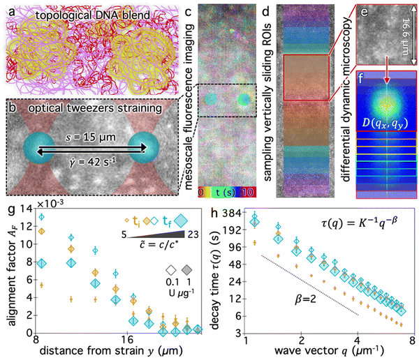
We now seek to determine how the blend composition impacts the ability of the surrounding molecules to couple their motion to a local strain, and how this coupling decays with distance from the strain. We use OpTiDDM45 to cyclically sweep an optically trapped probe through the DNA solution and simultaneously image fluorescent-labeled DNA molecules in the solution in a field-of-view (FOV) that encompasses the horizontally aligned strain path (Fig. 2a and b) and extends 35 μm vertically above and below the strain path (Fig. 2c). We fix the strain distance (s = 15 μm) and rate (![[small gamma, Greek, dot above]](https://www.rsc.org/images/entities/i_char_e0a2.gif) = 42 s−1) to be large compared to the intrinsic lengthscales and timescales of the system and match previously used parameters that maximize the coupling in similar systems.28,45 Specifically, the strain rate corresponds to Weissenberg numbers of Wi ≈ 11 and Wi ≈ 176 and Deborah numbers of De ≈ 0.8 and De ≈ 12.6 for
= 42 s−1) to be large compared to the intrinsic lengthscales and timescales of the system and match previously used parameters that maximize the coupling in similar systems.28,45 Specifically, the strain rate corresponds to Weissenberg numbers of Wi ≈ 11 and Wi ≈ 176 and Deborah numbers of De ≈ 0.8 and De ≈ 12.6 for ![[c with combining tilde]](https://www.rsc.org/images/entities/i_char_0063_0303.gif) ≃ 5 and
≃ 5 and ![[c with combining tilde]](https://www.rsc.org/images/entities/i_char_0063_0303.gif) ≃ 23, respectively (see Methods). We are thus well within the nonlinear regime and probing the viscoelastic response of the polymers. We expect the flow field induced by the probe to be primarily shear-dominated,58,59 but also exhibit signatures of extensional flow.42,60
≃ 23, respectively (see Methods). We are thus well within the nonlinear regime and probing the viscoelastic response of the polymers. We expect the flow field induced by the probe to be primarily shear-dominated,58,59 but also exhibit signatures of extensional flow.42,60
Fig. 2c qualitatively depicts the strain-induced deformation field for the starting composition (Fig. 2a), showing more pronounced motion near the strain and reduced motion further from the strain. We note that the signal-to-noise is low and single molecules are not easily resolved. This effect is a consequence of including a relatively high fraction of labeled molecules in solution to ensure ample statistics within small spatially resolved region-of-interests (ROIs), and is one of our motivations for using differential dynamic microscopy (DDM), rather than, e.g., particle-tracking or particle-image-velocimetry, to quantify DNA dynamics.
Specifically, we divide the FOV into (16 μm)2 ROIs centered at 20 vertical positions from y0 = 8 μm to yf = 27 μm (Fig. 2d), and perform DDM on each ROI (Fig. 2e–h). As described in Methods, DDM converts a time-series into stacks of image differences which encode information about how correlated two images separated by a given lag time Δt are, which can be analyzed to extract dynamics.16,61–63 In practice, DDM transforms image differences to Fourier space to compute image structure functions  (Fig. 2e and f) that quantify correlations in density fluctuations at a given spatial frequency, or wave vector
  , as a function of Δt (Fig. 2f). As described in the following sections, from
  we determine the (1) extent to which the direction of DNA motion aligns with the strain direction, which we quantify by the alignment factor AF(y,
 ![[c with combining tilde]](https://www.rsc.org/images/entities/i_char_0063_0303.gif) ) (Fig. 2g), and (2) type and rate of DNA motion, which we quantify by analyzing the q-dependent DDM decay time τ(q, y,
) (Fig. 2g), and (2) type and rate of DNA motion, which we quantify by analyzing the q-dependent DDM decay time τ(q, y, ![[c with combining tilde]](https://www.rsc.org/images/entities/i_char_0063_0303.gif) ) (Fig. 2h). Fig. 2g and h shows these metrics measured near the beginning and end of each stoichiometric digestion (0.1 and 1 U μg−1), demonstrating that composition generally has a significant effect on the dynamics. We also observe that both metrics display a similar non-monotonic dependence on
) (Fig. 2h). Fig. 2g and h shows these metrics measured near the beginning and end of each stoichiometric digestion (0.1 and 1 U μg−1), demonstrating that composition generally has a significant effect on the dynamics. We also observe that both metrics display a similar non-monotonic dependence on ![[c with combining tilde]](https://www.rsc.org/images/entities/i_char_0063_0303.gif) , with the strongest alignment and longest decay time occurring at intermediate
, with the strongest alignment and longest decay time occurring at intermediate ![[c with combining tilde]](https://www.rsc.org/images/entities/i_char_0063_0303.gif) values. In the absence of topological conversion, one may expect these metrics to increase monotonically with
values. In the absence of topological conversion, one may expect these metrics to increase monotonically with ![[c with combining tilde]](https://www.rsc.org/images/entities/i_char_0063_0303.gif) , insofar as stronger connectivity and increased steric hindrances are expected to slow motion (increasing τ) and enhance affine response to strain (increasing AF). We investigate the functional form of these non-monotonic dependences and their underlying mechanisms in the remaining sections. We choose to characterize composition primarily by
, insofar as stronger connectivity and increased steric hindrances are expected to slow motion (increasing τ) and enhance affine response to strain (increasing AF). We investigate the functional form of these non-monotonic dependences and their underlying mechanisms in the remaining sections. We choose to characterize composition primarily by ![[c with combining tilde]](https://www.rsc.org/images/entities/i_char_0063_0303.gif) rather than the mass fraction of linearized molecules ϕL because
rather than the mass fraction of linearized molecules ϕL because ![[c with combining tilde]](https://www.rsc.org/images/entities/i_char_0063_0303.gif) accounts for the fraction of all three topologies in a single quantity. Conversely, a single ϕL value could correspond to multiple compositions with varying relative fractions of ring and supercoiled molecules, which we expect to have a marked effect on dynamics due to their differing sizes and propensities for threading.28,64
accounts for the fraction of all three topologies in a single quantity. Conversely, a single ϕL value could correspond to multiple compositions with varying relative fractions of ring and supercoiled molecules, which we expect to have a marked effect on dynamics due to their differing sizes and propensities for threading.28,64
Strain alignment and propagation are maximized at intermediate reduced concentrations
For isotropic motion, such as Brownian motion, is radially symmetric, and taking the azimuthal average over
  provides a 1D image structure function,
  that can be fit to models to determine the type and rate of motion.16,62 However, for motion that has a preferred direction,
  is anisotropic, with higher correlations being weighted more heavily along the director axis,45 a feature that we leverage to quantify the degree of alignment. Specifically, we compute the alignment factor AF of the image structure function relative to the qx-axis by computing weighted azimuthal integrals of D(qx, qy, Δt), i.e., integrals over θ where θ = tan−1(qy/qx):
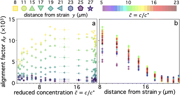
  (see Methods, Fig. 2g).65,66AF increases from 0 (for purely isotropic motion) as motion becomes more aligned along the strain direction (x-axis).45 We find that AF decays with increasing distance from the strain y for all compositions, as expected (Fig. 3a). However, the degree of alignment closest to the strain (y0 = 8 μm), and the extent to which alignment persists as y increases, displays a non-monotonic dependence on composition (Fig. 2g and 3).
As shown in Fig. 3a, AF(y0) initially increases as ![[c with combining tilde]](https://www.rsc.org/images/entities/i_char_0063_0303.gif) increases from the initial value of
increases from the initial value of ![[c with combining tilde]](https://www.rsc.org/images/entities/i_char_0063_0303.gif) i ≃ 5.4 until
i ≃ 5.4 until ![[c with combining tilde]](https://www.rsc.org/images/entities/i_char_0063_0303.gif) ≃ 10 where AF reaches a maximum. As
≃ 10 where AF reaches a maximum. As ![[c with combining tilde]](https://www.rsc.org/images/entities/i_char_0063_0303.gif) increases further, the alignment factor becomes largely independent of composition, with AF values remaining substantially higher than that for
increases further, the alignment factor becomes largely independent of composition, with AF values remaining substantially higher than that for ![[c with combining tilde]](https://www.rsc.org/images/entities/i_char_0063_0303.gif) i. This behavior persists until the highest
i. This behavior persists until the highest ![[c with combining tilde]](https://www.rsc.org/images/entities/i_char_0063_0303.gif) values (
values (![[c with combining tilde]](https://www.rsc.org/images/entities/i_char_0063_0303.gif) ≳ 22), in which the solutions comprise nearly all linear chains (Fig. 1d), at which point we see a subsequent drop in AF. This general non-monotonic dependence of
≳ 22), in which the solutions comprise nearly all linear chains (Fig. 1d), at which point we see a subsequent drop in AF. This general non-monotonic dependence of ![[c with combining tilde]](https://www.rsc.org/images/entities/i_char_0063_0303.gif) on AF holds for increasing y values until y ≃ 17 μm, at which point AF becomes increasingly insensitive to composition, and is essentially statistically insignificant for y > 20 μm.
on AF holds for increasing y values until y ≃ 17 μm, at which point AF becomes increasingly insensitive to composition, and is essentially statistically insignificant for y > 20 μm.
This effect can also be seen clearly in Fig. 3b which shows AF as a function of y for different blend compositions, denoted by the rainbow colorscale. For y < 20 μm, the non-monotonic ![[c with combining tilde]](https://www.rsc.org/images/entities/i_char_0063_0303.gif) dependence is shown by the maximum and minimum values being green (
dependence is shown by the maximum and minimum values being green (![[c with combining tilde]](https://www.rsc.org/images/entities/i_char_0063_0303.gif) ≃ 10) and purple (
≃ 10) and purple (![[c with combining tilde]](https://www.rsc.org/images/entities/i_char_0063_0303.gif) i ≈ 5) while the red tones that denote
i ≈ 5) while the red tones that denote ![[c with combining tilde]](https://www.rsc.org/images/entities/i_char_0063_0303.gif) ≳ 17 reside between these extrema. Two other features that are apparent are: the decrease in composition dependence for y > 20 μm, seen as a much smaller spread in the data compared to y < 20 μm; and the enhanced y dependence for
≳ 17 reside between these extrema. Two other features that are apparent are: the decrease in composition dependence for y > 20 μm, seen as a much smaller spread in the data compared to y < 20 μm; and the enhanced y dependence for ![[c with combining tilde]](https://www.rsc.org/images/entities/i_char_0063_0303.gif) ≃ 10 compared to higher or lower values, with AF dropping by a factor of ∼15 as y increases, versus ∼5 and ∼10 for
≃ 10 compared to higher or lower values, with AF dropping by a factor of ∼15 as y increases, versus ∼5 and ∼10 for ![[c with combining tilde]](https://www.rsc.org/images/entities/i_char_0063_0303.gif) i ≈ 5 and
i ≈ 5 and ![[c with combining tilde]](https://www.rsc.org/images/entities/i_char_0063_0303.gif) f ≈ 23.
f ≈ 23.
These results suggest that interactions between linear and circular molecules, which are absent for ![[c with combining tilde]](https://www.rsc.org/images/entities/i_char_0063_0303.gif) i and
i and ![[c with combining tilde]](https://www.rsc.org/images/entities/i_char_0063_0303.gif) f (Fig. 1d) provide more effective mechanisms for coupling to the strain compared to linear–linear and circular–circular interactions. As described in the Introduction, numerous previous studies have shown strong evidence of threading of rings by linear chains, which dramatically slows relaxation processes and enhances shear-thinning and viscosity. These features align with the enhanced stretching of threaded rings, as compared to entangled linear or ring polymers, reported by several studies.5,40,45,60 Likewise, we can understand the increased strain alignment at intermediate
f (Fig. 1d) provide more effective mechanisms for coupling to the strain compared to linear–linear and circular–circular interactions. As described in the Introduction, numerous previous studies have shown strong evidence of threading of rings by linear chains, which dramatically slows relaxation processes and enhances shear-thinning and viscosity. These features align with the enhanced stretching of threaded rings, as compared to entangled linear or ring polymers, reported by several studies.5,40,45,60 Likewise, we can understand the increased strain alignment at intermediate ![[c with combining tilde]](https://www.rsc.org/images/entities/i_char_0063_0303.gif) values as arising from threaded rings being maximally stretched along the strain direction due to the rigid constraints imposed by the threading chains that strongly resist strain-induced flow. In other words, as the ring is pulled along the strain path, the constraints (threadings) pull against the strain, thereby entropically stretching the threaded ring in the strain direction. A similar effect can occur for entangled linear polymers that are constrained by entanglements from neighboring chains. However, these constraints are expected to be less persistent than threadings, relaxing via reptation versus constraint release, rendering the entropic stretching weaker. We note that due to the nonlinear nature of the straining, we expect convective constraint release (CCR) to play a role in the dynamics for all entangled systems, regardless of the degree of threading.48,57 CCR, which reduces the local entanglement density, likely counteracts the propensity for chains to stretch along the strain direction, thereby dampening the strain-coupling we observe.
values as arising from threaded rings being maximally stretched along the strain direction due to the rigid constraints imposed by the threading chains that strongly resist strain-induced flow. In other words, as the ring is pulled along the strain path, the constraints (threadings) pull against the strain, thereby entropically stretching the threaded ring in the strain direction. A similar effect can occur for entangled linear polymers that are constrained by entanglements from neighboring chains. However, these constraints are expected to be less persistent than threadings, relaxing via reptation versus constraint release, rendering the entropic stretching weaker. We note that due to the nonlinear nature of the straining, we expect convective constraint release (CCR) to play a role in the dynamics for all entangled systems, regardless of the degree of threading.48,57 CCR, which reduces the local entanglement density, likely counteracts the propensity for chains to stretch along the strain direction, thereby dampening the strain-coupling we observe.
Collectively, this topological dependence on stretching is a plausible mechanism for the initial strong increase in alignment as ![[c with combining tilde]](https://www.rsc.org/images/entities/i_char_0063_0303.gif) increases from
increases from ![[c with combining tilde]](https://www.rsc.org/images/entities/i_char_0063_0303.gif) i ≃ 5.4 to
i ≃ 5.4 to ![[c with combining tilde]](https://www.rsc.org/images/entities/i_char_0063_0303.gif) max ≃ 10, followed by a modest decrease and greater spread as
max ≃ 10, followed by a modest decrease and greater spread as ![[c with combining tilde]](https://www.rsc.org/images/entities/i_char_0063_0303.gif) increases further to
increases further to ![[c with combining tilde]](https://www.rsc.org/images/entities/i_char_0063_0303.gif) f ≃ 23. Namely, as the degree of overlap increases and supercoiled molecules are replaced with linear chains (Fig. 1d), the entanglement density increases substantially and threading events become more pervasive. However, further increases in the degree of overlap (beyond
f ≃ 23. Namely, as the degree of overlap increases and supercoiled molecules are replaced with linear chains (Fig. 1d), the entanglement density increases substantially and threading events become more pervasive. However, further increases in the degree of overlap (beyond ![[c with combining tilde]](https://www.rsc.org/images/entities/i_char_0063_0303.gif) max ≃ 10), a result of rings being converted to linear chains, come at the cost of threading events, so serve to weaken alignment. It is important to note that if increased alignment was primarily a result of increased overlap/entanglement density, then AF should increase monotonically with
max ≃ 10), a result of rings being converted to linear chains, come at the cost of threading events, so serve to weaken alignment. It is important to note that if increased alignment was primarily a result of increased overlap/entanglement density, then AF should increase monotonically with ![[c with combining tilde]](https://www.rsc.org/images/entities/i_char_0063_0303.gif) . Instead, the non-monotonic dependence on
. Instead, the non-monotonic dependence on ![[c with combining tilde]](https://www.rsc.org/images/entities/i_char_0063_0303.gif) is direct evidence of topological effects dictating the strength of strain coupling. This conjecture is further supported by the larger drop in alignment that only occurs at the very highest
is direct evidence of topological effects dictating the strength of strain coupling. This conjecture is further supported by the larger drop in alignment that only occurs at the very highest ![[c with combining tilde]](https://www.rsc.org/images/entities/i_char_0063_0303.gif) values (
values (![[c with combining tilde]](https://www.rsc.org/images/entities/i_char_0063_0303.gif) ≳ 22) where the solutions comprise nearly all linear chains (Fig. 1d and 3a).
≳ 22) where the solutions comprise nearly all linear chains (Fig. 1d and 3a).
Finally, we note that the region of composition space in which the degree of alignment is maximized is that in which the fraction of linear chains ϕL surpasses that of supercoiled molecules ϕS (Fig. 1d, ![[c with combining tilde]](https://www.rsc.org/images/entities/i_char_0063_0303.gif) > 7.6) but remains lower than the fraction of rings ϕR (
> 7.6) but remains lower than the fraction of rings ϕR (![[c with combining tilde]](https://www.rsc.org/images/entities/i_char_0063_0303.gif) < 14). We also note that the maximally aligned composition (
< 14). We also note that the maximally aligned composition (![[c with combining tilde]](https://www.rsc.org/images/entities/i_char_0063_0303.gif) max ≃ 10) occurs at the point at which supercoiled molecules are completely eliminated, at which point we expect that all molecules (rings and linear chains) likely participate in threading events.
max ≃ 10) occurs at the point at which supercoiled molecules are completely eliminated, at which point we expect that all molecules (rings and linear chains) likely participate in threading events.
Strain-induced superdiffusivity is facilitated by rings and maximized for ring-linear blends
The data shown in Fig. 3 suggests that the strain induces athermal directed motion of the DNA, which is maximized at an intermediate overlap of![[c with combining tilde]](https://www.rsc.org/images/entities/i_char_0063_0303.gif) max ≃ 10 and significant for distances out to y ≈ 20 μm. To shed light on the nature of this athermal motion and the extent to which the strain dynamics can fingerprint onto the DNA, we turn to analyzing the q-dependent decay time τ(q), as described above and in Methods (Fig. 2h). τ(q) typically displays power-law dependence on wave vector q, τ(q) = K−1q−β, where β = 2, 1 < β < 2 and β = 1 denote diffusive, superdiffusive and ballistic dynamics, respectively.16,45,67 The larger τ(q) is for a given q, the slower the dynamics, which is quantified by the generalized transport coefficient K. Higher K values generally indicate faster motion; and for purely diffusive or ballistic motion, K respectively equates to the diffusion coefficient or speed.
max ≃ 10 and significant for distances out to y ≈ 20 μm. To shed light on the nature of this athermal motion and the extent to which the strain dynamics can fingerprint onto the DNA, we turn to analyzing the q-dependent decay time τ(q), as described above and in Methods (Fig. 2h). τ(q) typically displays power-law dependence on wave vector q, τ(q) = K−1q−β, where β = 2, 1 < β < 2 and β = 1 denote diffusive, superdiffusive and ballistic dynamics, respectively.16,45,67 The larger τ(q) is for a given q, the slower the dynamics, which is quantified by the generalized transport coefficient K. Higher K values generally indicate faster motion; and for purely diffusive or ballistic motion, K respectively equates to the diffusion coefficient or speed.
We first examine dynamics closest to the strain (y0 = 8 μm), where we observe the most pronounced alignment (Fig. 4a and b). We find that τ(q) curves for all compositions obey power-law scaling that approximately aligns with diffusive behavior (β = 2). However, the magnitude of τ(q) is generally highest for ![[c with combining tilde]](https://www.rsc.org/images/entities/i_char_0063_0303.gif) ≈ 10–12 and lowest for
≈ 10–12 and lowest for ![[c with combining tilde]](https://www.rsc.org/images/entities/i_char_0063_0303.gif) i ≈ 5, following a similar non-monotonic trend as AF. These features can be seen more clearly by examining the functional form of τq2, which is a horizontal line for β = 2 and adopts increasingly positive slopes as β decreases to more superdiffusive exponents (Fig. 4b). The relative rate of motion can also be approximated as the inverse of τq2. All compositions appear to display modest superdiffusivity over at least some region of q space, which appears to be most extreme for intermediate compositions. The mobility also markedly slows as
i ≈ 5, following a similar non-monotonic trend as AF. These features can be seen more clearly by examining the functional form of τq2, which is a horizontal line for β = 2 and adopts increasingly positive slopes as β decreases to more superdiffusive exponents (Fig. 4b). The relative rate of motion can also be approximated as the inverse of τq2. All compositions appear to display modest superdiffusivity over at least some region of q space, which appears to be most extreme for intermediate compositions. The mobility also markedly slows as ![[c with combining tilde]](https://www.rsc.org/images/entities/i_char_0063_0303.gif) increases from its initial value to
increases from its initial value to ![[c with combining tilde]](https://www.rsc.org/images/entities/i_char_0063_0303.gif) max ≃ 10.
max ≃ 10.
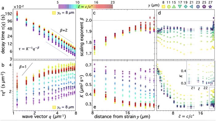 | ||
Fig. 4 Digestion of supercoiled DNA enhances strain-coupled superdiffusivity while slowing transport. (a) DDM decay time τ(q) versus wave vector q, evaluated at y0 = 8 μm, for varying blend compositions, characterized by ![[c with combining tilde]](https://www.rsc.org/images/entities/i_char_0063_0303.gif) , represented as cool to warm colors for , represented as cool to warm colors for ![[c with combining tilde]](https://www.rsc.org/images/entities/i_char_0063_0303.gif) i ≃ 5 (purple) to i ≃ 5 (purple) to ![[c with combining tilde]](https://www.rsc.org/images/entities/i_char_0063_0303.gif) f ≃ 23 (dark red). Green and yellow data points which are extremal for nearly all q values, correspond to f ≃ 23 (dark red). Green and yellow data points which are extremal for nearly all q values, correspond to ![[c with combining tilde]](https://www.rsc.org/images/entities/i_char_0063_0303.gif) ≈ 10. Dashed scaling bars denote diffusive (β = 2) scaling exponents associated with the expected power-law relation τ(q) = K−1q−β. (b) Data shown in (a) plotted as τq2versus q, which is independent of q for diffusive motion (β = 2) and displays an increasingly positive slope as motion becomes more superdiffusive, bounded above by ballistic scaling β = 1 (solid line). (c) and (d) Scaling exponent β versus (c) distance y and (d) blend composition ≈ 10. Dashed scaling bars denote diffusive (β = 2) scaling exponents associated with the expected power-law relation τ(q) = K−1q−β. (b) Data shown in (a) plotted as τq2versus q, which is independent of q for diffusive motion (β = 2) and displays an increasingly positive slope as motion becomes more superdiffusive, bounded above by ballistic scaling β = 1 (solid line). (c) and (d) Scaling exponent β versus (c) distance y and (d) blend composition ![[c with combining tilde]](https://www.rsc.org/images/entities/i_char_0063_0303.gif) , determined from fitting τ(q) for each blend composition and distance to τ(q) = K−1q−β. Dashed horizontal line denotes diffusive scaling and data that falls below is superdiffusive. (e) and (f) Transport coefficient K versus (e) distance y and (f) blend composition , determined from fitting τ(q) for each blend composition and distance to τ(q) = K−1q−β. Dashed horizontal line denotes diffusive scaling and data that falls below is superdiffusive. (e) and (f) Transport coefficient K versus (e) distance y and (f) blend composition ![[c with combining tilde]](https://www.rsc.org/images/entities/i_char_0063_0303.gif) , determined from the same fits used for (c) and (d). K values are in units of μm2 s−α, where α = 2/β. Inset in (f) is zoom-in of high , determined from the same fits used for (c) and (d). K values are in units of μm2 s−α, where α = 2/β. Inset in (f) is zoom-in of high ![[c with combining tilde]](https://www.rsc.org/images/entities/i_char_0063_0303.gif) data enclosed in the main plot, showing increase in K when all circular DNA is digested (highest data enclosed in the main plot, showing increase in K when all circular DNA is digested (highest ![[c with combining tilde]](https://www.rsc.org/images/entities/i_char_0063_0303.gif) ). Note that lower scaling exponents β (more superdiffusive) generally correlate nonintuitively with lower transport coefficients K (slower motion). Colors, symbols and error bars are as in Fig. 3. ). Note that lower scaling exponents β (more superdiffusive) generally correlate nonintuitively with lower transport coefficients K (slower motion). Colors, symbols and error bars are as in Fig. 3. | ||
To quantitatively examine these features and determine the extent to which they persist for molecules that are increasingly farther from the strain, we fit the data for each composition and distance to τ(q) = K−1q−β and evaluate β and K as functions of y (Fig. 4c–f). We observe that for all compositions, β increases with increasing distance y, extending from superdiffusive values as low as β ≈ 1.7 at y0 to diffusive scaling at the largest distance. Insofar as superdiffusivity can be taken as an indicator of strain-coupling, which adds a component of directed ballistic motion to the otherwise thermal diffusive motion of the DNA, this result is intuitive and corroborates our alignment factor analysis (Fig. 3). Moreover, β values display a similar non-monotonic dependence on composition, with ![[c with combining tilde]](https://www.rsc.org/images/entities/i_char_0063_0303.gif) ≈ 10 exhibiting the most pronounced superdiffusivity for nearly all y values, while
≈ 10 exhibiting the most pronounced superdiffusivity for nearly all y values, while ![[c with combining tilde]](https://www.rsc.org/images/entities/i_char_0063_0303.gif) i and
i and ![[c with combining tilde]](https://www.rsc.org/images/entities/i_char_0063_0303.gif) f blends have higher (less superdiffusive) β values. Also mirroring the alignment factor analysis, we observe a significant uptick in β near
f blends have higher (less superdiffusive) β values. Also mirroring the alignment factor analysis, we observe a significant uptick in β near ![[c with combining tilde]](https://www.rsc.org/images/entities/i_char_0063_0303.gif) f, where we expect blends to be nearly devoid of rings. This non-trivial composition dependence can also be seen clearly by examining β versus
f, where we expect blends to be nearly devoid of rings. This non-trivial composition dependence can also be seen clearly by examining β versus ![[c with combining tilde]](https://www.rsc.org/images/entities/i_char_0063_0303.gif) for the different distances (Fig. 4d), which display global minima at
for the different distances (Fig. 4d), which display global minima at ![[c with combining tilde]](https://www.rsc.org/images/entities/i_char_0063_0303.gif) max ≈ 10 for distances out to y ≈ 20 μm, a feature that is most pronounced closest to the strain.
max ≈ 10 for distances out to y ≈ 20 μm, a feature that is most pronounced closest to the strain.
Perhaps less intuitive is the larger effect of composition on β at both small and large distances compared to intermediate y values, which can be seen by the larger spread in the data at the left and right edges (low and high y) of Fig. 4c compared to the middle values. Examination of Fig. 4d reveals that this large spread is due to distinct trends for close versus far distances. Namely, at small y values, the spread is due to the large decrease in β values as ![[c with combining tilde]](https://www.rsc.org/images/entities/i_char_0063_0303.gif) increases to ∼10, while for large distances this spread is from a larger increase in β values at the very highest
increases to ∼10, while for large distances this spread is from a larger increase in β values at the very highest ![[c with combining tilde]](https://www.rsc.org/images/entities/i_char_0063_0303.gif) values compared to the other distances. The spread in values extends to β > 2, indicative of subdiffusive motion, which has been reported for entangled linear DNA.45 This finding suggests that even a small number of rings in the blend may be sufficient to enhance strain coupling, in accord with recent observations that a small fraction of rings in synthetic ring-linear blends substantially increases the melt viscosity.46
values compared to the other distances. The spread in values extends to β > 2, indicative of subdiffusive motion, which has been reported for entangled linear DNA.45 This finding suggests that even a small number of rings in the blend may be sufficient to enhance strain coupling, in accord with recent observations that a small fraction of rings in synthetic ring-linear blends substantially increases the melt viscosity.46
To shed further light on these results, we also examine the dependence of transport coefficients K on ![[c with combining tilde]](https://www.rsc.org/images/entities/i_char_0063_0303.gif) and y (Fig. 4e and f). We observe similar non-monotonic dependence on
and y (Fig. 4e and f). We observe similar non-monotonic dependence on ![[c with combining tilde]](https://www.rsc.org/images/entities/i_char_0063_0303.gif) as for the scaling exponents (Fig. 4c and d) and alignment factor (Fig. 3), with K decreasing substantially from
as for the scaling exponents (Fig. 4c and d) and alignment factor (Fig. 3), with K decreasing substantially from ![[c with combining tilde]](https://www.rsc.org/images/entities/i_char_0063_0303.gif) i to
i to ![[c with combining tilde]](https://www.rsc.org/images/entities/i_char_0063_0303.gif) max followed by a modest increase as
max followed by a modest increase as ![[c with combining tilde]](https://www.rsc.org/images/entities/i_char_0063_0303.gif) increases to
increases to ![[c with combining tilde]](https://www.rsc.org/images/entities/i_char_0063_0303.gif) f (Fig. 4e and f). Also consistent with the trends of the other metrics, K generally decreases with increasing distance from the strain, and this decrease is more pronounced for higher
f (Fig. 4e and f). Also consistent with the trends of the other metrics, K generally decreases with increasing distance from the strain, and this decrease is more pronounced for higher ![[c with combining tilde]](https://www.rsc.org/images/entities/i_char_0063_0303.gif) values. Finally, at the highest
values. Finally, at the highest ![[c with combining tilde]](https://www.rsc.org/images/entities/i_char_0063_0303.gif) values (
values (![[c with combining tilde]](https://www.rsc.org/images/entities/i_char_0063_0303.gif) > 21), we observe a more substantial uptick in K values, similar to the trends observed for β and AF.
> 21), we observe a more substantial uptick in K values, similar to the trends observed for β and AF.
While the general trends are consistent across metrics, the relationship between K and β appears complex and perhaps counter to expectations. One may expect that increased superdiffusivity, manifested as lower β values (within the range 2 ≥ β ≥ 1), should result in generally faster motion, described by a higher transport coefficient K. This relation is indeed what we observe for the y dependence: β increases and K decreases with increasing y values (Fig. 4c and e). This dependence suggests that it is the strain-coupling that primarily dictates the expected inverse relationship, with molecules closer to the strain being more strongly stretched along the strain path, resulting in faster and more directed motion. This strain-coupling decays as we move further from the strain site. Conversely, the coupled dependence of K and β on ![[c with combining tilde]](https://www.rsc.org/images/entities/i_char_0063_0303.gif) displays an opposite trend, whereby increasing β values correlate with increasing K values. In other words, as motion becomes ostensibly more superdiffusive (strain-coupled) it actually appears to be slower, i.e., the rate of motion is smaller. This effect can be seen in Fig. 4c and e, where
displays an opposite trend, whereby increasing β values correlate with increasing K values. In other words, as motion becomes ostensibly more superdiffusive (strain-coupled) it actually appears to be slower, i.e., the rate of motion is smaller. This effect can be seen in Fig. 4c and e, where ![[c with combining tilde]](https://www.rsc.org/images/entities/i_char_0063_0303.gif) ≈ 10 blends have the lowest values of both β (Fig. 4c) and K (Fig. 4e) among the different compositions; and in Fig. 4d and f which shows that both values generally decrease from
≈ 10 blends have the lowest values of both β (Fig. 4c) and K (Fig. 4e) among the different compositions; and in Fig. 4d and f which shows that both values generally decrease from ![[c with combining tilde]](https://www.rsc.org/images/entities/i_char_0063_0303.gif) i to
i to ![[c with combining tilde]](https://www.rsc.org/images/entities/i_char_0063_0303.gif) max, followed by a modest increase.
max, followed by a modest increase.
We conjecture that this positive correlation between β and K arises from the slowing of quiescent thermal relaxation modes due to threading events that likewise enhance strain-coupling. Stronger constraints lead to stronger strain-coupling, captured by increased alignment and superdiffusivity. However, they also more strongly suppress thermal motion, which contributes to the transport coefficient in a non-trivial way. When constraints are weaker and/or threading is limited, we expect faster thermal transport (higher K) but less strain-induced motion (higher β, lower AF). Thus, for low ![[c with combining tilde]](https://www.rsc.org/images/entities/i_char_0063_0303.gif) we expect K and β to be generally high and to display the weakest dependence on y, as we see in Fig. 4c–e.
we expect K and β to be generally high and to display the weakest dependence on y, as we see in Fig. 4c–e.
To quantitatively verify this effect, we estimate the diffusion coefficients for the lowest and highest concentration cases as the transport coefficients at the largest y distance, where β values are near 2 (diffusive) and we expect little effect of the strain on dynamics. The values for ![[c with combining tilde]](https://www.rsc.org/images/entities/i_char_0063_0303.gif) ≈ 5 and
≈ 5 and ![[c with combining tilde]](https://www.rsc.org/images/entities/i_char_0063_0303.gif) ≈ 23 blends are K ≃ 0.65 μm2 s−1 and K ≃ 0.3 μm2 s−1, respectively. Closest to the strain, these transport coefficients increase to K ≃ 0.7 μm2 s−1.1 and K ≃ 0.45 μm2 s−1.2. The ∼50% increase in K for
≈ 23 blends are K ≃ 0.65 μm2 s−1 and K ≃ 0.3 μm2 s−1, respectively. Closest to the strain, these transport coefficients increase to K ≃ 0.7 μm2 s−1.1 and K ≃ 0.45 μm2 s−1.2. The ∼50% increase in K for ![[c with combining tilde]](https://www.rsc.org/images/entities/i_char_0063_0303.gif) ≈ 23 is significantly higher than the ∼8% increase for
≈ 23 is significantly higher than the ∼8% increase for ![[c with combining tilde]](https://www.rsc.org/images/entities/i_char_0063_0303.gif) ≈ 5. We can therefore conclude that the correlated decrease in K and β for high
≈ 5. We can therefore conclude that the correlated decrease in K and β for high ![[c with combining tilde]](https://www.rsc.org/images/entities/i_char_0063_0303.gif) compared to low
compared to low ![[c with combining tilde]](https://www.rsc.org/images/entities/i_char_0063_0303.gif) blends is a result of, respectively, suppressed diffusion and increased strain-coupling.
blends is a result of, respectively, suppressed diffusion and increased strain-coupling.
Moreover, the speed of the moving probe is v = 45 μm s−1 and the strain distance is s = 15 μm, so the resulting Peclet numbers are Pe ≈ vs/D ≈ 103. As such, if the polymers were completely coupled to the strain, then thermal motion would indeed be negligible. However, the extent to which the polymers couple to the strain depends on the degree to which the polymers are sterically constrained (e.g., entangled, threaded) and the extent to which CCR reduces the density of constraints. For less entangled blends, the coupling is weaker, so thermal motion contributes more to the dynamics on the timescale of the strain.
Transport deviates more strongly from diffusive behavior at larger lengthscales and for blends without supercoiled molecules
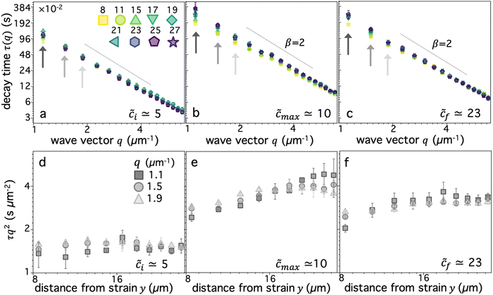
In the previous section, we approximated all τ(q) curves as obeying a simple power-law with a single q-independent scaling exponent β. However, closer inspection of τ(q) for varying distances reveals deviations from a single scaling law at small q values (q < 2 μm−1), which are more pronounced closer to the strain (Fig. 5a–c). This weaker scaling, indicative of more pronounced superdiffusivity, suggests that strain-coupling is more pronounced for larger lengthscale dynamics, i.e., at scales λ = 2πq−1 > 3 μm. This effect may indicate the necessity for many molecules to be sterically interacting, via entanglements/constraints, for dynamics to effectively couple to the strain, which we expect to only occur at scales several times larger than the polymer coil sizes, .57
.57
We also find that the deviation towards enhanced superdiffusivity is most pronounced for ![[c with combining tilde]](https://www.rsc.org/images/entities/i_char_0063_0303.gif) max ≃ 10 (Fig. 5b) and weakest for
max ≃ 10 (Fig. 5b) and weakest for ![[c with combining tilde]](https://www.rsc.org/images/entities/i_char_0063_0303.gif) i ≃ 5 (Fig. 5a). This result is coupled with the slowest and fastest transport, respectively, which can be seen by examining τq2 for the three smallest q values (Fig. 5d–f), where lower/higher values indicate faster/slower transport. These data also clearly show that the maximally strain-coupled composition (
i ≃ 5 (Fig. 5a). This result is coupled with the slowest and fastest transport, respectively, which can be seen by examining τq2 for the three smallest q values (Fig. 5d–f), where lower/higher values indicate faster/slower transport. These data also clearly show that the maximally strain-coupled composition (![[c with combining tilde]](https://www.rsc.org/images/entities/i_char_0063_0303.gif) max ≃ 10) is also the one with the largest dependence of mobility on distance from the strain site. Namely, τq2 displays the largest increase with increasing y, signifying the strongest strain-coupling; and this dependence is strongest for the smallest q value. This result corroborates the physical picture that strain-coupling is a many-polymer phenomena that requires numerous constraints and interactions to effectively propagate stress.
max ≃ 10) is also the one with the largest dependence of mobility on distance from the strain site. Namely, τq2 displays the largest increase with increasing y, signifying the strongest strain-coupling; and this dependence is strongest for the smallest q value. This result corroborates the physical picture that strain-coupling is a many-polymer phenomena that requires numerous constraints and interactions to effectively propagate stress.
Entropic stretching and superdiffusive dynamics are linked effects that drive coupling of DNA dynamics to the strain
The results described in the previous sections suggest a direct linkage between alignment and superdiffusivity, which are both reporters of strain-coupling, while transport coefficients are sensitive to both strain-induced directed motion and quiescent thermal fluctuations. Moreover, the non-monotonic dependence of these metrics on blend composition demonstrates that DNA blends with comparable ring and linear topologies and devoid of supercoiled constructs, occurring at reduced concentrations of![[c with combining tilde]](https://www.rsc.org/images/entities/i_char_0063_0303.gif) ≈ 10–12, exhibit emergent strain-coupling due to pervasive threading events. The dynamics of blends with lower
≈ 10–12, exhibit emergent strain-coupling due to pervasive threading events. The dynamics of blends with lower ![[c with combining tilde]](https://www.rsc.org/images/entities/i_char_0063_0303.gif) values, which have non-zero fractions of supercoiled constructs, are highly sensitive to variations in
values, which have non-zero fractions of supercoiled constructs, are highly sensitive to variations in ![[c with combining tilde]](https://www.rsc.org/images/entities/i_char_0063_0303.gif) , with metrics substantially increasing (AF) or decreasing (β, K) with increasing
, with metrics substantially increasing (AF) or decreasing (β, K) with increasing ![[c with combining tilde]](https://www.rsc.org/images/entities/i_char_0063_0303.gif) . Because supercoiled molecules are not expected to participate in threading events,20,28,64 this result corroborates the importance of threading in strain-coupling. The sensitivity is likely amplified by the increasing overlap as smaller supercoiled molecules are replaced with larger linear ones, increasing the entanglement density.
. Because supercoiled molecules are not expected to participate in threading events,20,28,64 this result corroborates the importance of threading in strain-coupling. The sensitivity is likely amplified by the increasing overlap as smaller supercoiled molecules are replaced with larger linear ones, increasing the entanglement density.
For concentrations above ![[c with combining tilde]](https://www.rsc.org/images/entities/i_char_0063_0303.gif) ≃ 10, the dependence on
≃ 10, the dependence on ![[c with combining tilde]](https://www.rsc.org/images/entities/i_char_0063_0303.gif) is much weaker due to competing effects of increasing overlap and entanglements and reducing threading probability. The latter appears to dominate the strain-coupling, resulting in metrics generally decreasing (AF) or increasing (β, K) for
is much weaker due to competing effects of increasing overlap and entanglements and reducing threading probability. The latter appears to dominate the strain-coupling, resulting in metrics generally decreasing (AF) or increasing (β, K) for ![[c with combining tilde]](https://www.rsc.org/images/entities/i_char_0063_0303.gif) > 10. Again, highlighting the importance of threading, we observe a greater change in all metrics (AF, β, K) for
> 10. Again, highlighting the importance of threading, we observe a greater change in all metrics (AF, β, K) for ![[c with combining tilde]](https://www.rsc.org/images/entities/i_char_0063_0303.gif) ≳ 22 blends, which have immeasurably low ring content, compared to 10 ≲
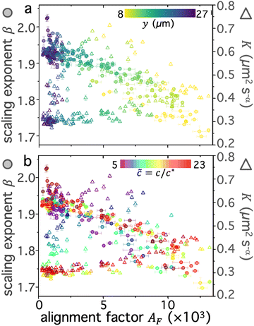
≳ 22 blends, which have immeasurably low ring content, compared to 10 ≲ ![[c with combining tilde]](https://www.rsc.org/images/entities/i_char_0063_0303.gif) < 22 blends that maintain a measurable fraction of rings. To summarize and more closely examine the correlations between the different metrics and their dependence on blend composition and distance, we evaluate pairings of scaling exponents and transport coefficients with their corresponding alignment factor for all distances (Fig. 6a) and compositions (Fig. 6b).
< 22 blends that maintain a measurable fraction of rings. To summarize and more closely examine the correlations between the different metrics and their dependence on blend composition and distance, we evaluate pairings of scaling exponents and transport coefficients with their corresponding alignment factor for all distances (Fig. 6a) and compositions (Fig. 6b).
As shown in Fig. 6a, the scaling exponent and alignment factor are generally inversely correlated for all distances, with the highest β and lowest AF values occurring at the largest distance yf = 27 μm and the lowest/highest β/AF occurring at y0 = 8 μm. This relation is consistent with the results discussed above. Conversely, K does not appear to be strongly correlated with alignment, but rather exhibits a large spread in values with the highest ones occurring at intermediate alignment.
To understand these results, we examine the dependence of ![[c with combining tilde]](https://www.rsc.org/images/entities/i_char_0063_0303.gif) on correlations between these metrics (Fig. 6b). We find that the large spread in K values at intermediate alignment factors (Fig. 6a) appears to be a signature of low
on correlations between these metrics (Fig. 6b). We find that the large spread in K values at intermediate alignment factors (Fig. 6a) appears to be a signature of low ![[c with combining tilde]](https://www.rsc.org/images/entities/i_char_0063_0303.gif) blends (blue and purple triangles) that are weakly constrained and exhibit the weakest strain-coupling. These compositions likewise generally display higher (less subdiffusive) scaling exponents compared to higher
blends (blue and purple triangles) that are weakly constrained and exhibit the weakest strain-coupling. These compositions likewise generally display higher (less subdiffusive) scaling exponents compared to higher ![[c with combining tilde]](https://www.rsc.org/images/entities/i_char_0063_0303.gif) blends. This behavior is distinct from that for blends with
blends. This behavior is distinct from that for blends with ![[c with combining tilde]](https://www.rsc.org/images/entities/i_char_0063_0303.gif) ≳ 10, in which the transport coefficients generally increase and scaling exponents β decrease with increasing alignment. Additionally, we note that while all
≳ 10, in which the transport coefficients generally increase and scaling exponents β decrease with increasing alignment. Additionally, we note that while all ![[c with combining tilde]](https://www.rsc.org/images/entities/i_char_0063_0303.gif) ≳ 10 blends display similar correlations of metrics,
≳ 10 blends display similar correlations of metrics, ![[c with combining tilde]](https://www.rsc.org/images/entities/i_char_0063_0303.gif) max ≈ 10 blends have lower values of β and K values than for blends with
max ≈ 10 blends have lower values of β and K values than for blends with ![[c with combining tilde]](https://www.rsc.org/images/entities/i_char_0063_0303.gif) ≳ 15 across the full range of measured alignment factors. Finally, we find that the large cluster of data points that exhibit diffusive (β ≈ 2), isotropic (AF ≈ 0) motion, which occurs at the largest y values (Fig. 6a), are predominantly from the highest
≳ 15 across the full range of measured alignment factors. Finally, we find that the large cluster of data points that exhibit diffusive (β ≈ 2), isotropic (AF ≈ 0) motion, which occurs at the largest y values (Fig. 6a), are predominantly from the highest ![[c with combining tilde]](https://www.rsc.org/images/entities/i_char_0063_0303.gif) blends (dark red tones), which are comprised of primarily linear chains. This effect further corroborates our interpretation that the presence of rings, at even a very small fraction, substantially enhance long-range stress propagation and entropic stretching, in line with bulk rheology observations of even low fractions of rings enhancing viscosity of ring-linear blends.46
blends (dark red tones), which are comprised of primarily linear chains. This effect further corroborates our interpretation that the presence of rings, at even a very small fraction, substantially enhance long-range stress propagation and entropic stretching, in line with bulk rheology observations of even low fractions of rings enhancing viscosity of ring-linear blends.46
Conclusions
Topological polymer blends exhibit fascinating emergent properties that continue to be a topic of great interest and debate. The roles that threading of circular polymers by linear chains and other neighboring rings,34,36,38,43,44,60 and steric entanglements between and among circular and linear polymers,31,46,54,68 play in the emergent properties remain poorly understand. The extent to which supercoiling alters these roles has also been scarcely studied.41,64,69 Here, we shed important new light on these open questions by measuring the spatially resolved mechanical response of circular-linear DNA blends with high compositional resolution. We leverage time-controlled enzymatic linearization of circular and supercoiled DNA to resolve over 70 different topological compositions, which we delineate by a unique single quantity, the reduced concentration![[c with combining tilde]](https://www.rsc.org/images/entities/i_char_0063_0303.gif) , that captures the relative fractions of all three topologies: ring, supercoiled and linear.
, that captures the relative fractions of all three topologies: ring, supercoiled and linear.
We visualize the dynamics of DNA comprising blends in response to local strains, and quantify the extent to which the strain fingerprints onto the DNA dynamics. We identify this coupling as deviations from isotropic Brownian motion, which manifest in our analysis as alignment of DNA motion along the strain path, superdiffusivity, and substantial dependence of metrics on the distance from the strain path. We observe robust non-monotonic dependences of all strain-coupling metrics on blend composition, which show extrema at ![[c with combining tilde]](https://www.rsc.org/images/entities/i_char_0063_0303.gif) ≈ 10, which comprises ϕR ≈ 68%, ϕL ≈ 32%, and ϕS ≈ 0. We rationalize this emergent behavior as arising from increased constraints imposed by threadings that facilitate entropic stretching of circular polymers along the strain path.5,43,60,70 Our findings reveal intriguing new information regarding the optimization of topological blend composition for specific performance metrics; and have important implications in the design of materials that can couple efficiently to manufacturing processes via ample stretching and distribution of imposed stresses.
≈ 10, which comprises ϕR ≈ 68%, ϕL ≈ 32%, and ϕS ≈ 0. We rationalize this emergent behavior as arising from increased constraints imposed by threadings that facilitate entropic stretching of circular polymers along the strain path.5,43,60,70 Our findings reveal intriguing new information regarding the optimization of topological blend composition for specific performance metrics; and have important implications in the design of materials that can couple efficiently to manufacturing processes via ample stretching and distribution of imposed stresses.
Methods
DNA preparation
We prepare solutions of double-stranded circular DNA of length 5.9 kbp (1.97 μm, N = 19.6) via replication of pYES2 plasmid constructs in Escherichia coli, alkaline lysis, and purification, following well-established protocols described previously.49 Following purification, we resuspend the DNA in nanopure deionized water and concentrate the solution using vacuum rotary evaporation to achieve a mass concentration of c ≃ 12 mg mL−1. Immediately following replication, the solution comprises purely supercoiled molecules (ϕS ≃ 1), but the subsequent purification process introduces nicks into a fraction of the supercoiled constructs, which allow the supercoils to unwind, resulting in relaxed circular (ring) topology. As such, the resulting DNA solution is a topological blend of ∼65% relaxed circular (ring) (ϕR ≃ 0.65), ∼35% supercoiled (ϕS ≃ 0.35), and ∼0% linear (ϕL ≃ 0) molecules (Fig. 1b and c). We quantify c and topological mass fraction ϕR,S,Lvia gel electrophoresis and band intensity analysis using Life Technologies E-Gel Imager and Gel Quant Express software.20For all measurements, we dilute the stock DNA solution to c ≃ 6 mg mL−1 to match concentrations used in previous works20,28,45 and achieve sufficient degree of polymer coil overlap (Fig. 1d). To determine the degree of overlap we compute the coil overlap concentration by modifying the expression for the overlap concentration for monodisperse polymer solutions, c* = (3/4π)(M/NA)RG−3 where M is the DNA molecular weight, to account for the different coil sizes of the different topologies: c* = (3/4π)(M/NA)/(ϕLRG,L3 + ϕRRG,R3 + ϕSRG,S3).20,41 Using previously reported radius of gyration values of RG,S ≃ 103 nm, RG,R ≃ 113 nm and RG,L ≃ 179 nm for ring, supercoiled and linear topologies (Fig. 1a),20,50,52 we determine an initial overlap concentration of  . The initial reduced concentration
  which provides a measure of the degree of polymer overlap, is
 ![[c with combining tilde]](https://www.rsc.org/images/entities/i_char_0063_0303.gif) i ≈ 5.3.
i ≈ 5.3.
At this initial concentration, we do not expect the DNA to be entangled, since it is below the nominal entanglement concentration ![[c with combining tilde]](https://www.rsc.org/images/entities/i_char_0063_0303.gif) i ≈ 6.20,53 The longest relaxation timescale in this semidilute unentangled regime is the Rouse time, which we compute to be τR ≈ 2NRG2/πD0 ≈ 261 ms, where N = 19.6 and D0 ≃ 1.53 μm2 s−1 is the tracer diffusion coefficient.50,57 At the highest concentration of
i ≈ 6.20,53 The longest relaxation timescale in this semidilute unentangled regime is the Rouse time, which we compute to be τR ≈ 2NRG2/πD0 ≈ 261 ms, where N = 19.6 and D0 ≃ 1.53 μm2 s−1 is the tracer diffusion coefficient.50,57 At the highest concentration of ![[c with combining tilde]](https://www.rsc.org/images/entities/i_char_0063_0303.gif) i ≈ 23, in which the polymers are well entangled, the longest relaxation timescale is the disengagement time, which we compute to be τD ≈ 3ZτR ≈ 4.2 s, where Z ≃ (c/ce)5/4 ≃ 5 is the entanglement density.57,71
i ≈ 23, in which the polymers are well entangled, the longest relaxation timescale is the disengagement time, which we compute to be τD ≈ 3ZτR ≈ 4.2 s, where Z ≃ (c/ce)5/4 ≃ 5 is the entanglement density.57,71
To image the blends during measurements, we fluorescent-label a small batch of the purified DNA with covalent dye MFP488 (MirusBio) that has excitation/emission peaks at 501/523 nm. We use the manufacturer-supplied Label IT Labeling Kit and corresponding protocols to label molecules at a dye to basepair ratio of 1:5.
Restriction endonuclease
We use the high-fidelity restriction endonuclease BamHI (New England BioLabs) to linearize circular DNA in solution. BamHI cleaves circular pYES2 constructs at a single recognition site, converting each ring and supercoiled molecule to linear topology. To ensure that we can achieve sufficient sampling across the entire composition space (from purely circular to purely linear), we perform measurements with two different BamHI:DNA stoichiometries: 0.1 U μg−1 and 1 U μg−1 (Fig. 1b and c).Gel electrophoresis
To characterize the topological state of DNA solutions during linearization of circular DNA via BamHI, we use direct current agarose gel electrophoresis to separate the different topologies. Specifically, we prepare 40 μL samples of c ≃ 6 mg mL−1 DNA, 0.1% Tween, and either 0.1 U μg−1 or 1 U μg−1 BamHI, suspended in CutSmart Buffer (0.5 M potassium acetate, 0.2 M Tris–acetate, 0.1 M magnesium acetate; New England BioLabs), We incubate the samples at RT for 240 min, during which we remove a 1 μL aliquot from each reaction in regular intervals and quench the aliquot with TE buffer (100 mM Tris–HCl (pH 8), 1 mM EDTA) and gel loading dye. We load 50 ng of DNA from each ‘kinetic aliquot’ onto a 1% agarose gel prepared with TAE (Tris–acetate–EDTA) buffer.20,37 We run the gel at 5 V cm−1 for 2.5 hours, allowing for separation of the DNA into distinct bands corresponding to ring, supercoiled, and linear topologies (Fig. 1b). To quantify the fraction of each topology at each interval, we perform standard band intensity analysis as described above,20 using intensity as a proxy for mass to determine the relative DNA mass in each band of a given lane.Sample preparation
For all optical tweezers experiments, we prepare samples as described above, and add 10 μg mL−1 of MFP488-labeled DNA and a trace amount of polystyrene beads of radius r = 2.25 μm (Polysciences, Inc.) (Fig. 2a and b). We coat beads with AlexaFluor594-BSA (ThermoFisher) to prevent DNA adsorption and allow for fluorescence imaging with different excitation/emission spectra than for DNA. Labeling beads and DNA tracers with spectrally distinct fluorophores ensures that the beads do not contribute to the DNA signal used in DDM analysis. To mitigate photobleaching, we add an oxygen-scavenging system comprising 45 μg mL−1 glucose, 43 μg mL−1 glucose oxidase, 7 μg mL−1 catalase, and 5 μg mL−1 β-mercaptoethanol.We mix the solution by pipetting up and down with a wide-bore pipet tip, and add BamHI last, marking the time at which it is added as t = 0. The buffer conditions and temperature (20 °C) provide good solvent conditions for the DNA.71–75 We construct sample chambers, measuring 20 × 3 × 0.1 mm3, using a microscope glass slide and coverslip, both coated with BSA to prevent DNA adsorption, and separated by two layers of double-sided tape. We introduce DNA samples into chambers via capillary action, using a wide-bore pipette tip, after which we seal chambers with epoxy.
OpTiDDM instrumentation and measurements
We use a custom-built optical trap formed from a 1064 nm Nd:YAG fiber laser (Manlight) focused with a 60× 1.4 NA objective (Olympus) and integrated into an Olympus IX71 epifluorescence microscope, as described previously.58,59 To image the MPF488-labeled DNA and AlexaFluor594-labeled microspheres in the samples we use 490/525 nm and 530/575 nm excitation/emission filter cubes and an ORCA-Flash 4.0 LT+ CMOS camera (Hamamatsu). To impose a local strain in the sample, we use a piezoelectric actuator mirror (PI USA) to move the trap relative to the sample chamber while keeping the 554 × 128 square-pixel (16.6 μm × 72 μm) field-of-view (FOV) that we use for DDM analysis fixed and centered at the resting trap position.45As shown in Fig. 2b, the microrheological strain program we apply consists of repeatedly sweeping the trapped bead back and forth horizontally (along the x-axis) at constant speed through a strain distance s = 15 μm. We pause between each 15 μm sweep for a fixed cessation time of 3 s to allow the polymers to relax. We perform all measurements at a speed of v = 40 μm s−1, which equates to a strain rate of ![[small gamma, Greek, dot above]](https://www.rsc.org/images/entities/i_char_e0a2.gif) = 42 s−1via the relation
= 42 s−1via the relation  .45,76 We chose this speed based on our previous work that showed that this rate allowed for the most pronounced strain coupling in comparable DNA solutions.45 For reference, the corresponding Weissenberg numbers for the initial and final concentrations (
![[c with combining tilde]](https://www.rsc.org/images/entities/i_char_0063_0303.gif) i ≈ 5,
i ≈ 5, ![[c with combining tilde]](https://www.rsc.org/images/entities/i_char_0063_0303.gif) i ≈ 23) are Wi ≈
i ≈ 23) are Wi ≈ ![[small gamma, Greek, dot above]](https://www.rsc.org/images/entities/i_char_e0a2.gif) τR ≈ 11 and Wi ≈
τR ≈ 11 and Wi ≈ ![[small gamma, Greek, dot above]](https://www.rsc.org/images/entities/i_char_e0a2.gif) τD ≈ 176. Considering that the time for the probe to complete one 15 μm strain is tp = 0.33 s, the corresponding Deborah numbers for these two extremal concentrations are De = τR/tp ≈ 0.8 and De = τD/tp ≈ 12.6. Therefore, we expect to be in the nonlinear regime and to be probing the viscoelastic response of the polymers.
τD ≈ 176. Considering that the time for the probe to complete one 15 μm strain is tp = 0.33 s, the corresponding Deborah numbers for these two extremal concentrations are De = τR/tp ≈ 0.8 and De = τD/tp ≈ 12.6. Therefore, we expect to be in the nonlinear regime and to be probing the viscoelastic response of the polymers.
We perform each oscillatory strain measurement for 50 s, during which we capture a time-series of images of the labeled DNA in the sample at 60 fps (Fig. 2b–e). We perform measurements every ∼1–10 min, depending on the digestion rate, for 4 hours, resulting in >80 measurements that are each performed with a new particle in a new location in the sample chamber, separated by >200 μm from the previous location. All data shown is the average across at least two replicates and three consecutive measurements. Vertical and horizontal error bars are standard error from averaging across replicates and consecutive measurements, respectively.
Differential dynamic microscopy (DDM)
As previously described in detail,45 to determine how the strain-induced DNA dynamics depend on the orthogonal distance y from the strain path, we divide the 554 × 128 square-pixel FOV into 128 × 128 square-pixel (16.6 μm)2 ROIs, centered horizontally at the midpoint of the s = 15 μm strain path and shifted along the ±y direction in 16-pixel increments, with the bottom edge of the first ROI at y = 0 (and its center at y0 = 8 μm) and the farthest ROI centered at yf = 27 μm. We analyze 10 ROIs in each of the +y and −y directions and average the ± data for each y, as they exhibit expectedly statistically indistinguishable dynamics.We use custom-written scripts (Python) to perform DDM analysis, which takes two-dimensional Fourier transforms of differences between images, separated by a range of lag times Δt, to quantify how the correlation of density fluctuations decays with Δt.67 We quantify this correlation as a function of the 2D wave vector ![[q with combining right harpoon above (vector)]](https://www.rsc.org/images/entities/i_char_0071_20d1.gif) = (qx, qy) via the image structure function D(
= (qx, qy) via the image structure function D(![[q with combining right harpoon above (vector)]](https://www.rsc.org/images/entities/i_char_0071_20d1.gif) , Δt) (Fig. 2e and f).
, Δt) (Fig. 2e and f).
To determine the extent to which the DNA dynamics are preferentially aligned along the strain path (x-axis) (Fig. 2 and 3), we compute an alignment factor AF with respect to the strain path (x-axis) by computing weighted azimuthal integrals of D(qx, qy, Δt), i.e., integrals over θ where θ = tan−1(qy/qx):  (Fig. 2g).65,66 Here, θ is defined relative to the x-axis such that isotropic and completely x-aligned dynamics correspond to AF = 0 and AF = A(q)/(A(q) + 2B(q)), respectively, where A(q) and B(q) are amplitude and background terms that we determine from DDM analysis, as described below. Larger AF values indicate more alignment. To obtain a single AF value for each distance y, we average over Δt = 0.17–1 s and q = 1–7 μm−1, where there is no statistically significant dependence of AF on these parameters.
To determine the type and rate of motion of the DNA, we radially average each D(![[q with combining right harpoon above (vector)]](https://www.rsc.org/images/entities/i_char_0071_20d1.gif) , Δt) (Fig. 2f) to get a 1D image structure function that can be described by D(q, Δt) = A(q)[1 − f(q, Δt)] + B(q), where f(q, Δt) is the intermediate scattering function (ISF). We model the ISF as a stretched exponential: f(q, Δt) = e−(Δt/τ(q))δ where τ(q) is the decay time and δ the stretching exponent.
, Δt) (Fig. 2f) to get a 1D image structure function that can be described by D(q, Δt) = A(q)[1 − f(q, Δt)] + B(q), where f(q, Δt) is the intermediate scattering function (ISF). We model the ISF as a stretched exponential: f(q, Δt) = e−(Δt/τ(q))δ where τ(q) is the decay time and δ the stretching exponent.
By evaluating the functional form of τ(q) determined from fitting the ISF, we analyze the extent to which τ(q) can be described by power-law scaling τ(q) ∼ q−β where the scaling exponent β describes the type of motion (Fig. 2h, 4 and 5). Specifically, β = 2 and β = 1 are indicative of diffusive and ballistic motion, respectively, and 1 < β < 2 indicates superdiffusion.16,77,78
Author contributions
RMRA conceived the project, guided the experiments, interpreted the data, wrote the manuscript and prepared figures. KRP performed the experiments, analyzed and interpreted the data, and prepared figures. RJM developed analysis software and helped guide experiments and analysis and write the manuscript.Data availability
All data shown in the manuscript is provided in ESI.† Raw data from which the presented data was determined will be made available upon reasonable request to the corresponding author.Conflicts of interest
The authors declare no competing interests.Acknowledgements
This research was funded by grants from the Air Force Office of Scientific Research (AFOSR-FA9550-17-1-0249, AFOSR-FA9550-21-1-0361) and National Science Foundation (DMREF DMR 2119663) awarded to RMRA, and National Institutes of Health (R15GM123420, 2R15GM123420-02) awarded to RJM and RMRA.References
- G. S. Grest, T. Ge, S. J. Plimpton, M. Rubinstein and T. C. O’Connor, Entropic Mixing of Ring/Linear Polymer Blends, ACS Polym. Au, 2023, 3(2), 209–216, DOI:10.1021/acspolymersau.2c00050.
- Y. Kobayashi, Y. Doi, S. S. Abdul Rahman, E. Kim, T.-H. Kim, A. Takano and Y. Matsushita, SANS Study of Ring Topology Effects on the Miscibility of Polymer Blends, Macromolecules, 2018, 51(5), 1885–1893, DOI:10.1021/acs.macromol.7b02359.
- Z. Bartczak and A. Galeski, Mechanical Properties of Polymer Blends, in Polymer Blends Handbook, ed. L. A. Utracki and C. A. Wilkie, Springer Netherlands, Dordrecht, 2014, pp. 1203–1297 DOI:10.1007/978-94-007-6064-6_13.
- S. H. Goh, Miscible Polymer Blends, in Polymer Blends Handbook, ed. L. A. Utracki and C. A. Wilkie, Springer Netherlands, Dordrecht, 2014, pp. 1915–2151 DOI:10.1007/978-94-007-6064-6_24.
- T. C. O’Connor, T. Ge and G. S. Grest, Composite Entanglement Topology and Extensional Rheology of Symmetric Ring-Linear Polymer Blends, J. Rheol., 2022, 66(1), 49–65, DOI:10.1122/8.0000319.
- E. Lattuada, T. Pietrangeli and F. Sciortino, Interpenetrating Gels in Binary Suspensions of DNA Nanostars, J. Chem. Phys., 2022, 157(13), 135101, DOI:10.1063/5.0117047.
- S. Thomas, D. Durand, C. Chassenieux and P. Jyotishkumar, Handbook of Biopolymer-Based Materials: From Blends and Composites to Gels and Complex Networks, John Wiley & Sons, 2013 Search PubMed.
- S. Banerjee, M. L. Gardel and U. S. Schwarz, The Actin Cytoskeleton as an Active Adaptive Material, Annu. Rev. Condens. Matter Phys., 2020, 11(1), 421–439, DOI:10.1146/annurev-conmatphys-031218-013231.
- M. Drechsler, F. Giavazzi, R. Cerbino and I. M. Palacios, Active Diffusion and Advection in the Drosophila Ooplasm Result from the Interplay of the Actin and Microtubule Cytoskeletons, bioRxiv, 2017, 7, 098590, DOI:10.1101/098590.
- D. A. Fletcher and R. D. Mullins, Cell Mechanics and the Cytoskeleton, Nature, 2010, 463(7280), 485–492, DOI:10.1038/nature08908.
- O. Wintner, N. Hirsch-Attas, M. Schlossberg, F. Brofman, R. Friedman, M. Kupervaser, D. Kitsberg and A. Buxboim, A Unified Linear Viscoelastic Model of the Cell Nucleus Defines the Mechanical Contributions of Lamins and Chromatin, Adv. Sci., 2020, 7(8), 1901222, DOI:10.1002/advs.201901222.
- F. Erdel, M. Baum and K. Rippe, The Viscoelastic Properties of Chromatin and the Nucleoplasm Revealed by Scale-Dependent Protein Mobility, J. Phys.: Condens. Matter, 2015, 27(6), 064115, DOI:10.1088/0953-8984/27/6/064115.
- C.-H. Yu, S. Redemann, H.-Y. Wu, R. Kiewisz, T. Y. Yoo, W. Conway, R. Farhadifar, T. Müller-Reichert and D. Needleman, Central-Spindle Microtubules Are Strongly Coupled to Chromosomes during Both Anaphase A and Anaphase B, Mol. Biol. Cell, 2019, 30(19), 2503–2514, DOI:10.1091/mbc.E19-01-0074.
- W. Alt and M. Dembo, Cytoplasm Dynamics and Cell Motion: Two-Phase Flow Models, Math. Biosci., 1999, 156(1), 207–228, DOI:10.1016/S0025-5564(98)10067-6.
- E. H. Barriga and R. Mayor, Adjustable Viscoelasticity Allows for Efficient Collective Cell Migration, Semin. Cell Dev. Biol., 2019, 93, 55–68, DOI:10.1016/j.semcdb.2018.05.027.
- R. Cerbino and P. Cicuta, Perspective: Differential Dynamic Microscopy Extracts Multi-Scale Activity in Complex Fluids and Biological Systems, J. Chem. Phys., 2017, 147(11), 110901, DOI:10.1063/1.5001027.
- B. Alberts, Molecular Biology of the Cell, Garland, 2004 Search PubMed.
- Y. S. Kim, B. Kundukad, A. Allahverdi, L. Nordensköld, P. S. Doyle and J. R. C. van der Maarel, Gelation of the Genome by Topoisomerase II Targeting Anticancer Agents, Soft Matter, 2013, 9(5), 1656–1663, 10.1039/C2SM27229F.
- M. Jinek, K. Chylinski, I. Fonfara, M. Hauer, J. A. Doudna and E. Charpentier, A Programmable Dual-RNA–Guided DNA Endonuclease in Adaptive Bacterial Immunity, Science, 2012, 337(6096), 816–821, DOI:10.1126/science.1225829.
- D. Michieletto, P. Neill, S. Weir, D. Evans, N. Crist, V. A. Martinez and R. M. Robertson-Anderson, Topological Digestion Drives Time-Varying Rheology of Entangled DNA Fluids, Nat. Commun., 2022, 13(1), 4389, DOI:10.1038/s41467-022-31828-w.
- A. Pingoud, G. G. Wilson and W. Wende, Type II Restriction Endonucleases—a Historical Perspective and More, Nucleic Acids Res., 2014, 42(12), 7489–7527, DOI:10.1093/nar/gku447.
- O. A. Saleh, B. Jeon and T. Liedl, Enzymatic Degradation of Liquid Droplets of DNA Is Modulated near the Phase Boundary, Proc. Natl. Acad. Sci. U. S. A., 2020, 117(28), 16160–16166, DOI:10.1073/pnas.2001654117.
- L.-Y. Zhou, J. Fu and Y. He, A Review of 3D Printing Technologies for Soft Polymer Materials, Adv. Funct. Mater., 2020, 30(28), 2000187, DOI:10.1002/adfm.202000187.
- D. Kokkinis, F. Bouville and A. R. Studart, 3D Printing of Materials with Tunable Failure via Bioinspired Mechanical Gradients, Adv. Mater., 2018, 30(19), 1705808, DOI:10.1002/adma.201705808.
- T. G. Mezger, The Rheology Handbook, Vincentz Network, 2006 Search PubMed.
- C. L. C. Chan, J. M. Taylor and E. C. Davidson, Design of Soft Matter for Additive Processing, Nat. Synth., 2022, 1(8), 592–600, DOI:10.1038/s44160-022-00115-3.
- F. M. Haque and S. M. Grayson, The Synthesis, Properties and Potential Applications of Cyclic Polymers, Nat. Chem., 2020, 12(5), 433–444, DOI:10.1038/s41557-020-0440-5.
- P. Neill, N. Crist, R. McGorty and R. Robertson-Anderson, Enzymatic Cleaving of Entangled DNA Rings Drives Scale-Dependent Rheological Trajectories, Soft Matter, 2024, 20(12), 2750–2766, 10.1039/D3SM01641B.
- D. Kong, S. Banik, M. J. San Francisco, M. Lee, R. M. Robertson Anderson, C. M. Schroeder and G. B. McKenna, Rheology of Entangled Solutions of Ring–Linear DNA Blends, Macromolecules, 2022, 55(4), 1205–1217, DOI:10.1021/acs.macromol.1c01672.
- G. B. McKenna, D. Chen, S. C. H. Mangalara, D. Kong and S. Banik, Some Open Challenges in Polymer Physics, Polym. Eng. Sci., 2022, 62(5), 1325–1355, DOI:10.1002/pen.25938.
- M. A. Ubertini, J. Smrek and A. Rosa, Entanglement Length Scale Separates Threading from Branching of Unknotted and Non-Concatenated Ring Polymers in Melts, Macromolecules, 2022, 55(23), 10723–10736, DOI:10.1021/acs.macromol.2c01264.
- W. J. Weigand, A. Messmore, J. Tu, A. Morales-Sanz, D. L. Blair, D. D. Deheyn, J. S. Urbach and R. M. Robertson-Anderson, Active Microrheology Determines Scale-Dependent Material Properties of Chaetopterus Mucus, PLoS One, 2017, 12(5), e0176732, DOI:10.1371/journal.pone.0176732.
- J. Roovers and P. M. Toporowski, Microheterogeneity in Miscible Blends of 1,2-Polybutadiene and 1,4-Polyisoprene, Macromolecules, 1992, 25(13), 3454–3461, DOI:10.1021/ma00039a023.
- D. Michieletto, N. Nahali and A. Rosa, Glassiness and Heterogeneous Dynamics in Dense Solutions of Ring Polymers, Phys. Rev. Lett., 2017, 119(19), 197801, DOI:10.1103/PhysRevLett.119.197801.
- D. Michieletto and T. Sakaue, Dynamical Entanglement and Cooperative Dynamics in Entangled Solutions of Ring and Linear Polymers, ACS Macro Lett., 2021, 10(1), 129–134, DOI:10.1021/acsmacrolett.0c00551.
- Y. Zhou, C. D. Young, M. Lee, S. Banik, D. Kong, G. B. McKenna, R. M. Robertson-Anderson, C. E. Sing and C. M. Schroeder, Dynamics and Rheology of Ring-Linear Blend Semidilute Solutions in Extensional Flow: Single Molecule Experiments, J. Rheol., 2021, 65(4), 729–744, DOI:10.1122/8.0000219.
- J. Marfai, R. J. McGorty and R. M. Robertson-Anderson, Cooperative Rheological State-Switching of Enzymatically-Driven Composites of Circular DNA And Dextran, Adv. Mater., 2023, 35(46), 2305824, DOI:10.1002/adma.202305824.
- J. Smrek, K. Kremer and A. Rosa, Threading of Unconcatenated Ring Polymers at High Concentrations: Double-Folded vs Time-Equilibrated Structures, ACS Macro Lett., 2019, 8(2), 155–160, DOI:10.1021/acsmacrolett.8b00828.
- D. G. Tsalikis and V. G. Mavrantzas, Size and Diffusivity of Polymer Rings in Linear Polymer Matrices: The Key Role of Threading Events, Macromolecules, 2020, 53(3), 803–820, DOI:10.1021/acs.macromol.9b02099.
- D. Parisi, M. Kaliva, S. Costanzo, Q. Huang, P. J. Lutz, J. Ahn, T. Chang, M. Rubinstein and D. Vlassopoulos, Nonlinear Rheometry of Entangled Polymeric Rings and Ring-Linear Blends, J. Rheol., 2021, 65(4), 695–711, DOI:10.1122/8.0000186.
- R. Peddireddy K., M. Lee, Y. Zhou, S. Adalbert, S. Anderson, M. Schroeder C. and M. Robertson-Anderson, R., Unexpected Entanglement Dynamics in Semidilute Blends of Supercoiled and Ring DNA, Soft Matter, 2020, 16(1), 152–161, 10.1039/C9SM01767D.
- K. R. Peddireddy, M. Lee, C. M. Schroeder and R. M. Robertson-Anderson, Viscoelastic Properties of Ring-Linear DNA Blends Exhibit Nonmonotonic Dependence on Blend Composition, Phys. Rev. Res., 2020, 2(2), 023213, DOI:10.1103/PhysRevResearch.2.023213.
- K. R. Peddireddy, R. Clairmont and R. M. Robertson-Anderson, Polymer Threadings and Rigidity Dictate the Viscoelasticity of Entangled Ring-Linear Blends and Their Composites with Rigid Rod Microtubules, J. Rheol., 2023, 67(1), 125–138, DOI:10.1122/8.0000529.
- A. F. Katsarou, A. J. Tsamopoulos, D. G. Tsalikis and V. G. Mavrantzas, Dynamic Heterogeneity in Ring-Linear Polymer Blends, Polymers, 2020, 12(4), 752, DOI:10.3390/polym12040752.
- K. R. Peddireddy, R. Clairmont, P. Neill, R. McGorty and R. M. Robertson-Anderson, Optical-Tweezers-Integrating-Differential-Dynamic-Microscopy Maps the Spatiotemporal Propagation of Nonlinear Strains in Polymer Blends and Composites, Nat. Commun., 2022, 13(1), 5180, DOI:10.1038/s41467-022-32876-y.
- D. Parisi, J. Ahn, T. Chang, D. Vlassopoulos and M. Rubinstein, Stress Relaxation in Symmetric Ring-Linear Polymer Blends at Low Ring Fractions, Macromolecules, 2020, 53(5), 1685–1693, DOI:10.1021/acs.macromol.9b02536.
- J. D. Halverson, G. S. Grest, A. Y. Grosberg and K. Kremer, Rheology of Ring Polymer Melts: From Linear Contaminants to Ring-Linear Blends, Phys. Rev. Lett., 2012, 108(3), 038301, DOI:10.1103/PhysRevLett.108.038301.
- P. Khanal, K. R. Peddireddy, J. Marfai, R. McGorty and R. M. Robertson-Anderson, DNA Topology Dictates Emergent Bulk Elasticity and Hindered Macromolecular Diffusion in DNA-Dextran Composites, J. Rheol., 2022, 66(4), 699–715, DOI:10.1122/8.0000447.
- S. Laib, R. M. Robertson and D. E. Smith, Preparation and Characterization of a Set of Linear DNA Molecules for Polymer Physics and Rheology Studies, Macromolecules, 2006, 39(12), 4115–4119, DOI:10.1021/ma0601464.
- R. M. Robertson, S. Laib and D. E. Smith, Diffusion of Isolated DNA Molecules: Dependence on Length and Topology, Proc. Natl. Acad. Sci. U. S. A., 2006, 103(19), 7310–7314, DOI:10.1073/pnas.0601903103.
- D. E. Smith, T. T. Perkins and S. Chu, Dynamical Scaling of DNA Diffusion Coefficients, Macromolecules, 1996, 29(4), 1372–1373, DOI:10.1021/ma951455p.
- D. R. Latulippe and A. L. Zydney, Radius of Gyration of Plasmid DNA Isoforms from Static Light Scattering, Biotechnol. Bioeng., 2010, 107(1), 134–142, DOI:10.1002/bit.22787.
- R. M. Robertson and D. E. Smith, Self-Diffusion of Entangled Linear and Circular DNA Molecules: Dependence on Length and Concentration, Macromolecules, 2007, 40(9), 3373–3377 CrossRef CAS.
- T. Ge, S. Panyukov and M. Rubinstein, Self-Similar Conformations and Dynamics in Entangled Melts and Solutions of Nonconcatenated Ring Polymers, Macromolecules, 2016, 49(2), 708–722, DOI:10.1021/acs.macromol.5b02319.
- R. M. Robertson and D. E. Smith, Direct Measurement of the Confining Forces Imposed on a Single Molecule in a Concentrated Solution of Circular Polymers, Macromolecules, 2007, 40(24), 8737–8741, DOI:10.1021/ma071440e.
- A. Y. Grosberg, Annealed Lattice Animal Model and Flory Theory for the Melt of Non-Concatenated Rings: Towards the Physics of Crumpling, Soft Matter, 2013, 10(4), 560–565, 10.1039/C3SM52805G.
- M. Doi and S. F. Edwards, The Theory of Polymer Dynamics, Clarendon Press, 1988 Search PubMed.
- C. D. Chapman and R. M. Robertson-Anderson, Nonlinear Microrheology Reveals Entanglement-Driven Molecular-Level Viscoelasticity of Concentrated DNA, Phys. Rev. Lett., 2014, 113(9), 098303, DOI:10.1103/PhysRevLett.113.098303.
- R. M. Robertson-Anderson, Optical Tweezers Microrheology: From the Basics to Advanced Techniques and Applications, ACS Macro Lett., 2018, 7(8), 968–975, DOI:10.1021/acsmacrolett.8b00498.
- Q. Huang, J. Ahn, D. Parisi, T. Chang, O. Hassager, S. Panyukov, M. Rubinstein and D. Vlassopoulos, Unexpected Stretching of Entangled Ring Macromolecules, Phys. Rev. Lett., 2019, 122(20), 208001, DOI:10.1103/PhysRevLett.122.208001.
- R. Cerbino, F. Giavazzi and M. E. Helgeson, Differential Dynamic Microscopy for the Characterization of Polymer Systems, J. Polym. Sci., 2022, 60(7), 1079–1089, DOI:10.1002/pol.20210217.
- R. Cerbino and V. Trappe, Differential Dynamic Microscopy: Probing Wave Vector Dependent Dynamics with a Microscope, Phys. Rev. Lett., 2008, 100(18), 188102, DOI:10.1103/PhysRevLett.100.188102.
- D. M. Wulstein, K. E. Regan, R. M. Robertson-Anderson and R. McGorty, Light-Sheet Microscopy with Digital Fourier Analysis Measures Transport Properties over Large Field-of-View, Opt. Express, 2016, 24(18), 20881–20894, DOI:10.1364/OE.24.020881.
- J. Smrek, J. Garamella, R. Robertson-Anderson and D. Michieletto, Topological Tuning of DNA Mobility in Entangled Solutions of Supercoiled Plasmids, Sci. Adv., 2021, 7(20), eabf9260, DOI:10.1126/sciadv.abf9260.
- L. M. Walker and N. J. Wagner, SANS Analysis of the Molecular Order in Poly(γ-Benzyl l-Glutamate)/Deuterated Dimethylformamide (PBLG/d-DMF) under Shear and during Relaxation, Macromolecules, 1996, 29(6), 2298–2301, DOI:10.1021/ma951127p.
- Z. Varga and J. W. Swan, Large Scale Anisotropies in Sheared Colloidal Gels, J. Rheol., 2018, 62(2), 405–418, DOI:10.1122/1.5003364.
- H. N. Verwei, G. Lee, G. Leech, I. I. Petitjean, G. H. Koenderink, R. M. Robertson-Anderson and R. J. McGorty, Quantifying Cytoskeleton Dynamics Using Differential Dynamic Microscopy, JoVE, 2022, 184, e63931, DOI:10.3791/63931.
- J. Wang and T. Ge, Crazing Reveals an Entanglement Network in Glassy Ring Polymers, Macromolecules, 2021, 54(16), 7500–7511, DOI:10.1021/acs.macromol.1c01080.
- B. A. Krajina and A. J. Spakowitz, Large-Scale Conformational Transitions in Supercoiled DNA Revealed by Coarse-Grained Simulation, Biophys. J., 2016, 111(7), 1339–1349, DOI:10.1016/j.bpj.2016.07.045.
- T. C. O’Connor, T. Ge, M. Rubinstein and G. S. Grest, Topological Linking Drives Anomalous Thickening of Ring Polymers in Weak Extensional Flows, Phys. Rev. Lett., 2020, 124(2), 027801, DOI:10.1103/PhysRevLett.124.027801.
- S. Banik, D. Kong, M. J. San Francisco and G. B. McKenna, Monodisperse Lambda DNA as a Model to Conventional Polymers: A Concentration-Dependent Scaling of the Rheological Properties, Macromolecules, 2021, 54(18), 8632–8654, DOI:10.1021/acs.macromol.0c02537.
- D. Michieletto, P. Neill, S. Weir, D. Evans, N. Crist, V. A. Martinez and R. M. Robertson-Anderson, Topological Digestion Drives Time-Varying Rheology of Entangled DNA Fluids, Nat. Commun., 2022, 13(1), 4389, DOI:10.1038/s41467-022-31828-w.
- R. M. Robertson, S. Laib and D. E. Smith, Diffusion of Isolated DNA Molecules: Dependence on Length and Topology, Proc. Natl. Acad. Sci. U. S. A., 2006, 103(19), 7310–7314, DOI:10.1073/pnas.0601903103.
- S. Pan, D. At Nguyen, T. Sridhar, P. Sunthar and J. Ravi Prakash, Universal Solvent Quality Crossover of the Zero Shear Rate Viscosity of Semidilute DNA Solutions, J. Rheol., 2014, 58(2), 339–368, DOI:10.1122/1.4861072.
- J. R. Prakash, Universal Dynamics of Dilute and Semidilute Solutions of Flexible Linear Polymers, Curr. Opin. Colloid Interface Sci., 2019, 43, 63–79, DOI:10.1016/j.cocis.2019.03.001.
- T. M. Squires and T. G. Mason, Fluid Mechanics of Microrheology, Annu. Rev. Fluid Mech., 2010, 42(1), 413–438, DOI:10.1146/annurev-fluid-121108-145608.
- Y. Gao, J. Kim and M. E. Helgeson, Microdynamics and Arrest of Coarsening during Spinodal Decomposition in Thermoreversible Colloidal Gels, Soft Matter, 2015, 11(32), 6360–6370, 10.1039/C5SM00851D.
- D. Germain, M. Leocmach and T. Gibaud, Differential Dynamic Microscopy to Characterize Brownian Motion and Bacteria Motility, Am. J. Phys., 2016, 84(3), 202–210, DOI:10.1119/1.4939516.
Footnote |
| † Electronic supplementary information (ESI) available. See DOI: https://doi.org/10.1039/d4sm01065e |
| This journal is © The Royal Society of Chemistry 2024 |


