Ex situ vapor phase boron doping of silicon nanowires using BBr3
Gregory S.
Doerk
,
Gabriella
Lestari
,
Fang
Liu
,
Carlo
Carraro
and
Roya
Maboudian
*
Department of Chemical Engineering, University of California, Berkeley, CA 94720. E-mail: maboudia@berkeley.edu
First published on 22nd May 2010
Abstract
An ex situ vapor phase technique for doping vapor–liquid–solid grown silicon nanowires (NWs) based on the reduction of BBr3 by H2 has been demonstrated. Electron microscope images show that the excellent crystal quality of the nanowires is preserved with minimal alteration of their surface morphology. Fano resonance in the Raman spectra for single nanowires indicates that active boron concentrations over two orders of magnitude and as high as 1020 cm−3 are achievable in a well-controlled manner, with excellent axial uniformity. Electrical resistance measurements from single nanowires confirm that incorporated boron is electrically active, and doping of epitaxial bridging Si NWs is successfully demonstrated. By avoiding the pitfalls of nonuniform concentration profiles and drastic morphological changes that often accompany in situ boron doping, this technique provides a valuable alternative doping route for the development of single Si NW devices in a reliable manner.
1 Introduction
Well-controlled doping is crucial to development of promising silicon nanowire-based electronic,1,2 thermoelectric,3,4 photovoltaic,5–7 and nanoelectromechanical8 devices. In situ doping has been the primary means of introducing electrically active impurities into vapor–liquid–solid (VLS) grown nanowires,9–12 but is subject to notable limitations. For instance the dopants are often confined to a surface layer, resulting in non-uniform radial and axial dopant distributions.13–18 Furthermore, the enhancement in vapor-solid growth that is typical during in situ boron doping can lead to tapering,13,16,19 sawtooth faceting,20 or even substantial amorphous shell growth.21 It would also be highly advantageous to develop doping strategies usable with nanowires (NWs) in a variety of geometries. As an example, epitaxial bridging Si NWs exhibit very low contact resistances22 and rigid mechanical contacts,23 and are ideal geometries for devices like mechanical resonators8 or horizontal wrap-gate field effect transistors24 but the direct integration of NW growth with device fabrication requires extremely well-controlled growth that may not be possible with in situ B doping.This motivates efforts to dope NWs ex situ, particularly for the case of boron doping. Ion implantation has been successfully used to dope Si NWs25,26 but is an expensive, low-throughput method that may be limited to the geometries in which nanowires are accessible to doping (due to shadowing by other materials). Spin-on-dopants can achieve high active dopant concentrations in horizontal Si NWs,27 but can damage or even remove NWs that are oriented vertically or are bridging two electrodes across a trench. Recently, Ingole et al. demonstrated an ex situ proximity diffusion doping method based on the vapor phase transport of B2O3 from a spin-on-dopant film source, followed by its reduction to elemental B (with concomitant Si oxidation) on the Si surface.28 Boron concentrations in the Si NWs ranged from 1018–1020 cm−3, but the necessary Si oxidation limits the minimum diameter of Si NWs that can be doped by this method.
Here we report an ex situ vapor phase doping method based on the hydrogen reduction of boron tribromide. Axially homogeneous active boron concentrations from 1018–1020 cm−3 are achieved with minimal effects on the NW morphologies or crystal quality. In addition, the technique can be readily extended to bridging Si NWs at temperatures as low as 700 °C for use in a variety of nanoelectronic or nanoelectromechanical devices.
2 Experimental
Au particles or films as catalysts for VLS growth of Si NWs were primarily deposited on silicon by galvanic displacement. Si(111) dice, p-type and 7 mm × 7 mm, were degreased by sequential sonication in acetone, isopropanol (IPA) and de-ionized (DI) water, followed by drying under a N2 stream. They were then placed in a UV ozone cleaner (Jelight UVO-Cleaner® Model No. 42) for 5 min and immersed in 10![[thin space (1/6-em)]](https://www.rsc.org/images/entities/char_2009.gif) :
:![[thin space (1/6-em)]](https://www.rsc.org/images/entities/char_2009.gif) 1 DI water
1 DI water![[thin space (1/6-em)]](https://www.rsc.org/images/entities/char_2009.gif) :
:![[thin space (1/6-em)]](https://www.rsc.org/images/entities/char_2009.gif) 48% hydrofluoric acid (10
48% hydrofluoric acid (10![[thin space (1/6-em)]](https://www.rsc.org/images/entities/char_2009.gif) :
:![[thin space (1/6-em)]](https://www.rsc.org/images/entities/char_2009.gif) 1 HF) for more than 20 s to remove the native oxide. Gold film deposition was accomplished by immersion in a plating solution of 1 mM potassium tetrachloroaurate (KAuCl4) and HF over 20 times in excess dissolved in DI water for one minute, followed by drying in N2. At these concentrations, the Au film is deposited at a rate of approximately 1.7 nm min−1.29 To examine the effect of nanowire areal density on the doping process and for epitaxial bridging growth in silicon-on-insulator (SOI) microtrenches, Si NWs were also grown from 50 nm Au colloids (Ted Pella Inc., 4.5 × 10¹⁰ mL−1). These Au colloids were deposited by immersing the samples in a diluted acidified suspension of Au colloids (10
1 HF) for more than 20 s to remove the native oxide. Gold film deposition was accomplished by immersion in a plating solution of 1 mM potassium tetrachloroaurate (KAuCl4) and HF over 20 times in excess dissolved in DI water for one minute, followed by drying in N2. At these concentrations, the Au film is deposited at a rate of approximately 1.7 nm min−1.29 To examine the effect of nanowire areal density on the doping process and for epitaxial bridging growth in silicon-on-insulator (SOI) microtrenches, Si NWs were also grown from 50 nm Au colloids (Ted Pella Inc., 4.5 × 10¹⁰ mL−1). These Au colloids were deposited by immersing the samples in a diluted acidified suspension of Au colloids (10![[thin space (1/6-em)]](https://www.rsc.org/images/entities/char_2009.gif) :
:![[thin space (1/6-em)]](https://www.rsc.org/images/entities/char_2009.gif) 1
1![[thin space (1/6-em)]](https://www.rsc.org/images/entities/char_2009.gif) :
:![[thin space (1/6-em)]](https://www.rsc.org/images/entities/char_2009.gif) 0.275 DI H2O
0.275 DI H2O![[thin space (1/6-em)]](https://www.rsc.org/images/entities/char_2009.gif) :
:![[thin space (1/6-em)]](https://www.rsc.org/images/entities/char_2009.gif) Au colloids
Au colloids![[thin space (1/6-em)]](https://www.rsc.org/images/entities/char_2009.gif) :
:![[thin space (1/6-em)]](https://www.rsc.org/images/entities/char_2009.gif) HF) for 1–5 min. Additionally, the doping process was also applied to VLS-grown Si NWs catalyzed by galvanically displaced Pt particles or films to assess whether it is compatible with this more CMOS-suitable catalyst.30 The details of the Pt deposition on Si via galvanic displacement were reported in a previous publication.30
HF) for 1–5 min. Additionally, the doping process was also applied to VLS-grown Si NWs catalyzed by galvanically displaced Pt particles or films to assess whether it is compatible with this more CMOS-suitable catalyst.30 The details of the Pt deposition on Si via galvanic displacement were reported in a previous publication.30
After metal catalyst deposition, Si NWs were synthesized from SiCl4 precursor via VLS growth in a manner described previously.31,32 Briefly, samples were placed in a quartz tube, hot-wall, atmospheric pressure chemical vapor deposition reactor, with nanowire growth occurring at 800–850 °C for 5–30 min for Au catalysts. SiCl4 was transported into the reactor by bubbling 50 sccm of carrier gas (10% H2 in Ar) through liquid SiCl4 held at 0 °C, while 120–200 sccm of the carrier gas flowed directly to the reactor. Pt-catalyzed Si NWs were grown at 970 °C for 5–15 min with 6 sccm of carrier gas passing through the SiCl4 bubbler and 270 sccm of carrier gas proceeding directly to the reactor.
Before doping, Au-catalyzed Si NWs were immersed in an iodine/potassium iodide (KI) solution (4![[thin space (1/6-em)]](https://www.rsc.org/images/entities/char_2009.gif) :
:![[thin space (1/6-em)]](https://www.rsc.org/images/entities/char_2009.gif) 1
1![[thin space (1/6-em)]](https://www.rsc.org/images/entities/char_2009.gif) :
:![[thin space (1/6-em)]](https://www.rsc.org/images/entities/char_2009.gif) 40 KI
40 KI![[thin space (1/6-em)]](https://www.rsc.org/images/entities/char_2009.gif) :
:![[thin space (1/6-em)]](https://www.rsc.org/images/entities/char_2009.gif) I2
I2![[thin space (1/6-em)]](https://www.rsc.org/images/entities/char_2009.gif) :
:![[thin space (1/6-em)]](https://www.rsc.org/images/entities/char_2009.gif) H2O) for at least 30 min to selectively etch and remove as much Au as possible. Otherwise Au migrates rapidly along the full length of the NWs at temperatures above approximately 600 °C,31,33 with dramatic effects on their surface morphologies34 and potentially on their electrical properties. After Au etching, the samples were cleaned in a UV ozone cleaner for 15 min, immersed in 10
H2O) for at least 30 min to selectively etch and remove as much Au as possible. Otherwise Au migrates rapidly along the full length of the NWs at temperatures above approximately 600 °C,31,33 with dramatic effects on their surface morphologies34 and potentially on their electrical properties. After Au etching, the samples were cleaned in a UV ozone cleaner for 15 min, immersed in 10![[thin space (1/6-em)]](https://www.rsc.org/images/entities/char_2009.gif) :
:![[thin space (1/6-em)]](https://www.rsc.org/images/entities/char_2009.gif) 1 HF for one min, rinsed with DI water, and dried under a N2 stream. Pt-catalyzed NWs did not undergo this step.
1 HF for one min, rinsed with DI water, and dried under a N2 stream. Pt-catalyzed NWs did not undergo this step.
Doping was conducted in the same reactor as for NW growth but with a different quartz tube to prevent the possibility of B contamination during NW growth. The doping process consists of two stages. In the first “prediffusion” stage, 6 sccm carrier gas was bubbled through liquid BBr3 held at 0 °C while 270 sccm of carrier gas passed directly to the reactor, at a set temperature between 600 to 800 °C for 1–5 min. The sample could then be removed from the reactor (after cooling down) or left in for the second “drive-in” stage. Drive-in occurred at a controlled temperature between 700 to 850 °C for times ranging from 10 to 60 min in Ar gas only.
Sheet resistance measurements on flat samples were performed using a Signatone four-point probe apparatus. Scanning electron microscope images were taken using a Novelx MySEM low-voltage system, while transmission electron micrographs were acquired at 200 kV using a FEI Tecnai transmission electron microscope (TEM). For Raman spectrometry Si NWs were sonicated off their growth substrates into ethanol and drop cast from suspension onto Si dice covered with an evaporated Au or Ag film, upon which they generally laid flat. Raman spectrometry measurements (JYHoriba LabRAM) were performed in backscattering configuration with an excitation line provided by a HeNe laser (632.8 nm wavelength) through an Olympus BX41 100X confocal microscope (numerical aperture = 0.8). The power at the sample was 1–2.5 mW, as controlled by a density filter for a spot size less than 1 μm. NWs were examined individually to avoid convoluted effects from overlapping spectra of different NWs and were aligned along the laser polarization direction to maximize the intensity of the Raman spectra due to the large polarization anisotropy arising from the NW/air dielectric mismatch.35 Raman maps were obtained using a high-resolution piezoelectric stage.
For electrical measurements, NWs were drop cast from suspension in ethanol onto Si chips covered with 510 nm of chemical vapor deposited silicon oxide and 110 nm silicon nitride with metal electrodes patterned by photolithography on top. Contacts to individual NWs were defined by electron beam lithography on a JEOL 6400 SEM with the Nanometer Pattern Generation System (NPGS). The pattern was written in poly(methyl methacrylate) (PMMA) and developed in a solution of 3![[thin space (1/6-em)]](https://www.rsc.org/images/entities/char_2009.gif) :
:![[thin space (1/6-em)]](https://www.rsc.org/images/entities/char_2009.gif) 1 isopropanol
1 isopropanol![[thin space (1/6-em)]](https://www.rsc.org/images/entities/char_2009.gif) :
:![[thin space (1/6-em)]](https://www.rsc.org/images/entities/char_2009.gif) methyl isobutyl ketone (MIBK). Native oxide was then removed from the NW surfaces by immersing the samples in 10
methyl isobutyl ketone (MIBK). Native oxide was then removed from the NW surfaces by immersing the samples in 10![[thin space (1/6-em)]](https://www.rsc.org/images/entities/char_2009.gif) :
:![[thin space (1/6-em)]](https://www.rsc.org/images/entities/char_2009.gif) 1 HF for more than 10 s. Immediately afterwards the samples were loaded into a high-vacuum Thermionics VE 100 electron beam evaporator and Ti/Au (100 nm/100 nm) was deposited. No annealing step was performed after pattern lift-off. Electrical measurements were performed in air using either a probe station with a HP4145B Semiconductor Parameter Analyzer or a Signatone S-1160 probe station with a Keithley 2400 Sourcemeter controlled by Keithley Labtracer 2.0 software.
1 HF for more than 10 s. Immediately afterwards the samples were loaded into a high-vacuum Thermionics VE 100 electron beam evaporator and Ti/Au (100 nm/100 nm) was deposited. No annealing step was performed after pattern lift-off. Electrical measurements were performed in air using either a probe station with a HP4145B Semiconductor Parameter Analyzer or a Signatone S-1160 probe station with a Keithley 2400 Sourcemeter controlled by Keithley Labtracer 2.0 software.
The fabrication of trenched SOI device substrates for epitaxial bridging Si NW growth was described in a previous publication.36 Before electrical testing (but after doping, if done) the samples were immersed in 10![[thin space (1/6-em)]](https://www.rsc.org/images/entities/char_2009.gif) :
:![[thin space (1/6-em)]](https://www.rsc.org/images/entities/char_2009.gif) 1 HF for 10 min, rinsed with DI H2O and dried under a N2 stream.
1 HF for 10 min, rinsed with DI H2O and dried under a N2 stream.
3 Results and discussion
For doping Si, boron is frequently deposited on Si surfaces in the form of B2O3. This is reduced on the Si surface by the oxidation of the Si, resulting in a B rich SiOx layer. The elemental boron is then diffused into the Si and activated (incorporated into a substitutional site) during a high temperature “drive-in” anneal. For nanoscale Si structures, this may have limited use due to the need to oxidize (and consume) part of the silicon. On the other hand, B may be deposited directly in elemental form by reduction in H2 according to the following net reaction:37| BBr3 + 3/2H2 → B + 3HBr | (1) |
However, care must be taken since HBr, much like HCl, can etch Si at elevated temperatures.38,39
To determine the general efficacy of this doping method we performed four-point probe measurements on flat Si(100) samples with initially high sheet resistance after 1 min prediffusions at various temperatures (Fig. 1). A steady drop by nearly two orders of magnitude is found in sheet resistance from 180 ± 91 kΩ/□ for the undoped sample to 2.6 ± 0.3 kΩ/□ for the sample prediffused at 800 °C. In this stage a large dose is introduced into the Si in a thin layer but most of it remains inactive. To drive-in the boron, each sample was covered with 50 nm evaporated SiOx (to prevent contamination or the introduction of more boron) and annealed in Ar at 850 °C for 30 min. After this the sheet resistance was substantially reduced. The sheet resistance difference among the samples was also reduced, with a range of only 2.0 ± 0.2 kΩ/□ for the sample prediffused at 600 °C to 0.66 ± 0.15 kΩ/□ for the sample prediffused at 800 °C.
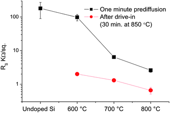 | ||
| Fig. 1 Sheet resistance measurements for flat bulk Si samples after one minute prediffusions for various temperatures, followed by 30 min drive-ins at 850 °C. The samples were covered with ∼50 nm evaporated SiOx before drive-in. | ||
Given that the junction depth is likely dominated by the drive-in step, this indicates the active boron concentration only varies by approximately a factor of 3 or less across the prediffusion temperature range, suggesting that the process is generally insensitive to prediffusion temperature. Nonetheless, the low doping temperatures used in this process are valuable for minimizing thermal budget in device fabrication. No change in sheet resistance from the undoped samples was measured for prediffusion temperatures below 600 °C for the conditions used in this study. Conditions are expected to be very different for NWs which have high surface to volume ratios and cylindrical geometries with diameters smaller than expected junction depths. Furthermore, when translated to doping NWs, the efficiency of this process is expected to be strongly impacted by their areal density due to reactant depletion effects.
Raman spectrometry provides a rapid, quantitative and non-destructive method to measure the active B concentrations in Si with sub-micron resolution.13,40 Thus, we have employed it to probe the capability to dope Si NWs by this process. Interference between discrete phonon Raman scattering and continuous electronic Raman scattering from intravalence band hole transitions gives rise to a Fano resonance in the Raman spectra for Si around the characteristic optical phonon frequency.41 Quantitative doping information can be extracted by fitting the spectra with Fano line shapes:
 | (2) |
 | (3) |
In this equation A is a fitting constant, ωo is the phonon frequency, Γ is the line width, and q is commonly called the asymmetry parameter. Both ωo and Γ are independent of the excitation line wavelength but are affected by other factors such as sample temperature42 and stress.43
The reciprocal of the asymmetry parameter, 1/q, is proportional to the ratio of the Raman tensor for electronic scattering to the Raman tensor for phonon scattering41 and, as a result, is also approximately proportional to the free carrier concentration for active boron concentrations in the range of 1019 cm−3.44 The q parameter is also a function of the excitation line wavelength and thus must be calibrated for the laser. However, both ωo and Γ are functions of the free carrier concentration but not the wavelength. Using the data available in Cerdeira et al.,41 their relationship to active boron concentration was determined and then approximate reference points for the relationship between 1/q and the active boron concentration in our doped Si NWs were obtained. A 1/q value of 0.09 corresponds to an active B concentration of ∼2 × 1019 cm−3 and a 1/q value of 0.5 corresponds to ∼1 × 1020 cm−3. We note that these values nearly match those of Imamura et al. who estimated a 1/q values of 0.1 and 0.5 for concentrations of ∼2 × 1019 cm−3 and ∼1 × 1020 cm−3, respectively, for the same laser wavelength.13,40
Fig. 2(a) shows the Raman spectra for typical Si NWs subjected to different doping conditions. The spectra have been shifted vertically for ease of viewing, and all spectra except that for the undoped Si NW are shown with a corresponding Fano line shape, which clearly fit the data well. For a prediffusion (1 min) and drive-in (10 min) temperature as low as 750 °C, the 1/q value is 0.079 ± 0.002, indicating that the average active boron concentration in the wire is approximately 1–2 × 1019 cm−3. As expected, increasing the prediffusion temperature to 800 °C and the drive-in temperature and time to 850 °C and 30 min, respectively, can increase the active boron concentration to ∼5 × 1019 cm−3. The same conditions applied to Si NWs grown from ∼50 nm Au colloids (and therefore more spaced apart from those grown from an Au film) give rise to NWs with Raman spectra characterized by very high 1/q values. For example, the top-most spectra is typical for such NWs and exhibits a 1/q value of 0.78 ±.01, indicating that active boron concentrations greater than 1 × 1020 cm−3 —approaching, or possibly exceeding the equilibrium solid solubility limit45,46 —are attainable. This demonstrates the range of achievable active dopant concentrations and the importance of accounting for areal density in controlling dopant dosing.
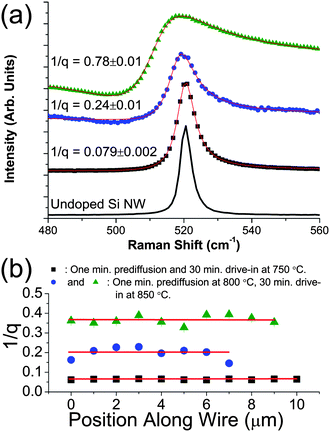 | ||
| Fig. 2 (a) Raman spectra for Si NWs subjected to different doping conditions, shifted vertically from each other for ease of viewing. All spectra except the top one (green triangles) correspond to Si NWs grown from galvanically displaced Au films. The doping conditions for each curve are (from bottom to top): undoped Si NW (black line); one min prediffusion at 750 °C, 10 min drive-in at 750 °C (black squares); one min prediffusion at 800 °C, 30 min drive-in at 850 °C (blue circles); one min prediffusion at 800 °C, 30 min drive-in at 850 °C, NWs grown from 50 nm Au colloids (green triangles). Solid red lines are the fits to a Fano line shape (eqn (2)). (b) Fano fit asymmetry (1/q) as a function of position along the axes of doped NWs grown from Au films. Doping conditions are included in the figure. The red lines represent approximate mean values for 1/q, corresponding to active B concentrations of (top to bottom) ∼ 7.3 × 1019 cm−3, ∼ 4.1 × 1019 cm−3, and ∼ 1.6 × 1019 cm−3. The error bars are comparable to symbol size. | ||
A common issue with in situ doping is the lack of both axial and radial doping uniformity,13,16,17 but it may be possible to control this with ex situ techniques. Fig. 2(b) shows 1/q values measured for spectra taken 1 μm apart along the axes of three Si NWs with different average 1/q values (and hence different average active boron concentrations). The active boron concentration does not deviate much from the average values, marked by red lines that correspond to approximately 7.3 × 1019 cm−3, 4.1 × 1019 cm−3, and 1.6 × 1019 cm−3 from highest to lowest. This shows the excellent axial doping uniformity achievable through this ex situ doping technique. On the other hand, Raman mapping alone does not provide information about the radial doping profile; however, boron diffusivity may be enhanced by several orders of magnitude when the boron concentration exceeds the intrinsic free carrier concentration47 —an important effect at low temperatures. Combined with the high surface-to-volume ratio and cylindrical geometry of NWs, along with the fact that ex situ methods decouple dopant incorporation from diffusion, we expect that a higher degree of radial uniformity may be achievable through ex situ rather than in situ doping. We intend to design and perform further experiments to examine this supposition in the future.
Electron microscopy enables one to examine the structural properties of the NWs doped using BBr3. Fig. 3(a) shows a SEM image of undoped Si NWs grown from galvanically displaced Au film, and Fig. 3(b) shows Si NWs grown in the same manner after a 5 min prediffusion and 1 h drive-in, both at 775 °C. The estimated active boron concentration for these wires is greater than 2 × 1019 cm−3. Though there has been some attenuation in the number of NWs (primarily from the iodine pretreatment), these SEM images demonstrate that there is no discernible change in morphology for the majority of doped NWs.
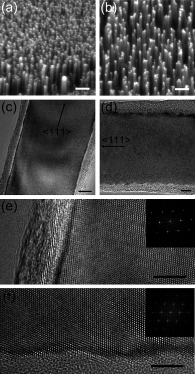 | ||
| Fig. 3 Electron microscope images of doped and undoped Si NWs. (a) SEM image of undoped Si NWs (scale bar = 2 μm). (b) SEM image of Si NWs doped through a 1 min prediffusion and a 1 h drive-in, both at 775 °C (scale bar = 2 μm). (c) TEM image of an undoped Si NW (scale bar = 10 nm). (d) TEM image of a Si NW doped through a 1 min prediffusion at 800 °C and a 30 min drive-in at 850 °C, with an expected active boron concentration of ∼5 × 1019 cm−3 (scale bar = 10 nm). (e–f) High magnification TEM images of sidewall segments for the undoped NW in (c) and the doped NW in (d), respectively. The scale bar is 5 nm for both images. The inset Fourier transforms in (e) and (f) indicate that each of these wires possesses a <111> growth direction, and that a high quality single crystal is retained through the doping process. | ||
More detailed structural information was obtained through TEM. Fig. 3(c) shows a representative TEM of an undoped wire viewed along a <110> direction that permits viewing of the 112 NW sidewalls edge-on.34 The NW sidewalls are smooth though occasionally NWs exhibit facets similar to those reported previously48 (we find these facets are much more common in NWs grown from Au colloids), and they are covered with less than ∼5 nm of native oxide. Fig. 3(d) shows a TEM image of a NW from a sample with NWs doped to approximately ∼5 × 1019 cm−3, also viewed along a <110> direction. The surface oxide layer is slightly thicker (typically 5 to 10 nm) on these NWs, possibly due to a small amount of oxidation that occurs during the drive-in stage. Higher magnification TEM images (Fig. 3(e) and (f)) of sidewall segments from the above undoped and doped Si NWs, respectively, show that while there are no dramatic morphological changes in the NWs as a result of doping (e.g. tapering or faceting), the process does induce a degree of surface roughening. Analyses of Fig. 3(e) and (f) indicate that the doping process induces a root mean squared (RMS) NW sidewall profile roughness of ∼ 0.4 nm, compared with a RMS roughness value of ∼ 0.2 nm for the undoped Si NW. Analyses of similar high magnification images of other doped and undoped Si NWs yield similar results. Such roughening may have important implications for NW thermal and electrical properties, especially for decreasing NW diameters. For instance, surface roughening has been shown to dramatically reduce Si NW thermal conductivities,4 a fact beneficial to the employment of Si NWs in thermoelectric devices. On the other hand, surface roughening may decrease the electronic mobility of Si NWs for diameters below approximately 10 nm,49 hindering the aggressive scaling of Si NW-based electronics. Fourier transforms of Fig. 3(e) and (f) shown in the inset of each image demonstrate that the wires retain the excellent quality needed for a variety of device applications.
Two-point electrical measurements were performed to confirm the conductivity enhancement afforded by this doping process. Fig. 4 shows characteristic current–voltage curves for an undoped Pt-catalyzed Si NW and a Pt-catalyzed Si NW from a sample that underwent a 5 min prediffusion followed by a 30 min drive-in, both at 750 °C. The undoped wire (diameter ≈ 120 nm) exhibits rectifying contacts and a resistivity of 120 Ω cm (measured from 4.5–5.0 V). Assuming bulk Si hole mobility,50 its free carrier concentration is estimated at ∼1 × 1014 cm−3. On the other hand, the doped wire (diameter ≈ 50 nm) exhibits ohmic contacts and an apparent resistivity of 0.046 Ω cm (resistance = 1 MΩ), which provides an estimated active B concentration of ∼8 × 1017 cm−3, assuming bulk Si hole mobility.50 Fano fitting of other NWs from the same sample imply an average estimated active boron concentration between ∼6 × 1018 cm−3 and ∼1 × 1019 cm−3, though this is at the lower detection limit for this technique. This range of concentrations suggests that the NW resistivity should be approximately one order of magnitude lower. However, factors such as surface oxide thickness (unmeasured by SEM imaging), size dependent mobilities, surface interface states,50,51 catalyst contamination,52 dopant deactivation53 and contact resistances can substantially increase resistivity in silicon nanowires beyond that expected from the intentional impurity concentration alone. Indeed, higher-than-expected four point resistivities have been measured for Si NWs B-doped in situ10 or ex situ,28 attributed in part to the reasons highlighted above (except contact resistance).
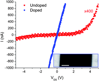 | ||
| Fig. 4 Current–voltage curves for an undoped Pt-catalyzed Si NW (red squares) and a Pt-catalyzed Si NW doped through a 5 min prediffusion followed by a 30 min drive-in at 750 °C (blue circles). Inset: SEM image of the doped Si NW whose data is shown (scale bar = 1 μm). | ||
Finally, the versatility of this vapor phase ex situ doping method is exemplified in its capability to dope bridging Si NWs. We exposed a SOI sample with as-grown Si NWs bridging B-doped single-crystal Si electrodes (1018 cm−3) to a prediffusion step for 1 min and drive-in for 15 min, both at 700 °C. Fig. 5(a) is a SEM image showing a set of three bridging NWs after doping, all 3 μm long (dictated by the trench width) and an average diameter of 95 nm. Debris from NWs that did not grow properly underneath the bottom NW does not bridge the electrodes and thus should not participate in electrical conduction. Fig. 5(b) shows the current–voltage characteristic curve for this set of NWs. Assuming that all three NWs participate in electrical conduction, the apparent resistivity of 5.6 Ω cm per NW yields an estimated active boron concentration of only ∼2 × 1015 cm−3. However, several of the previously mentioned factors can increase resistivity even more substantially at dopant concentrations below 1018 cm−3.51,53
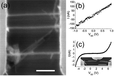 | ||
| Fig. 5 (a) SEM image of epitaxial bridging Au-catalyzed Si NWs doped as described in the text (scale bar = 1 μm). (b) Current–voltage curve for the set of NWs shown in (a). (c) Current–voltage response for a single undoped epitaxial bridging Si NW, shown in the inset SEM image (scale bar = 1 μm). | ||
To substantiate the claim of doping these NWs, we may compare their current–voltage response to that of an undoped epitaxial bridging Si NW, shown in Fig. 5(c). An estimated resistivity from the linear regions of the curve of 140 Ω cm implies a free carrier concentration of ∼1 × 1014 cm−3. More importantly, this NW shows very strong rectifying behavior due to the potential barrier imposed by the difference between the near-intrinsic Fermi level in the NW and the Fermi level of the doped contact pads, in contrast to the ohmic behavior of the doped NWs. The low areal density of NWs on these substrates and their double-clamped geometry make them more sensitive to the particular doping conditions. As a result, attempts at attaining higher doping levels for these bridging NWs resulted in significant damage to them, possibly from increased stress or Si etching from gaseous HBr. Process optimization should render much more highly doped epitaxial bridging Si NWs in the future.
4 Conclusions
In summary, we have successfully doped Si NWs ex situ with boron through the reduction of BBr3 by H2 to deposit elemental boron on the Si surface, followed by a drive-in anneal to further diffuse and activate the boron. Analysis of the Fano resonance in the Raman spectra for single doped NWs reveal that boron concentrations from less than 1019 cm−3 to 1020 cm−3 are attainable in a well-controlled manner, though the areal density of the NWs on the sample is found to have a large impact. Raman mapping exhibits the excellent doping uniformity in this technique. The crystallinity of the NWs remains unaltered, with minimal changes in their surface morphology. Electrical measurements confirm the conductivity enhancement as a result of intentional boron doping for Si NWs grown from metal catalyst films or epitaxially bridging Si NWs in SOI microtrenches. The fine control of boron concentration magnitude and uniformity afforded by this ex situ vapor phase method makes it a valuable alternative to in situ doping in the development of a range of applications based on Si NWs or other nanomaterials.Acknowledgements
We thank Velimir Radmilovic for use of the facilities at the National Center for Electron Microscopy, Lawrence Berkeley National Laboratory. This work was supported by the National Science Foundation under Grant No. DMR-0804646 and Grant No. EEC-0832819 (Center of Integrated Nanomechanical Systems).References
- Y. Cui and C. Lieber, Science, 2001, 291, 851–853 CrossRef CAS.
- J. Xiang, W. Lu, Y. Hu, Y. Wu, H. Yan and C. Lieber, Nature, 2006, 441, 489–493 CrossRef CAS.
- A. Boukai, Y. Bunimovich, J. Tahir-Kheli, J. Yu, W. Goddard and J. Heath, Nature, 2008, 451, 168–171 CrossRef CAS.
- A. Hochbaum, R. Chen, R. Delgado, W. Liang, E. Garnett, M. Najarian, A. Majumdar and P. Yang, Nature, 2008, 451, 163–U5 CrossRef CAS.
- B. Tian, X. Zheng, T. Kempa, Y. Fang, N. Yu, G. Yu, J. Huang and C. Lieber, Nature, 2007, 449, 885–U8 CrossRef CAS.
- E. Garnett and P. D. Yang, J. Am. Chem. Soc., 2008, 130, 9224–9225 CrossRef CAS.
- S. Boettcher, J. Spurgeon, M. Putnam, E. Warren, D. Turner-Evans, M. Kelzenberg, J. Maiolo, H. Atwater and N. Lewis, Science, 2010, 327, 185–187 CrossRef CAS.
- R. He, X. Feng, M. Roukes and P. Yang, Nano Lett., 2008, 8, 1756–1761 CrossRef.
- Y. Cui, X. Duan, J. Hu and C. Lieber, J. Phys. Chem. B, 2000, 104, 5213–5216 CrossRef CAS.
- K. Lew, L. Pan, T. Bogart, S. Dilts, E. Dickey, J. Redwing, Y. Wang, M. Cabassi, T. Mayer and S. Novak, Appl. Phys. Lett., 2004, 85, 3101–3103 CrossRef CAS.
- Y. Wang, K. Lew, T. Ho, L. Pan, S. Novak, E. Dickey, J. Redwing and T. Mayer, Nano Lett., 2005, 5, 2139–2143 CrossRef CAS.
- H. Schmid, M. Bjork, J. Knoch, S. Karg, H. Riel and W. Riess, Nano Lett., 2009, 9, 173–177 CrossRef CAS.
- G. Imamura, T. Kawashima, M. Fujii, C. Nishimura, T. Saitoh and S. Hayashi, Nano Lett., 2008, 8, 2620–2624 CrossRef CAS.
- D. Perea, E. Hemesath, E. Schwalbach, J. Lensch-Falk, P. Voorhees and L. Lauhon, Nat. Nanotechnol., 2009, 4, 315–319 CrossRef CAS.
- J. Allen, D. Perea, E. Hemesath and L. Lauhon, Adv. Mater., 2009, 21, 3067–3072 CrossRef CAS.
- R. Schlitz, D. Perea, J. Lensch-Falk, E. Hemesath and L. Lauhon, Appl. Phys. Lett., 2009, 95, 162101 CrossRef.
- E. Koren, Y. Rosenwaks, J. Allen, E. Hemesath and L. Lauhon, Appl. Phys. Lett., 2009, 95, 092105 CrossRef.
- P. Xie, Y. Hu, Y. Fang, J. Huang and C. Lieber, Proc. Natl. Acad. Sci. U. S. A., 2009, 106, 15254–15258 CrossRef CAS.
- E. Garnett, W. Liang and P. Yang, Adv. Mater., 2007, 19, 2946–2950 CrossRef CAS.
- F. Li, P. Nellist and D. Cockayne, Appl. Phys. Lett., 2009, 94, 263111 CrossRef.
- L. Pan, K. Lew, J. Redwing and E. Dickey, J. Cryst. Growth, 2005, 277, 428–436 CrossRef CAS.
- A. Chaudhry, V. Ramamurthi, E. Fong and M. Islam, Nano Lett., 2007, 7, 1536–1541 CrossRef CAS.
- A. San Paulo, J. Bokor, R. Howe, R. He, P. Yang, D. Gao, C. Carraro and R. Maboudian, Appl. Phys. Lett., 2005, 87, 053111 CrossRef.
- E. Garnett, Y. Tseng, D. Khanal, J. Wu, J. Bokor and P. Yang, Nat. Nanotechnol., 2009, 4, 311–314 CrossRef CAS.
- A. Colli, A. Fasoli, C. Ronning, S. Pisana, S. Piscanec and A. Ferrari, Nano Lett., 2008, 8, 2188–2193 CrossRef CAS.
- S. Hoffmann, J. Bauer, C. Ronning, T. Stelzner, J. Michler, C. Ballif, V. Sivakov and S. Christiansen, Nano Lett., 2009, 9, 1341–1344 CrossRef CAS.
- R. Beckman, E. Johnston-Halperin, N. Melosh, J. Green and J. Heath, J. Appl. Phys., 2004, 96, 5921–5923 CrossRef CAS.
- S. Ingole, P. Aella, P. Manandhar, S.C., E. A. Akhadov, D. Smith and S. Picraux, J. Appl. Phys., 2008, 103, 104302 CrossRef.
- L. Magagnin, R. Maboudian and C. Carraro, J. Phys. Chem. B, 2002, 106, 401–407 CrossRef CAS.
- M. Cerruti, G. Doerk, G. Hernandez, C. Carraro and R. Maboudian, Langmuir, 2010, 26, 432–437 CrossRef CAS.
- G. Doerk, N. Ferralis, C. Carraro and R. Maboudian, J. Mater. Chem., 2008, 18, 5376–5381 RSC.
- D. Gao, R. He, C. Carraro, R. Howe, P. Yang and R. Maboudian, J. Am. Chem. Soc., 2005, 127, 4574–4575 CrossRef CAS.
- J. Hannon, S. Kodambaka, F. Ross and R. Tromp, Nature, 2006, 440, 69–71 CrossRef CAS.
- G. Doerk, V. Radmilovic and R. Maboudian, Appl. Phys. Lett., 2010, 96, 123117 CrossRef.
- J. Fréchette and C. Carraro, Phys. Rev. B: Condens. Matter Mater. Phys., 2006, 74, 161404 CrossRef.
- A. San Paulo, N. Arellano, J. Plaza, R. He, C. Carraro, R. Maboudian, R. Howe, J. Bokor and P. Yang, Nano Lett., 2007, 7, 1100–1104 CrossRef.
- E. Haupfear and L. Schmidt, Chem. Eng. Sci., 1994, 49, 2467–2481 CrossRef CAS.
- Z. Walker and E. Ogryzlo, J. Appl. Phys., 1991, 69, 2635–2638 CrossRef CAS.
- S. Sharma, T. Kamins and R. Williams, J. Cryst. Growth, 2004, 267, 613–618 CrossRef CAS.
- G. Imamura, T. Kawashima, M. Fujii, C. Nishimura, T. Saitoh and S. Hayashi, J. Phys. Chem. C, 2009, 113, 10901–10906 CrossRef CAS.
- F. Cerdeira, T. Fjeldly and M. Cardona, Phys. Rev. B: Solid State, 1973, 8, 4734–4745 CrossRef CAS.
- G. Doerk, C. Carraro and R. Maboudian, Phys. Rev. B: Condens. Matter Mater. Phys., 2009, 80, 073306 CrossRef.
- E. Anastassakis, A. Pinczuk, E. Burnstein, F. Pollak and M. Cardona, Solid State Commun., 1970, 8, 133–138 CrossRef CAS.
- V. Magidson and R. Beserman, Phys. Rev. B: Condens. Matter Mater. Phys., 2002, 66, 195206 CrossRef.
- G. Vick and K. Whittle, J. Electrochem. Soc., 1969, 116, 1142–1144 CrossRef CAS.
- G. Glass, H. Kim, P. Desjardins, N. Taylor, T. Spila, Q. Lu and J. Greene, Phys. Rev. B: Condens. Matter Mater. Phys., 2000, 61, 7628–7644 CrossRef CAS.
- R. Fair, J. Electrochem. Soc., 1975, 122, 800–805 CrossRef CAS.
- F. Ross, J. Tersoff and M. Reuter, Phys. Rev. Lett., 2005, 95, 146104 CrossRef CAS.
- E. Ramayya, D. Vasileska, S. Goodnick and I. Knezevic, J. Appl. Phys., 2008, 104, 063711 CrossRef.
- K. Seo, S. Sharma, A. Yasseri, D. Stewart and T. Kamins, Electrochem. Solid-State Lett., 2006, 9, G69–G72 CrossRef CAS.
- V. Schmidt, S. Senz and U. Gosele, Appl. Phys. A: Mater. Sci. Process., 2006, 86, 187–191 CrossRef.
- J. Allen, E. Hemesath, D. Perea, J. Lensch-Falk, Z. Y. Li, F. Yin, M. Gass, P. Wang, A. Bleloch, R. Palmer and L. Lauhon, Nat. Nanotechnol., 2008, 3, 168–173 CrossRef CAS.
- M. Björk, H. Schmid, J. Knoch, H. Riel and W. Riess, Nat. Nanotechnol., 2009, 4, 103–107 CrossRef.
| This journal is © The Royal Society of Chemistry 2010 |
