 Open Access Article
Creative Commons Attribution 3.0 Unported Licence
Unique catanionic vesicles as a potential “Nano-Taxi” for drug delivery systems. In vitro and in vivo biocompatibility evaluation†
Soledad Stagnolib,
M. Alejandra Lunaa,
Cristian C. Villa‡
a,
Fabrisio Alustizab,
Ana Niebylskib,
Fernando Moyanoa,
N. Mariano Correa *a and
R. Darío Falcone
 a
a
aDepartamento de Química, Universidad Nacional de Río Cuarto, Agencia Postal #3, C.P. X5804BYA Río Cuarto, Argentina. E-mail: mcorrea@exa.unrc.edu.ar
bDepartamento de Biología Molecular, Universidad Nacional de Río Cuarto, Agencia Postal #3, C.P. X5804BYA Río Cuarto, Argentina
First published on 17th January 2017
Abstract
We evaluate in vitro and in vivo toxicity and stability in an acidic environment of new vesicles formed by the catanionic surfactant bis(2-ethylhexyl) sulfosuccinate benzyl-n-hexadecyldimethylammonium (AOT-BHD) in order to investigate their potential application as an oral drug delivery system. Unilamellar vesicles were spontaneously formed by dissolving AOT-BHD in water and their toxicity was evaluated through in vitro and in vivo assays. Cell membrane permeability assays (hemolytic activity, Trypan blue assay) and cellular survival or proliferation (MTT assay) were performed. The results showed that only the highest concentration of vesicles tested (2 mg mL−1) diminished the red blood cells' resistance. In vivo toxicity evaluation was carried out on mice through lethal dose 50 (LD50) experiments. The safety for living organisms in doses lower than 0.05 mg mL−1 and the acid pH stability makes our AOT-BHD vesicles a very promising candidate for oral drug delivery.
Introduction
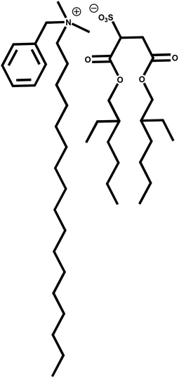
An emerging field of supramolecular chemistry, catanionic surfactants, arises from the binding of equimolar quantities of anionic and cationic surfactants.1,2 It is known that mixtures of these surfactants may have considerable synergistic benefits in their properties and applications.3 It has been shown that these molecules have several unique characteristics that differ substantially from their individual components. In this sense, it was demonstrated that various catanionic mixtures can form different types of organized systems spontaneously, including unilamellar vesicles. Previous work in our lab has demonstrated that a mixture of the surfactants sodium bis(2-ethylhexyl) sulfosuccinate (Na-AOT) and benzyl-n-hexadecyldimethylammonium chloride (BHDC) forms a new catanionic surfactant, bis (2-ethylhexyl) sulfosuccinate-benzyl-n-hexadecyldimethylammonium (AOT-BHD) (Fig. 1) by removing their counterions.4 This system has the ability to form unilamellar vesicles spontaneously, without adding energy to the system, which is an advantage compared to traditional methods that require mechanical techniques to obtain unilamellar vesicles. Interesting, the size of the vesicles do not depend on the surfactant concentration working at concentration below 6 mg mL−1.4 In addition, we previously demonstrated that spontaneous large AOT-BHD vesicles have a bilayer with low polarity, high viscosity, more electron donor capacity and less proton permeability that can encapsulate different kinds of dyes without perturbing the assemble.5 These properties produce large incorporation of ionic and nonionic molecules with partition constants that are higher than in conventional vesicles.5 Those results showed that AOT-BHD vesicles are very promising to use as nanocarrier in pharmacologic, cosmetic and foods fields.5A drug delivery system is a formulation that is capable of introducing a therapeutic substance into the body in a manner that enhances its safety and efficacy over others methods of drug administration. The improved efficacy can be due to greater localization, enhanced bioavailability or sustained duration of drug action.6 Vesicles have been used in various administrative routes, such as intravascular,7 oral,8 pulmonary,9 ocular,10 among others. Particularly, oral administration offers numerous advantages such as being painless and easily self-administrable. The human gastro-intestinal tract, however, forms a formidable barrier due to low stomach pH and enzymatic degradation.11 Peptides or proteins (e.g. insulin, hormones, some vaccines and antineoplastic drugs) are the most commonly used but they cannot be administered orally, due to their degradation in the stomach since they do not resist such acidic medium. Therefore, oral administration is a challenge that requires more highly innovative and sophisticated delivery systems.11 As the AOT-BHD vesicles are not formed by phospholipids (which are hydrolyzes in the stomach with the consequent vesicle disruption), it is expected that their behavior in the human body will be different to the traditional vesicles. One of the key differences could be found if these vesicles can tolerate extreme pH conditions of digestive system which would enable use by oral via. Interesting, non-phospholipids based vesicles are becoming very promising candidates for oral drug delivery due to their ability to carry hydrophilic drugs by encapsulation in the aqueous inner pool or hydrophobic drugs by intercalation into hydrophobic domains.12–14
The development of nanocompounds however, must go accompanied by studies to know their effects in living organism and environment. The shape, size, material type, purity, electric charge, structural characteristics, dose, route of administration, concentration in the target organ and duration of action, are some of the variables that will determine their potential toxicity, which would be seen in the disruption of membranes or membrane potential, protein oxidation, genotoxicity, interruption in the energy transmission, reactive oxygen species increment, release of toxic components and inflammatory processes.15 An ideal drug delivery system should be biodegradable, biocompatible, and unassociated with incidental adverse effects.16 Implementation of new technologies in drug production implies the realization of different tests including toxicological studies to determine the safety of their use.
The safety assessment of a compound or formulation is carried out through in vitro and in vivo experiments, even though ethical and economic reasons the use of in vitro assays is preferred to minimize use of animals, results may differ greatly from those obtained in vivo due to physiological conditions in cells cultures are very different from living organisms, and because of that they are irreplaceable.17,18
Numerous nanosized drug carriers have been extensively investigated in drug delivery.19,20 However, despite the improved pharmacokinetic properties and the reduced adverse effects,19–21 currently drug delivery has only achieved modest therapeutic benefits.22,23 Thus, the design of nanocarriers with more efficient drug delivery and thus higher therapeutic efficacy is still a pressing need. Due to the promising physicochemical properties4,5 of the AOT-BHD vesicles we propose them as potential vehicle for drug administration. Thus, the aim of this work is to evaluate in vitro and in vivo toxicity and the stability in acidic pH of AOT-BHD vesicles in order to investigate their potential application as drug delivery system via oral.
Experimental
AOT-BHD vesicles formation
AOT-BHD was obtained as previously reported4 using an equimolar mixture of sodium 1,4-bis (2-ethylhexyl) sulfosuccinate (Na-AOT) and benzyl-n-hexadecyldimethylammonium chloride (BHDC), both from Sigma (>99% purity). The formation of AOT-BHD was confirmed by 1H NMR technique. The catanionic surfactant was dried under vacuum for 4 hours before its use.A stock solution of AOT-BHD vesicles (10 mg mL−1) were prepared by weighting an appropriated amount of AOT-BHD and diluting with of 0.9% NaCl saline aqueous solution to obtain the desire concentration. An opalescent solution was obtained by hand shaking during two minutes at room temperature. The formation of the AOT-BHD vesicles was confirmed by the DLS technique, obtained a similar diameter as previously reported.4 All samples were prepared and used immediately after preparation.
DOPC vesicles preparation
The vesicle suspension was typically prepared as follows: the stock lipid solution was prepared by taking the appropriate amount of 1,2-dioleoyl-sn-glycero-3-phosphocholine (DOPC) in chloroform (Sintorgan HPLC). After the solvent was evaporated and the film was dried under reduced pressure, large multilamellar vesicles were obtained by hydrating the dry lipid–dye film using 0.9% NaCl saline solution, through mixing (Vortex-2-Genie) for about 5 min at room temperature. The resulting solution of the large multilamellar vesicles had the desired lipid concentration. To prepare large unilamellar vesicles (LUVs), the large multilamellar vesicles suspension was extruded 10 times (Extruder, Lipex biomembranes) through two stacked polycarbonate filters of pore size 200 nm under nitrogen pressure up to 3.4 atm. The monodispersity of the LUV size achieved through this technique was previously checked.24 The unilamellar nature of the pure DOPC vesicles prepared by using the extrusion method21 was previously confirmed24–26 by measuring the extent of quenching by Mn2+ of their 31P NMR signals.27 All samples were used immediately after preparation.Characterization of catanionic vesicles
The apparent diameters (dapp) of AOT-BHD vesicles were determined by DLS. Cleanliness of the cuvettes used for measurements was of crucial importance for obtaining reliable and reproducible data.28 Cuvettes were washed with ethanol, and then with doubly distilled water and dried with acetone. Prior to use the samples were filtered three times to avoid dust or particles presents in the original solution using a nylon membrane for the vesicles samples. Prior to data acquisition, the samples were equilibrated in the DLS instrument for 10 min at 37 °C. To obtain valid results from DLS measurements requires knowledge of the system's refractive index and viscosity in addition to well-defined conditions. The refractive indices and viscosities were assumed to be the same as neat water.29 Multiple samples at each size were made, and thirty independent size measurements were made for each individual sample at the scattering angle of 90°. The instrument was calibrated before and during the course of experiments using several different size standards. Thus, we are confident that the magnitudes obtained by DLS measurements can be taken as statistically meaningful for all the systems investigated. The algorithm used was CONTIN and the DLS experiments shown that the polidispersity of the catanionic vesicles sizes were less than 5%.
In vitro toxicity
Citotoxicity was evaluated throughout membrane permeability changes30 (hemolytic activity, Trypan blue assay) and cellular survival (MTT assay).Peripheral human blood samples were obtained from healthy donors under informed consent as established by bioethics standards specified by the bioethics committee of the UNRC, endorsed by the CIEIS of the Province of Córdoba (Res 13/2015).
Equal amounts of leukocytes suspension and trypan blue ((3Z,3′Z)-3,3′-[(3,3′-dimethylbiphenyl-4,4′-diyl) di(1Z) hydrazin-2-yl-1-ylidene] bis (5-amino-4-oxo-3,4-dihydronaphthalene-2,7-disulfonic acid)) solution 0.4% w/v were mixed and counting was performed using a Neubauer hemocytometer distinguishing between dead cells (with membrane disruption, dyed blue) of the living cells with intact membranes (uncolored).33 Cell viability was calculated using eqn (1):
 | (1) |
All measurements were repeated five times for a statistical analysis.
A MTT assay for DOPC vesicles was performed testing the same concentrations that AOT-BHD vesicles to compare toxicity of our system with other vesicles of known low toxicity.
In vivo toxicity
Balb-c mice (25–30 g) of both sexes provided by Molecular Biology Department bioterio (UNRC) were used for in vivo toxicity evaluation. The animals were maintained in cages in standard conditions (20 ± 2 °C in a 12 h light–dark cycle) and were provided with free access to standard pellet chow and water. Procedures concerning animal treatments and experiments in this study were reviewed and approved by the Animal Use Ethics Committee of the UNRC (CoEdi) (Res. 13/2015) being in accordance with the bioethics guidelines of the European Economic Community and the Public Health Service Guide of U.S., National Institutes of Health, regarding care and use of Laboratory Animals, minimization of suffering, and number of animal used.Results and discussion
Surface zeta potential
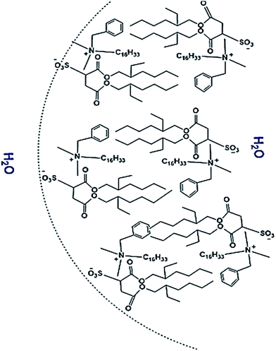
Zeta potential is one of the crucial parameters for determining the vesicles stability and interaction with biological system. Our findings showed that vesicles have a zeta potential of −34 mV at the three different AOT-BHD concentration investigated indicating that anionic surfactant-groups are exposed to the outer face of the vesicle bilayer as it is schematically shown in Fig. 2 at any surfactant concentration. Moreover, the DLS experiments give a dapp value for the AOT-BHD vesicles around 80 nm with a polydispersity value of 0.1.AOT-BHD vesicles stability to acidic pH
Fig. 3A shows the dapp values of the AOT-BHD vesicles at different times during the acidic pH exposition. The size of AOT-BHD vesicles reminds stable during the first 90 minutes of exposition with a mean diameter of 95 nm and, after that, the size decreases sharply to almost half of the original diameter. A similar behavior was observed in the polydispersity values of the prepared vesicles (Fig. 3B). During the first 90 minutes of exposition, polydispersity reminds almost constant and then increases sharply. It is clear that at longer times to 90 minutes, the system turns unstable under acidic conditions. However, liquid formulations remain a short time in the stomach, less than 90 minutes, which make the system propitious for oral administration since molecules sensible to acidic pHs can pass to the duodenum and be incorporated into the intestinal cells without altering their biological function.36,37 Herein it is one remarkable difference with the vesicles obtained using traditional phospholipids which do not resist the low pH values because they hydrolyze and the system breaks down (DOPC vesicles results, not shown). The chemical structure of AOT-BHD and its bilayer properties recently shown,5 do not seem to suffer the same lysis process that DOPC vesicles in acidic environments (pH ≤ 2). Looking at the AOT-BHD structure it can be seen that there is an ester group in the AOT moiety. Although esters are non-very much reactive to the hydrolysis, the presence of such amount of protons during 90 minutes may accelerate the hydrolysis reaction, changing the surfactant structure, mainly the packing parameter and destroying the vesicles. Another possible explanation is that, the vesicles external charge in the presence of high ionic strength may be masked allowing the vesicles flocculation.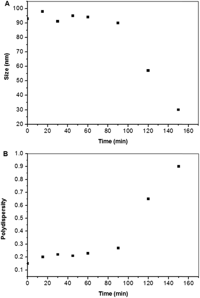 | ||
| Fig. 3 (A) AOT-BHD vesicles sizes at different times during the HCl/KCl pH 2.1 solution exposition. (B) Polydispersity of AOT-BHD vesicles at different times during the pH 2.1 exposition. | ||
In vitro toxicity evaluation
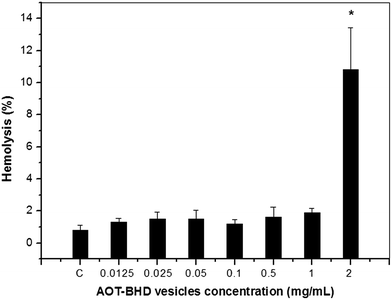 | ||
| Fig. 4 Hemolysis percentage at different concentrations of AOT-BHD vesicles. Means ± SE (error bars) are represented. *p = 0.00007 vs. control (C). In the ESI, it is provided the raw data as Table S1.† | ||
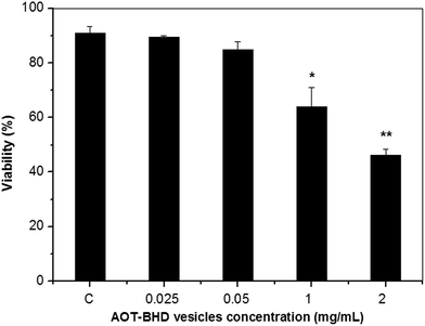 | ||
| Fig. 5 Trypan blue exclusion assay in leukocytes incubated with different concentrations of AOT-BHD vesicles. Means ± SE are represented. *p = 0.004 vs. control (C) **p = 0.00006 vs. control (C). In the ESI, it is provided the raw data as Table S2.† | ||
Viability decreased 21% with 1 mg mL−1 and 46.7% in samples incubated with 2 mg mL−1 of AOT-BHD vesicles, indicating a clear relationship between percent of viability and concentration of vesicles.
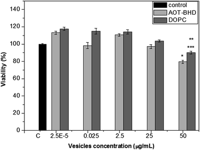 | ||
| Fig. 6 Fibroblasts mitochondrial enzyme activity evaluation (MTT assay). Means ± SE are represented. *p = 0.005 vs. C; **p = 0.038 vs. C ***p = 0.0013 vs. 0.05 mg mL−1 AOT-BHD. In the ESI, it is provided the raw data as Table S3.† | ||
At minor concentrations, a tendency to enzyme activity increases was observed for both systems, AOT-BHD and DOPC vesicles, but the difference vs. control is not statistically significant.
In vivo toxicity evaluation
Factors that showed no statistically significant difference were grouped in the same treatment population (doses and sex).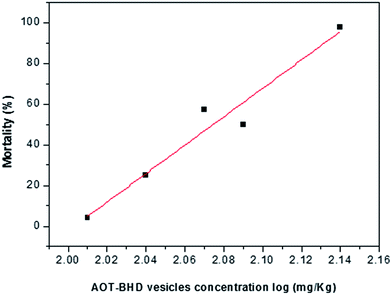 | ||
| Fig. 7 Mortality percentage of mice treated with different doses of AOT-BHD vesicles after 24 hours. In the ESI, it is provided the raw data as Table S4.† | ||
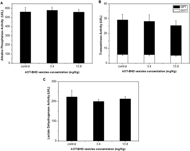 | ||
| Fig. 8 Serum enzymatic activity in animals treated AOT-BHD vesicles (3.4 and 13.8 mg kg−1 from animal) during 30 days. A – AP, B – GPT and GOT, C – LDH. In the ESI, it is provided the raw data as Table S5.† | ||
| Treatment | Food intake (g) | Water intake (mL) | Daily weight gain (mg) |
|---|---|---|---|
| Control | 3.71 ± 0.23 | 9.53 ± 1.23 | 41.6 ± 19.0 |
| 3.4 mg kg−1 | 3.81 ± 0.21 | 7.70 ± 0.58 | 40.5 ± 29.0 |
| 13.7 mg kg−1 | 3.97 ± 0.26 | 8.01 ± 0.88 | 77.7 ± 22.0 |
Our findings showed that the vesicles apparent diameter (dapp) value was 80 nm being consistent with the results found by Villa et al.5 We also observed a negative charge on the outer layer (−34 mV) that may be explained by the exposition of sulfonate groups corresponding to headgroup anionic surfactant.
Enhanced serum stability and longer blood residence time have been found to depend substantially on size, composition, and surface properties.38 It has been shown that the superficial charge of nanoparticles is a crucial parameter which defines stability of the system and, as a consequence, its biocompatibility.38 In previous studies, it has been observed that positively charged vesicles showed greater toxic effects in comparison with anionic systems.39 These toxic effects have several causes, one of them being the inhibitory activity over key enzymes which are involved in the regulation of cellular signaling, such as protein kinase C (PKC).40 On the other hand, through electrostatic attraction, positive charged systems are more unstable in biological media, given their interaction with serum proteins such as lipoproteins, albumin and immunoglobulin. This may causes opsonization, aggregation or nonspecific adsorption and creates disturbances in their biodistribution and cellular interaction.41,42 The delay in opsonization substantially improves the average lifetime in circulation of nanoparticles.43
On the other hand, stability in stomach like conditions was evaluated. Diameter and polydispersity changes observed for AOT-BHD vesicles after 90 minutes in acidic media (pH = 2) may be attributed to its degradation due to the extreme pH condition and strong ionic force of buffer solution. Nevertheless, it is important to notice that this only occurs after a long period of time confirming our first assessment about the resistance of the AOT-BHD vesicles to these conditions. These results are the first step in understanding the behavior of these vesicles in pH conditions (pH = 1.0–2.1) found in the stomach. The results are very promising for potential application as drug delivery system administered orally.
To evaluate biocompatibility, several tests were performed. RBC hemolysis measurement is a simple and widely used method to study surfactant–membrane interaction. If cell membrane is damaged, hemoglobin will be released from red blood cell to plasma. A quantitative measure of released hemoglobin can give an indication of potential damage of vesicles to RBC which is a good indicator of vesicles' toxicity at in vitro conditions.44
We observed that only the highest concentration of vesicles tested diminished the red blood cells resistance, showing a change in the cell membrane permeability. Interestingly more dilute vesicles solutions showed similar hemolysis percentages to control, which would be indicative of little or no damage to cell membranes. Similar results were obtained from Trypan blue and MTT assays. Increasing AOT-BHD vesicles concentration from 0.05 mg mL−1 to 2 mg mL−1 results in an increase in membrane and mitochondrial destabilization, but concentrations lower than 0.05 mg mL−1 showed the same values that control, which evidences the low toxicity of the system at these concentrations. Scheme 1 shows a possible explanation of what happens when different concentrations of vesicles interact with membranes (plasma or mitochondrial). We propose that AOT-BHD vesicles are incorporated to cells by endocytosis at low concentrations. On the other hand, when the concentration is higher, the membrane cannot make this process and, as results, the vesicles interact strongly with the membrane resulting in the membrane deformation and permeability changes leading to cell death.
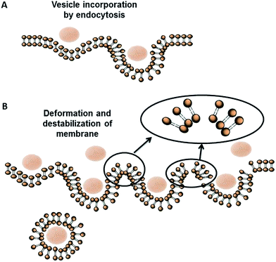 | ||
| Scheme 1 Schematic representation of AOT-BHD vesicle–membrane interaction. (A) Low concentration of AOT-BHD vesicles, (B) high vesicle concentration. | ||
A comparative analysis between AOT-BHD and DOPC vesicles showed a similar behavior at the same doses. It is noteworthy that in vitro studies with DOPC-based liposomes reported a lack of toxicity toward fibroblasts, hematopoietic and bone marrow cells.45 However, concentrations in which AOT-BHD vesicles are innocuous according to our in vitro studies, are highly cytotoxic for others nanocarrier systems, including nanoparticles.46–48
Even though a broad exploration of in vitro toxicity has been carried out for different nanomaterials in vivo studies of toxicity remain limited.49 Lethal dose,50 LD50, is the dose capable of causing death of 50% of the animal population and it is frequently used to determine toxicity of a substance in a living organism since reduces the amount of required tests. Our results shows that the obtained value of LD50 (118.03% mg kg−1) is higher than the one obtained for other systems such as polymeric nanoparticles of 110 nm (40 and 75 mg kg−1).50 Moreover, it was observed that administration of different doses of vesicles (3.4 and 13.8 mg kg−1) during 30 days turned out to be innocuous, whereas the same concentrations results toxic for other nanomaterials proposed as drug vehicles.51,52 Additional tests to determine the safety of the system were carried out through quantification of biochemical markers in blood, such as GPT, GOT, AP and LDH, as well as behavioral studies. The release of cytosolic enzymes into the blood is an indicator of tissue lesion; a muscle, liver, heart, kidney or skeleton lesion could significantly increase the content of these enzymes.53 However, our studies did not register differences in any of the enzymatic parameters between control and treated groups. In the behavioral assays, no significant changes were observed. Therefore, these results indicate that AOT-BHD catanionic vesicles administered one time only, or chronically (for 30 days), produce no detectable toxic effects, which means they are innocuous as far as the studied doses are concerned.
Innocuousness observed in both in vitro (≤0.05 mg mL−1) and in vivo (≤118.6 mg kg−1) studies could be associated with the fact that anionic systems are stable in biological media due to electrostatic repulsion with serum proteins,38 and also decreases its immunogenicity, increase its biological compatibility and its permanence in the cardiovascular system.39 In addition, the innocuousness of our system can also be explained because of its shape (spherical) and its size (80 nm). It has previously been reported that the interaction of particles with cells is known to be strongly influenced by particle size and shape.54 Nanoparticles smaller than 50 nm significantly decrease cellular viability.55 This difference depends on the particle mechanisms for interacting with and internalizing the cell.54,56 Particles with a size smaller than 50 nm are internalized through mechanisms of direct penetration or passive density gradient diffusion causing the disruption of lipid membranes, leading to loss of cytoplasmic substance and culminating in cell death.54,56 On the other hand, the internalization of nanoparticles bigger than 50 nm depends on endocytosis of cell-surface receptors without damaging the cellular membrane ultrastructure.57 This means that catanionic vesicles biocompatibility observed in vitro and in vivo may be closely associated with system properties (charge, size and shape) as well as with the administered doses.
Conclusions
The results of in vitro (globular resistance, MTT, trypan blue) and in vivo (dose lethal 50 and toxicity chronic) and stomach like pH simulation studies have demonstrated the stability of our system to acid pH and safety to living systems due to the low toxicity observed in doses lower than 0.05 mg mL−1 in vitro or 118.6 mg kg−1 in vivo. This characteristic makes our AOT-BHD vesicles highly biocompatible and very promising candidate for oral drug delivery, based in its unique bilayer properties.Acknowledgements
Financial support from the Consejo Nacional de Investigaciones Científicas y Técnicas (CONICET, PIP CONICET 112-201101-00204), Universidad Nacional de Río Cuarto, Agencia Nacional de Promoción Científica y Técnica (PICT 2012-0232, PICT 2015-0585), and Ministerio de Ciencia y Tecnología, gobierno de la provincia de Córdoba (PID-2013) is gratefully acknowledged. N. M. C., R. D. F. and F. M. hold a research position at CONICET. C. C. V., S. S., M. A. L. thanks CONICET for a research fellowship. We thank M. Farías and Miguel Bueno collaboration.References
- B. Jönsson, P. Jokela, A. Khan, B. Lindman and A. Sadaghiani, Langmuir, 1991, 7, 889–895 CrossRef.
- M. Blesic, M. Swadźba-Kwaśny, J. D. Holbrey, J. N. Canongia Lopes, K. R. Seddonab and L. P. Rebelo, Phys. Chem. Chem. Phys., 2009, 11, 4260–4268 RSC.
- (a) K. Yang, L. Z. Zhu and B. S. Xing, Environ. Sci. Technol., 2006, 40, 4274–4280 CrossRef CAS PubMed; (b) A. A. Dar, G. M. Rather, S. Ghosh and A. R. Das, J. Colloid Interface Sci., 2008, 322, 572–581 CrossRef CAS PubMed.
- C. C. Villa, F. Moyano, M. Ceolin, J. J. Silber, R. D. Falcone and N. M. Correa, Chem.–Eur. J., 2012, 18, 15598–15601 CrossRef CAS PubMed.
- C. C. Villa, N. M. Correa, J. J. Silber, F. Moyano and R. D. Falcone, Phys. Chem. Chem. Phys., 2015, 17, 17112–17121 RSC.
- J. V. Natarajan, C. Nugrahaand, X. W. Ng and S. Venkatraman, J. Controlled Release, 2014, 193, 122–138 CrossRef CAS PubMed.
- A. Gabizon, H. Shmeeda, A. T. Horowitz and S. Zalipsky, Adv. Drug Delivery Rev., 2004, 56, 1177–1192 CrossRef CAS PubMed.
- M. M. Niu, Y. N. Tan, P. P. Guan, L. Hovgaard, Y. Lu, J. P. Qi, R. Y. Lian, X. Y. Li and W. Wu, Int. J. Pharm., 2014, 460, 119–130 CrossRef CAS PubMed.
- S. Chattopadhyay, J. Liposome Res., 2013, 23, 255–267 CrossRef CAS PubMed.
- Y. K. Dai, R. Zhou, L. Liu, Y. Lu, J. P. Qi and W. Wu, Int. J. Nanomed., 2013, 8, 1921–1933 Search PubMed.
- A. C. Hunter, J. Elsom, P. P. Wibroe and S. M. Moghimi, Nanomedicine, 2012, 1, 5–20 Search PubMed.
- G. Kumar and P. Rajeshwarrao, Acta Pharmacol. Sin., 2011, 1, 208–219 CrossRef.
- K. Valdés, M. J. Morilla, E. Romero and J. Chávez, Colloids Surf., B, 2014, 117, 1–6 CrossRef PubMed.
- B. Tah, P. Pal and G. B. Talapatra, J. Lumin., 2014, 145, 81–87 CrossRef CAS.
- D. Gutiérrez-Praena, A. Jos, S. Pichardo, M. Puerto, E. Sánchez-Granados, A. Grilo and A. Cameán, Rev. Toxicol., 2009, 26, 87–92 Search PubMed.
- J. Pietkiewicz, K. Zielinska, J. Saczko, J. Kulbacka, M. Majkowski and K. A. Wilk, Eur. J. Pharm. Sci., 2010, 39, 322–335 CrossRef CAS PubMed.
- S. D. Gettings, R. A. Lordo, K. L. Hintze, D. M. Bagley, P. L. Casterton, M. Chudkowski, R. D. Curren, J. L. Demetrulias, I. C. Dipasquale, L. K. Earl, P. I. Feder, C. L. Galli, S. M. Glaza, V. C. Gordon, J. Janus, P. J. Kurtz, K. D. Marenus, J. Moral, W. J. W. Pape, K. J. Renskers, L.-A. Rheins, M. T. Roddy, M. G. Rozen, J. P. Tedeschi and J. Zyracki, Food Chem. Toxicol., 1996, 34, 79–117 CrossRef CAS PubMed.
- M. Worth-Balls, Altern. Lab. Anim., 2002, 30(1), 1–125 Search PubMed.
- R. Tong, D. A. Christian, L. Tang, H. Cabral, J. R. Baker Jr, K. Kataoka, D. E. Discher and J. Cheng, MRS Bull., 2009, 34, 422–431 CrossRef CAS.
- F. Danhier, O. Feron and V. Preat, J. Controlled Release, 2010, 148, 135–146 CrossRef CAS PubMed.
- M. E. R. O'Brien, N. Wigler, M. Inbar, R. Rosso, E. Grischke, A. Santoro, R. Catane, D. G. Kieback, P. Tomczak, S. P. Ackland, F. Orlandi, L. Mellars, L. Alland, C. Tendler and C. B. C. S. Grp, Ann. Oncol., 2004, 15, 440–449 CrossRef.
- E. P. Winer, D. A Berry, S. Woolf, W. Duggan, A. Kornblith, L. N. Harris, L. N. Michaelson, J. A. Kirshner, C. F. Fleming, M. C. Perry, M. L. Graham, S. A. Sharp, R. Keresztes, C. I. Henderson, C. Hudis, H. Muss and L. Norton, J. Clin. Oncol., 2004, 22, 2061–2068 CrossRef CAS PubMed.
- W. J. Gradishar, S. Tjulandin, N. Davidson, H. Shaw, N. Desai, P. Bhar, M. Hawkins and J. O'Shaughnessy, J. Clin. Oncol., 2005, 23, 7794–7803 CrossRef CAS PubMed.
- T. Parasassi, G. De Stasio, A. D'Ubaldo and E. Gratton, Biophys. J., 1990, 57, 1179 CrossRef CAS PubMed.
- S. Rex, Biophys. Chem., 1996, 58, 75–85 CrossRef CAS PubMed.
- N. Asgharian, X. Wu, R. L. Meline, B. Derecskei, H. Cheng and Z. A. Schelly, J. Mol. Liq., 1997, 72, 315–322 CrossRef CAS.
- M. J. Hope, M. B. Bally, G. Webb and P. R. Cullis, Biochim. Biophys. Acta, 1985, 812, 55–65 CrossRef CAS.
- M. A. Sedgwick, A. M. Trujillo, N. Hendricks, N. E. Levinger and D. C. Crans, Langmuir, 2011, 27, 948–954 CrossRef CAS PubMed.
- (a) J. P. Blitz, J. L. Fulton and R. D. Smith, J. Phys. Chem., 1988, 92, 2707–2710 CrossRef CAS; (b) H. B. Bohidar and M. Behboudina, Colloids Surf., A, 2001, 178, 313–323 CrossRef CAS.
- M. Bottini, S. Bruckner, K. Nika, N. Bottini, S. Bellucci, A. Magrini, A. Bergamaschi and T. Mustelin, Toxicol. Lett., 2006, 160, 121–126 CrossRef CAS PubMed.
- J. Liu, Y. Jiang, Y. Cui, C. Xu, X. Ji and Y. Luan, Int. J. Pharm., 2014, 473, 560–571 CrossRef CAS PubMed.
- S. Y. Brendler-Schwaab, P. Schmezer, U. Liegibel, S. Weber, K. Michalek, A. Tompa and B. L. Pool-Zobel, Toxicol. In Vitro, 1994, 8, 1285–1302 CrossRef CAS PubMed.
- M. Butler, Cell counting and viability measurements, In: Animal Cell Biotechnology Methods and Protocols, ed. Nigel Jenkins, Humana Press Inc., New Jersey, 1999, pp. 131–144 Search PubMed.
- R. I. Freshney, Culture of Animal Cells: A Manual of Basic Technique, Wiley-Liss, New York, 4th edn, 2002, pp. 149–175 Search PubMed.
- J. Weyermann, D. Lochmann and A. Zimmer, Int. J. Pharm., 2005, 288, 369–376 CrossRef CAS PubMed.
- V. G. Kassil. NASSA Technical Document Collection. 1965, p. 1 Search PubMed.
- C. A. Chang, D. R. McKenna and I. T. Beck, Gut, 1968, 9, 420–424 CrossRef CAS PubMed.
- B. B. Karakoçaka, R. Raliyaa, J. T. Davisb, S. Chavalmanea, W.-N. Wanga, N. Ravia and P. Biswasa, Toxicol. in Vitro, 2016, 37, 61–69 CrossRef PubMed.
- C. M. Goodman, C. D. McCusker, T. Yilmaz and V. M. Rotello, Bioconjugate Chem., 2004, 15, 897–900 CrossRef CAS PubMed.
- L. V. Hongtao, Z. Shubiao, W. Bing, C. Shaohvi and Y. Jie, J. Controlled Release, 2006, 114, 100–109 CrossRef PubMed.
- M. E. Davis, Z. Chen and D. M. Shin, Nat. Rev. Drug Discovery, 2008, 7, 771–782 CrossRef CAS PubMed.
- M. C. Woodle, Adv. Drug Delivery Rev., 1998, 32, 139–152 CrossRef CAS PubMed.
- G. Hassan Dar, V. Gopal and N. Rao Madhusualhana, Mol. Pharmaceutics, 2014, 12, 610–620 Search PubMed.
- D. R. Nogueira, M. Mitjans, M. R. Infante and M. P. Vinardell, Acta Biomater., 2011, 7, 2846–2856 CrossRef CAS PubMed.
- G. Navarro, S. Essex and V. Torchilin, in DNA and RNA nanobiotechnologies in medicine: diagnosis and treatments of diseases, J. Springer, Berlin. 2003, pp. 241–262 Search PubMed.
- D. Fischer, Y. Li, B. Ahlemeyer, J. Krieglstein and T. Kissel, Biomaterials, 2003, 24, 1121–1131 CrossRef CAS PubMed.
- J. S. Choi, E. J. Lee, H. S. Jang and J. S. Park, Bioconjugate Chem., 2001, 12, 108–113 CrossRef CAS PubMed.
- J. Liu, Y. Jiang, Y. Cui, C. Xu, X. Ji and Y. Luan, Int. J. Pharm., 2014, 473, 560–571 CrossRef CAS PubMed.
- L. V. Hongtao, Z. Shubiao, W. Bing, C. Shaohvi and Y. Jie, J. Controlled Release, 2006, 114, 100–109 CrossRef PubMed.
- J. P. Plard and D. Bazile, Colloids Surf., B, 1999, 16, 173–183 CrossRef CAS.
- X. D. Zhang, H. Y. Wu, D. Wu, Y. Y. Wang, J. H. Chang, Z. B. Zhai, A. M. Meng, P. X. Liu, L. A. Zhang and F. Y. Fan, Int. J. Nanomed., 2010, 5, 771–781 CrossRef CAS PubMed.
- Z. Liu, J. T. Robinson, X. Sun and H. Dai, J. Am. Chem. Soc., 2008, 130, 10876–10877 CrossRef CAS PubMed.
- V. Tzankova, C. Gorinova, M. Kondeva-Burdina, R. Simeonova, S. Philipov, S. Konstantinov, P. Petrov Dimitar Galabov and K. Yoncheva, Food Chem. Toxicol., 2016, 97, 1–10 CrossRef CAS PubMed.
- S. E. A. Gratton, P. A. Ropp, P. D. Pohlhaus, J. C. Luft, V. J. Madden, M. E. Napier and J. M. DeSimone, Proc. Natl. Acad. Sci. U. S. A., 2008, 105, 11613–11618 CrossRef CAS PubMed.
- M. Tarantola, A. Pietuch, D. Schneider, J. Rother, E. Sunnick, C. Sonnichsen, J. Wegener and A. Janshoff, Nanotoxicology, 2011, 5, 254–268 CrossRef CAS PubMed.
- S. Nangia and R. Sureshkumar, Langmuir, 2012, 28, 17666–17671 CrossRef CAS PubMed.
- B. D. Chithrani, A. A. Ghazani and W. C. W. Chan, Nano Lett., 2006, 6, 662–668 CrossRef CAS PubMed.
Footnotes |
| † Electronic supplementary information (ESI) available. See DOI: 10.1039/c6ra27020d |
| ‡ Present address: Programa de Química, Universidad del Quindío, Carrea 15 calle 14 norte, Armenia, Colombia. |
| This journal is © The Royal Society of Chemistry 2017 |

